Article

Crystal Structure of New 1-Phenyl-Substituted Tribenzsilatranes

by
Vitalijs Romanovs
1,*,
Sergey Belyakov
1,*,
Evgeniya Doronina
2,
Valery Sidorkin
2,*,
Thierry Roisnel
3 and
Viatcheslav Jouikov
3
1
Latvian Institute of Organic Synthesis, Aizkraukles 21, LV-1006 Riga, Latvia
2
A. E. Favorsky Irkutsk Institute of Chemistry, Siberian Branch of the Russian Academy of Sciences, 1 Favorsky str., 664033 Irkutsk, Russia
3
UMR 6226-ISCR, University of Rennes, 35042 Rennes, France
*
Authors to whom correspondence should be addressed.
Crystals 2023, 13(5), 772; https://doi.org/10.3390/cryst13050772
Submission received: 1 April 2023 / Revised: 29 April 2023 / Accepted: 4 May 2023 / Published: 6 May 2023
(This article belongs to the Special Issue Feature Papers in Organic Crystalline Materials)

Abstract

:
The family of practically requested “common” silatrane derivatives of triethanolamine X-Si(OCH2CH2)3N, 1, was enlarged with the first representatives of 3,4,6,7,10,11-tribenzo-2,8,9trioxa-5-aza-1-silatricyclo(3.3.3.0^1,5^)undecanes X-Si(O-para-R-C6H3)3N, tribenzsilatranes 2 (R = H (a), Me (b), F (c)), carrying the substituent R in the side aromatic rings. These compounds were prepared via the transesterification of phenyl trimethoxysilane with the corresponding triphenol amines and studied using XRD and DFT calculations. These derivatives of 1-X-(4-R-2,2′,2′′-nitrilotriphenoxy)silane are expected to have, as their parent “common” silatranes 1, diverse biological and pharma activities. A common characteristic feature of the molecular structures of both 1 and 2 is the presence of an intramolecular dative bond N→Si whose existence is evidenced by geometric and quantum topological (AIM) criteria. In the crystals, the length of this bond (dSiN) is noticeably longer in tribenzsilatranes than in 1. The results of DFT B3PW91/6-311++G(d,p) calculations suggest the reason for this to be the more rigid nature of the potential functions of the N→Si bond deformation in 2 compared to 1. The relative degree of “softness”/”hardness” of the potential functions can be assessed from the difference in the calculated values of dSiN in isolated molecules 1 and 2a–c and in their crystals.

1. Introduction

The most remarkable specificity of “common” silatranes X-Si(OCH2CH2)3N (1) is a dative bond between the nitrogen atom and silicon, which predetermines their unique structure, unusual spectral characteristics and reactivity, as well as a wide range of biological activity [1,2,3,4,5,6]. Moreover, experimental and theoretical studies of 1 indicate the extreme softness of the potential functions of deformation of the N→Si bond [6,7,8,9,10]. According to high-precision CCSD(T) calculations, only ca. 0.02 eV is needed to modify the Si⋯N distance by as much as 0.1 Å [9]. As a result, silatranes 1 have a characteristic feature: hypersensitivity to internal (nitrogen environment) and external (phase state, solvent, temperature) factors [1,2,6,7,8,9,10]. For instance, the Si⋯N contact length in X-Si(OCH2CH2)3N (X = H, Me, F) in the gas phase, according to electron diffraction data and the results of quantum chemical calculations, exceeds the corresponding value in the crystalline phase (from XRD) by more than 0.25 Å [1,7,10,11,12,13,14].
Upon substitution of lateral ethylene chains in 1 for phenylene fragments (i.e., going to tribenzsilatranes X-Si(OC6H4)3N (2)), a noticeable elongation of the Si⋯N contact is observed (according to XRD data [2,15,16,17]). This elongation can be related to the increased rigidity of the atrane cage (in other words, to the profile of the potential function of deformation of the N→Si bond).
As an alternative explanation, the redistribution of electronic effects in the side chains (substitution of alkane carbon atoms for more electronegative aromatic ones) can also be advanced [17]. However, neither of these explanations have yet received convincing support. It is also important to emphasize that of the five known tribenzsilatranes 2 (X = Cl, CH2Cl, Ph, Si(SiMe3)3 and Si(SiMe3)2SiMe2SiMe2Si(SiMe3)3) [15,16,17], there is not a single structure bearing the substituents in the side phenylene groups. Meanwhile, preparing such derivatives and studying them by means of X-ray diffractometry and quantum chemistry could undoubtedly contribute to a deeper understanding of the role played by electronic and steric effects in the formation of the atrane backbone of molecules 2 and, thus, to their more meaningful and efficient practical use.
In this regard, we realized the synthesis of the derivatives of 1-phenyl-tribenzsilatranes Ph-Si(O-para-R-C6H3)3N 2 (R = H, Me, F), carried out their X-ray diffraction analysis and the related quantum chemical calculations.

2. Materials and Methods

2.1. Single Crystal X-ray Diffraction

The X-ray diffraction data for compounds of 2a (R = H), 2b (R = Me) and 2c (R = F) were collected on automatic four-circle diffractometers(Bruker-AXS GmbH, Champs-sur-Marne, France), D8 VENTURE Bruker AXS and APEXII Kappa-CCD (Bruker-AXS), using molybdenum monochromatic Mo-Kα radiation (λ = 0.71073 Å).

2.2. Modeling and Quantitative Analysis of Crystal Structures

The crystal structures were determined through direct methods with the SHELXT [18] structure solution program using Intrinsic Phasing and refined with the SHELXL refinement package [19].
For the structures with good R-factors (2b and 2c), H atoms were partly localized from Fourier difference series and on the basis of the geometrical considerations. The positions of H atoms were refined within the riding model (refining the C atom carrying the given H atom at a constant C-H distance).

2.3. Computational Details

Optimization of the structure of isolated molecules of 1-phenylsilatrane 1 and 1-phenyl-tribenzsilatranes 2a–c was carried out through the density functional theory method DFT B3PW91/6-311++G(d,p). The literature [10] attests that this method perfectly reproduces the known experimental gas-phase (electron diffraction) geometries of “common” silatranes X-Si(OCH2CH2)3N (X = H, Me, F). The value of the mean arithmetic error (MAE = 0.02) when using the B3PW91 method for describing the Si⋯N contact length in these compounds suggests that this method is almost as good as high-precision CCSD due to the lucky compensation of errors.
The optimized structures corresponded to the minima on the potential energy surface, as was confirmed by the positive eigenvalues of the corresponding hessians.
The calculations were performed using the package Gaussian 09 [20].
The analysis of the MP2(full)/ 6-311++G(d,p) electron distribution of ρ(r) in 1, 2a–c was performed with the atoms-in-molecules (AIM) approach [21], using the AIMALL program [22]. AIM estimation of the energy [23,24,25,26] of dative contact N→Si (ESiN) was performed according to the well-proven [27,28,29,30,31,32,33,34,35] relation ESiN = −V(rc)/2, with V(rc) being the potential energy density at the bond critical point bcp(SiN). Natural bond orbital (NBO) [36] analysis was performed using the NBO program NBO 5.0 [37] via the interface FIREFLY [38] on HF/6-311++G(d,p) molecular orbitals.
The degree of pentacoordination of silicon (ηe) in 2a–c was calculated using Tamao’s formula: η e = ( 1 120 1 / 3 n = 1 3 φ n 120 109.5 ) × 100 % (φ is the angle between the equatorial bonds to Si) [39].

2.4. Synthesis of the Compounds

The title tribenzsilatranes were prepared through the transesterification of commercially available phenyl trimethoxysilane. The reaction requires the use of dibutyl ether (n-Bu2O), a polar yet non-coordinating solvent, because the starting triphenolamines easily coordinate with solvents such as dimethylformamide to form, along with some amount of the desired product, a dimethylformamide-coordinated amine that crystallizes during the workup. The progress of the reaction was monitored through TLC. After synthesis, the products were rinsed with pentane and purified through a celite/activated-carbon filter, then recrystallized from carbon tetrachloride.
1-Ph Tribenzsilatrane (2a) was prepared in 58% yield as described in [40] and had identical physical constants. The syntheses of tribenzsilatranes 2b and 2c followed the same protocol, i.e., heating the mixture of the amine (1 eq.) and the trialkoxysilane (1.3 eq.) for 18 h at 130 °C in an inert argon atmosphere. Dry dibutyl ether (distilled prior to use from sodium benzophenone ketyl) was used as a solvent. After cooling down to room temperature, the solid was filtered off, rinsed with pentane, purified through a carbon–celite filter and then recrystallized from carbon tetrachloride to give monocrystalline material suitable for XRD. In total, three tribenzsilatranes were synthesized with good yields (58–80%).
1-Ph p-Me-tribenzsilatrane (2b). The mixture of the triphenolamine (0.10 g, 0.30 mmol) and the trialkoxysilane (0.09 mL 0.39 mmol) was heated for 18 h at 130 °C in an argon atmosphere. Dry dibutyl ether was used as a solvent. The formed solid was filtered off, washed with pentane, then dissolved in a minimal amount of CH2Cl2 to be purified through a carbon–celite filter, and then recrystallized from carbon tetrachloride to give the desired compound 2b as colorless needles (0.08 g, 63%).
1H NMR: (400 MHz, CDCl3) δH: 8.12–8.05 (m, 2H), 7.68 (d, J = 8.1 Hz, 3H), 7.54–7.47 (m, 3H), 6.92 (d, J = 2.0 Hz, 3H), 6.79 (dd, J = 8.2, 1.9 Hz, 3H), 2.28 (s, 9H). 13C NMR: (101 MHz, CDCl3) δC: 153.2, 139.4, 135.0, 134.5, 129.7, 127.8, 125.4, 122.9, 118.4, 21.4. Anal. calcd. for C27H23NO3Si: C, 74.11; H, 5.30; N, 3.20. Found: C, 74.38; H, 5.34; N, 3.66.
1-Ph p-F-tribenzsilatrane (2c). The mixture of the amine (0.15 g, 0.43 mmol) with the trialkoxysilane (0.13 mL, 0.56 mmol) in dry dibutyl ether was heated for 18 h at 130 °C under argon. After the reaction was over, the solid was filtered off, rinsed with pentane, purified through a carbon–celite filter and then recrystallized from carbon tetrachloride to give the desired benzsilatrane 2c as green needles (0.17 g, 80%).
1H NMR: (400 MHz, CDCl3) δH: 8.07–7.97 (m, 2H), 7.69 (dd, J = 8.8, 5.6 Hz, 3H), 7.54–7.46 (m, 3H), 6.82 (dd, J = 9.3, 2.8 Hz, 3H), 6.72 (m, J = 8.8, 8.2, 2.9 Hz, 3H). 13C NMR: (101 MHz, CDCl3) δC: 134.9, 128.0, 127.0, 126.9, 109.8, 109.5, 106.2, 105.9. Anal. calcd. for C24H14F3NO3Si: C, 64.14; H, 3.14; N, 3.12. Found: C, 64.33; H, 3.27; N, 3.54.

3. Results

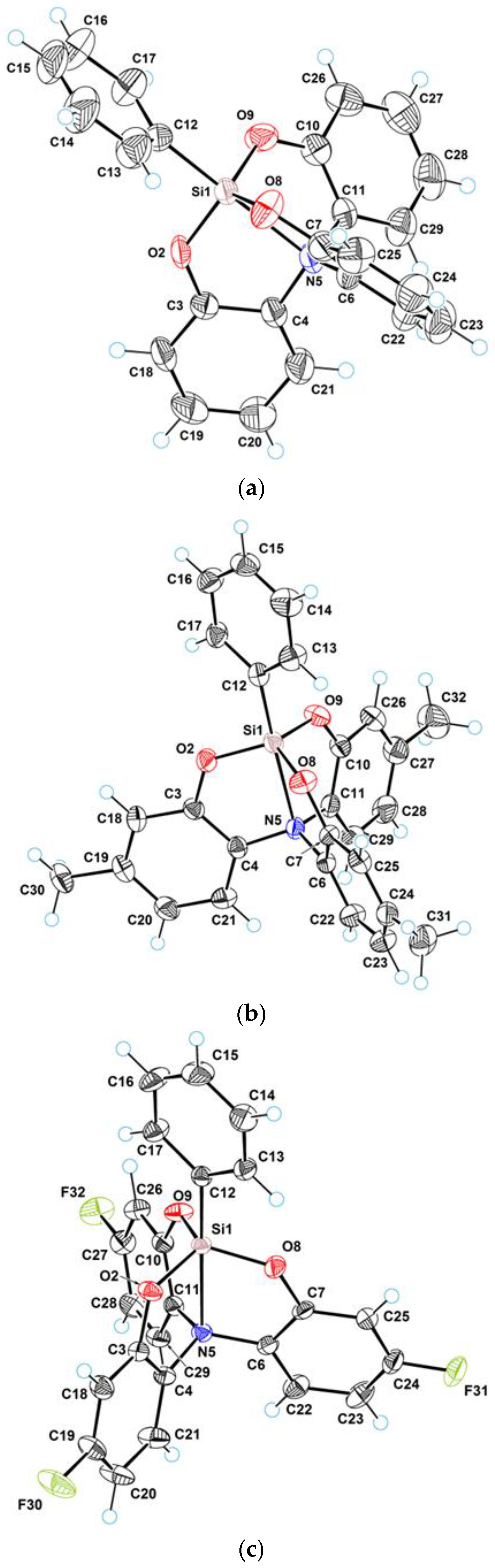
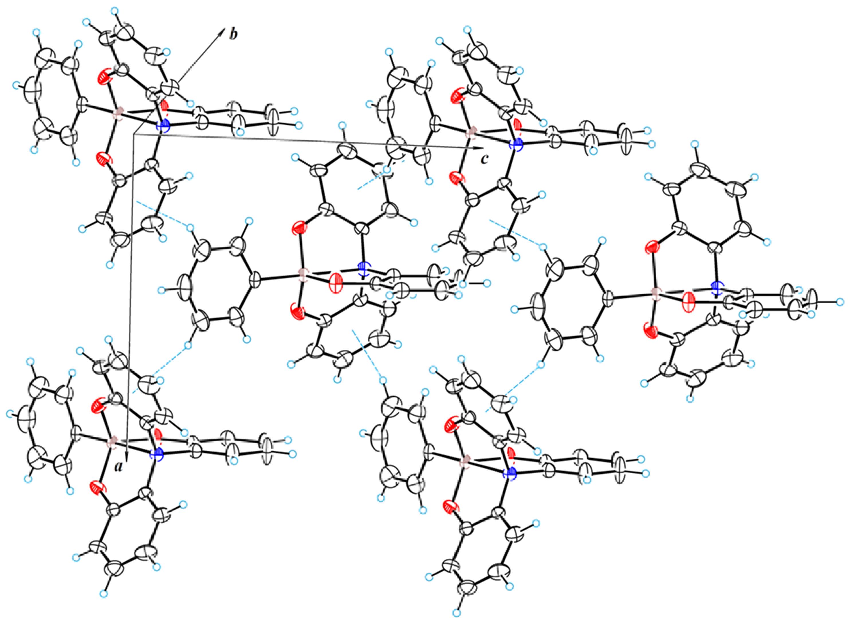
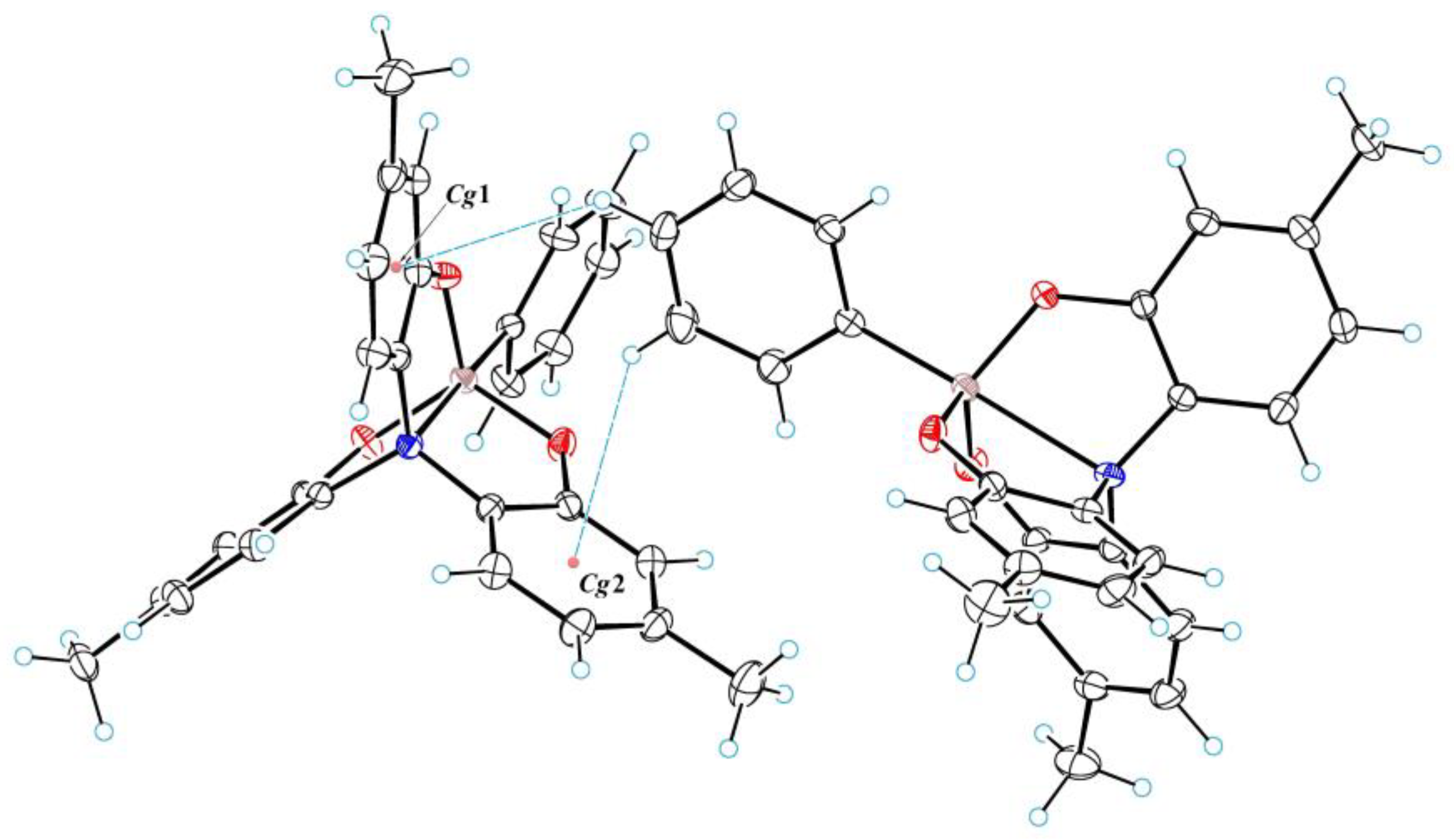
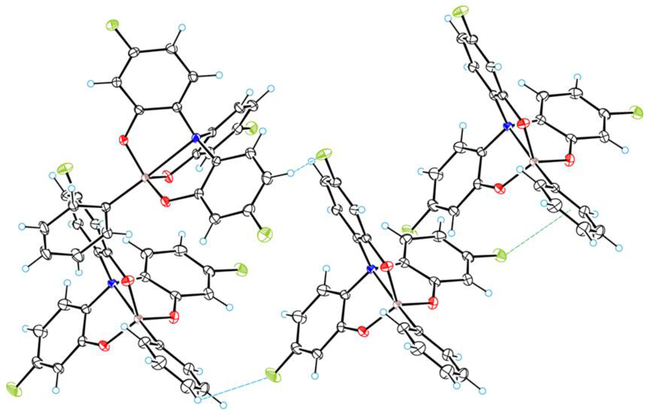
3.1. Crystal Structure of 2a–c

The main crystal data for the three prepared phenyl tribenzsilatranes are listed in Table 1.
Figure 1 shows a perspective view of molecules 2a–c with thermal ellipsoids, and the atom numbering scheme follows in the text. The crystal structure of 2a has been described in [15]. These crystals of 2a belong to the rhombic pyramidal crystal class (space group Cmc21). In the crystal structure, molecules 2a lie in the special positions (in mirror symmetry planes m). Therefore, atoms Si1, O2, C3, C4, N5, C12, C15, C18, C19, C20 and C21 are in these planes. Figure 2 illustrates a fragment of the molecular packing of 2a, revealing C–H⋯π interactions that were not described in the earlier work [15]. In this crystal structure, the C14–H group (as well as the symmetrically equivalent C16–H group) forms an intermolecular hydrogen bond of the CH⋯π type with the aromatic ring. The geometric parameters of this bond are as follows: C⋯Cg (−x + ½, −y + ½, z − ½) = 3.673(15) Å, H⋯Cg = 2.88 Å, C–H⋯Cg = 144°, where Cg is the centroid of ring C6–C7–C25–C24–C23–C22. By means of these bonds, the molecular layers are formed in the crystal structure. These layers are located in an arrangement parallel to the glide symmetry plane c.
An interesting feature of the molecular packing of 2a is that it almost lacks voids in its crystal structure. In fact, only small cavities were revealed in the crystal structure. These cavities are located practically on the screw axes of symmetry 21 and, thus, are repeated through half of the translation along the crystallographic axis c (see Figure S1 Supplementary Materials). The total volume of these cavities in the unit cell amounts to 18.6 Å3. Such a small volume of the voids leads to the fact that the density of 2a is relatively high (d = 1.349 g/cm3).
The crystal structures of 2b and 2c are drastically different from 2a. The crystals of 2b and 2c belong to the prismatic crystal class 2/m. In these crystals, molecules 2b and 2c lie in general positions. The crystal structure of 2b also contains C–H⋯π interactions similar to the structure 2a. The C15–H and C16–H groups form such interactions with aromatic rings. These interactions are shown in Figure 3. The geometric parameters of these bond are as follows: C15⋯Cg1 (−x + 2, y − ½, −z + ½) = 3.819(5) Å, H⋯Cg = 3.08 Å, C–H⋯Cg = 135°; C16⋯Cg2 (−x + 2, y − ½, −z + ½) = 3.857(5) Å, H⋯Cg = 3.09 Å, C–H⋯Cg = 139°, where Cg1 and Cg2 are the centroids of rings C10–C11–C29–C28–C27–C26 and C3–C4–C21–C20–C19–C18, respectively. Thus, these bonds are weaker than the C–H⋯π bonds in the 2a structure, but in 2b, a pair of neighboring molecules are connected by two such bonds. By means of these bonds, the molecular chains in 2b are formed along the monoclinic axis.
In contrast to 2a, the volume of voids in the crystal structure of 2b is quite significant and amounts to 68.4 Å3 in the unit cell. These voids are located in the general positions of the unit cell near the centers of inversion (see Figure S2 Supplementary Materials). The volume of one void region is, thus, 17.1 Å3, which corresponds to the volume of small molecules (such as water). However, due to the fact that substance 2b is hydrophobic, crystal hydrates have not been formed. The presence of the relatively large cavities leads to a decrease in the density of 2b (d = 1.253 g/cm3) in comparison with 2a.
In the crystal structure of 2c, the strongest intermolecular interactions are due to fluorine atoms. These contacts are depicted in Figure 4 (dotted lines). The interactions F30⋯H–C15(x,y − 1,z) and F32⋯H–C23 (x − ½, −y + ½, z − ½) can be described as hydrogen bonds of the CH⋯F type. The parameters of these bond are as follows: F30⋯C15 = 3.257(2) Å, F⋯H = 2.90 Å, C–H⋯F = 104°; F32⋯C23 = 3.167(2) Å, F⋯H = 3.12 Å, C–H⋯F = 84°. The third interaction of the F⋯π type is F31⋯Cg (x−1,y,z), where Cg is the centroid of the ring C12–C13–C14–C15–C16–C17. The distance F31⋯Cg amounts to 3.180(2) Å. Due to these intermolecular interactions, the crystal structure of 2c forms a three-dimensional molecular framework.
The presence of this kind of interaction in the crystal cell 2c is also supported by quantum topological AIM analysis (see Figure S3 Supplementary Materials), detecting bcp(3, −1) critical points in the corresponding inter-atomic regions F⋯H and F⋯C.
In the structure of 2c, the voids occupy a smaller volume compared to 2b (see Figure S4 Supplementary Materials), which is still larger than in 2a. Their total volume per unit cell is 39.6 Å3, which leads to an increase in the density of the substance in comparison with 2b. Considering that in the molecular structure of 2c, instead of hydrogen atoms, there are three fluorine atoms, which have a Van der Waals radius close to that of hydrogen but are 19 times heavier than hydrogen, the compound 2c has the highest density of the three tribenzsilatranes studied (d = 1.454 g/cm3).

3.2. Geometry of Silatrane Cage in 2a–c

Table 2 (for more complete data, see Table S1 Supplementary Materials) presents selected bond lengths and angles characterizing the geometry of the silatrane cage in tribenzsilatranes 2. The common characteristic feature of their molecular structure is the presence of an intramolecular donor–acceptor bond N→Si. Its existence is evidenced by both geometric (the length of the Si⋯N contact is less than the sum of the Van der Waals radii of Si and N atoms, 3.65 Å) and by quantum topological (AIM) criteria (the presence of a bond critical point (3, −1) in the inter-atomic region Si⋯N, see Figure 5).
Judging by the properties of the detected bcp(3, −1), the N→Si bond in phenyl tribenzsilatranes 2a–c (just as in phenylsilatrane 1) formally belongs to the intermediate type of inter-atomic interactions (Table 3). Indeed, the positive sign of the Laplacian ∇2ρ(rc) suggests that this bond is ionic, while according to the negative sign of the electron energy density E(rc), it is covalent [41,42].
As expected [31,32,33], the geometric and quantum topological descriptors of the dative bond N→Si of the molecules 2 (as well as 1) change coherently. For instance, the shortening of the N→Si bond length (dSiN) is accompanied by an increase in the electron density ρ(r) in the corresponding bcp (SiN) and by an increase in the energy of the dative bond N→Si (ESiN) (Table 3).
As noted above (see Introduction), the replacement of ethylene fragments in “ordinary” silatrane 1 (for crystal structure of 1, see Figure S5 Supplementary Materials) with phenylene fragments (i.e., the transition to tribenzsilatranes 2) leads to a noticeable elongation (according to XRD data [2]) of the Si⋯N (dSiN) contact. Moreover, this feature is fundamentally independent of the effect of crystal polymorphism and of the quality of crystals (the magnitude of the R-factor). For instance, for the three polymorph modifications α, β and γ of the “common” phenylsilatrane Ph-Si(OCH2CH2)3N, the Si⋯N distance is dSiN = 2.193 Å (α) [43], 2.156 Å (β) [44] and 2.132 Å (γ) [45], while for tribenzsilatrane Ph-Si(OC6H4)3N, 2a, dSiN = 2.329 Å (R-factor = 0.1105, this work) and 2.344 Å (R-factor = 0.0599 [15]). The ratio dSiN (1) < dSiN (2) also holds for tribenzsilatranes 2b,c containing, in contrast to 2a, para-substituents in the side phenylene groups (see Table 2).
Based on the literature [2,17], this fact can be explained by the relatively high energy requirements, ΔE, for the N→Si bond deformation in 2a–c compared to those in 1. Indeed, according to the calculations performed, it takes less energy to shorten the Si⋯N contact length by 0.1 Å in 1 than in 2 (see Figure 6). With this, it is important to emphasize that, judging by the value of ΔE, the “softness” of the potential function E = f(dSiN) for structures 2ac is comparable (Figure S6 Supplementary Materials). This fact does not allow us to explain the observed increase in the solid-phase dSiN in the series 2a, 2b, 2c with confidence. On the other hand, taking the difference in the calculated values of dSiN in isolated molecules 1 and 2a–c and in crystals, ΔdSiNgas-solid (see Table 2), as a measure of the relative “softness”/“hardness” of potential functions [7,10], one obtains the series ΔdSiNgas-solid (Å): 0.32–0.38 (1) > 0.12–0.13 (2a) > 0.09 (2b) > 0.08 (2c) (regardless of the polymorphism of 1 and the value of the R-factor for 2a, see above). According to this series, the isolated molecule 2c is characterized by the longest Si⋯N contact, which means that it is relatively less sensitive to the effects of the crystal field (a “harder” potential function). Note that according to [7], the more “rigid” character of the potential function of tribenzsilatrane 2c should manifest itself in the lower sensitivity of its 15N, 29Si NMR spectral properties to the nature of the solvent. Let us underline that due to the significant difference between the calculated gas-phase and experimental solid-phase geometries of 1 and 2a–c, the patterns of change in their dSiN under the influence of internal factors (the nature of X, R) may be inconsistent (Table 2 and Table 3).
Using the results of the NBO analysis, we failed to relate the increase in dSiN in the series 2a, 2b, 2c with the obvious difference in electronic effects in the -O-para-R-C6H3- side chains of 2a–c, likely due to their multi-orbital structure.
Early theoretical works [2,27] have shown the N→Si bond length to depend on many factors, including the packing of silatrane molecules in crystals. Therefore, the total energy of intermolecular interactions in the crystal cell, which, as demonstrated above (see Section 3.1), are expected to be fundamentally different for 2a, 2b and 2c, should be considered as an energy criterion ΔE of the impact on the 2a–c geometry upon their transition from the isolated state to the solid phase.
Formally, upon transition of an isolated molecule into a crystal, the structural rearrangement can occur not only towards contraction, but also towards elongation of the Si⋯N contact. However, as has been repeatedly demonstrated in the literature (see review [27] and references therein), the increase in the dipole moment µ of the polar molecular structures containing dative bonds favors their energetically favorable deformation. In the fundamental plan, this enhances the interaction of a single molecule with its environment. In cases of 1 and 2, only the reduction in the Si→N bond length leads to an increase in the dipole moment µ.
For “ordinary” silatranes X-Si(OCH2CH2)3N [2,46,47], a satisfactory relationship between geometric parameters of the coordination site XSiO3N (the N→Si bond length dSiN, the displacement of the silicon atom from the equatorial plane O3–ΔSi, the displacement of the nitrogen atom from the C3 plane formed by the three carbon atoms associated with it, ΔN, and the degree of pentacoordination of the silicon atom, ηe) has been demonstrated for a number of examples. As dSiN decreases, the coordination node deforms towards the trigonal–bipyramidal bond configuration of the central silicon atom, i.e., both the degree of pentacoordination of Si (and its planarity) and tetrahedrality of N (its maximum shift to Si) increase. A similar relationship between the parameters of the coordination site CSiO3N with a change in dSiN is also observed in the case of tribenzsilatranes 2a-c (Table 2).
Note that in “common” silatranes 1 [44,46], the atrane cage adopts a zigzag conformation. This is not possible in molecules of 2a-c; due to the presence of aromatic rings, the atrane cages are rigid and the torsion angles Si–O–C–C, O–C–C–N and C–C–N→Si scarcely differ from zero (Table S1 Supplementary Materials).

4. Conclusions

Three tribenzsilatranes 2, derivatives of 3,4,6,7,10,11-tribenzo-2,8,9-trioxa-5-aza-1-silatricyclo(3.3.3.0^1,5^)undecanes with a phenyl group at silicon and substituted at the side aromatic cycles with the para-R group (R = H, Me, F), have been synthesized and studied using X-ray diffractometry and DFT calculations. Like “common” silatranes 1, their parent derivatives with OCH2CH2 side chains, these tribenzsilatranes have an intramolecular dative bond N→Si whose existence is unequivocally evidenced by geometric and quantum topological (AIM) criteria and whose nature can be defined as ion-covalent. With this, the length of the N⋯Si contact (dSiN) in the crystals of tribenzsilatranes is remarkably longer than in 1. Using DFT B3PW91/6-311++G(d,p) calculations, we have shown that this feature stems from more rigid potential functions of deformation of the N→Si bond in 2 compared to 1. The character (soft/hard) of the potential functions can be assessed from the difference in the calculated values of dSiN in isolated molecules 2a–c and in their crystals. Meanwhile, multiple intermolecular interactions such as the C-H⋯π (with the aromatic rings) and C-H⋯F types as well as of the F⋯π type were revealed through XRD and supported by detecting bcp(3, −1) critical points in the quantum topological AIM analysis of these structures. These findings underline that the correct description of the structure and properties of the N→Si bond in 2 and in their derivatives with other substituents is only possible upon careful consideration of the total energy of all intermolecular interactions in the crystal cell. Further work on the synthesis and study of the related structures is in progress.

Supplementary Materials

The following supporting information can be downloaded at: https://www.mdpi.com/article/10.3390/cryst13050772/s1, Figure S1: The voids in the molecular packing of 2a; Figure S2: The voids in the molecular packing of 2b; Figure S3: Crystal cell graph for 2c from AIM analysis; Figure S4: The voids in the molecular packing of 2c; Table S1: XRD Experimental and B3PW91/6-311++G(d,p) calculated (italics) selected geometric parameters of molecules 2a–c; Figure S5: ORTEP drawing of phenylsilatrane (1); Figure S6: Potential functions of deformation of the N→Si bond in “common” silatrane Ph-Si(OCH2CH2)3N 1 and tribenzsilatranes 2a–c.

Author Contributions

V.S. conceived and designed the experiments and conceptualized the work; E.D. ran the theoretical calculations and prepared the manuscript for publication; S.B. provided crystal structure analysis and reviewed and edited the manuscript; V.J. provided acquisition of funding and supervision of the research; T.R. conducted the X-ray analysis; V.R. realized the synthesis of the compounds and reviewed and edited the manuscript. All authors have read and agreed to the published version of the manuscript.

Funding

VR thanks the support from the European Regional Development Fund ERDF–Latvia (No. 1.1.1.2/VIAA/3/19/577); VJ and SB are grateful to PHC OSMOSE Project 48362YK.

Data Availability Statement

The data presented in this study are available in Supplementary Materials.

Acknowledgments

The crystal structures were obtained at the platform of X-ray diffractometry of the University of Rennes. The quantum chemistry calculations were performed within the research project of the Russian Academy of Sciences No 121021000264-1. The authors are grateful to the Irkutsk Supercomputer Center of SB RAS (http://hpc.icc.ru/) (accessed on 1 March 2023) for providing computational resources of the HPC-cluster “Akademik V.M. Matrosov”.

Conflicts of Interest

The authors declare no conflict of interest.

References

  1. Verkade, J.G. Main group atranes: Chemical and structural features. Coord. Chem. Rev. 1994, 137, 233–295. [Google Scholar] [CrossRef]
  2. Pestunovich, V.; Kirpichenko, S.; Voronkov, M. The Chemistry of Organic Silicon Compounds; Rappoport, Z., Apeloig, Y., Eds.; Wiley: Chichester, UK, 1998; Volume 2, pp. 1447–1537. [Google Scholar]
  3. Voronkov, M.G.; Baryshok, V.P. Use of Silatranes for Medicine and Agriculture; Tolstikov, G.A., Ed.; Publishing House of the Siberian Branch of Russian Academy of Sciences: Novosibirsk, Russia, 2005; p. 258. [Google Scholar]
  4. D’yakov, V.M.; Voronkov, M.G.; Kazimirovskaya, V.B.; Loginov, S.V.; Rasulov, M.M. Organosilicon Chemistry VI: From Molecules to Materials; Auner, N., Weis, J., Eds.; Wiley-VCH Verlag GmbH: Weinheim, Germany, 2008; pp. 588–594. [Google Scholar]
  5. Puri, J.K.; Singh, R.; Chahal, V.K. Silatranes: A review on their synthesis, structure, reactivity and applications. Chem. Soc. Rev. 2011, 40, 1791–1840. [Google Scholar] [CrossRef] [PubMed]
  6. Sidorkin, V.F.; Belogolova, E.F.; Wang, Y.; Jouikov, V.; Doronina, E.P. Electrochemical Oxidation and Radical Cations of Structurally Non-rigid Hypervalent Silatranes: Theoretical and Experimental Studies. Chem. Eur. J. 2017, 8, 1910–1919. [Google Scholar] [CrossRef]
  7. Belogolova, E.F.; Sidorkin, V.F. Correlation among the Gas-Phase, Solution, and Solid-Phase Geometrical and NMR Parameters of Dative Bonds in the Pentacoordinate Silicon Compounds. 1-Substituted Silatranes. J. Phys. Chem. A 2013, 117/25, 5365–5376. [Google Scholar] [CrossRef] [PubMed]
  8. Belogolova, E.F.; Liu, G.; Doronina, E.P.; Ciborowski, S.M.; Sidorkin, V.F.; Bowen, K.H. “Outlaw” Dipole-Bound Anions of Intra-Molecular Complexes. J. Phys. Chem. Lett. 2018, 9, 1284–1289. [Google Scholar] [CrossRef] [PubMed]
  9. Sidorkin, V.F.; Belogolova, E.F.; Doronina, E.P.; Liu, G.; Ciborowski, S.M.; Bowen, K.H. “Outlaw” Dipole-Bound Anions of Intra-Molecular Complexes. J. Am. Chem. Soc. 2020, 142, 2001–2011. [Google Scholar] [CrossRef]
  10. Belogolova, E.F.; Shlykov, S.A.; Eroshin, A.V.; Doronina, E.P.; Sidorkin, V.F. The hierarchy of ab initio and DFT methods for describing an intramolecular non-covalent Si···N contact in the silicon compounds using electron diffraction geometries. Phys. Chem. Chem. Phys. 2021, 23, 2762–2774. [Google Scholar] [CrossRef]
  11. Shen, Q.; Hilderbrandt, R.L. The structure of methyl silatrane (1-methyl-2,8,9-trioxa-5-aza-1-silabicyclo(3.3.3)undecane) as determined by gas phase electron diffraction. J. Mol. Struct. 1980, 64, 257–262. [Google Scholar] [CrossRef]
  12. Forgacs, G.; Kolonits, M.; Hargittai, I. The gas-phase molecular structure of 1-fluorosilatrane from electron diffraction. Struct. Chem. 1990, 1, 245–250. [Google Scholar] [CrossRef]
  13. Shishkov, I.F.; Khristenko, L.V.; Rudakov, F.M.; Golubinskii, A.V.; Vilkov, L.V.; Karlov, S.S.; Zaitseva, G.S.; Samdal, S. Molecular Structure of Silatrane Determined by Gas Electron Diffraction and Quantum-Mechanical Calculations. Struct. Chem. 2004, 15, 11–16. [Google Scholar] [CrossRef]
  14. Korlyukov, A.A.; Antipin, M.Y.; Bolgova, Y.I.; Trofimova, O.M.; Voronkov, M.G. Chemical bonding in the crystal structure of 1-hydrosilatrane. Russ. Chem. Bull. Int. Ed. 2009, 58, 25–30. [Google Scholar] [CrossRef]
  15. Boer, F.P.; Turley, J.W.; Flynn, J.J. Structural studies of pentacoordinate silicon. II. Phenyl(2′2′,2′′-nitrilotriphenoxy)silane. J. Am. Chem. Soc. 1968, 90, 5102–5105. [Google Scholar] [CrossRef]
  16. Chen, W.; Wu, G.L.; Luo, Y. Jiegou Huaxue (Chin.). Chin. J. Struct. Chem. 1987, 6, 165. [Google Scholar]
  17. Meshgi, M.A.; Zaitsev, K.V.; Vener, M.V.; Churakov, A.V.; Baumgartner, J.; Marschner, C. Hypercoordinated Oligosilanes Based on Aminotrisphenols. ACS Omega 2018, 3, 10317. [Google Scholar] [CrossRef] [PubMed]
  18. Sheldrick, G.M. SHELXT—Integrated space-group and crystal-structure determination. Acta Crystallogr. Section A 2015, 71, 3–8. [Google Scholar] [CrossRef] [PubMed]
  19. Sheldrick, G.M. Crystal structure refinement with SHELXL. Acta Crystallogr. Section C 2015, 71, 3–8. [Google Scholar]
  20. Frisch, M.J.; Trucks, G.W.; Schlegel, H.B.; Scuseria, G.E.; Robb, M.A.; Cheeseman, J.R.; Scalmani, G.; Barone, V.; Mennucci, B.; Petersson, G.A.; et al. Gaussian 09, Revision C.01; Gaussian Inc.: Wallingford, CT, USA, 2010. [Google Scholar]
  21. Bader, R.F.W. Atoms in Molecules: A Quantum Theory; Clarendon Press: Oxford, UK, 1990; p. 458. [Google Scholar]
  22. Todd, A.; Keith, T.K. AIMAll (Version 19.02.13); Gristmill Software: Overland Park, KS, USA, 2019. [Google Scholar]
  23. Espinosa, E.; Molins, E.; Lecomte, C. Hydrogen bond strengths revealed by topological analyses of experimentally observed electron densities. Chem. Phys. Lett. 1998, 285, 170–173. [Google Scholar] [CrossRef]
  24. Espinosa, E.; Lecomte, C.; Molins, E. Experimental electron density overlapping in hydrogen bonds: Topology vs. energetics. Chem. Phys. Lett. 1999, 300, 745–748. [Google Scholar] [CrossRef]
  25. Espinosa, E.; Molins, E. Retrieving interaction potentials from the topology of the electron density distribution: The case of hydrogen bonds. J. Chem. Phys. 2000, 113, 5686–5694. [Google Scholar] [CrossRef]
  26. Espinosa, E.; Alkorta, I.; Rozas, I.; Elguero, J.; Molins, E. About the evaluation of the local kinetic, potential and total energy densities in closed-shell interactions. Chem. Phys. Lett. 2001, 336, 457–461. [Google Scholar] [CrossRef]
  27. Korlyukov, A.A. Coordination compounds of tetravalent silicon, germanium and tin: The structure, chemical bonding and intermolecular interactions in them. Russ. Chem. Rev. 2015, 84, 422–440. [Google Scholar] [CrossRef]
  28. Lyssenko, K.A.; Korlyukov, A.A.; Golovanov, D.G.; Ketkov, S.Y.; Antipin, M.Y. Estimation of the Barrier to Rotation of Benzene in the (η6-C6H6)2Cr Crystal via Topological Analysis of the Electron Density Distribution Function. J. Phys. Chem. A 2006, 110, 6545–6551. [Google Scholar] [CrossRef] [PubMed]
  29. Zhurova, E.A.; Stash, A.I.; Tsirelson, V.G.; Zhurov, V.V.; Bartashevich, E.V.; Potemkin, V.A.; Pinkerton, A.A. Atoms-in-Molecules Study of Intra- and Intermolecular Bonding in the Pentaerythritol Tetranitrate Crystal. J. Am. Chem. Soc. 2006, 128, 14728–14734. [Google Scholar] [CrossRef] [PubMed]
  30. Sidorkin, V.F.; Doronina, E.P.; Belogolova, E.F. A New Approach to the Design of Neutral 10-C-5 Trigonal-Bipyramidal Carbon Compounds: A “π-Electron Cap” Effect. Chem. Eur. J. 2013, 19, 10302–10311. [Google Scholar] [CrossRef]
  31. Sidorkin, V.F.; Doronina, E.P. Cage Silaphosphanes with a P→Si Dative Bond. Organometallics 2009, 28, 5305–5315. [Google Scholar] [CrossRef]
  32. Doronina, E.P.; Sidorkin, V.F.; Lazareva, N.F. (PO→Si) Chelates of Silylmethyl Derivatives of Phosphoric Acids R2P(O)ZCH2SiMe3−nHaln (n = 1−3; Z = O, NMe, CH2, S). Organometallics 2010, 29, 3327–3340. [Google Scholar] [CrossRef]
  33. Sidorkin, V.F.; Belogolova, E.F.; Doronina, E.P. Assignment of photoelectron spectra of silatranes: First ionization energies and the nature of the dative Si←N contact. Phys. Chem. Chem. Phys. 2015, 17, 26225–26237. [Google Scholar] [CrossRef]
  34. Doronina, E.P.; Sidorkin, V.F.; Belogolova, E.F.; Jouikov, V. Isomer-selective dative bond O→M (M = Si, Ge) for designing new photochromic hemi-indigo systems. J. Organomet. Chem. 2018, 858, 89–96. [Google Scholar] [CrossRef]
  35. Doronina, E.P.; Jouikov, V.V.; Sidorkin, V.F. Molecular Design of Silicon-Containing Diazenes: Absorbance of E and Z Isomers in the Near-Infrared Region. Chem. Eur. J. 2022, 28, e202201508. [Google Scholar] [CrossRef]
  36. Reed, A.E.; Curtiss, L.A.; Weinhold, F. Intermolecular interactions from a natural bond orbital, donor-acceptor viewpoint. Chem. Rev. 1988, 88, 899–926. [Google Scholar] [CrossRef]
  37. Glendening, E.D.; Badenhoop, J.K.; Reed, A.E.; Carpenter, J.E.; Bohmann, J.A.; Morales, C.M.; Weinhold, F. NBO 5.G; Theoretical Chemistry Institute, University of Wisconsin: Madison, WI, USA, 2004; Available online: https://nbo7.chem.wisc.edu/ (accessed on 20 March 2023).
  38. Granovsky, A.A. Firefly Version 8. Available online: http://classic.chem.msu.su/gran/firefly/index.html (accessed on 20 March 2023).
  39. Tamao, K.; Hayashi, T.; Ito, Y.; Shiro, M. Pentacoordinate anionic bis(siliconates) containing a fluorine bridge between two silicon atoms. Synthesis, solid-state structures, and dynamic behavior in solution. Organometallics 1992, 11, 2099–2114. [Google Scholar] [CrossRef]
  40. Frye, C.L.; Vincent, G.A.; Hauschildt, G.L. Pentacoordinate Silicon Derivatives. III.1 2,2′2′-Nitrilotriphenol, a New Chelating Agent. J. Am. Chem. Soc. 1966, 88, 2727–2730. [Google Scholar] [CrossRef]
  41. Olsson, L.; Ottosson, C.-H.; Cremer, D. Properties of R3SiX Compounds and R3Si+ Ions: Do Silylium Ions Exist in Solution? J. Am. Chem. Soc. 1995, 117, 7460–7479. [Google Scholar] [CrossRef]
  42. Ottosson, C.-H.; Cremer, D. Intramolecularly Stabilized Phenylsilyl and Anthrylsilyl Cations. Organometallics 1996, 15, 5309–5320. [Google Scholar] [CrossRef]
  43. Turley, J.W.; Boer, F.P. Structural Studies of Pentacoordinate Silicon. I. Phenyl-(2,2′,2′′-nitrilotriethoxy)silane. J. Am. Chem. Soc. 1968, 90, 4026–4030. [Google Scholar] [CrossRef]
  44. Parkanyi, L.; Simon, K.; Nagy, J. Crystal and Molecular Structure of β-l-Phenylsilatrane, C12H17O3NSi. Acta Cryst. 1974, 30, 2328–2332. [Google Scholar] [CrossRef]
  45. Parkanyi, L.; Nagy, J.; Simon, K. Crystal and Molecular Structure of γ−l-Phenylsilatrane, Some Structural Features of Silatranes. J. Organomet. Chem. 1975, 101, 11–18. [Google Scholar] [CrossRef]
  46. Pestunovich, V.; Lukevics, E.; Pudova, O.; Sturkovich, R. Molecular Structure of Organosilicon Compounds; John Wiley: Chichester, NY, USA, 1989; p. 359. [Google Scholar]
  47. Greenberg, A.; Wu, G. Structural Relationships in Silatrane Molecules. Struct. Chem. 1990, 1, 79–85. [Google Scholar] [CrossRef]
Figure 1. ORTEP diagram for molecules 2a (a), 2b (b) and 2c (c) showing atomic labels and 50% probability displacement ellipsoids. Hydrogen atoms are shown as small spheres of arbitrary radii.
Crystals 13 00772 g001
Figure 2. Part of the crystal structure of 2a, showing the formation of a layer built with CH⋯π hydrogen bonds. Hydrogen atoms are shown as small spheres of arbitrary radii.
Crystals 13 00772 g002
Figure 3. CH⋯π hydrogen bonds in the crystal structure 2b.
Crystals 13 00772 g003
Figure 4. Intermolecular interactions involving fluorine atoms in the crystal structure 2c.
Crystals 13 00772 g004
Figure 5. MP2(full)/6-311++G(d,p) molecular graph of 2c. The small green spheres represent the bcp(3, −1) bond critical points, and the red spheres represent ring critical points rcp(3, 1).
Crystals 13 00772 g005
Figure 6. Potential functions of the deformation of N→Si bond in “common” silatrane Ph-Si(OCH2CH2)3N 1 and in tribenzsilatrane 2c.
Crystals 13 00772 g006
Table 1. Crystal data and structure refinement parameters for 2a–c.
Parameter2a2b2c
Empirical formulaC24H17NO3Si C27H23NO3Si C24H14F3NO3Si
Formula weight, Mr395.47437.55449.45
Temperature (K)296(2)150(2)150(2)
DiffractometerD8 VENTURE
Bruker AXS
APEXII
Bruker AXS
APEXII
Bruker AXS
Crystal size (mm3)0.80 × 0.36 × 0.270.33 × 0.24 × 0.100.58 × 0.42 × 0.09
Crystal systemorthorhombicmonoclinicmonoclinic
Space groupCmc21P21/cP21/n
a (Å)11.127(6)10.086(1)9.571(2)
b (Å)14.714(4)12.433(1)12.308(2)
c (Å)11.892(3)19.107(2)17.430(2)
β (°)90104.594(6)91.533(5)
Unit cell volume (Å3)1946.9(13)2318.7(4)2052.5(5)
Molecular multiplicity444
Absorption coefficient (mm−1)0.1470.1300.169
F(000)824920920
Calculated density (g/cm3)1.3491.2531.454
max (°)55.051.055.0
Reflections collected626615,74911,432
Independent reflections with I > 2σ(I)190123513348
Number of refined parameters137292289
R-factor0.11050.06360.0417
CCDC number224780622478032247804
Table 2. XRD Experimental and B3PW91/6-311++G(d,p) calculated (italics) selected geometric parameters of molecules 2a–c (bond lengths in Å, angles in °).
Parameter2a2b2c
exp.calc.exp.calc.exp.calc.
Si1–O21.654(8)1.6811.657(2)1.6841.655(2)1.683
Si1–C121.863(11)1.8611.850(3)1.8631.852(3)1.857
O2–C31.342(14)1.3531.370(4)1.3531.369(4)1.349
C3–C41.400(15)1.3991.387(4)1.3981.392(4)1.400
N5–C41.443(13)1.4381.446(4)1.4401.444(4)1.437
O2–Si1–O8117.1(3)115.0116.0(1)115.4116.2(1)114.7
O2–Si1–C12100.3(4)102.198.9(1)102.2102.5(1)103.2
Si1–O2–C3126.3(7)126.4125.7(2)126.1125.9(2)126.8
O2–C3–C4118.2(11)119.7118.1(3)119.6119.2(3)120.0
C3–C4–N5113.3(10)114.7113.9(3)114.8114.4(3)115.0
C4–N5–C6116.5(7)117.1114.7(2)117.3116.8(2)117.4
N5–Si1–C12179.8(4)179.6178.2(1)179.6178.0(1)179.5
Si1–N52.329(9)2.4632.358(3)2.4512.411(3)2.494
ΔN0.300(9)0.3570.292(4)0.2470.267(4)0.230
ΔSi0.284(7)0.2430.286(3)0.3460.318(3)0.372
ηe72.458.472.260.965.854.9
Σ(O-Si-O)351.4(3)346.9351.3(1)347.7349.3(1)345.8
Σ(C-N-C)347.4(7)351.6348.2(2)351.3350.0(2)352.5
Table 3. Geometric * parameters of the coordination node CSiO3N and quantum topological ** characteristics at the bond critical point bcp(SiN) of 1 and 2a-c as isolated molecules and in crystal.
Compound dSiNηeρ(r)2ρ(r)E(rc)ESiNRef.
1gas2.515540.2350.861−0.078.9
crystalα2.193860.3701.984−0.1722.6[43]
β2.156870.3912.554−0.1825.1[44]
γ2.132880.4042.908−0.1826.7[45]
2agas2.463580.2560.737−0.0810.2
crystal2.344720.2990.866−0.1314.6[15]
2.329720.3110.823−0.1415.5this work
2bgas2.451610.2610.701−0.0910.5
crystal2.358720.2960.745−0.1213.9this work
2cgas2.494550.2440.786−0.079.4
crystal2.411660.2740.749−0.1011.9this work
* Inter-atomic distance Si⋯N (dSiN, Å) and degree of pentacoordination of silicon (ηe, %). ** Electron density (ρ(rc), e/Å3), Laplacian (∇2ρ(rc), e/Å5), electron energy density (E(rc), hartree/Å3) in bcp(SiN) and energy of the dative bond N→Si (ESiN, kcal/mol).
Disclaimer/Publisher’s Note: The statements, opinions and data contained in all publications are solely those of the individual author(s) and contributor(s) and not of MDPI and/or the editor(s). MDPI and/or the editor(s) disclaim responsibility for any injury to people or property resulting from any ideas, methods, instructions or products referred to in the content.

