Article

Crystal Structure and Spectroscopic Analysis of 3-Diethoxyphosphoryl-28-[1-(1-deoxy-β-D-glucopyranosyl)-1H-1,2,3-triazol-4-yl]carbonylbetulin

1
Department of Organic Chemistry, Faculty of Pharmaceutical Sciences in Sosnowiec, Medical University of Silesia, Katowice, 4 Jagiellońska Str., 41-200 Sosnowiec, Poland
2
Department of Physics of Crystals, Institute of Physics, University of Silesia, 4 Uniwersytecka Str., 40-007 Katowice, Poland
*
Author to whom correspondence should be addressed.
Crystals 2023, 13(10), 1488; https://doi.org/10.3390/cryst13101488
Submission received: 19 September 2023 / Revised: 1 October 2023 / Accepted: 10 October 2023 / Published: 13 October 2023
(This article belongs to the Special Issue Feature Papers in Biomolecular Crystals in 2022-2023)

Abstract

:
The molecular structure of 3-diethoxyphosphoryl-28-[1-(1-deoxy-β-D-glucopyranosyl)-1H-1,2,3-triazol-4-yl]carbonylbetulin was investigated through various experimental and theoretical methods. X-ray diffraction, Hirshfeld surface, experimental and calculated FT-IR spectra analysis, molecular electrostatic potential (MEP) and molecular orbital (HOMO and LUMO) were used for the analyses. It was found that the introduction of a triazole substituent affects the arrangement of molecules in the crystal structure and the formation of hydrogen bonds. The Hirshfeld surface analysis shows that the red regions are concentrated near groups, which create hydrogen bonds, which means that the hydrogen bonds are strong. The analysis of HOMO and LUMO orbitals and the chemical reactivity descriptors shows that the compound is kinetically and chemically stable. A molecular electrostatic potential map was used to analyze the electrophilic and nucleophilic area in the molecule.

1. Introduction

Betulin (3β-lup-20(29)-en-3,28-diol) belongs to naturally occurring lupane-type pentacyclic triterpenoids. This compound is most often isolated from birch bark. Interest in this substance of natural origin results from its diverse biological activity, including anti-cancer, anti-inflammatory, hepatoprotective, anti-HIV, anti-fungal and anti-bacterial effects [1,2,3,4,5,6,7]. Betulin is slightly toxic to normal cells [1]. The lack of clinical use of betulin is due to its low bioavailability and poor solubility in the aqueous environment. The improvement of betulin water solubility is associated with modifications of its structure leading to the formation of new derivatives. The chemical methods used for this purpose usually concern the C3 and C28 positions of the betulin scaffold (Figure 1a). The hydroxyl groups present in the structure of betulin are most often subjected to oxidation, etherification and acetylation reactions [7].
Phosphates are involved in a variety of biochemical processes, including the biosynthesis and polymerization of nucleic acids, glycolysis and lipid biosynthesis. The phosphate group is often used in prodrugs of therapeutic agents. Phosphate prodrugs are most often obtained by modifying acidic oxygen atoms with metabolically labile protecting groups. In this way, charge-neutral compounds with increased lipophilicity and reduced sensitivity to phosphoesterase are obtained [8].
A combination of two active moieties, such as betulin scaffold and phosphoryl group, allowed a new group of compounds showing high biological activity to be obtained [9,10]. The introduction of a diethyl phosphate group at the C3 carbon atom of the betulin molecule gave 3-diethoxyphosphorylbetulin, which exhibits low anticancer activity against MV-4-11 leukemic cells (IC50 = 25.60 µM) (Figure 1b). Moreover, the obtained derivative demonstrates a three-times lower activity against MV-4-11 cells compared to betulin [9]. For this reason, the structure modifications were made by introducing an acetylene group or a triazole ring [9,10].
The alkyne group, which occurs in many clinically used drugs, is involved in interaction with biological target proteins, like MAO, tyrosine kinases, BACE1, steroid receptors, mGlu5 receptors, FFA1/GPR40, and HIV-1 RT [11]. Many alkyne derivatives of betulin with significant anticancer activity have been described in the literature [12,13,14]. Typically, the terminal alkyne is transformed into 1,4-disubstituted 1,2,3-triazole in the Husigen cycloaddition. The 1,2,3-triazole moiety is a known pharmacophore in medicinal substances like radezolid, tazobactam, carboxyamidotriazole and cefatrizine. Triazole derivatives have antifungal, antiviral, antibacterial, antituberculosis, antiallergic, antidiabetic and neuroprotective properties. Many compounds with the 1,2,3-triazole structure also have significant anticancer properties [15,16,17]. In recent years, the synthesis and biological activity of many triazole derivatives of betulin have been described. The introduction of this moiety affects the physicochemical properties of betulin derivatives, such as water solubility, lipophilicity, and the ability to interact with a biological target [18,19,20,21,22].
The current work is a continuation of research that our team has previously published. Previous studies have described the synthesis and anticancer activity of the title compound. In the series of tested triazoles, 3-diethoxyphosphoryl-28-[1-(1-deoxy-β-D-glucopyranosyl)-1H-1,2,3-triazol-4-yl]carbonylbetulin has shown significant activity against the tested cell line [9]. Hence, it would be interesting to discover the crystal structure of the compound. The X-ray study allows the determination of the interaction between molecules in a crystal unit. According to the literature, the hydrogen bond occurring in the crystal structure influence the stability and chemical reactivity of the compound after its dissolution [23,24]. The intermolecular interaction occurring in crystal structure was confirm by the Hirshfeld surface. The study supplemented the analysis of experimental and calculated FT-IR spectroscopic frequencies of the title molecule. Moreover, the highest occupied molecular orbital (HOMO) and the lowest unoccupied molecular orbital (LUMO), as well as the global reactivity descriptor and molecular electrostatic potential (MEP) surface analysis of the title compound are reported.

2. Materials and Methods

2.1. Synthesis of 3-Diethoxyphosphoryl-28-[1-(1-deoxy-β-D-glucopyranosyl)-1H-1,2,3-triazol-4-yl]carbonylbetulin

The synthesis of the title compound has been described in the literature [9]. Briefly, the 3-diethoxyphosphorylbetulin 1 (0.58 g; 1 mmol) was dissolved in dichloromethane (5 mL). After cooling to −10 °C, the propiolic acid (0.074 mL, 1.18 mmol) and solution of N,N′-dicyclohexylcarbodiimide (DCC) (0.246 g, 1.18 mmol), 4-dimethylaminopyridine (DMAP) (0.01 g, 0.08 mmol) in dichloromethane (1 mL) were added. After 24 h, the reaction mixture was removed under reduced pressure. The crude product was purified using column chromatography. The 3-diethoxyphosphoryl-28-propynoylbetulin 2 obtained with 63% yield (0.396 g, 0.63 mmol). The chemical structure was confirmed by 1H and 13C NMR spectra, and was compatible with literature data [9]. Next, the 3-diethoxyphosphoryl-28-propynoylbetulin (0.396 g, 0.63 mmol) was dissolved in toluene (2 mL) and the solution of 1-deoxy-β-D-glucopyranosyl azide (0.69 mmol), and the coper iodide (I) (0.069 mmol, 0.014 g) in toluene (1 mL) was added. The reaction mixture was heated under reflux for 72 h. After this time, the solvent was evaporated under reduced pressure. The crude product 3 was purified by the column chromatography, obtaining colorless product with 70% yield. The chemical structure was confirmed by 1H and 13C NMR spectra, which were compatible with literature data [9].

2.2. X-ray Diffraction Analysis

The pure compound was dissolved in an acetone/DMF (v/v 10:1) mixture. After evaporating the mixture at room temperature, a colorless crystal was obtained. The X-ray diffraction measurement of selected crystal was carried out the Xcalibur kappa diffractometer with the Sapphire3 CCD detector and CuKα radiation. The crystal structure was determined using the SHELXS-2013 and refined using SHELXL-2014/6 program [25]. The H-bonds were found through analysis of the crystal structure using Mercury 4.1.0 program. The obtained results were refined using Shelx program. The crystallographic data have been stored in the Cambridge Crystallographic Data Centre (CCDC) as CCDC-1853753. These data can be obtained free of charge from the CCDC via www.ccdc.cam.ac.uk/data_request/cif (5 September 2023).

2.3. Hirshfeld Surface Analysis

The percentage contributions for intermolecular contacts in title compound were obtained using Hirshfield surface analyses; the 3D view and 2D fingerprint plots were performed by Crystal-Explorer v.3.1 program [26]. The normalized contact distance (dnorm) is defined as the combination of di and de value. The di is the distance of the closest internal nucleus to the surface and de is the distance from the point to the closest exterior nucleus to the surface [27].

2.4. FT-IR Spectra

The FT-IR spectrometer RXross equipped with the attenuated total reflection (ATR) diamond accessory was used for measurement. The 64 scans were accumulated, with resolution of 2 cm−1 (digital resolution 0.482 cm−1) in the spectral range of 400-3800 cm−1. To calculate the absorbance, the ATR correction was used to transform the reflectance spectrum to a log (1/R) absorption spectrum.

2.5. Computational Details

The optimized chemical structure of compound was determined using the density functional theory (DFT/B3LYP/6-311+G(d,p)) method implemented in the Gaussian 09 program package [28]. The initial geometry of compound 3 was taken from the X-ray crystallographic data. Comparing the calculated and X-ray molecular structure shows that the betulin scaffold and sugar moiety show good capability (Figure S1A,B), while the triazole moiety has poor capability in calculated and experimental molecular structure (Figure S1B). This effect is caused by different values of the torsion angle between C17 and C28, and O2 and C31 atoms, which in calculation and experimental molecular structure is equal 120.57° and −124.73°, respectively (Figure S1B). The optimized structure of molecule was used to calculate the IR spectrum. The calculated vibration was scaled by 0.967 factor [29]. The obtained calculated structure was used to determine the HOMO–LUMO energy, quantum chemical descriptors, and the molecular electrostatic potential [30]. All obtained results were visualized in the GaussView, Version 5 software package [31].

3. Results and Discussion

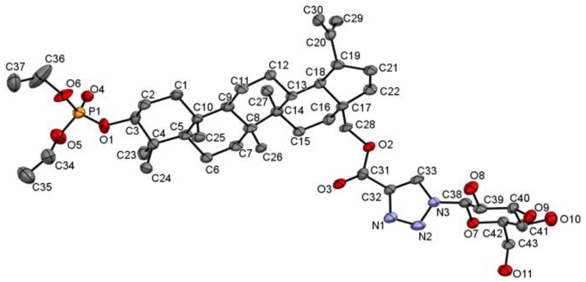
3.1. Crystal Structure

Scheme 1 presents the synthesis of 3-diethoxyphosphoryl-28-[1-(1-deoxy-β-D-glucopyranosyl)-1H-1,2,3-triazol-4-yl]carbonylbetulin 3. In the first stage, the 3-diethoxyphosphorylbetulin 1 was converted to 3-diethoxyphosphoryl-28-propynoylbetulin 2. The 1,2,3-triazole derivative 3 was obtained by the cycloaddition reaction of compound 2 with 1-deoxy-β-D-glucopyranosyl azide.
The chemical structure of compound 3 was designated by the 1H NMR, 13C NMR and IR spectroscopy. Additionally, its crystal structure was investigated using the X-ray diffraction method. Figure 2 presents the structure of derivative 3 and atom numbering. The crystal parameters, experimental data and refinement details are presented in Table S1. The bond length and bond angles are collected in Tables S2–S4.
Compound 3 crystalizes in a monoclinic P21 space group. The arrangement analysis shows that the six-membered rings of the betulin scaffold have a chair conformation and the five-membered ring exhibits the conformation of the twisted envelope. The triazole ring is almost flat, while the sugar moiety has a chair conformation. The β-D-glucopyranose ring could exist as two conformers. As seen in Figure S2, the β-D-glucopyranose ring in the title compound have 4C1 conformation.
The crystal structure of derivative 2 has been described in the literature [9]. The arrangement of the betulin scaffold in 2 and 3 are similar (Figure S2). However, the introduction of triazole substituent influences the arrangement of substituent at the C3 and C28 positions of the betulin scaffold. The angles C3-O1-P1 and C17-C28-O2 are equal 122.9° and 110.2° in compound 2 and 125.4° and 108.5° in compound 3. The results show that changing the substituent at the C-28 position influence the arrangement of the phosphoryl group at the C3 position (Figure S1).
The elementary unit consists of two molecules (Z = 2). In each unit, the molecules are arranged alternately to each other, which means that the triazole moiety is located opposite the diethoxyphosphoryl group. In the crystal structure, the molecules are arranged in two layers (Figure 3). In each layer, they are distributed in such a way that the sugar moieties are localized next to the diethoxyphosphoryl group.
As seen in Table 1 and Figure 3, the hydroxyl groups at the C40 and C39 positions create hydrogen bonds, as H-bond acceptors. According to the Jeffrey’s hydrogen bonds classification, the O8-H8···O4 and O9-H9···O4 interactions are moderately strong [23].

3.2. Hirshfeld Surface

Hirshfeld surface analysis is useful method for the identification of all contacts in crystal structure. Surface color represents the different dnorm values. The negative value is represented in red color, the positive dnorm in blue color. Gray means that dnorm is zero, and the surface indicates contacts equal to van der Waals radii [32,33].
As seen in Figure 4, the red regions are localized near the hydroxyl group at sugar moiety and oxygen atom of the phosphoryl group. The red regions are concentrated near groups which create hydrogen bonds, which means that the hydrogen bonds are strong. The di, de, surface index, curvedness index and fragment patch maps are presented in Figure S3. The index of dnorm, di and de were obtained in the range of −0.6198 and 2.0124, 0.7288 and 2.9994, 0.7293 and 2.8637, respectively. Shape index, curvedness index and fragment patch were determined in the ranges of −1.0000 to 1.0000, −4.0000 to 0.4000 and 0 to 17, respectively.
Figure 5 shows 2D fingerprint plots of compound 3 with the percentage contribution of each interaction on the surface of 3. The dominant intermolecular interaction is HH contact, with a percentage of 68.5% (Figure 5a). The HO contact, which is involved in hydrogen bonding, accounts for 19.1% of the total Hirshfeld surface (Figure 5b). The other contacts show an inferior contribution (Figure 5c,d). Moreover, the phosphorus atom does not have intermolecular interaction with other atoms.

3.3. Vibrational Assignments

The title compound, composed of 103 atoms, exhibits 378 vibrations, which were analyzed using experimental and calculated FT-IR spectra (Figure 6). The calculated spectrum reproduces the experimental ones well, and the coefficient of correlation is equal to 0.9971 (Figure S4). Most of vibrations are ring vibrations, which are difficult to identify in the experimental spectrum. For this reason, Table 2 presents the selected band associated with the O-H, P=O, N=N, C-N and C-H vibrations.
In the region between 3000 cm−1 and 3500 cm−1, stretching vibrations of O-H and sp3-hybridized of C-H band are observed [23,34,35]. In calculated spectrum, a peak at 3509 cm−1 is associated with the stretching vibration of hydroxyl group. The stretching vibration of the C-H group at betulin moiety is observed at 3413 cm−1. In the experimental spectrum, a broad band at 3387 cm−1, which was associated with stretching vibration of C-H and OH groups. This effect could be related to a hydrogen bond, which creates the hydroxyl group at sugar moiety. The stretching vibration of the sp2-hybridized of C–H (=C–H) in isopropenyl group at the C19 of betulin moiety is seen at 2970 cm−1 and 2968 cm−1 in experimental and calculated spectra, respectively. The C-H bending vibrations in-plane and out-of-plane are observed in the range of 1500–1000 cm−1 and 950–800 cm−1, respectively [34,36]. The peak at 1450 cm−1 is associated with in-plane bending vibration of C-H in betulin moiety. In the experimental spectrum, the in-plane C-H bending vibration of the ethyloxy group and sugar moiety are observed at 1367 cm−1 and 1230 cm−1, respectively. In the calculated spectrum, these peaks are localized at 1409 cm−1 and 1209 cm−1, respectively. The out-of-plane C-H bending vibration of the betulin and sugar moieties are observed at 1065 cm−1 and 983 cm−1, respectively. Based on the calculated spectrum, the peak at 1167 cm−1 is associated with out-of-plane C-H bending vibration of the triazole ring.
The carbonyl stretching vibration is observed as a strong peak in the region of 1800–1600 cm−1 [34,37]. The C=O stretching vibration is observed at 1721 cm−1 and 1669 cm−1 in the experimental and calculated spectra, respectively. In the experimental FT-IR spectrum of tested compound, the broad peak at 1132 cm−1 is associated with the stretching vibration of the C-O and N=N groups. The in-plane C-O bending vibration is observed at 880 cm−1 and 897 cm−1 in the experimental and calculated spectra, respectively.
In the experimental spectrum, the highest peak is observed at 1016 cm−1. Comparing the calculated and experimental spectra allows this peak to be identified as the P=O stretching vibration. The high intensity of this peak could be due to the formation of the hydrogen bond between phosphoryl group and hydroxyl group at sugar moiety [23,38].

3.4. Chemical Reactivity Descriptors

The reactivity of chemical molecules has been characterized by the Frontier Molecular Orbitals (FMO) study. According to the FMO theory, the most important are the most occupied (HOMO) and least unoccupied (LUMO) orbitals. The location of these orbitals and their energy influence the interaction between molecules [39,40,41]. The orbitals were designated by the Gaussian program [28].
The HOMO orbital is mainly delocalized near the isopropenyl group and five-membered ring of betulin scaffold, while the LUMO orbital is delocalized near a triazole ring and ester group at the C-28 position of betulin (Figure 7). The distribution of the HOMO and LUMO indicates that the molecular system has moderate charge transfer capabilities. The computation of the energy gap provides information about a molecule’s kinetic stability and reactivity. A small HOMO–LUMO energy gap indicates heightened reactivity, as electron transitions between these orbitals are favorable, while a larger gap implies kinetic stability [42,43]. The energy gap between LUMO and HOMO orbitals is equal to 4.946 eV, which means that compound 3 has a low chemical reactivity and high kinetic stability (Table 3).
The chemical potential (μ) defines the ability of compound to be an acceptor or donor of electrons. As seen in Table 1, the low chemical potential shows that molecule 3 has an electron-donor role and a great ability to donate electrons to adjacent molecules. Comparing the ionization potential (I) and electron affinity (A) shows that the I value is higher than the A value. The results determine that compound 3 is a better electron donor than electron acceptor [44]. The electrophilicity index (ω) is higher than 1.5 eV, which means that 3 is nucleophile [45]. The global softness and hardness are equal to 0.202 eV and 2.473 eV, respectively. The analysis of the chemical descriptor shows that the molecule is kinetically stable and hard, with a low capacity to accept electrons and a high capacity to donate them.

3.5. MEP Analysis

The molecular electrostatic potential (MEP) method is a computational technique employed in quantum chemistry to analyze the distribution of electrostatic charges within a molecule. It offers valuable insights into the reactivity and chemical behavior of molecular systems by mapping the electrostatic potential field around a molecule in three-dimensional space [46]. The color of MEP maps displays the different values of electrostatic potential. The blue color means the most positive electrostatic potential; red color marks the most negative potential. The potential values increase in the order red < orange < yellow < green < blue [47].
The MEP map of compound 3 shows that the negative potential is localized in two main areas. The first is near the oxygen atom of the phosphoryl group at the C3 position, and the second is near the ester group at the C28 position of the betulin scaffold (Figure 8). The blue region is localized near the hydrogen atoms of sugar and the triazole moiety. The dominated color is green, denoting electrostatic neutral region.
Comparing the MEP maps of molecules 2 [48] and 3 shows that the introduction of triazole substituent at the C28 position of the betulin scaffold significantly increases the negative electrostatic potential in this area. The substituent at the C3 position does not influence the electrostatic potential of phosphoryl group.

4. Conclusions

In the present research, the X-ray structural and chemical reactivity descriptors of the 3-diethoxyphosphoryl-28-[1-(1-deoxy-β-D-glucopyranosyl)-1H-1,2,3-triazol-4-yl]carbonylbetulin 3 was described. The Hirshfeld surface analysis shows that the hydrogen bonds between the phosphoryl group at the C3 position of the betulin scaffold and the hydroxyl group at the sugar moiety are strong.
The experimental and calculated FT-IR spectra show a good correlation, and the coefficient of correlation is equal to 0.9971. The most important difference was observed in the 1016 cm−1 region. In this region, a high intensity peak is observed, which is associated with the P=O stretching vibration. The high intensity of this peak could be due to the formation of the H-bond.
The analysis of chemical reactivity descriptors shows that the title compound is characterized by high kinetic and chemical stability. The molecular potential map shows that the nucleophilic regions are localized near the oxygen atoms of the phosphoryl group at the C3 position and ester group at the C28 position of the betulin scaffold.

Supplementary Materials

The following supporting information can be downloaded at: https://www.mdpi.com/article/10.3390/cryst13101488/s1, Figure S1: Overlap the calculated (pink molecule) and X-ray (blue molecule) molecular structures of compound 3 in (a) betulin scaffold and (b) sugar moiety; Figure S2: Atom by atom superimposition of the compounds 2 (blue molecule) and 3 (violet molecule); Figure S3: Hirshfeld surface mapped with a. dnorm, b. di, c. de, d. shape index, e. curvedness index and f. fragment patch for compound 3; Figure S4: The linear regression between the experimental and calculated wavenumber; Table S1: Experimental data and refinement details of compound 3; Table S2: Experimental bond length in crystal structure of compound 3; Table S3: Experimental bond angle in crystal structure of compound 3; Table S4: Experimental torsion angle in crystal structure of compound 3.

Author Contributions

Conceptualization and writing—original draft preparation, M.K.-T.; methodology, E.B., E.C. and M.K.; visualization, A.S. All authors have read and agreed to the published version of the manuscript.

Funding

This research was funded by the Medical University of Silesia in Katowice, Poland. Grants: PCN-2-027/K/2/F, PCN-1-042/K/2/F and PCN-1-044/K/2/F.

Data Availability Statement

Samples of the compound 3 is available from the authors.

Conflicts of Interest

The authors declare no conflict of interest.

References

  1. Zhao, J.; Li, R.; Pawlak, A.; Henklewska, M.; Sysak, A.; Wen, L.; Yi, J.E.; Obmińska-Mrukowicz, B. Antitumor activity of betulinic acid and betulin in canine cancer cell lines. In Vivo 2018, 32, 1081–1088. [Google Scholar] [CrossRef]
  2. Schwiebs, A.; Radeke, H.H. Immunopharmacological activity of betulin in inflammation-associated carcinogenesis. Anticancer Agents Med. Chem. 2018, 18, 645–651. [Google Scholar] [CrossRef]
  3. Buko, V.; Zavodnik, I.; Palecz, B.; Stepniak, A.; Kirko, S.; Shlyahtun, A.; Misiuk, W.; Belonovskaya, E.; Lukivskaya, O.; Naruta, E.; et al. Betulin/2-hydroxypropyl-β-cyclodextrin inclusion complex: Physicochemical characterization and hepatoprotective activity. J. Mol. Liq. 2020, 309, 113118. [Google Scholar] [CrossRef]
  4. Chaniad, P.; Sudsai, T.; Septama, A.W.; Chukaew, A.; Tewtrakul, S. Evaluation of anti-HIV-1 integrase and anti-Inflammatory activities of compounds from betula alnoides buch-ham. Adv. Pharmacol. Sci. 2019, 2019, 2573965. [Google Scholar] [CrossRef] [PubMed]
  5. Javed, S.; Mahmood, Z.; Khan, K.M.; Sarker, S.D.; Javaid, A.; Khan, I.H.; Shoaib, A. Lupeol acetate as a potent antifungal compound against opportunistic human and phytopathogenic mold Macrophomina phaseolina. Sci. Rep. 2021, 11, 8417. [Google Scholar] [CrossRef] [PubMed]
  6. Zhang, L.; Ma, Z.; Wang, R.; Zhu, M. Synthesis and characterization of methacrylate-functionalized betulin derivatives as antibacterial comonomer for dental restorative resins. ACS Biomater. Sci. Eng. 2021, 7, 3132–3140. [Google Scholar] [CrossRef]
  7. Li, J.; Jiang, B.; Chen, C.; Fan, B.; Huang, H.; Chen, G. Biotransformation of betulin by Mucor subtilissimus to discover anti-inflammatory derivatives. Phytochemistry 2019, 166, 112076. [Google Scholar] [CrossRef]
  8. Wiemer, A.J. Metabolic efficacy of phosphate prodrugs and the remdesivir paradigm. ACS Pharmacol. Transl. Sci. 2020, 3, 613–626. [Google Scholar] [CrossRef]
  9. Chrobak, E.; Kadela-Tomanek, M.; Bębenek, E.; Marciniec, K.; Wietrzyk, J.; Trynda, J.; Pawełczak, B.; Kusz, J.; Kasperczyk, J.; Chodurek, E.; et al. New phosphate derivatives of betulin as anticancer agents: Synthesis, crystal structure, and molecular docking study. Bioorg. Chem. 2019, 87, 613–628. [Google Scholar] [CrossRef]
  10. Chrobak, E.; Jastrzębska, M.; Bębenek, E.; Kadela-Tomanek, M.; Marciniec, K.; Latocha, M.; Wrzalik, R.; Kusz, J.; Boryczka, S. Molecular structure, in vitro anticancer study and molecular docking of new phosphate derivatives of betulin. Molecules 2021, 26, 737. [Google Scholar] [CrossRef] [PubMed]
  11. Talele, T.T. Acetylene group, friend or foe in medicinal chemistry. J. Med. Chem. 2020, 63, 5625–5663. [Google Scholar] [CrossRef]
  12. Boryczka, S.; Bębenek, E.; Wietrzyk, J.; Kempińska, K.; Jastrzębska, M.; Kusz, J.; Nowak, M. Synthesis, structure and cytotoxic activity of new acetylenic derivatives of betulin. Molecules 2013, 18, 4526–4543. [Google Scholar] [CrossRef]
  13. Bębenek, E.; Kadela-Tomanek, M.; Chrobak, E.; Wietrzyk, J.; Sadowska, J.; Boryczka, S. New acetylenic derivatives of betulin and betulone, synthesis and cytotoxic activity. Med. Chem. Res. 2017, 26, 1–8. [Google Scholar] [CrossRef] [PubMed]
  14. Amiri, S.; Dastghaib, S.; Ahmadi, M.; Mehrbod, P.; Khadem, F.; Behrouj, H.; Aghanoori, M.R.; Machaj, F.; Ghamsari, M.; Rosik, J.; et al. Betulin and its derivatives as novel compounds with different pharmacological effects. Biotechnol. Adv. 2020, 38, 107409. [Google Scholar] [CrossRef]
  15. Jiang, X.; Hao, X.; Jing, L.; Wu, G.; Kang, D.; Liu, X.; Zhan, P. Recent applications of click chemistry in drug discovery. Expert. Opin. Drug Discov. 2019, 14, 779–789. [Google Scholar] [CrossRef] [PubMed]
  16. Alam, M.M. 1,2,3-Triazole hybrids as anticancer agents: A review. Arch. Pharm. 2022, 355, e2100158. [Google Scholar] [CrossRef] [PubMed]
  17. Xu, Z.; Zhao, S.J.; Liu, Y. 1,2,3-Triazole-containing hybrids as potential anticancer agents: Current developments, action mechanisms and structure-activity relationships. Eur. J. Med. Chem. 2019, 183, 111700. [Google Scholar] [CrossRef]
  18. Bębenek, E.; Jastrzębska, M.; Kadela-Tomanek, M.; Chrobak, E.; Orzechowska, B.; Zwolińska, K.; Latocha, M.; Mertas, A.; Czuba, Z.; Boryczka, S. Novel triazole hybrids of betulin: Synthesis and biological activity profile. Molecules 2017, 22, 1876–1898. [Google Scholar] [CrossRef]
  19. Kadela-Tomanek, M.; Jastrzębska, M.; Marciniec, K.; Chrobak, E.; Bębenek, E.; Latocha, M.; Kuśmierz, D.; Boryczka, S. Design, synthesis and biological activity of 1,4-quinone moiety attached to betulin derivatives as potent DT-diaphorase substrate. Bioorg. Chem. 2021, 106, 104478. [Google Scholar] [CrossRef]
  20. Grishko, V.V.; Tolmacheva, I.A.; Nebogatikov, V.O.; Galaiko, N.V.; Nazarov, A.V.; Dmitriev, M.V.; Ivshina, I.B. Preparation of novel ring-A fused azole derivatives of betulin and evaluation of their cytotoxicity. Eur. J. Med. Chem. 2017, 125, 629–639. [Google Scholar] [CrossRef]
  21. Csuk, R.; Deigner, H.P. The potential of click reactions for the synthesis of bioactive triterpenes. Bioorg. Med. Chem. Lett. 2019, 29, 949–958. [Google Scholar] [CrossRef] [PubMed]
  22. Kadela-Tomanek, M.; Jastrzębska, M.; Marciniec, K.; Chrobak, E.; Bębenek, E.; Boryczka, S. Lipophilicity, pharmacokinetic properties, and molecular docking study on SARS-CoV-2 target for betulin triazole derivatives with attached 1,4-quinone. Pharmaceutics 2021, 13, 781. [Google Scholar] [CrossRef]
  23. Steiner, T. The hydrogen bond in the solid state. Angew. Chem. Int. Ed. Engl. 2002, 4, 49–76. [Google Scholar] [CrossRef]
  24. Nishiyama, Y.; Sugiyama, J.; Chanzy, H.; Langan, P. Crystal structure and hydrogen bonding system in cellulose I(alpha) from synchrotron X-ray and neutron fiber diffraction. J. Am. Chem. Soc. 2003, 125, 14300–14306. [Google Scholar] [CrossRef] [PubMed]
  25. Sheldrick, G. A short history of SHELX. Acta Cryst. A 2008, 64, 112–122. [Google Scholar] [CrossRef]
  26. Wolff, S.; Grimwood, D.J.; McKinnon, J.; Jayatilaka, D.; Spackman, M. CrystalExplorer 3.0.; University of Western Australia: Perth, Australia, 2001. [Google Scholar]
  27. Spackman, M.; Jayatilaka, D. Hirshfeld surface analysis. CrystEngComm 2009, 11, 19–32. [Google Scholar] [CrossRef]
  28. Frisch, M.J.; Trucks, G.W.; Schlegel, H.B.; Scuseria, G.E.; Robb, M.A.; Cheeseman, J.R.; Scalmani, G.; Barone, V.; Mennucci, B.; Petersson, G.A.; et al. Gaussian 16, Revision A. 03; Gaussian Inc.: Wallingford, CT, USA, 2016. [Google Scholar]
  29. Foresman, J.; Frisch, E. Exploring Chemistry with Electronic Structure Methods: A Guide to Using Gaussian; Gaussian, Inc.: Pittsburg, PA, USA, 1996. [Google Scholar]
  30. Politzer, P.; Laurence, P.; Jayasuriya, K. Molecular electrostatic potentials: An effective tool for the elucidation of biochemical phenomena. Environ. Health Perspect. 1985, 61, 191–202. [Google Scholar] [CrossRef] [PubMed]
  31. Dennington, R.; Keith, T.; Millam, J. GaussView Version 5; Semichem Inc.: Shawnee Mission, KS, USA, 2009. [Google Scholar]
  32. Guerroudj, A.R.; Boukabcha, N.; Benmohammed, A.; Dege, N.; Belkafouf, N.E.H.; Khelloul, N.; Djafri, A.; Chouaih, A. Synthesis, crystal structure, vibrational spectral investigation, intermolecular interactions, chemical reactivity, NLO properties and molecular docking analysis on (E)-N-(4-nitrobenzylidene)-3-chlorobenzenamine: A combined experimental and theoretical study. J. Mol. Struct. 2021, 1240, 130589. [Google Scholar]
  33. Anthony, L.A.; Rajaraman, D.; Sundararajan, G.; Suresh, M.; Nethaji, P.; Jaganathan, R.; Poomani, K. Synthesis, crystal structure, Hirshfeld surface analysis, DFT, molecular docking and molecular dynamic simulation studies of (E)-2,6-bis(4-chlorophenyl)-3-methyl-4-(2-(2,4,6-trichlorophenyl)hydrazono)piperidine derivatives. J. Mol. Struct. 2022, 1266, 133483. [Google Scholar] [CrossRef]
  34. Silverstein, R.; Webster, F.; Kiemle, D.; Bryce, D. Spectrometric Identification of Organic Compounds, 8th ed.; Wiley: New York, NY, USA, 2014. [Google Scholar]
  35. Krishnakumar, V.; Dheivamalar, S.; Xavier, R.J.; Balachandran, V. Analysis of vibrational spectra of 4-amino-2,6-dichloropyridine and 2-chloro-3,5-dinitropyridine based on density functional theory calculations. Spectrochim. Acta A Biomol. Spectosc. 2006, 65, 147–154. [Google Scholar] [CrossRef]
  36. Kalsi, P.S. Spectroscopy of Organic Compounds; New Age International: New York, NY, USA, 2007. [Google Scholar]
  37. Boukabcha, N.; Djafri, A.; Megrouss, Y.; Tamer, O.; Avcı, D.; Tuna, M.; Dege, N.; Chouaih, A.; Atalay, Y.; Djafri, A.F. Hamzaoui, Synthesis, crystal structure, spectroscopic characterization and nonlinear optical properties of (Z)-N’-(2,4-dinitrobenzylidene)-2-(quinolin-8-yloxy) acetohydrazide. J. Mol. Struct. 2019, 1194, 112–123. [Google Scholar] [CrossRef]
  38. Socrates, G. Infrared and Raman Characteristic Group Frequencies; Wiley-Blackwell: Hoboken, NJ, USA, 2004. [Google Scholar]
  39. Muruganantham, N.; Sivakumar, R.; Anbalagan, N.; Gunasekaran, V.; Leonard, J.T. Synthesis, anticonvulsant and antihypertensive activities of 8-substituted quinoline derivatives. Biol. Pharm. Bull. 2004, 27, 1683–1687. [Google Scholar] [CrossRef]
  40. Saral, A.; Sudha, P.; Muthu, S.; Sevvanthi, S.; Sangeetha, P.; Selvakumari, S. Vibrational spectroscopy, quantum computational and molecular docking studies on 2-chloroquinoline-3-carboxaldehyde. Heliyon 2021, 7, e07529. [Google Scholar] [CrossRef] [PubMed]
  41. Manoj, K.P.; Elangovan, N.; Chandrasekar, S. Synthesis, XRD, hirshfeld surface analysis, ESP, HOMO-LUMO, quantum chemical modeling and anticancer activity of di(p-methyl benzyl)(dibromo)(1,10-phenanthroline) tin(IV) complex. Inorg. Chem. Commun. 2022, 139, 109324. [Google Scholar] [CrossRef]
  42. Parvathy, G.; Kaliammal, R.; Velsankar, K.; Kumar, M.K.; Sankaranarayanan, K.; Sudhahar, S. Studies on structural, optical, homo-lumo and mechanical properties of piperazinium p-hydroxybenzoate monohydrate single crystal for nonlinear optical applications. Chem. Phys. Lett. 2020, 758, 137934. [Google Scholar] [CrossRef]
  43. Aihara, J. Reduced HOMO−LUMO gap as an Index of kinetic stability for polycyclic aromatic hydrocarbons. J. Phys. Chem. A 1999, 103, 7487–7495. [Google Scholar] [CrossRef]
  44. Morell, C.; Grand, A.; Toro-Labbé, A.; Chermette, H. Is hyper-hardness more chemically relevant than expected? J. Mol. Model. 2013, 19, 2893–2900. [Google Scholar] [CrossRef]
  45. Domingo, L.R.; Aurell, M.J.; Pérez, P.; Contreras, R. Quantitative characterization of the global electrophilicity power of common diene/dienophile. Tetrahedron 2002, 58, 4417–4423. [Google Scholar] [CrossRef]
  46. Buvaneswari, M.; Santhakumari, R.; Usha, C.; Jayasree, R.; Sagadevan, S. Synthesis, growth, structural, spectroscopic, optical, thermal, DFT, HOMO–LUMO, MEP, NBO analysis and thermodynamic properties of vanillin isonicotinic hydrazide single crystal. J. Mol. Struct. 2021, 1243, 130856. [Google Scholar] [CrossRef]
  47. Kumar, C.S.C.; Balachandran, V.; Fun, H.K.; Chandraju, S.; Quah, C.K. Synthesis, crystal growth, single crystal X-ray analysis and vibrational spectral studies of (2E)-3-(2-chloro-4-fluorophenyl)-1-(3,4-dimethoxyphenyl)prop-2-en-1-one: A combined DFT study. J. Mol. Struct. 2015, 1100, 299–310. [Google Scholar] [CrossRef]
  48. Pęcak, P.; Chrobak, E.; Kadela-Tomanek, M. The applicat ion of the RP-TLC method and in silico studies to the evaluation of pharmacokinet ic parameters of acylated betulin derivatives. In Badania i rozwój Młodych Naukowców w Polsce. Nauki Medyczne i Nauki o Zdrowiu. Część 3; Młodzi Naukowcy: Poznan, Poland, 2021; pp. 72–79. [Google Scholar]
Figure 1. Chemical structure of (a) betulin; (b) 3-diethoxyphosphorylbetulin.
Crystals 13 01488 g001
Scheme 1. The synthesis of compound 3.
Crystals 13 01488 sch001
Figure 2. Molecular structure of compound 3.
Crystals 13 01488 g002
Figure 3. Crystal arrangement of compound 3. View along b axis.
Crystals 13 01488 g003
Figure 4. Hirshfeld surface mapped with dnorm.
Crystals 13 01488 g004
Figure 5. 2D fingerprint plots of (a) HH; (b) OH; (c) NH; (d) CH.
Crystals 13 01488 g005
Figure 6. Experimental (black line) and calculated (red line) FT-IR spectra in the range 400–3800 cm−1 for compound 3.
Crystals 13 01488 g006
Figure 7. HOMO–LUMO orbitals of molecule 3.
Crystals 13 01488 g007
Figure 8. The MEP map of compound 3.
Crystals 13 01488 g008
Table 1. Parameters (Å, degree) of the hydrogen bonds for compound 3.
D–H···AD–HH···AD···A<(DHA)
O8-H8···O4 i0.841.9502.780169.47
O9-H9···O4 i0.841.9622.796172.56
i: x, −1 + y, 1 + z.
Table 2. Band assignments of experimental and calculated FT-IR spectra for compound 3.
Experimental (cm−1)Calculated (cm−1)Assignment
33873509
3413
νO-H
νC-H
29682970νC-Hisopropenyl
29402945νC-Hbetulin
28722849νC-Hbetulin
17211669νC=O
14501496δC-Hbetulin
13911427νC-Ntriazole
13671409δC-Hethyl
12301209δC-Hsugar
11671166δC-Htriazol
11321126νN=N
νC-O
10651087δC-Hbetulin
10161036νP=O
983993δC-Hsugar
880897δC-O
Table 3. Calculated chemical reactivity descriptors.
Parameters [eV]6-311+G(d.p)
EHOMO−6.526
ELUMO−1.580
ΔELUMO−EHOMO4.946
Ionization potential (I)6.526
Electron affinity (A)1.580
Hardness (η)2.473
Softness (S)0.202
Chemical potential (μ)−4.053
Electronegativity (ϰ)4.053
Electrophilicity index (ω)3.321
Disclaimer/Publisher’s Note: The statements, opinions and data contained in all publications are solely those of the individual author(s) and contributor(s) and not of MDPI and/or the editor(s). MDPI and/or the editor(s) disclaim responsibility for any injury to people or property resulting from any ideas, methods, instructions or products referred to in the content.

