Article

Succinyl and Adipoyl Dihydrazones: A Solid-State, Solution and Antibacterial Study

1
Department of Chemistry, Faculty of Science, University of Zagreb, Horvatovac 102a, 10000 Zagreb, Croatia
2
Department of Chemistry and Biochemistry, School of Medicine, University of Zagreb, Šalata 3, 10000 Zagreb, Croatia
*
Author to whom correspondence should be addressed.
Crystals 2022, 12(8), 1175; https://doi.org/10.3390/cryst12081175
Submission received: 31 July 2022 / Revised: 12 August 2022 / Accepted: 18 August 2022 / Published: 21 August 2022

Abstract

:
A series of aryl-functionalized alkyl dihydrazones was prepared by condensation of succinyl or adipoyl dihydrazide and selected ortho-hydroxybenzaldehydes (2-hydroxybenzaldehyde, 2-hydroxy-1-naphthaldehyde, 2,3-dihydroxybenzaldehyde, and 2,4-dihydroxybenzaldehyde) in solution. The obtained products were structurally characterized in the solid state by single-crystal X-ray diffraction (SC-XRD), thermal analysis (TGA-DSC), and Fourier transform infrared (FTIR) spectroscopy and in DMSO-d6 solution by nuclear magnetic resonance (NMR) techniques. Combined FTIR and crystal structure data point to a N–NH–C=O tautomeric form of the hydrazone parts as well as the enol-imino tautomeric form of the aldehyde residues and a robust trans-syn conformation for the structurally investigated ones. While the molecules retain the same tautomeric form in the DMSO-d6 solution, they adopt several conformations, due to rotations around Car–C, C–N, and N–N bonds. The compounds show exceptional thermal stability, with a complex degradation pattern. Slight differences in thermal behavior correlate to alkyl chain length and aryl substituents. The in vitro cytotoxic activity of prepared dihydrazones was evaluated on THP-1 and HepG2 cell lines, while their antibacterial activity was tested against Staphylococcus aureus, Enterococcus faecalis, Escherichia coli, and Moraxella catarrhalis bacteria. All compounds proved to be non-cytotoxic, and some exhibited moderate antibacterial activity.

1. Introduction

In the last few decades, hydrazones have emerged as a class of compounds associated with a multitude of specific functions [1,2,3]. Owing to the stability, modularity, and acid-base properties of the hydrazone R1R2C=N–NHR3 linkage, such systems have been well acclaimed as anion receptors [4], sensors [5], and molecular switches [6].On the other hand, compounds of this kind have shown biological activities relevant to the treatment of different conditions, such as inflammation, cancer, or Alzheimer’s disease [7,8,9,10,11,12,13]. Moreover, their propensity to isomerization makes them appropriate for the construction of metal-organic assemblies, which have been extensively investigated for their biological and catalytic potential [14,15,16].
The introduction of more than one hydrazonic subunit within the same molecule can give superior functions, e.g., sensor action, bioactivity, or biding capacity, in comparison with the monohydrazone compounds [17,18]. This is intrinsically related to greater isomeric and tautomeric prosperity as well as redox activity of the systems [19,20,21]. Such behavior can be even more pronounced if the molecular fragments connecting hydrazone subunits are flexible (e.g., alkyl joints), as they allow spatially separated hydrazone parts to approach closely [18,22,23].
Development of novel antibacterial agents or modification of existing ones is nowadays one of the vital directions in medicinal chemistry, where hydrazone scaffolds are considered as promising candidates [10,24,25]. While most such studies have been devoted to monohydrazone compounds (Supplementary Materials, Scheme S1), those evaluating derivatives with two or more hydrazone subunits are less numerous and mostly targeted at their metal complexes [26,27,28,29]. Against this background, we chose to explore succinyl- and adipoyl-based dihydrazones, enumerated in Scheme 1, in the solid state and in solution, while complementing their behavior with cytotoxic and antibacterial activity assays.
In the present study, we unveil efficient synthetic protocols towards the title compounds, accompanied by a thorough solid-state analysis of the resulting compounds via Fourier transform infrared (FTIR) spectroscopy, simultaneous thermogravimetric analysis and differential scanning calorimetry (TGA-DSC), and X-ray diffraction (XRD). A detailed nuclear magnetic resonance (NMR) study revealed that, in DMSO-d6 solution, all compounds offered several isomers, while their hydrazonic subunits assumed the same tautomeric form as established in the solid state. Finally, the examined dihydrazones proved to be non-cytotoxic, while some of the derivatives showed moderate antibacterial activity against the selected bacterial strains.

2. Materials and Methods

2.1. Synthesis

Succinic dihydrazide, adipic dihydrazide, salicylaldehyde, 2,3-dihydroxybenzaldehyde, 2,4-dihydroxybenzaldehyde, and 2-hydroxy-1-naphtaldehyde were purchased from TCI and used as received. Ethanol, used in syntheses, was purchased from Kemika (Zagreb, Croatia).
Dihydrazones were prepared by condensation of two equivalents of aldehyde with one equivalent of alkyl dihydrazide in boiling ethanol by the following general procedure: 2.0 mmol of aldehyde was dissolved in 20 mL of hot ethanol. Then, 1.0 mmol of appropriate alkyl dihydrazide was added as a solid to the solution. The resulting suspension was refluxed for two hours with vigorous stirring. After cooling, the solids were filtered and dried on air.

2.1.1. Synthesis of H4L1

Obtained by the reaction of salicylaldehyde and succinic dihydrazide. Yield: 0.29 g (82%). White powder. Anal. Calcd. for C18H18N4O4 (354.39): C, 61.00%, H, 5.12%, N, 15.81%. Found: C, 61.19%, H, 4.87%, N, 15.40%. IR spectroscopy: 1670, 1651 υ(C=O); 1621, 1608 υ(C=N); 1573 υ(C=C); 1486 δ(N−H); 1268 υ(C(O)−N); 1203 υ(C−O). 1H NMR: δ 8.29, 8.35, 10.13, 10.14, 11.17, 11.21, 11.28, 11.29, 11.7, 11.72. 13C NMR: δ 141.4, 141.5, 146.7, 146.9, 156.7, 157.7, 168.1, 168.4, 173.4, 173.5. 15N NMR: δ 174, 179.2, 302.5, 312. Detailed assignation in the Supplementary Material.

2.1.2. Synthesis of H4L2

Obtained by the reaction of 2-hydroxy-1-naphthaldehyde and succinic dihydrazide. Yellow powder. Yield: 0.28 g (62%). Anal. Calcd. for C26H22N4O4 (454.51): C, 68.70%, H, 4.88%, N, 12.33%. Found: C, 68.41%, H, 4.59%, N, 11.88%. IR (ATR, cm−1): 1660 υ(C=O); 1623 υ(C=N); 1595 υ(C=C); 1467 δ(N−H); 1279 υ(C(O)−N); 1204 υ(C−O). 1H NMR: δ 8.93, 8.96, 9.16, 9.18, 11.14, 11.18, 11.3, 11.38, 11.76, 11.83, 11.85, 12.59, 12.61, 12.63. 13C NMR: δ 142.7, 142.8, 145.1, 145.2, 145.3, 158.2, 167.9. 15N NMR: δ 173.4, 179.6, 296.3, 307.7. Detailed assignation in the Supplementary Material.

2.1.3. Synthesis of H4L3

Obtained by the reaction of 2,3-dihydroxybenzaldehyde and succinic dihydrazide. Yield: 0.24 g (62%). Off-white powder. Anal. Calcd. for C18H18N4O6 (386.39): C, 55.95%, H, 4.70%, N, 14.50%. Found: C, 55.72%, H, 4.96%, N, 14.19%. IR (ATR, cm−1): 1669, 1642 υ(C=O); 1623, 1612 υ(C=N); 1587 υ(C=C); 1471 δ(N−H); 1268 υ(C(O)−N); 1205 υ(C−O). 1H NMR: δ 8.26, 8.27, 8.31, 9.16, 9.17, 9.43, 9.45, 9.48, 9.53, 11.01, 11.06, 11.3, 11.31, 11.7, 11.72. 13C NMR: δ 142.4, 142.5, 145.6, 146.4, 147.6, 168, 168.3, 173.2, 173.4. 15N NMR: δ 172.4, 179, 301, 310. Detailed assignation in the Supplementary Material.

2.1.4. Synthesis of H4L4

Obtained by the reaction of 2,4-dihydroxybenzaldehyde and succinic dihydrazide. Yield: 0.27 g (70%). Off-white powder. The sample was dried before elemental analysis. Anal. Calcd. for C18H18N4O6 (386.39): C, 55.95%, H, 4.70%, N, 14.5%. Found: C, 56.01%, H, 4.70%, N, 14.14%. IR (ATR, cm−1): 1651 υ(C=O); 1628, 1613 υ(C=N); 1590 υ(C=C), 1472 δ(N−H); 1270 υ(C(O)−N); 1221 υ(C−O). 1H NMR: δ 8.13, 8.14, 8.21, 9.89, 10.15, 11.09, 11.1, 11.35, 11.48, 11.51. 13C NMR: δ 142.8, 147.7, 147.8, 158.6, 159.8, 160.7, 161, 167.7, 167.9, 172.8, 173. 15N NMR: δ 172, 177.8, 291, 300. Detailed assignation in the Supplementary Material.

2.1.5. Synthesis of H4L5

Obtained by the reaction of salicylaldehyde and adipic dihydrazide. White powder. Yield: 0.28 g (73%). Anal. Calcd. for C20H22N4O4 (382.44): C, 62.81%, H, 5.80%, N, 14.65%. Found: C, 62.84%, H, 5.61%, N, 14.83%. IR (ATR, cm−1): 1664 υ(C=O); 1610 υ(C=N); 1556 υ(C=C); 1489 δ(N−H); 1273 υ(C(O)−N); 1219 υ(C−O). 1H NMR: δ 8.27, 8.35, 10.13, 11.2, 11.22, 11.58, 11.6. 13C NMR: δ 141.2, 147, 156.8, 157.8, 168.7, 174.1, 174.2. 15N NMR: δ 174.4, 180.6, 303, 312.7. Detailed assignation in the Supplementary Material.

2.1.6. Synthesis of H4L6

Obtained by the reaction of 2-hydroxy-1-naphthaldehyde and adipic dihydrazide. Yellow powder. Yield: 0.29 g (60%). Anal. Calcd. for C28H26N4O4 (482.56): C, 69.69%, H, 5.43%, N, 11.61%. Found: C, 69.92%, H, 5.72%, N, 11.42%. IR (ATR, cm−1): 1654 υ(C=O); 1621 υ(C=N); 1570, 1551 υ(C=C); 1466 δ(N−H); 1282 υ(C(O)−N); 1240 υ(C−O). 1H NMR: δ 8.93, 8.94, 9.12, 9.17, 11.3, 11.7, 12.6. 13C NMR: δ 142.7, 145.4, 157.3, 158.6, 168.6, 173.7. 15N NMR: δ 174.3, 180.6, 296.1, 308.1. Detailed assignation in the Supplementary Material.

2.1.7. Synthesis of H4L7

Obtained by the reaction of 2,3-dihydroxybenzaldehyde and adipic dihydrazide. Off-white powder. Yield: 0.18 g (43%). Anal. Calcd. for C20H22N4O6 (414.44): C, 57.96%, H, 5.35%, N,13.52%. Found: C, 57.67%, H, 5.60%, N, 13.37%. IR (ATR, cm−1): 1652 υ(C=O); 1612 υ(C=N); 1564 υ(C=C); 1488 δ(N−H); 1258 υ(C(O)−N); 1220 υ(C−O). 1H NMR: δ 8.25, 8.3, 9.43, 9.44, 9.46, 11.07, 11.08, 11.22, 11.23, 11.61. 13C NMR: δ 142.2, 145.6, 145.9, 146.4, 147.8, 168.7, 174.1, 177. 15N NMR: δ 174.5, 179.9, 300.9, 311.3. Detailed assignation in the Supplementary Material.

2.1.8. Synthesis of H4L8

Obtained by the reaction of 2,4-dihydroxybenzaldehyde and adipic dihydrazide. Off-white powder. Yield: 0.16 g (39%). Anal. Calcd. for C20H22N4O6 (414.44): C, 57.96%, H, 5.35%, N, 13.52%. Found: C, 58.42%, H, 5.40%, N, 13.53%. IR (ATR, cm−1): 1673 υ(C=O); 1629, 1606 υ(C=N); 1561 υ(C=C); 1447 δ(N−H); 1268 υ(C(O)−N); 1216 υ(C−O). 1H NMR: δ 8.13, 8.22, 10, 11, 11.03, 11.4, 11.43. 13C NMR: δ 142.6, 147.9, 158.5, 159.8, 160.7, 161, 168.4, 173.7. 15N NMR: δ 173.4, 178.4, 291.5, 300.8. Detailed assignation in the Supplementary Material.

2.2. Methods

Elemental analyses (C, H, N) were provided by the Analytical Services Laboratory of the Ruđer Bošković Institute, Zagreb, Croatia. Powder patterns of the samples were collected on a Panalytical Empyrean diffractometer (Panalytical, Almelo, The Netherlands) using Cu Kα radiation, PIXcel3D detector, and automatic divergent optics on zero-background sample holders in Bragg−Brentano geometry. The patterns were collected in the 2θ range between 4° and 40°, with a step size of 0.0066° and counting time of 32.7 s/px. Data were evaluated using X’Pert Highscore v4.9 software (Panalytical, Almelo, The Netherlands). FTIR ATR attenuated total reflectance spectra were recorded on a Nicolet iS50 spectrometer (Thermo Scientific, Waltham, MA, USA) equipped with diamond ATR stage in the spectral range of 4000 to 400 cm−1. A total of 16 scans were co-added for each spectrum at 1 cm−1 resolution, with a total acquisition time of ~30 s. Data were evaluated using OMNIC software (version 8, Thermo Scientific, Waltham, MA, USA). Combined thermogravimetric analyses/differential scanning calorimetry (TGA/DSC) was conducted on a Mettler-Toledo TGA/DSC 3+ thermobalance (Mettler-Toledo, Columbus, SAD) with aluminium crucibles under dynamic nitrogen stream of 50 mL min−1 in a temperature range between 25 °C and 300 °C, while the heating rate was set to 10 °C·min−1. The results of experiments were processed with Mettler Toledo STARe Evaluation Software (version 16.10, Mettler-Toledo, Columbus, SAD).
High-quality single crystals of H4L4·2H2O H4L6 and H4L7 were grown from the DMSO solutions. Diffracted intensities were collected on a Rigaku XtaLAB Synergy diffractometer (Rigaku Oxford Diffraction: Oxford, UK) equipped with Dualflex source (Cu Kα radiation, λ = 1.54184 Å) and a HyPix detector using ω-scans. The crystal was kept at 170 K during data collection. Data were prepared using the CrysAlis program package (1.171.41.92a; Rigaku Oxford Diffraction: Oxford, UK) [30]. A summary of general and crystal data, intensity data collection, and final refinement parameters is presented in Table S1 (Supplementary Material). The structures were solved with dual space methods using SHELXT [31]. The refinement procedure by full-matrix least-squares methods based on F2 values against all reflections included anisotropic displacement parameters for all non-H atoms. Hydrogen atoms bound to carbon atoms were placed in geometrically idealized positions and refined by the use of the riding model with Uiso = 1.2Ueq of the connected carbon atom or as ideal CH3 groups with Uiso = 1.5Ueq. Hydrogen atoms attached to heteroatoms were located in the difference Fourier maps at the final stages of the refinement procedure. Their coordinates were refined freely but with restrained N−H distances of 0.86(2) and O–H distances of 0.82(2) A. All refinements were performed using SHELXL [32]. The SHELX programs operated within the Olex2 suite [33]. Geometrical calculations and molecular graphics were performed with Mercury [34]. Crystals of other compounds/solid phases were found unsuitable for single-crystal diffraction analysis due to small size and/or low crystal quality.
NMR spectroscopy. 1D (1H, 13C-DEPTq) and 2D (COSY, 1H-13C HSQC, 1H-13C HMBC, 1H–15N HSQC, 1H–15N HMBC) solution-state NMR spectra were recorded on a Bruker Avance III HD 400 MHz/54 mm Ascend spectrometer (Bruker, Billerica, USA) equipped with a 5 mm PA BBI 1H/D BB Z-GRAD probehead using standard Bruker pulse programs in the range of 25 °C to 75 °C. DMSO-d6 was used as solvent and TMS as an internal standard for proton and carbon chemical shifts. Nitrogen chemical shifts were extracted from 2D 1H–15N HSQC/HMBC spectra and reported relative to liquid ammonia.
In vitro biological studies. The cytotoxic properties of examined dihydrazones were tested against acute monocytic leukemia (THP-1, ATCC TIB-202) and hepatocellular carcinoma (HepG2, ATCC HB-8065) human cell lines using the MTS assay [35] according to the protocol that was provided in a previous study [36]. Their antibacterial activity was tested as described previously [36] against two Gram-positive, i.e., Staphylococcus aureus (ATCC 29213) and Enterococcus faecalis (ATCC 29212), and two Gram-negative, i.e., Escherichia coli (ECM 1556) and Moraxella catarrhalis (ATCC 23246), bacterial strains by the broth microdilution method and according to CLSI guidelines [37].

3. Results and Discussion

3.1. Synthesis and Solid-State Characterization

Synthesis of the title compounds was achieved by condensation of alkyl dihydrazide and the appropriate aldehyde in a 1:2 molar ratio and proceeded straightforwardly. Ethanol was chosen as the ecologically most benign solvent, although syntheses can be performed in, e.g., methanol with comparable outcomes. Since the starting alkyl dihydrazides and resulting dihydrazones are poorly soluble in any solvent other than DMSO, to assure reaction completion, mixtures were vigorously stirred and heated for two hours.
Except for H4L4, which crystallizes as the dihydrate, i.e., H4L4·2H2O, as determined by X-ray diffraction (vide infra), analysis of the remaining members of this series gave no concrete evidence for the presence of crystal solvents and were treated as essentially non-solvated. H4L4·2H2O is not stable at ambient conditions and starts to lose crystal water after exposure to such conditions (Supplementary Material, Figure S4), while the other compounds show a mostly featureless and flat m(T) curve up to 170 °C (Figure 1, Supplementary Material, Figures S1–S9). Nevertheless, prepared dihydrazones show an interesting contrast in thermal behavior above 170 °C. Even though all of the investigated compounds show relatively high thermal stability, those derived from succinic dihydrazide start decomposing at around 200 °C, while the ones derived from adipic dihydrazide exhibit much higher decomposition onset points at around 270 °C. It must be pointed out that TG and DSC curves reveal that these compounds do not simply melt but experience complex phase transitions and degradation processes upon heating (Supplementary Material, Figures S1–S8), in some cases coupled with several endo- and exothermic events, as seen for H4L4 (Supplementary Material, Figure S4, Note S1). It should be noted that the observed patterns seen on the DSC curves do not seem to reflect straightforwardly the nature of the aldehydic or hydrazonic residues of the investigated compounds. Indeed, in-depth insight into the pathways and mechanisms of thermal degradation for the investigated compounds will require a significant experimental and theoretical effort. Similar complexity in thermal behavior was previously established for a series of carbohydrazides, close relatives of compounds investigated here [36,38]. Finally, it is interesting to note that compounds derived from dihydroxybenzaldehydes are not so resilient to heating as their salicylaldehyde or napthaldehyde counterparts.
FTIR spectroscopy. All FTIR-ATR spectra (Supplementary Material, Figures S10–S17) of the investigated compounds are fairly similar and strongly suggest that all isolated compounds exist in the solid state in the same tautomeric form, the enol-imino one, when considering aldehyde residues, and have their hydrazone linkages in the N–NH–C=O form. In all cases, the spectra are characterized by a set of superimposed bands in the region between ≈2800 cm−1 and ≈3500 cm−1, which are assigned to stretching of N–H, O–H, and C–H functionalities. Stretching vibrations of the amide C=O group (amide I band) are observed around 1660 cm−1, while the stretching vibrations of the imine C=N and the C=Car moieties of the aldehyde residues appear at ≈1620 cm−1 and ≈1550 cm−1, respectively. Comparing the position of the amide I band in the spectra of H4L1H4L8 with those observed for similar compounds suggests the involvement of the C=O moiety in extensive hydrogen bonding [36,38,39]. For H4L2,39 H4L4, H4L6, and H4L7, this is unambiguously confirmed by structural studies via X-ray diffraction (vide infra). Finally, the bands arising from the N–H deformations appear at ca. 1470 cm−1, whereas the stretching vibrations of the C(O)–N and C–O functionalities are found at ≈1260 cm−1 and ≈1210 cm−1, respectively.
Single-crystal X-ray diffraction. During this study, H4L2·DMSO, H4L4·2H2O, H4L6, and H4L7 were obtained in the form of crystals of suitable size and quality for single-crystal X-ray diffraction experiments (Supplementary Materials, Figures S18–S21). The measured crystal structure of H4L2·DMSO is essentially identical to the one reported in the literature (Supplementary Materials, Note S2) [39]. Careful analysis of relevant bond lengths and angles (Supplementary Materials, Tables S2 and S3) reveals that the investigated compounds assume a N–NH–C=O form, considering their hydrazone parts, while the aldehyde parts exist in the enol-imino form (Figure 2). As expected for aroylhydrazones derived from ortho-hydroxybenzaldehydes, an intramolecular hydrogen bond O1–H1···N1 stabilizing the molecular structures is established in all cases, while the molecules adopt trans-syn conformation (Supplementary Material, Table S4, Figures S22–S31). Nevertheless, there are subtle conformational differences in the molecular structures of the investigated compounds, with H4L7 showing centrosymmetric geometry, H4L6 revealing two symmetrically independent but still centrosymmetric molecules (one being planar and the other skewed), and H4L4 failing to achieve inversion symmetry due to slight canting of the aryl rings. In the case of H4L6 and H4L7, which contain planar molecules, the distance between the two coplanar subunits of dihydrazone ligands is ≈0.77 Å for H4L7 and ≈0.68 Å for the planar molecule of H4L6. The skewed molecule of H4L6 has an interplanar angle of the phenyl rings only ca. 2°, but its interring distance is considerably shorter than in the planar molecule: 16.8 Å vs. 19.4 Å, respectively. It seems reasonable that a combination of planar and skewed molecules in the crystal structure facilitates the packing of bulky naphthyl substituents. Similarly, aryl subunits in H4L4 are not coplanar, forming an angle of ca. 6°, with a shorter intercentroid distance of 15.8 Å due to a shorter alkyl linker.
Crystal packing in H4L4·2H2O, H4L6, and H4L7 are dominated by hydrogen bonds established between the amide functionalities (–NH–C=O) of the molecules and remaining available hydrogen donors and acceptors (Figure 3; Supplementary Material, Table S4, Figures S32–S36). In H4L7, only two hydrogen bond motifs are achieved, one being a C 1 1 ( 10 ) chain formed between a C=O amide fragment and the 3-hydroxy group of an aryl fragment and the other being a C 1 1 ( 7 ) chain between an amide N-H moiety and the 2-hydroxy group of an aryl fragment [40]. However, these two simple interactions combine, affording complex hydrogen-bonded networks, such as a supramolecular   R 8 8 ( 60 ) ring consisting of only the C 1 1 ( 10 ) chain motif, a R 6 6 ( 70 ) supramolecular ring comprising the C 1 1 ( 7 ) chain motif only, and various supramolecular rings when the two motifs are considered in combination. In H4L6, hydrogen bonding scenarios are a bit simpler, as only the amide fragments can interact. Here, the main supramolecular motif is a C 4 4 ( 26 ) chain consisting of two pairs of symmetrically independent molecules interacting through amide subunits. Finally, the crystal structure of H4L4·2H2O shows quite a rich variety of hydrogen bond motifs due to the presence of water molecules. The supramolecular structure can be satisfactorily described as a layered one, relying on hydroxy functional groups and water molecules as hydrogen bond donors and the amide C=O groups and, again, water molecules as hydrogen bond acceptors. The para-hydroxy group is responsible for building the layer, forming, among others, a R 6 6 ( 78 ) supramolecular ring motif, while the interaction of an ortho-hydroxy group together with water molecules connects the layers into a three-dimensional supramolecular network. From the solid-state structural standpoint, it can be concluded that the obtained ligands have robust electronic and geometric features, deviating slightly from the most symmetric ones due to intermolecular interactions or packing constraints. This is quite the opposite of the behavior in solution.

3.2. NMR Spectroscopy

Tautomeric equilibria and conformational space for compounds H4L1H4L8 in DMSO-d6 solution were explored via NMR spectroscopy (Supplementary Material, Figures S37–S42). When carefully examining molecular structures of H4L1H4L8, it is clear that they can afford several tautomeric forms (Supplementary Material, Schemes S2 and S3). Due to intramolecular O–H···N proton transfer, typical for ortho-hydroxy Schiff bases and aroylhydrazones, aldehyde residues of each subunit of H4L1H4L8 can exist in the enol-imino or keto-amino tautomeric form (Supplementary Material, Schemes S2 and S3). Additionally, hydrazone functionality can undergo tautomeric equilibrium to yield the N–NH–C=O or N–N=C–OH form (Supplementary Material, Schemes S2 and S3). For the evaluation of such complex systems in solution, techniques of multinuclear (1H, 13C, 15N) NMR spectroscopy are irreplaceable and regularly employed [36,38,41,42,43,44,45,46]. Namely, nitrogen chemical shifts, δN, as well as carbon signals, δC, of the functionalities involved in the tautomerization will greatly differ. For example, the pure enol-imino form (considering aldehyde residues), where no intramolecular O–H···N hydrogen bonding occurs, will expectedly give δN signals around 330 ppm (referenced to the NH3(l)). In the case of intramolecular O–H···N hydrogen bond formation, the imine nitrogen δN signal will be shifted upfield, to ca. 280 ppm. However, the change in the tautomeric form will cause a dramatic δN upfield shift to values ranging from 140 ppm to 180 ppm. A comparable situation is observed for the carbon signals, δC, where typical values for the pure enol-imino form are ≈150 ppm, whereas those for the pure keto-amino form are expected to appear at about 180 ppm. However, one must keep in mind that, in the case of fast conversion of tautomers, only one set of signals, δN and δC, will appear, reflecting the time- and space-averaged contribution of each tautomer in the mixture. Similarly, shifts of δN and δC are expected to follow the change in the tautomeric form, i.e., N–NH–C=O/N–N=C–OH, of the hydrazone functionality. For the investigated compounds, δN values appear at ≈300 ppm and at ≈170 ppm for N1 and N2 atoms, respectively (Supplementary Material, Table S5 and S6). On the other hand, values of δC for the C2 and C8 atoms are found at ≈140 ppm and at ≈170 ppm, respectively (Supplementary Material, Table S5 and S6). This reveals that, in DMSO-d6 solution, H4L1–H4L8 predominantly occur as enol-imino tautomers, when considering both of their aldehyde subunits, while the related hydrazone parts assume the N–NH–C=O form.
Besides the potential to yield several tautomeric forms, H4L1–H4L8 can also afford an impressive collection of isomers in solution (Supplementary Material, Schemes S2 and S3). The total number of isomers existing in the solution at a given temperature will depend on the energy barrier of the rotation around particular bonds, which again will be reflected in the rate of conversion between isomers. While the rotation around the carbon–nitrogen bond of the –(C=O)–NH–linkage will give rise to cis and trans isomers, the rotations about N–N and Car–CH are associated with the presence of syn, anti, syn’, and anti’ conformers (Supplementary Material, Schemes S2 and S3) [23,47,48,49,50,51]. As expected by the presence of two subunits which can assume the same or different conformations, the NMR spectra of all compounds investigated here are extremely complex, showing that in each case there are several isomers/conformers present in the solution at 25 °C (Supplementary Material, Figures S37–S42). Namely, for compounds H4L1–H4L3 and H4L5–H4L7, for some atoms, up to four sets of signals were observed, while for H4L4 and H4L8, even six sets of signals could be distinguished (Supplementary Material, Tables S5 and S6). However, due to severe overlap of the corresponding signals, unambiguous assignment of the conformer types, without the aid of quantum chemical calculations, was not possible. Based on the comparison with the relevant literature data and considering significant spectral features, it seems reasonable to conclude that the detected isomers constitute two large groups, one of the cis and the other of the trans type, the trans one being more populated [47,48,49,50]. Within each group, we anticipate, there exist different isomers/conformers, syn, anti, and syn’ (considering only one subunit), due to rotations around other available bonds (Supplementary Material, Schemes S2 and S3). Such a scenario is supported by the temperature-dependent NMR measurements, which reveal that heating of the DMSO solutions for H4L1–H4L8 from 25 °C to 75 °C results in convergence (coalescence) of signals, leaving in all cases only two sets of signals, one related to the group of trans isomer(s) and the other to the group of cis isomer(s) (Figure 4, Supplementary Material, Figures S43 and S44).

3.3. In Vitro Cytotoxic and Antibacterial Activity

The dihydrazones reported here were tested for their cytotoxic activity against THP-1 and HepG2 cells, and their antibacterial activity was assessed on S. aureus, E. faecalis, E. coli, and M. catarrhalis bacterial strains. The bioassay results are summarized in Table 1.
The determined IC50 values (the concentration of the compound required to decrease cell viability by 50%) higher than 100 μmol L−1 show that all of the examined dihydrazones can be considered non-cytotoxic against the THP-1 and HepG2 cell lines. This result encouraged us to investigate the antibacterial activity of these compounds. In general, MIC values (the lowest concentration at which no growth of bacteria is observed) equal to 64 mg mL−1 or higher indicate that the examined dihydrazones show negligible or no antibacterial activity towards the tested bacterial isolates. Furthermore, some of the tested dihydrazones with MICs in the range 8–16 mg mL−1 demonstrate selective activity against M. catarrhalis and E. faecalis which can be characterized as substantial to intermediate. However, the anti-M. catarrhalis activity of these compounds is notably lower when compared to the relevant antibiotic azithromycin. When the effect of the aldehyde moiety on the activity is compared, the highest activity is exhibited by the dihydrazones derived from 1-hydroxy-2-naphthaldehyde and salicylaldehyde, while those originating from 3-hydroxy- and 4-hydroxysalicylaldehyde show the lowest activity. The enhanced antibacterial activity of H4L1, H4L2, H4L5, and H4L6 could be explained by the increased lipophilicity of these compounds and their easier passage through the cell membrane.

4. Conclusions

Alkyl dihydrazones were straightforwardly prepared by condensation of succinyl or adipoyl dihydrazide with selected ortho-hydroxybenzaldehydes utilizing conventional solution synthesis. FTIR and structural studies in the solid state revealed the enol-imino tautomeric form of the aldehyde residues and a N–NH–C=O tautomeric form of the hydrazone functionalities. For the structurally characterized compounds, trans-syn conformation was established, with slight differences in molecular geometry. Unlike in the solid state, in DMSO-d6 solution, molecules adopted several conformations while retaining the same tautomeric form, arising from rotations around C–N, N–N, and Car–C bonds. This study clearly demonstrates the conformational richness for this class of compounds, which certainly deserves further research, including a wider set of compounds and support by quantum chemical calculations.
None of the dihydrazones caused cytotoxic effects on THP-1 and HepG2 cells under the tested conditions. Some of the dihydrazones reported herein showed moderate anti-M. catarrhalis and anti-E. faecalis activity. These results suggest that the further modification of this type of compound could lead to new agents with enhanced antibacterial ability.

Supplementary Materials

The following supporting information can be downloaded at: https://www.mdpi.com/article/10.3390/cryst12081175/s1, Table S1: General and crystallographic data; Tables S2 and S3: Selected bond lengths and angles; Table S4: Geometry of hydrogen bonds and π···π interactions; Figures S1–S9: TGA and DSC thermograms; Figures S10–S17: IR-ATR spectra; Figures S18–S21: Comparison of PXRD patterns; Figures S22–S24: Molecular structures; Figures S25–S36: Crystal packings; Schemes S1 and S2: Structures and the NMR numbering schemes; Tables S5 and S6: 1H and 13C and 15N assignments; Figures S37–S44: NMR spectra.

Author Contributions

Investigation, formal analysis, writing—original draft preparation, E.T., V.D., K.P. and M.R.; visualization, E.T. and M.R., writing—review and editing, E.T., V.D., K.P., M.R. and V.V.; funding acquisition, project administration, supervision, V.V. All authors have read and agreed to the published version of the manuscript.

Funding

This work has been fully supported by the Croatian Science Foundation under the project (IP-2016-06-4221).

Institutional Review Board Statement

Not applicable.

Informed Consent Statement

Not applicable.

Data Availability Statement

Crystallographic datasets for the structures H4L4·2H2O, H4L6, and H4L7 are available through the Cambridge Structural Database with deposition numbers CCDC 2093077-2093079. These data can be obtained free of charge via https://www.ccdc.cam.ac.uk/structures/, accessed on 30 July 2022.

Acknowledgments

We are grateful to Ljubica Ljubić, Kristina Prezelj, mag. chem., for assistance in the synthetic procedures and Nikola Cindro for help with the NMR measurements. We acknowledge the support of project CIuK co-financed by the Croatian Government and the European Union through the European Regional Development Fund–Competitiveness and Cohesion Operational Program (Grant KK.01.1.1.02.0016).

Conflicts of Interest

The authors declare no conflict of interest.

References

  1. Tatum, L.A.; Su, X.; Aprahamian, I. Simple Hydrazone Building Blocks for Complicated Functional Materials. Acc. Chem. Res. 2014, 47, 2141–2149. [Google Scholar] [CrossRef] [PubMed]
  2. Hussain, I.; Ali, A. Exploring the Pharmacological Activities of Hydrazone Derivatives: A Review. J. Phytochem. Biochem. 2017, 1, 1–11. [Google Scholar]
  3. Sonawane, S.J.; Kalhapure, R.S.; Govender, T. Hydrazone Linkages in pH Responsive Drug Delivery Systems. Eur. J. Pharm. Sci. 2017, 99, 45–65. [Google Scholar] [CrossRef] [PubMed]
  4. Busschaert, N.; Caltagirone, C.; Van Rossom, W.; Gale, P.A. Applications of Supramolecular Anion Recognition. Chem. Rev. 2015, 115, 8038–8155. [Google Scholar] [CrossRef]
  5. Su, X.; Aprahamian, I. Hydrazone-based switches, metallo-assemblies and sensors. Chem. Soc. Rev. 2014, 43, 1963–1981. [Google Scholar] [CrossRef] [Green Version]
  6. Van Dijken, D.J.; Kovaříček, P.; Ihrig, S.P.; Hecht, S. Acylhydrazones as Widely Tunable Photoswitches. J. Am. Chem. Soc. 2015, 137, 14982–14991. [Google Scholar] [CrossRef]
  7. Li, L.-Y.; Peng, J.-D.; Zhou, W.; Qiao, H.; Deng, X.; Li, Z.-H.; Li, J.-D.; Fu, Y.-D.; Li, S.; Sun, K.; et al. Potent Hydrazone Derivatives Targeting Esophageal Cancer Cells. Eur. J. Med. Chem. 2018, 148, 359–371. [Google Scholar] [CrossRef]
  8. Verma, G.; Marella, A.; Shaquiquzzaman, M.; Akhtar, M.; Rahmat Ali, M.; Mumtaz Alam, M. A review exploring biological activities of hydrazones. J. Pharm. Bioallied Sci. 2014, 6, 69–80. [Google Scholar]
  9. Rollas, S.; Küçükgüzel, S.G. Biological activities of hydrazone derivatives. Molecules 2007, 12, 1910–1939. [Google Scholar] [CrossRef] [Green Version]
  10. De Oliveira Carneiro Brum, J.; Costa França, T.C.; LaPlante, S.R.; Figueroa Villar, J.D. Synthesis and Biological Activity of Hydrazones and Derivatives: A Review. Mini-Rev. Med. Chem. 2020, 20, 342–368. [Google Scholar] [CrossRef]
  11. Ullah, H.; Previtali, V.; Mihigo, H.B.; Twamley, B.; Rauf, M.K.; Javed, F.; Waseem, A.; Baker, R.J.; Rozas, I. Structure-activity relationships of new Organotin(IV) anticancer agents and their cytotoxicity profile on HL-60, MCF-7 and HeLa human cancer cell lines. Eur. J. Med. Chem. 2019, 181, 111544. [Google Scholar] [CrossRef] [PubMed]
  12. Omidi, S.; Kakanejadifard, A. A review on biological activities of Schiff base, hydrazone, and oxime derivatives of curcumin. RSC Adv. 2020, 10, 30186–30202. [Google Scholar] [CrossRef] [PubMed]
  13. Wahbeh, J.; Milkowski, S. The Use of Hydrazones for Biomedical Applications. SLAS Technol. 2019, 24, 161–168. [Google Scholar] [CrossRef]
  14. Ray, D.; Foy, J.T.; Hughes, R.P.; Aprahamian, I. A switching cascade of hydrazone-based rotary switches through coordination-coupled proton relays. Nat. Chem. 2012, 4, 757–762. [Google Scholar] [CrossRef] [PubMed]
  15. Ruben, M.; Lehn, J.-M.; Müller, P. Addressing metal centres in supramolecular assemblies. Chem. Soc. Rev. 2006, 35, 1056–1067. [Google Scholar] [CrossRef]
  16. Hardy, J.G. Metallosupramolecular grid complexes: Towards nanostructured materials with high-tech applications. Chem. Soc. Rev. 2013, 42, 7881–7899. [Google Scholar] [CrossRef] [Green Version]
  17. Zhao, L.; Niel, V.; Thompson, L.K.; Xu, Z.; Milway, V.A.; Harvey, R.G.; Miller, D.O.; Wilson, C.; Leech, M.; Howard, J.A.K.; et al. Self-assembled polynuclear clusters derived from some flexible polydentate dihydrazide ligands. Dalton Trans. 2004, 9, 1446–1455. [Google Scholar] [CrossRef]
  18. Stadler, A.-M.; Harrowfield, J. Bis-acyl-/aroyl-hydrazones as multidentate ligands. Inorg. Chim. Acta 2009, 362, 4298–4314. [Google Scholar] [CrossRef]
  19. Lu, C.; Htan, B.; Ma, C.; Liao, R.-Z.; Gan, Q. Acylhydrazone Switches: E/Z Stability Reversed by Introduction of Hydrogen Bonds. Eur. J. Org. Chem. 2018, 48, 7046–7050. [Google Scholar] [CrossRef]
  20. Landge, S.M.; Tkatchouk, E.; Benítez, D.; Lanfranchi, D.A.; Elhabiri, M.; Goddard, W.A.; Aprahamian, I. Isomerization Mechanism in Hydrazone-Based Rotary Switches: Lateral Shift, Rotation, or Tautomerization? J. Am. Chem. Soc. 2011, 133, 9812–9823. [Google Scholar] [CrossRef] [Green Version]
  21. Cvrtila, I.; Fanlo-Virgós, H.; Schaeffer, G.; Monreal Santiago, G.; Otto, S. Redox Control over Acyl Hydrazone Photoswitches. J. Am. Chem. Soc. 2017, 139, 12459–12465. [Google Scholar] [CrossRef] [PubMed] [Green Version]
  22. Borthakur, R.; Kumar, A.; Lemtur, A.; Lal, R.A. Synthesis, characterization and electrochemical properties of bis(μ2-perchlorato)tricopper(II) complexes derived from succinoyldihydrazones. RSC Adv. 2013, 3, 15139–15147. [Google Scholar] [CrossRef]
  23. Ranford, J.D.; Vittal, J.J.; Wang, Y.M. Dicopper(II) Complexes of the Antitumor Analogues Acylbis(salicylaldehyde hydrazones) and Crystal Structures of Monomeric [Cu2(1,3-propanedioyl bis(salicylaldehydehydrazone))(H2O)2]⋅(ClO4)2·3H2O and Polymeric [{Cu2(1,6-hexanedioyl bis(salicylaldehydehydrazone))(C2H5OH)2}m]⋅(ClO4)2m·m(C2H5OH). Inorg. Chem. 1998, 37, 1226–1231. [Google Scholar] [PubMed]
  24. Popiołek, Ł. Hydrazide–hydrazones as potential antimicrobial agents: Overview of the literature since 2010. Med. Chem. Res. 2017, 26, 287–301. [Google Scholar] [CrossRef] [Green Version]
  25. Kumar, P.; Narasimhan, B. Hydrazides/hydrazones as antimicrobial and anticancer agents in the new millennium. Mini Rev. Med. Chem. 2013, 13, 971–987. [Google Scholar] [CrossRef] [PubMed]
  26. Shujah, S.; Khalid, N.; Ali, S. Homobimetallic Organotin(IV) Complexes with Succinohydrazide Schiff Base: Synthesis, Spectroscopic Characterization, and Biological Screening. Russ. J. Gen. Chem. 2017, 87, 515–522. [Google Scholar] [CrossRef]
  27. Sedaghat, T.; Aminian, M.; Bruno, G.; Amiri Rudbari, H. Binuclear organotin(IV) complexes with adipic dihydrazones: Synthesis, spectral characterization, crystal structures and antibacterial activity. J. Organomet. Chem. 2013, 737, 26–31. [Google Scholar] [CrossRef]
  28. Sharma, M.P.; Varshney, V.K.; Sharma, R.C. Synthesis and antimicrobial study of some hydrazone metal complexes. Proc. Natl. Acad. Sci. USA 1991, 61, 447–452. [Google Scholar]
  29. González-García, C.; Mata, A.; Zani, F.; Antonia Mendiola, M.; López-Torres, E. Synthesis and antimicrobial activity of tetradentate ligands bearing hydrazone and/or thiosemicarbazone motifs and their diorganotin(IV) complexes. J. Inorg. Biochem. 2016, 163, 118–130. [Google Scholar] [CrossRef]
  30. CrysAlisPro Software System, version 1.171.41.92a; Rigaku Oxford Diffraction: Oxford, UK, 2020.
  31. Sheldrick, G.M. SHELXT-Integrated space-group and crystal-structure determination. Acta Cryst. A 2015, 71, 3–8. [Google Scholar] [CrossRef] [Green Version]
  32. Sheldrick, G.M. Crystal structure refinement with SHELXL. Acta Cryst. C 2015, 71, 3–8. [Google Scholar] [CrossRef] [PubMed]
  33. Dolomanov, O.V.; Bourhis, L.J.; Gildea, R.J.; Howard, J.A.K.; Puschmann, H. OLEX2: A complete structure solution, refinement and analysis program. J. Appl. Cryst. 2009, 42, 339–341. [Google Scholar] [CrossRef]
  34. Macrae, C.F.; Sovago, I.; Cottrell, S.J.; Galek, P.T.A.; McCabe, P.; Pidcock, E.; Platings, M.; Shields, G.P.; Stevens, J.S.; Towler, M.; et al. Mercury 4.0: From visualization to analysis, design and prediction. J. Appl. Cryst. 2020, 53, 226–235. [Google Scholar] [CrossRef] [PubMed] [Green Version]
  35. Mosmann, T. Rapid colorimetric assay for cellular growth and survival: Application to proliferation and cytotoxicity assays. J. Immunol. Methods 1983, 65, 55–63. [Google Scholar] [CrossRef]
  36. Rubčić, M.; Pisk, J.; Pičuljan, K.; Damjanović, V.; Lovrić, J.; Vrdoljak, V. Symmetrical disubstituted carbohydrazides: From solid-state structures to cytotoxic and antibacterial activity. J. Mol. Struct. 2019, 1178, 222–228. [Google Scholar] [CrossRef]
  37. CLSI Document M07-A8, Methods for Dilution Antimicrobial Susceptibility Tests for Bacteria that Grow Aerobically; Clinical and Laboratory Standards Institute: Wayne, PA, USA, 2009.
  38. Rubčić, M.; Galić, N.; Halasz, I.; Jednačak, T.; Judaš, N.; Plavec, J.; Šket, P.; Novak, P. Multiple solid forms of 1,5-bis(salicylidene)carbohydrazide: Polymorph-modulated thermal reactivity. Cryst. Growth Des. 2014, 14, 2900–2912. [Google Scholar] [CrossRef]
  39. Borthakur, R.; Kumar, A.; Lal, R.A. Synthesis and characterization of heterotrinuclear bis(μ2-chlorido)dicopper (II) mono zinc(II) complexes derived from succinoyldihydrazones. Spectrochim. Acta A Mol. Biomol. Spectrosc. 2014, 118, 94–101. [Google Scholar] [CrossRef]
  40. Etter, M.C.; MacDonald, J.C.; Bernstein, J. Graph-set analysis of hydrogen-bond patterns in organic crystals. Acta Cryst. B 1990, 46, 256–262. [Google Scholar] [CrossRef]
  41. Hansen, P.E.; Rozwadowski, Z.; Dziembowska, T. NMR Studies of Hydroxy Schiff Bases. Curr. Org. Chem. 2009, 13, 194–215. [Google Scholar] [CrossRef]
  42. Claramunt, R.M.; López, C.; Santa María, M.D.; Sanz, D.; Elguero, J. The use of NMR spectroscopy to study tautomerism. Prog. Nucl. Magn. Reson. Spectrosc. 2006, 49, 169–206. [Google Scholar] [CrossRef]
  43. Novak, P.; Jednačak, T.; Parlov Vuković, J.; Zangger, K.; Rubčić, M.; Galić, N.; Hrenar, T. Synthesis, Structural Characterization and Hydrogen Bonding of Mono(salicylidene)carbohydrazide. Croat. Chem. Acta 2012, 85, 451–456. [Google Scholar] [CrossRef]
  44. Schilf, W.; Kamieński, B.; Užarević, K. Nitrogen and carbon CPMAS NMR investigations of keto–enol tautomerism in asymmetric o-hydroxy Schiff bases. J. Mol. Struct. 2013, 1031, 211–215. [Google Scholar] [CrossRef]
  45. Užarević, K.; Rubčić, M.; Stilinović, V.; Kaitner, B.; Cindrić, M. Keto–enol tautomerism in asymmetric Schiff bases derived from p-phenylenediamine. J. Mol. Struct. 2010, 984, 232–239. [Google Scholar] [CrossRef]
  46. Božić, A.R.; Filipović, N.R.; Verbić, T.Ž.; Milčić, M.K.; Todorović, T.R.; Cvijetić, I.N.; Klisurić, O.R.; Radišić, M.M.; Marinković, A.D. A detailed experimental and computational study of monocarbohydrazones. Arab. J. Chem. 2020, 13, 932–953. [Google Scholar] [CrossRef]
  47. Caprice, K.; Aster, A.; Cougnon, F.B.L.; Kumpulainen, T. Untying the Photophysics of Quinolinium-Based Molecular Knots and Links. Chem. Eur. J. 2020, 26, 1576–1587. [Google Scholar] [CrossRef] [PubMed] [Green Version]
  48. Fischer, G. Chemical aspects of peptide bond isomerisation. Chem. Soc. Rev. 2000, 29, 119–127. [Google Scholar] [CrossRef]
  49. Hamzi, I.; Barhoumi-Slimi, T.M.; Abidi, R. Synthesis, Characterization, and Conformational Study of Acylhydrazones of α,β-Unsaturated Aldehydes. Heteroat. Chem. 2016, 27, 139–148. [Google Scholar] [CrossRef]
  50. Kumar, P.; Kadyan, K.; Duhan, M.; Sindhu, J.; Singh, V.; Singh Saharan, B. Design, synthesis, conformational and molecular docking study of some novel acyl hydrazone based molecular hybrids as antimalarial and antimicrobial agents. Chem. Cent. J. 2017, 11, 115. [Google Scholar] [CrossRef] [Green Version]
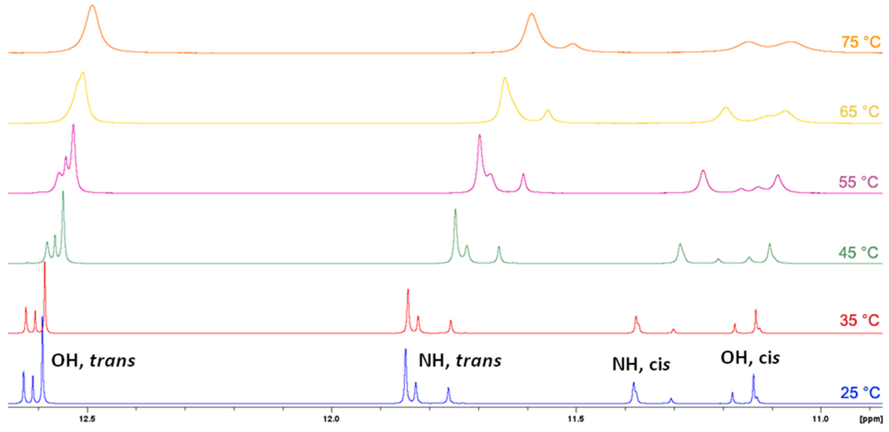
  51. In principle, E/Z isomerisation can also occur in hydrazones. However, this process usually proceeds under the influence of external stimuli, e.g., UV irradiation, and it is crucial that structure contains functionality that can stabilize generally less stable Z isomer through e.g., intramolecular hydrogen bond. For example see references 1, 5, 14 and 20. For the structures investigated here, the observed chemical shifts do not suggest the occurrence of Z isomer in solution. Also considering the structure of molecules, occurrence of Z isomer here should be disfavored due to the sterical hindrance it should sustain.
Scheme 1. The succinyl- and adipoyl-based dihydrazones.
Crystals 12 01175 sch001
Figure 1. Comparison of TG curves for dihydrazones on the common scale. Dihydrazones derived from succinic dihydrazide show significantly lower thermal stability.
Crystals 12 01175 g001
Figure 2. Molecular structures of: (a) H4L4·2H2O, (b) H4L6, and (c) H4L7 with the atom numbering schemes. Displacement ellipsoids are drawn at the 50% probability level. Hydrogen atoms are presented as spheres of arbitrarily small radii.
Crystals 12 01175 g002
Figure 3. Crystal packing in: (a) H4L7 viewed down the c-axis, (b) H4L6 viewed down the b-axis, and (c) H4L4·2H2O viewed down the a-axis.
Crystals 12 01175 g003
Figure 4. A portion of 1H NMR spectra of H4L2 in DMSO-d6 solution at different temperatures.
Crystals 12 01175 g004
Table 1. The IC50 values and minimum inhibitory concentrations (MIC) of the dihydrazones.
CompoundIC50/µmol L−1MIC (μg mL−1)
THP-1HepG2S. aureusE. faecalisE. coliM. catarrhalis
H4L1>100>100>25664>25616
H4L2>100>100>256161288
H4L3>100>100>256>256>256128
H4L4>100>100>256>256>256>256
H4L5>100>100>25616>256256
H4L6>100>100>256161616
H4L7>100>100>256>256>256>256
H4L8>100>100>256>256>256128
staurosporine0.3230.75----
azithromycin--280.250.25
Publisher’s Note: MDPI stays neutral with regard to jurisdictional claims in published maps and institutional affiliations.

