
Stable CsPbBr3 Nanocrystals—Decorated Nanoporous Gold for Optoelectronic Applications

 ,
,  , , , , , , and
Abstract
:1. Introduction
2. Materials and Methods
2.1. Synthesis and Purification of CsPbBr3 NCs
2.2. Synthesis of NP Au
2.2.1. Precursor Alloy Fabrication
2.2.2. NP Au Fabrication
2.3. Synthesis and Purification of CsPbBr3–Au Hybrid Structure
2.4. Characterization
3. Results and Discussion
4. Conclusions
Author Contributions
Funding
Institutional Review Board Statement
Informed Consent Statement
Data Availability Statement
Acknowledgments
Conflicts of Interest
References
- Protesescu, L.; Yakunin, S.; Bodnarchuk, M.I.; Krieg, F.; Caputo, R.; Hendon, C.H.; Yang, R.X.; Walsh, A.; Kovalenko, M.V. Nanocrystals of Cesium Lead Halide Perovskites (CsPbX 3, X = Cl, Br, and I): Novel Optoelectronic Materials Showing Bright Emission with Wide Color Gamut. Nano Lett. 2015, 15, 3692–3696. [Google Scholar] [CrossRef] [PubMed] [Green Version]
- Shaikh, J.S.; Shaikh, N.S.; Mali, S.S.; Patil, J.V.; Beknalkar, S.A.; Patil, A.P.; Tarwal, N.L.; Kanjanaboos, P.; Hong, C.K.; Patil, P.S. Quantum Dot Based Solar Cells: Role of Nanoarchitectures, Perovskite Quantum Dots, and Charge-Transporting Layers. ChemSusChem 2019, 12, 4724–4753. [Google Scholar] [CrossRef] [PubMed]
- Zhang, J.; Zhang, W.; Cheng, H.-M.; Silva, S.R.P. Critical review of recent progress of flexible perovskite solar cells. Mater. Today 2020, 39, 66–88. [Google Scholar] [CrossRef]
- Zhou, Y.; Zhao, Y. Chemical stability and instability of inorganic halide perovskites. Energy Environ. Sci. 2019, 12, 1495–1511. [Google Scholar] [CrossRef]
- Satta, J.; Casu, A.; Chiriu, D.; Carbonaro, C.M.; Stagi, L.; Ricci, P.C. Formation Mechanisms and Phase Stability of Solid-State Grown CsPbI3 Perovskites. Nanomaterials 2021, 11, 1823. [Google Scholar] [CrossRef]
- Cho, H.; Kim, Y.-H.; Wolf, C.; Lee, H.-D.; Lee, T.-W. Improving the Stability of Metal Halide Perovskite Materials and Light-Emitting Diodes. Adv. Mater. 2018, 30, 1704587. [Google Scholar] [CrossRef]
- Satta, J.; Melis, C.; Carbonaro, C.M.; Pinna, A.; Salado, M.; Salazar, D.; Ricci, P.C. Raman spectra and vibrational analysis of CsPbI3: A fast and reliable technique to identify lead halide perovskite polymorphs. J. Mater. 2021, 7, 127–135. [Google Scholar] [CrossRef]
- Dimitrijević, N.M.; Rajh, T.; Ahrenkiel, S.P.; Nedeljković, J.M.; Mićić, O.I.; Nozik, A.J. Charge Separation in Heterostructures of InP Nanocrystals with Metal Particles. J. Phys. Chem. B 2005, 109, 18243–18249. [Google Scholar] [CrossRef]
- Sheldon, M.T.; Trudeau, P.-E.; Mokari, T.; Wang, L.-W.; Alivisatos, A.P. Enhanced Semiconductor Nanocrystal Conductance via Solution Grown Contacts. Nano Lett. 2009, 9, 3676–3682. [Google Scholar] [CrossRef] [Green Version]
- Nawrot, K.C.; Wawrzyńczyk, D.; Bezkrovnyi, O.; Kępiński, L.; Cichy, B.; Samoć, M.; Nyk, M. Functional CdS-Au Nanocomposite for Efficient Photocatalytic, Photosensitizing, and Two-Photon Applications. Nanomaterials 2020, 10, 715. [Google Scholar] [CrossRef]
- Wei, H.; Ratchford, D.; Li, X.; Xu, H.; Shih, C.-K. Propagating Surface Plasmon Induced Photon Emission from Quantum Dots. Nano Lett. 2009, 9, 4168–4171. [Google Scholar] [CrossRef] [PubMed]
- Dey, S.; Zhao, J. Plasmonic Effect on Exciton and Multiexciton Emission of Single Quantum Dots. J. Phys. Chem. Lett. 2016, 7, 2921–2929. [Google Scholar] [CrossRef] [PubMed]
- Carretero-Palacios, S.; Jiménez-Solano, A.; Míguez, H. Plasmonic Nanoparticles as Light-Harvesting Enhancers in Perovskite Solar Cells: A User’s Guide. ACS Energy Lett. 2016, 1, 323–331. [Google Scholar] [CrossRef] [Green Version]
- Liao, J.-F.; Cai, Y.-T.; Li, J.-Y.; Jiang, Y.; Wang, X.-D.; Chen, H.-Y.; Kuang, D.-B. Plasmonic CsPbBr3–Au nanocomposite for excitation wavelength dependent photocatalytic CO2 reduction. J. Energy Chem. 2021, 53, 309–315. [Google Scholar] [CrossRef]
- Zhou, X.; Bao, C.; Li, F.; Gao, H.; Yu, T.; Yang, J.; Zhu, W.; Zou, Z. Hole-transport-material-free perovskite solar cells based on nanoporous gold back electrode. RSC Adv. 2015, 5, 58543–58548. [Google Scholar] [CrossRef]
- Ding, Y.; Zhang, Z. Introduction to nanoporous metals. In Nanoporous Metals for Advanced Energy Technologies; Springer International Publishing: Cham, Switzerland, 2016; pp. 1–35. [Google Scholar]
- Law, M.; Greene, L.E.; Johnson, J.C.; Saykally, R.; Yang, P. Nanowire dye-sensitized solar cells. Nat. Mater. 2005, 4, 455–459. [Google Scholar] [CrossRef]
- Ye, M.; Xin, X.; Lin, C.; Lin, Z. High efficiency dye-sensitized solar cells based on hierarchically structured nanotubes. Nano Lett. 2011, 11, 3214–3220. [Google Scholar] [CrossRef]
- Zhang, W.; Zhao, Y.; He, K.; Luo, J.; Li, G.; Liu, R.; Liu, S.; Cao, Z. Ultrathin nanoporous metal—semiconductor heterojunction photoanodes for visible light hydrogen evolution. Nano Res. 2018, 11, 2046–2057. [Google Scholar] [CrossRef]
- Zielasek, V.; Jürgens, B.; Schulz, C.; Biener, J.; Biener, M.M.; Hamza, A.V.; Bäumer, M. Gold Catalysts: Nanoporous Gold Foams. Angew. Chemie Int. Ed. 2006, 45, 8241–8244. [Google Scholar] [CrossRef]
- Ruffino, F.; Grimaldi, M.G. Nanoporous Gold-Based Sensing. Coatings 2020, 10, 899. [Google Scholar] [CrossRef]
- Kucheyev, S.O.; Hayes, J.R.; Biener, J.; Huser, T.; Talley, C.E.; Hamza, A.V. Surface-enhanced Raman scattering on nanoporous Au. Appl. Phys. Lett. 2006, 89, 053102. [Google Scholar] [CrossRef]
- Zhang, L.; Song, Y.; Fujita, T.; Zhang, Y.; Chen, M.; Wang, T.-H. Large Enhancement of Quantum Dot Fluorescence by Highly Scalable Nanoporous Gold. Adv. Mater. 2014, 26, 1289–1294. [Google Scholar] [CrossRef] [PubMed] [Green Version]
- Biener, J.; Wittstock, A.; Zepeda-Ruiz, L.A.; Biener, M.M.; Zielasek, V.; Kramer, D.; Viswanath, R.N.; Weissmüller, J.; Bäumer, M.; Hamza, A.V. Surface-chemistry-driven actuation in nanoporous gold. Nat. Mater. 2008, 8, 47–51. [Google Scholar] [CrossRef] [PubMed]
- Gonçalves, J.M.; Kumar, A.; da Silva, M.I.; Toma, H.E.; Martins, P.R.; Araki, K.; Bertotti, M.; Angnes, L. Nanoporous Gold-Based Materials for Electrochemical Energy Storage and Conversion. Energy Technol. 2021, 9, 2000927. [Google Scholar] [CrossRef]
- Jia, C.; Yin, H.; Ma, H.; Wang, R.; Ge, X.; Zhou, A.; Xu, X.; Ding, Y. Enhanced photoelectrocatalytic activity of methanol oxidation on TiO2-decorated nanoporous gold. J. Phys. Chem. C 2009, 113, 16138–16143. [Google Scholar] [CrossRef]
- Song, J.; Li, J.; Xu, L.; Li, J.; Zhang, F.; Han, B.; Shan, Q.; Zeng, H. Room-Temperature Triple-Ligand Surface Engineering Synergistically Boosts Ink Stability, Recombination Dynamics, and Charge Injection toward EQE-11.6% Perovskite QLEDs. Adv. Mater. 2018, 30, 1800764. [Google Scholar] [CrossRef]
- McCue, I.; Benn, E.; Gaskey, B.; Erlebacher, J. Dealloying and Dealloyed Materials. Annu. Rev. Mater. Res. 2016, 46, 263–286. [Google Scholar] [CrossRef]
- Pia, G.; Mascia, M.; Delogu, F. Kinetics of nanoporous Au formation by chemical dealloying. Scr. Mater. 2014, 76, 57–60. [Google Scholar] [CrossRef]
- Lutterotti, L. Total pattern fitting for the combined size–strain–stress–texture determination in thin film diffraction. Nucl. Instrum. Methods Phys. Res. Sect. B Beam Interact. Mater. At. 2010, 268, 334–340. [Google Scholar] [CrossRef]
- Qin, Z.; Dai, S.; Hadjiev, V.G.; Wang, C.; Xie, L.; Ni, Y.; Wu, C.; Yang, G.; Chen, S.; Deng, L.; et al. Revealing the Origin of Luminescence Center in 0D Cs 4 PbBr 6 Perovskite. Chem. Mater. 2019, 31, 9098–9104. [Google Scholar] [CrossRef] [Green Version]
- Yaffe, O.; Guo, Y.; Tan, L.Z.; Egger, D.A.; Hull, T.; Stoumpos, C.C.; Zheng, F.; Heinz, T.F.; Kronik, L.; Kanatzidis, M.G.; et al. Local Polar Fluctuations in Lead Halide Perovskite Crystals. Phys. Rev. Lett. 2017, 118, 136001. [Google Scholar] [CrossRef] [PubMed]
- Liu, M.; Zhao, J.; Luo, Z.; Sun, Z.; Pan, N.; Ding, H.; Wang, X. Unveiling Solvent-Related Effect on Phase Transformations in CsBr–PbBr 2 System: Coordination and Ratio of Precursors. Chem. Mater. 2018, 30, 5846–5852. [Google Scholar] [CrossRef]
- Xie, L.-Q.; Zhang, T.-Y.; Chen, L.; Guo, N.; Wang, Y.; Liu, G.-K.; Wang, J.-R.; Zhou, J.-Z.; Yan, J.-W.; Zhao, Y.-X.; et al. Organic–inorganic interactions of single crystalline organolead halide perovskites studied by Raman spectroscopy. Phys. Chem. Chem. Phys. 2016, 18, 18112–18118. [Google Scholar] [CrossRef] [PubMed]
- Balakrishnan, S.K.; Kamat, P.V. Au–CsPbBr 3 Hybrid Architecture: Anchoring Gold Nanoparticles on Cubic Perovskite Nanocrystals. ACS Energy Lett. 2017, 2, 88–93. [Google Scholar] [CrossRef]
- Xiao, J.; Qi, L. Surfactant-assisted, shape-controlled synthesis of gold nanocrystals. Nanoscale 2011, 3, 1383. [Google Scholar] [CrossRef]
- Millstone, J.E.; Hurst, S.J.; Métraux, G.S.; Cutler, J.I.; Mirkin, C.A. Colloidal Gold and Silver Triangular Nanoprisms. Small 2009, 5, 646–664. [Google Scholar] [CrossRef]
- Muniba; Naz, G.; Anjum, M.N.; Irfan, M.; Irfan, M.; Arshad, M.; Bajwa, S.Z.; Khan, W.S. Quats stabilized gold nanospheres for efficient ligand exchange procedure. Results Mater. 2020, 5, 100065. [Google Scholar] [CrossRef]
- Lakowicz, J.R. Principles of Fluorescence Spectroscopy; Lakowicz, J.R., Ed.; Springer: Boston, MA, USA, 2006; ISBN 978-0-387-31278-1. [Google Scholar]
- Luo, B.; Pu, Y.C.; Yang, Y.; Lindley, S.A.; Abdelmageed, G.; Ashry, H.; Li, Y.; Li, X.; Zhang, J.Z. Synthesis, Optical Properties, and Exciton Dynamics of Organolead Bromide Perovskite Nanocrystals. J. Phys. Chem. C 2015, 119, 26672–26682. [Google Scholar] [CrossRef]
- Wu, K.; Liang, G.; Shang, Q.; Ren, Y.; Kong, D.; Lian, T. Ultrafast interfacial electron and hole transfer from CsPbBr3 perovskite quantum dots. J. Am. Chem. Soc. 2015, 137. [Google Scholar] [CrossRef]
- Wu, Y.; Han, Q.; Wang, M.; Juan, F.; Hou, G.; Xun, F.; Wei, H.; Xu, J.; Cao, B. Enhanced photoluminescence of CsPbBr 3-x I x nanocrystals via plasmonic Au nanoarrays. Opt. Express 2021, 29, 36988–36996. [Google Scholar] [CrossRef]
- Chen, S.; Lyu, D.; Ling, T.; Guo, W. Reversible modulation of CsPbBr3 perovskite nanocrystal/gold nanoparticle heterostructures. Chem. Commun. 2018, 54, 4605–4608. [Google Scholar] [CrossRef] [PubMed]


| Element | Line Type | Atom % | Atom % Error |
|---|---|---|---|
| C K | K | 41.9 | 0.9 |
| N K | K | 23.2 | 2.6 |
| O K | K | 14.0 | 1.4 |
| Ag L | L | 1.0 | 0.1 |
| Cs L | L | 0.5 | 0.1 |
| Au L | L | 18.9 | 0.2 |
| Pb L | L | 0.5 | 0.1 |
| 100.0 |
| Element | Line Type | Atom % | Atom % Error |
|---|---|---|---|
| C K | K | 37.4 | 0.8 |
| N K | K | 19.9 | 2.1 |
| O K | K | 15.8 | 1.3 |
| Br K | K | 3.3 | 0.2 |
| Ag L | L | 4.9 | 0.1 |
| Cs L | L | 1.1 | 0.1 |
| Au L | L | 16.7 | 0.2 |
| Pb L | L | 0.9 | 0.1 |
| 100.0 |
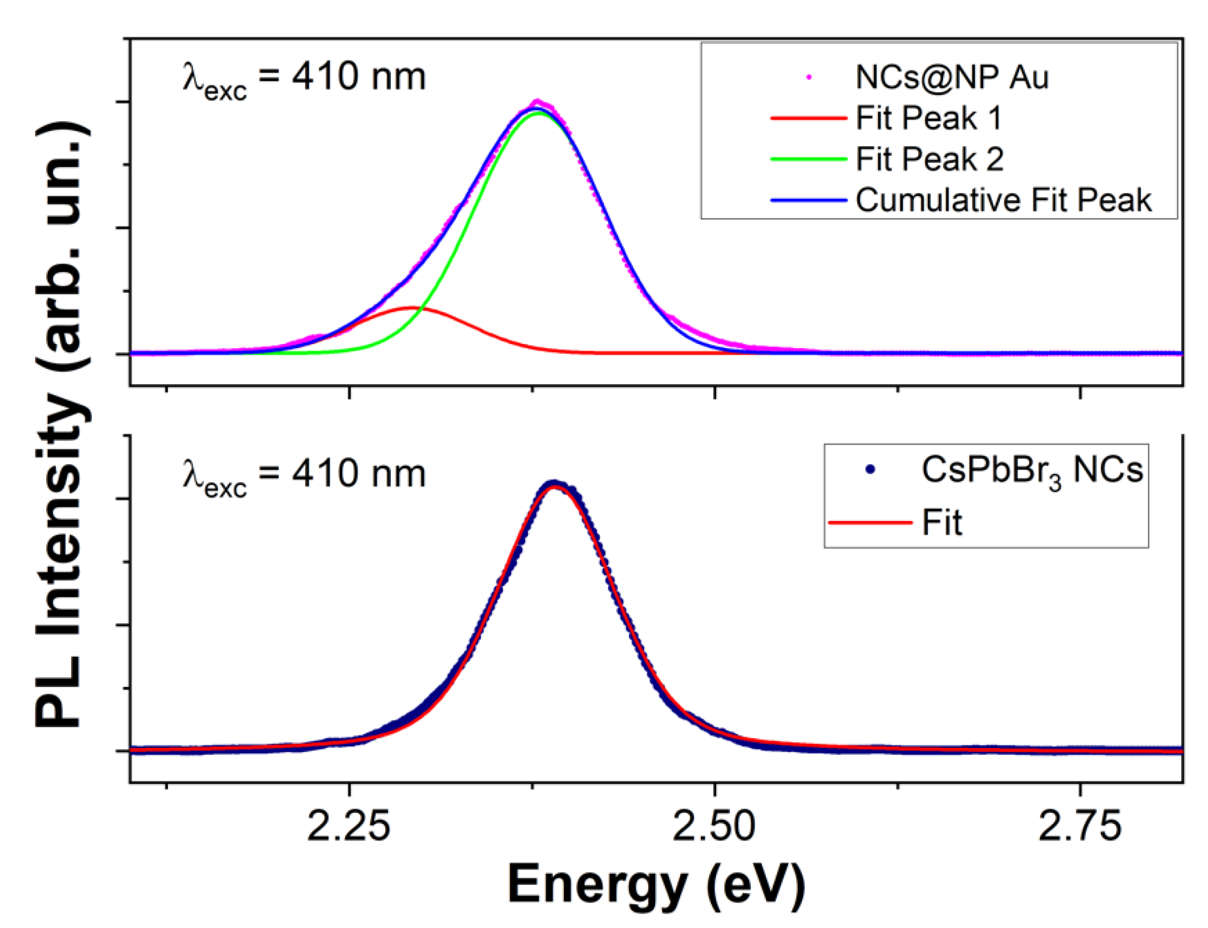
| Sample | Peak | xc [eV] | w [eV] |
|---|---|---|---|
| CsPbBr3 NCs | 2.39 | 0.097 | |
| NCs@NP Au | Peak 1 | 2.37 | 0.088 |
| Peak 2 | 2.29 | 0.081 | |
| Sample | [ns] | A1 | τ1 [ns] | A2 | τ2 [ns] |
|---|---|---|---|---|---|
| CsPbBr3 NCs | 3.6 (8) | 1445 (1) | 0.9 (6) | 144 (1) | 7.0 (6) |
| NCs@NP Au | 1.9 (3) | 1760 (1) | 0.9 (6) | 140 (1) | 4.4 (6) |
Publisher’s Note: MDPI stays neutral with regard to jurisdictional claims in published maps and institutional affiliations. |
© 2022 by the authors. Licensee MDPI, Basel, Switzerland. This article is an open access article distributed under the terms and conditions of the Creative Commons Attribution (CC BY) license (https://creativecommons.org/licenses/by/4.0/).
Share and Cite
Satta, J.; Pinna, A.; Pia, G.; Pilia, L.; Carbonaro, C.M.; Chiriu, D.; Stagi, L.; Abdullah, Q.A.; Ricci, P.C. Stable CsPbBr3 Nanocrystals—Decorated Nanoporous Gold for Optoelectronic Applications. Crystals 2022, 12, 863. https://doi.org/10.3390/cryst12060863
Satta J, Pinna A, Pia G, Pilia L, Carbonaro CM, Chiriu D, Stagi L, Abdullah QA, Ricci PC. Stable CsPbBr3 Nanocrystals—Decorated Nanoporous Gold for Optoelectronic Applications. Crystals. 2022; 12(6):863. https://doi.org/10.3390/cryst12060863
Chicago/Turabian StyleSatta, Jessica, Andrea Pinna, Giorgio Pia, Luca Pilia, Carlo Maria Carbonaro, Daniele Chiriu, Luigi Stagi, Qader Abdulqader Abdullah, and Pier Carlo Ricci. 2022. "Stable CsPbBr3 Nanocrystals—Decorated Nanoporous Gold for Optoelectronic Applications" Crystals 12, no. 6: 863. https://doi.org/10.3390/cryst12060863