Article

Molecular Self-Assembly of an Unusual Dinuclear Ruthenium(III) Complex Based on the Nucleobase Guanine

by
Marta Orts-Arroyo
,
Adriana Silvestre-Llora
,
Isabel Castro
and
José Martínez-Lillo
*
Instituto de Ciencia Molecular (ICMol), Universitat de València, c/Catedrático José Beltrán 2, Paterna, 46980 València, Spain
*
Author to whom correspondence should be addressed.
Crystals 2022, 12(4), 448; https://doi.org/10.3390/cryst12040448
Submission received: 7 March 2022 / Revised: 17 March 2022 / Accepted: 18 March 2022 / Published: 23 March 2022
(This article belongs to the Special Issue Self-Assembled Complexes: “Love at First Sight”)

Abstract

:
The study of crystal structures based on complexes containing purine nucleobases is a significant research subject, mainly regarding the diagnosis and treatment of some diseases and the investigation of genetic mutations and biochemical structures in life sciences. We have obtained and characterized a new dinuclear ruthenium(III) complex based on guanine with the formula [{Ru(µ-Cl)(µ-gua)}2Cl4]·2H2O (1) (gua = guanine). 1 was characterized by means of Fourier transform infrared spectroscopy (FT–IR), scanning electron microscopy and energy dispersive X-ray analysis (SEM–EDX), single-crystal X-ray diffraction (XRD), Hirshfeld surface analysis and cyclic voltammetry (CV). The study of its electrochemical properties allowed us to investigate the presence of guanine molecules when linked to the ruthenium(III) ion in 1. The well-resolved voltammetric response together with the reliability and stability achieved through 1 could provide a step forward to developing new ruthenium-based platforms, devices and modified electrodes adequate to study this purine nucleobase.

1. Introduction

Guanine is one of the main nitrogenous bases found in the nucleotides of the nucleic acids (DNA and RNA) present in the cells of living organisms and viruses. Besides being involved in the storage and the expression of genetic information, it participates in many other cellular biochemical processes and structures [1,2,3]. Given that this purine nucleobase can be present in the fluids of human beings, their levels have been proposed as direct and indirect biomarkers for a variety of diseases and pathologies, such as Alzheimer’s disease [4], epilepsy [5] and human immunodeficiency virus (HIV) infection [6,7]. Hence, sensitive and selective methods for determining both this and other purine nucleobases are being investigated in several research areas [8,9].
Ruthenium complexes have undergone significant development in different research areas for the last 20 years [10], displaying a wide variety of technological applications, and ranging from catalysis to anticancer drugs [11,12,13,14,15,16,17,18]. Following our ongoing investigation into biomolecule-based complexes [19,20,21,22,23,24,25,26,27], we have focused also on the purine nucleobase guanine (Scheme 1). There exists only one guanine-containing Ru(III) complex, of the formula trans-[RuCl4(Hgua)(DMSO)]·2H2O (Hgua = protonated guanine), which was reported in 2004 [28]. This compound was characterized by single-crystal X-ray diffraction and its in vitro antitumor activity was also evaluated, showing a mild antiproliferative effect but an interesting proadhesive effect [28]. Herein, we report the synthesis and characterization of a new guanine-bridged diruthenium(III) complex, of formula [{Ru(µ-Cl)(µ-gua)}2Cl4]·2H2O (1) (gua = guanine). To our knowledge, 1 is the first dinuclear ruthenium(III) compound based on guanine which has been reported so far.

2. Materials and Methods

2.1. Reagents and Instruments

Guanine (99.90%) was purchased from Acros Organics and the ruthenium precursor K2[RuCl5(H2O)] (99.95%) was acquired from Alfa Aesar. The elemental analyses (C, H, N) and X-ray microanalysis were performed through Central Service for the Support to Experimental Research (SCSIE) at the University of Valencia. Images and results of scanning electron microscopy (SEM-EDX) were obtained through a Hitachi S-4800 field emission scanning electron microscope, with 20 kV and 9.0 mm of accelerating voltage and working distance, respectively. Each infrared spectrum (FT-IR) was performed on KBr pellets and was obtained through a PerkinElmer Spectrum 65 FT-IR spectrometer in the 4000–500 range (cm−1) with 25 scans and a spectral resolution of 4 cm−1. The study of the electrochemical properties was carried out on 1 by means of an Autolab/PGSTAT 204 scanning potentiostat at a scan rate working in the 10–250 range (mVs−1) and with Metrohm electrodes. CV curves were obtained with a 0.1 M (NBu4)[PF6] solution, as supporting electrolyte, and a 10−3 M solution of 1 in dry N,N’-dimethylformamide (DMF). A glassy carbon disk of 0.32 cm2 was used as working electrode, polished with 1.0 μm diamond powder, which was sonicated and then washed with absolute ethanol and acetone, and air-dried. The AgCl/Ag reference electrode was separated from the test solution through a salt bridge with the solvent and supporting electrolyte. The platinum electrode was auxiliary. The study was performed in a standard electrochemical cell at 20 °C with flowing argon. The studied potential range was from −1.5 to +1.5 V vs. AgCl/Ag. Ferrocene (Fc) was added as an internal standard at the end of the measurements.

2.2. Preparation

Synthesis of [{Ru(µ-Cl)(µ-gua)}2Cl4]·2H2O (1)

K2[RuCl5(H2O)] (11.2 mg, 0.03 mmol) and guanine (6.80 mg, 0.03 mmol) were reacted through a solvothermal synthesis in HCl (4 mL, 3.0 M) at 90 °C for three days, then a 12 h cooling process took place to room temperature. Dark brown parallelepipeds of 1 were obtained. Yield: ca. 53%. Anal. Calcd. for C10H10Cl6N10O2Ru2 (1): C, 16.8; H, 1.4; N, 19.5. Found: C, 16.7; H, 1.7; N, 19.2. SEM-EDX: a molar ratio of 1:3 for Ru/Cl was found for 1. IR peaks (KBr pellets): 3419(s), 3325(m), 3208(m), 3114(m), 3062(m), 3012(m), 2922(m), 2853(m), 1708(vs), 1635(vs), 1594(m), 1559(m), 1540(s), 1506(w), 1457(m), 1394(s), 1270(w), 1210(m), 1124(w), 1095(w), 983(w), 873(m), 768(m), 704(w), 612(w), 573(w), 474(w) cm−1.

2.3. X-ray Diffraction Data Collection and Structure Refinement

Data collection from a single crystal of 1 (with dimensions of 0.46 × 0.13 × 0.09 mm3) was performed on a Bruker D8 Venture diffractometer with graphite-monochromated Mo–Kα radiation (λ = 0.71073 Å). Table 1 summarizes the crystal parameters and refinement results for 1. The structure of 1 was solved by standard direct methods and then completed by Fourier recycling through the SHELXTL program. The model was refined with version 2018/1 of SHELXL against F2 on all data by full-matrix least-squares [29]. The non-hydrogen atoms were anisotropically refined, and the H atoms of the guanine molecules were set in calculated positions and refined isotropically. The H atoms of the water molecules in 1 were neither detected nor included in the model, this fact being due to the thermal disorder observed on the water molecules, which increases the R1 and wR2 parameters in this system (Table 1). The graphic manipulations were performed with the DIAMOND program [30] of CCDC 2081706.

3. Results and Discussion

3.1. Synthetic Procedure

The ruthenium(III) precursor K2[RuCl5(H2O)] was made to react with guanine in hydrochloric acid (3.0 M) solutions, thus we prepared a new purine-based ruthenium(III) complex of the formula [{Ru(µ-Cl)(µ-gua)}2Cl4]·2H2O (1) (Figure 1). The synthetic procedure was carried out by means of heating this mixture at 90 °C in a solvothermal reaction and, in order to crystallize, the process continued for a further 12 h cooling step to room temperature. In this way, dark brown crystals of 1 were obtained with a satisfactory yield (53%). It is worth mentioning that compound 1 was also obtained by replacing hydrochloric acid with acetic acid, but the yield was too low (less than 10%). These results support the fact that this is an adequate synthesis to prepare purine-based dinuclear ruthenium(III) complexes.

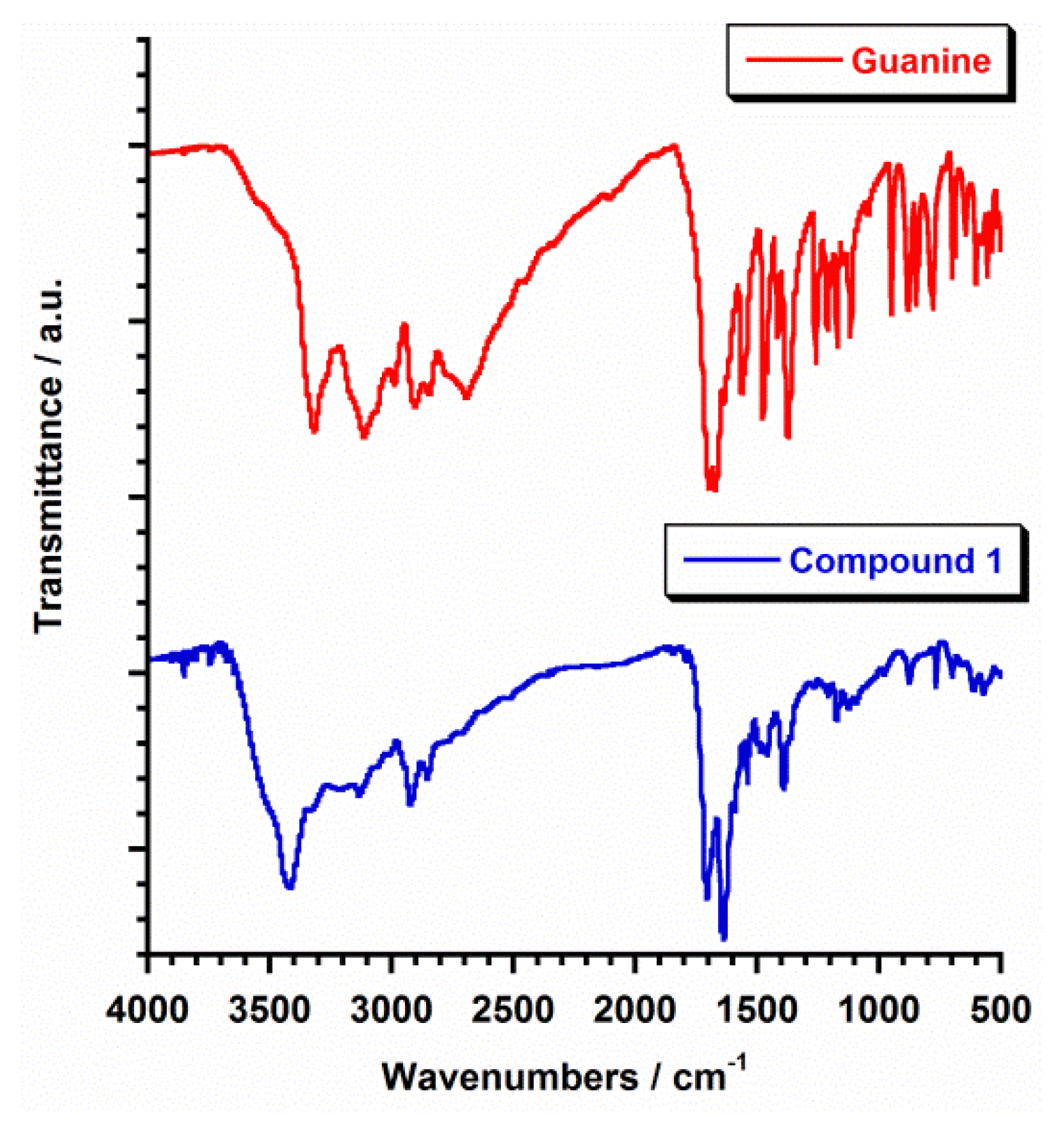
3.2. Infrared Spectroscopy

The infrared (IR) spectra of 1 and that of the free guanine ligand are given in Figure 2. The IR spectrum of guanine has been previously studied [31,32], so that it has been added in this work only for comparison. In general, the vibrational bands mainly associated with N-H symmetric (νs) and asymmetric (νas) stretching for the free guanine molecule were more intense and complex than those of 1 in the ca. 3800–2000 cm−1 region (Figure 2), which would be due to a more ordered hydrogen-bonding network in the crystalline solid for the free nucleobase [31,32]. In the case of the IR spectrum of 1, the values associated to the vibrational νas(NH2) (3325 cm−1) and νs(NH2) (3114 and 3062 cm−1) bands were very similar to those obtained for the free guanine [31,32] (Figure 2). In the 1800–500 cm−1 region, the most interesting features were the two strong vibrational bands associated to the stretching ν(C=N) and ν(C=C) and bending δ(NH2), mainly scissoring, which were found at 1695 and 1672 cm−1 for the guanine molecule [31,32]. In the IR spectrum of 1, these two vibrational bands were shifted to 1708 and 1635 cm−1, respectively, with this fact indicating the coordination of the Ru(III) metal ions to guanine molecules in compound 1 (Figure 2).

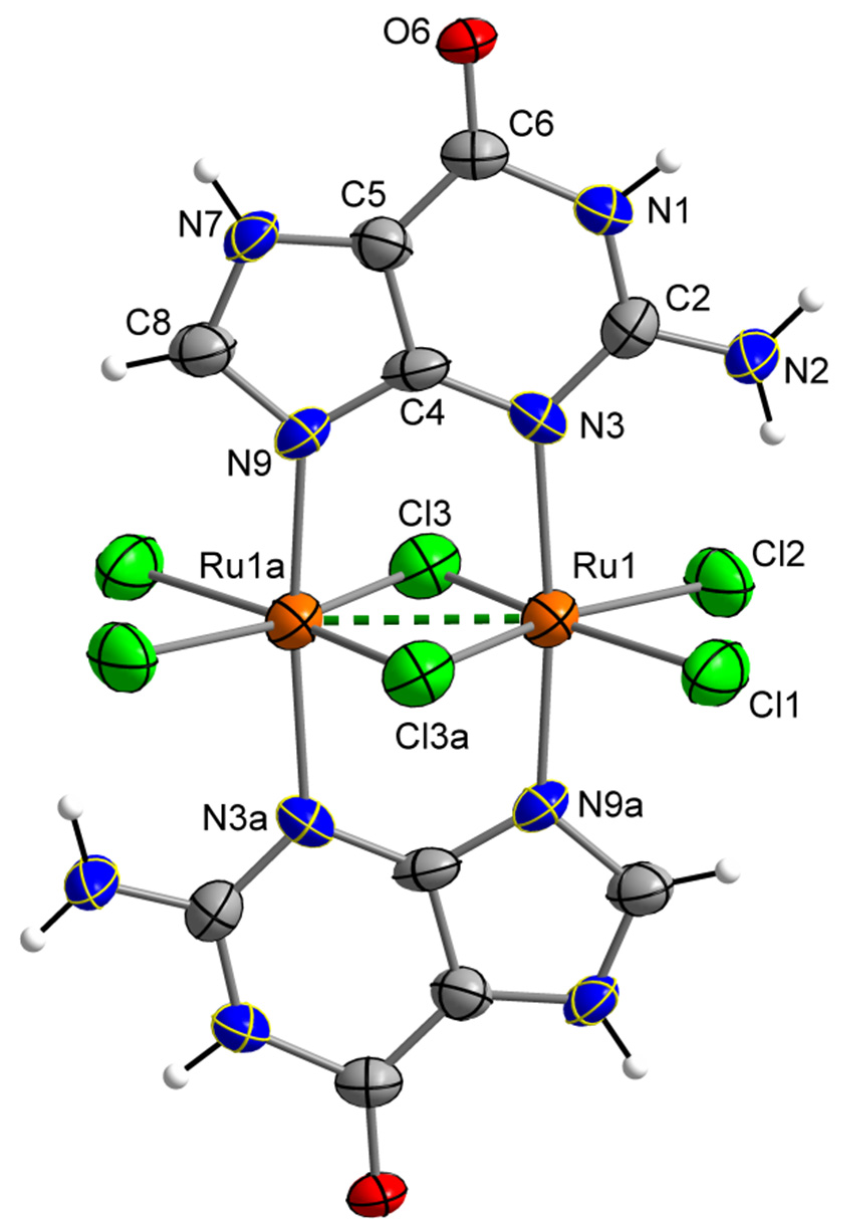
3.3. Description of the Crystal Structure

The crystal structure of 1 was obtained by single-crystal X-ray diffraction. A CSD survey revealed that 1 displays the first reported crystal structure based on a dinuclear Ru(III) compound containing guanine. Compound 1 crystallizes in the monoclinic system with space group C2/c (Table 1). The crystal structure of 1 is made up of neutral [{Ru(µ-Cl)(µ-gua)}2Cl4] units and H2O molecules. The asymmetric unit of 1 consists of half a [{Ru(µ-Cl)(µ-gua)}2Cl4] complex and one H2O molecule (Figure 1).
In this dinuclear complex, each Ru(III) ion is linked to four chloride ions and two nitrogen atoms (these are N3 and N9) from two guanine molecules in an almost regular Oh geometry. The two Ru(III) ions are connected to each other through two guanine molecules and two chloro-bridges (Figure 1). A very short intramolecular Ru···Ru distance [Ru(1)···Ru(1a) = 2.644 Å, (a) = −x−1/2, −y + 1/2, −z−2] indicates the formation of a metal–metal bond (dashed line in Figure 1). The average values of the Ru–Cl [2.318(1) Å] and Ru–N [2.103(1) Å] bond lengths are in agreement with those values published for previously reported Ru(III) systems with a similar metal environment [17,25].
In the crystal of 1, H-bonding interactions between neighboring guanine molecules [O(6)⋯N(7b) distance of ca. 2.745(1) Å, (b) = −x, −y, −z−1] afford chains of neutral [{Ru(µ-Cl)(µ-gua)}2Cl4] units (Figure 3). These chains are linked by additional H-bonds involving Cl anions and NH2 groups of adjacent guanine molecules [Cl(3)···N(2c) distance of ca. 3.345(1) Å, (c) = −x−1/2, y−1/2, −z−3/2], which set up a two-dimensional network. The cohesiveness of the structure is further strengthened by the presence of intermolecular Cl···π interactions among [{Ru(µ-Cl)(µ-gua)}2Cl4] units [Cl···π interactions ranging with 3.52–3.81 Å values]. The thus packed [{Ru(µ-Cl)(µ-gua)}2Cl4] units generate cavities with a diameter of ca. 13 Å, where most of the water molecules are located in 1 (Figure 4). The supramolecular network is supported by additional H-bonding interactions, which stabilize the crystal structure in compound 1.

3.4. Hirshfeld Surface Analysis

The intermolecular interactions of the neutral [{Ru(µ-Cl)(µ-gua)}2Cl4] complex were further studied through the CrystalExplorer program [33,34]. This program calculated the surfaces which allowed us the qualitative and quantitative investigation as well as the visualization of the main intermolecular contacts in 1 by mapping the distances from the surface to the nearest atom outside (de) and inside (di) this surface. Besides, a normalized contact distance called dnorm was also taken into account to overcome some limitations generated by the atom size [33,34]. The Hirshfeld surfaces for complex 1 are given in Figure 5, the shorter contacts being shown with red color [34]. The intermolecular H···O contacts generated among the N-H and carbonyl groups of adjacent guanine molecules are approximately 16% of the complete fingerprint plot (Figure 5). The Cl···H contacts involving Cl anions and N-H groups of neighboring dinuclear [{Ru(µ-Cl)(µ-gua)}2Cl4] units are the main interactions observed on the Hirshfeld surface, which covers ca. 24% (Figure 5). Finally, further O···H contacts involving solvent water molecules and N-H groups of the guanine molecules are close to the 6% of the fingerprint plot (Figure 5).

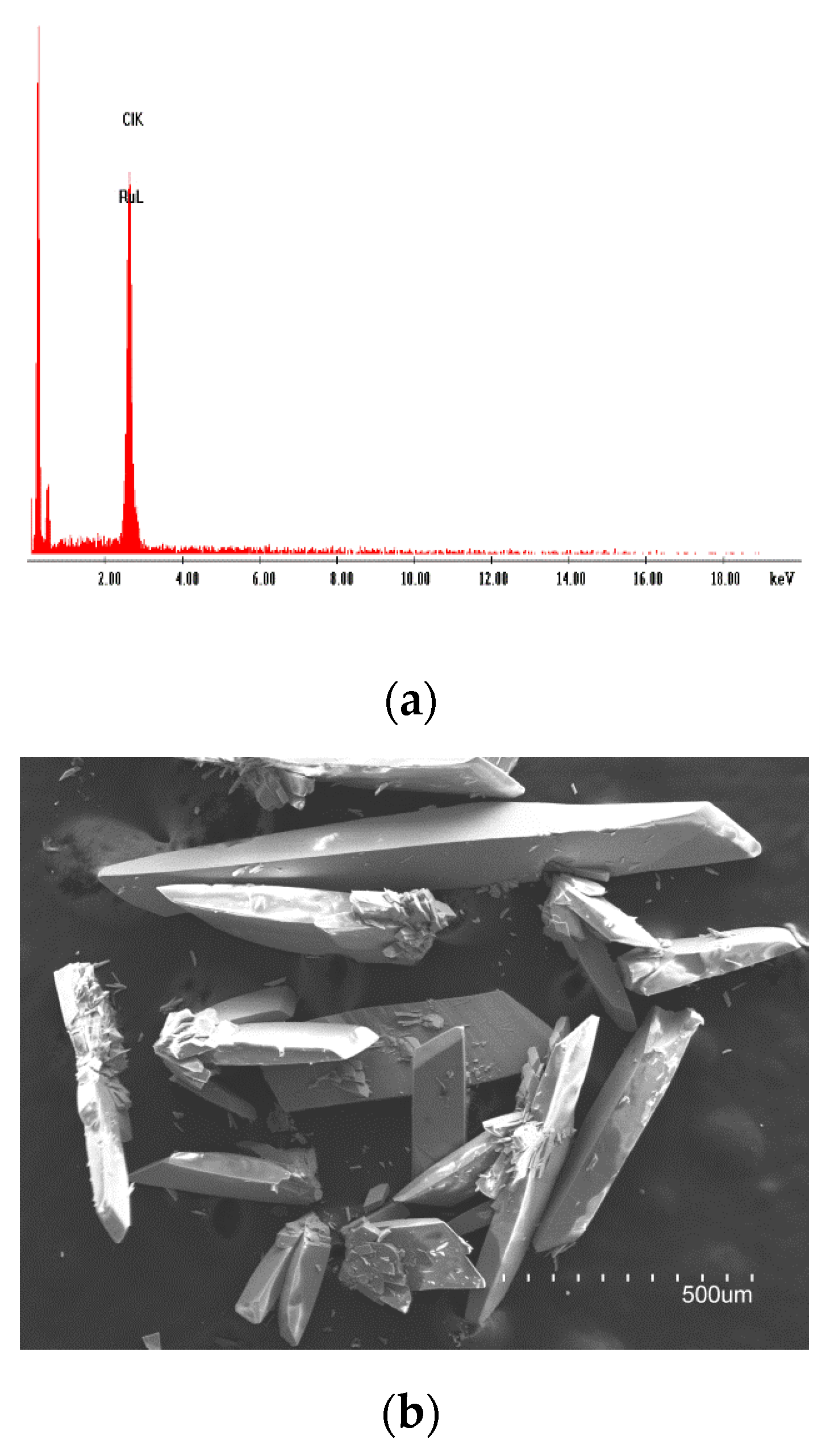
3.5. Scanning Electron Microscopy–Energy Dispersive X-ray Analysis

Compound 1 was studied by means of scanning electron microscopy and energy dispersive X-ray analysis (SEM-EDX), these analyses being carried out as previously performed for other ruthenium systems [35,36]. The results of the microanalysis gave a Ru/Cl molar ratio of 1:3 for 1. A recorded image of 1 is given in Figure 6. Crystals of 1 are shown as crystallized parallelepipeds in Figure 6.

3.6. Cyclic Voltammetry (CV)

The study of the electrochemical properties of compound 1 was performed employing cyclic voltammetry (CV) in N,N’-dimethylformamide (DMF), containing 0.1 M [NBu4][PF6], in the range of potential values of +1.5 and −1.5 V and at 20 °C. The CV curve for 1 is shown in Figure 7.
Dinuclear ruthenium compounds containing metal–metal bonds have been studied for many years [37]. They show redox properties that are strongly dependent upon the solvent and the supporting electrolyte and are generally well known [38,39,40]. So, this type of Ru–Ru compound could be an acceptable reference in electrochemical studies [38,39,40]. In the CV curve of 1, three reduction processes can be observed (Figure 7). The first two reduction peaks were found between 0.0 V and −0.50 V, which would be associated with the formation of the mixed-valent Ru(II)-Ru(III) species (at −0.30 V) and also to that of the Ru(II)-Ru(II) system (at −0.50 V). These reduction potential values are close to those published for other dinuclear ruthenium complexes [38,39,40]. Nevertheless, much more interesting would be the third detected reduction peak at −0.89 V, which would be generated by the influence of the guanine molecule, see inset in Figure 7. It is worth mentioning that this value assigned to guanine is in agreement with those previously reported for this purine nucleobase in electrochemistry research works performed through modified electrodes based on polyaniline-MnO2 [41] and graphite-WS2 [42], which have shown high accuracy and promising redox activity toward purine nucleobases [41,42]. In order to analyze the repeatability of the CV curve, it was measured five times. Indeed, a relative standard deviation value of ca. 1.2% for the current response was obtained for 1. In any case, these results could establish a first step to develop new sensor devices suitable for the detection of purine nucleobases as guanine [25]. Nevertheless, this is an early stage of the research and, therefore, this type of diruthenium(III) systems must be further investigated. The comparison with other methods together with the study of the nature of the samples to be measured will be addressed subsequently in future works [25].

4. Conclusions

An unusual guanine-based dinuclear ruthenium(III) complex, of the formula [{Ru(µ-Cl)(µ-gua)}2Cl4]·2H2O (1) (gua = guanine), was prepared and characterized. Compound 1 is the first dinuclear ruthenium(III) compound based on guanine reported so far. Compound 1 was characterized by FT–IR, SEM–EDX, single-crystal X-ray diffraction (XRD), Hirshfeld surface analysis and cyclic voltammetry (CV). The study of its electrochemical properties revealed well-resolved, potentially useful, current peaks, which allowed us to investigate the guanine when linked to the ruthenium ion in 1.

Supplementary Materials

The following supporting information can be downloaded at: https://www.mdpi.com/article/10.3390/cryst12040448/s1, X-ray crystallographic data in CIF format for compound 1.

Author Contributions

Conceptualization, I.C. and J.M.-L.; funding acquisition, I.C. and J.M.-L.; methodology, M.O.-A., A.S.-L., I.C. and J.M.-L.; investigation, M.O.-A., A.S.-L., I.C. and J.M.-L.; formal analysis, M.O.-A., A.S.-L., I.C. and J.M.-L.; writing—original draft preparation, J.M.-L.; writing—review and editing, J.M.-L. All authors have read and agreed to the published version of the manuscript.

Funding

This research was funded by Spanish Ministry of Science and Innovation (Grant number PID2019-109735GB-I00) and also by VLC-BIOCLINIC Program of the University of Valencia (Grant number PI-2021-007-DIRUGEN).

Institutional Review Board Statement

Not applicable.

Informed Consent Statement

Not applicable.

Data Availability Statement

The raw data that support the findings of this study are available from the corresponding author upon reasonable request.

Acknowledgments

M.O.-A. thanks the Spanish “FPI” PhD funding action plan.

Conflicts of Interest

The authors declare no conflict of interest.

References

  1. An, S.; Kumar, R.; Sheets, E.D.; Benkovic, S.J. Reversible Compartmentalization of de Novo Purine Biosynthetic Complexes in Living Cells. Science 2008, 320, 103–106. [Google Scholar] [CrossRef] [PubMed]
  2. Pedley, A.M.; Benkovic, S.J. A New View into the Regulation of Purine Metabolism—The Purinosome. Trends Biochem. Sci. 2017, 42, 141–154. [Google Scholar] [CrossRef] [PubMed] [Green Version]
  3. Rhodes, D.; Lipps, H.J. G-quadruplexes and their regulatory roles in biology. Nucleic Acids Res. 2015, 43, 8627–8637. [Google Scholar] [CrossRef] [Green Version]
  4. Huynh, R.A.; Mohan, C. Alzheimer’s Disease: Biomarkers in the Genome, Blood, and Cerebrospinal Fluid. Front. Neurol. 2017, 8, 102. [Google Scholar] [CrossRef] [PubMed] [Green Version]
  5. Beamer, E.; Lacey, A.; Alves, M.; Conte, G.; Tian, F.; de Diego-García, L.; Khalil, M.; Rosenow, F.; Delanty, N.; Dale, N.; et al. Elevated blood purine levels as a biomarker of seizures and epilepsy. Epilepsia 2021, 62, 817–828. [Google Scholar] [CrossRef]
  6. Kolgiri, V.; Patil, V.W. Protein carbonyl content: A novel biomarker for aging in HIV/AIDS patients. Braz. J. Infect. Dis. 2017, 21, 35–41. [Google Scholar] [CrossRef] [Green Version]
  7. Ellis, R.J.; Moore, D.J.; Sundermann, E.E.; Heaton, R.K.; Mehta, S.; Hulgan, T.; Samuels, D.; Fields, J.A.; Letendre, S.L. Nucleic acid oxidation is associated with biomarkers of neurodegeneration in CSF in people with HIV. Neurol. Neuroimmunol. Neuroinflamm. 2020, 7, e902. [Google Scholar] [CrossRef]
  8. Klampfla, C.W.; Himmelsbach, M.; Buchberger, W.; Klein, H. Determination of purines and pyrimidines in beer samples by capillary zone electrophoresis. Anal. Chim. Acta 2002, 454, 185–191. [Google Scholar] [CrossRef]
  9. Inazawa, K.; Sato, A.; Kato, Y.; Yamaoka, N.; Fukuuchi, T.; Yasuda, M.; Mawatari, K.; Nakagomi, K.; Kaneko, K. Determination and profiling of purines in foods by using HPLC and LC-MS. Nucleosides Nucleotides Nucleic Acids 2014, 33, 439–444. [Google Scholar] [CrossRef]
  10. Higgins, S. Regarding ruthenium. Nat. Chem. 2010, 2, 1100. [Google Scholar] [CrossRef]
  11. Bruneau, C.; Achard, M. Allylic ruthenium(IV) complexes in catalysis. Coord. Chem. Rev. 2012, 256, 525–536. [Google Scholar] [CrossRef]
  12. Furrer, J.; Süss-Fink, G. Thiolato-bridged dinuclear arene ruthenium complexes and their potential as anticancer drugs. Coord. Chem. Rev. 2016, 309, 36–50. [Google Scholar] [CrossRef]
  13. Zeng, L.; Gupta, P.; Chen, Y.; Wang, E.; Ji, L.; Chao, H.; Chen, Z.-S. The development of anticancer ruthenium(II) complexes: From single molecule compounds to nanomaterials. Chem. Soc. Rev. 2017, 46, 5771–5804. [Google Scholar] [CrossRef]
  14. Alessio, E. Thirty Years of the Drug Candidate NAMI-A and the Myths in the Field of Ruthenium Anticancer Compounds: A Personal Perspective. Eur. J. Inorg. Chem. 2017, 1549–1560. [Google Scholar] [CrossRef]
  15. Alessio, E.; Messori, L. NAMI-A and KP1019/1339, Two Iconic Ruthenium Anticancer Drug Candidates Face-to-Face: A Case Story in Medicinal Inorganic Chemistry. Molecules 2019, 24, 1995. [Google Scholar] [CrossRef] [Green Version]
  16. Chen, H.; Parkinson, J.A.; Parsons, S.; Coxall, R.A.; Gould, R.O.; Sadler, P.J. Organometallic Ruthenium(II) Diamine Anticancer Complexes:  Arene-Nucleobase Stacking and Stereospecific Hydrogen-Bonding in Guanine Adducts. J. Am. Chem. Soc. 2002, 124, 3064–3082. [Google Scholar] [CrossRef]
  17. Armentano, D.; Martínez-Lillo, J. Hexachlororhenate(IV) salts of ruthenium(III) cations: X-ray structure and magnetic properties. Inorg. Chim. Acta 2012, 380, 118–124. [Google Scholar] [CrossRef]
  18. Orts-Arroyo, M.; Castro, I.; Lloret, F.; Martínez-Lillo, J. Molecular Self-Assembly in a Family of Oxo-Bridged Dinuclear Ruthenium(IV) Systems. Cryst. Growth Des. 2020, 20, 2044–2056. [Google Scholar] [CrossRef]
  19. Escrivà, E.; García-Lozano, J.; Martínez-Lillo, J.; Nuñez, H.; Server-Carrió, J.; Soto, L.; Carrasco, R.; Cano, J. Synthesis, Crystal Structure, Magnetic Properties, and Theoretical Studies of [{Cu(mepirizole)Br}2(μ-OH)(μ-pz)] (Mepirizole = 4-Methoxy-2-(5-methoxy-3-methyl-1H-pyrazol-1-yl)-6-methylpyrimidine; pz = Pyrazolate), a Novel μ-Pyrazolato−μ-Hydroxo-Dibridged Copper(II) Complex. Inorg. Chem. 2003, 42, 8328–8336. [Google Scholar]
  20. Armentano, D.; Marino, N.; Mastropietro, T.F.; Martínez-Lillo, J.; Cano, J.; Julve, M.; Lloret, F.; De Munno, G. Self-Assembly of a Chiral Carbonate- and Cytidine-Containing Dodecanuclear Copper(II) Complex: A Multiarm-Supplied Globular Capsule. Inorg. Chem. 2008, 47, 10229–10231. [Google Scholar] [CrossRef]
  21. Marino, N.; Armentano, D.; Mastropietro, T.F.; Julve, M.; De Munno, G.; Martínez-Lillo, J. Cubane-Type CuII4 and MnII2MnIII2 Complexes Based on Pyridoxine: A Versatile Ligand for Metal Assembling. Inorg. Chem. 2013, 52, 11934–11943. [Google Scholar] [CrossRef] [PubMed]
  22. Armentano, D.; Barquero, M.A.; Rojas-Dotti, C.; Moliner, N.; De Munno, G.; Brechin, E.K.; Martínez-Lillo, J. Enhancement of Intermolecular Magnetic Exchange through Halogen···Halogen Interactions in Bisadeninium Rhenium(IV) Salts. Cryst. Growth Des. 2017, 17, 5342–5348. [Google Scholar] [CrossRef] [Green Version]
  23. Orts-Arroyo, M.; Castro, I.; Lloret, F.; Martínez-Lillo, J. Field-induced slow relaxation of magnetisation in two one-dimensional homometallic dysprosium(III) complexes based on alpha- and beta-amino acids. Dalton Trans. 2020, 49, 9155–9163. [Google Scholar] [CrossRef] [PubMed]
  24. Orts-Arroyo, M.; Ten-Esteve, A.; Ginés-Cárdenas, S.; Castro, I.; Martí-Bonmatí, L.; Martínez-Lillo, J. A gadolinium(III) complex based on the thymine nucleobase with properties suitable for magnetic resonance imaging. Int. J. Mol. Sci. 2021, 22, 4586. [Google Scholar] [CrossRef] [PubMed]
  25. Orts-Arroyo, M.; Castro, I.; Martínez-Lillo, J. Detection of Hypoxanthine from Inosine and Unusual Hydrolysis of Immunosuppressive Drug Azathioprine through the Formation of a Diruthenium(III) System. Biosensors 2021, 11, 19. [Google Scholar] [CrossRef] [PubMed]
  26. Sanchis-Perucho, A.; Orts-Arroyo, M.; Camús-Hernández, J.; Rojas-Dotti, C.; Escrivà, E.; Lloret, F.; Martínez-Lillo, J. Hexahalorhenate(IV) salts of protonated ciprofloxacin: Antibiotic-based single-ion magnets. CrystEngComm 2021, 23, 8579–8587. [Google Scholar] [CrossRef]
  27. Orts-Arroyo, M.; Sanchis-Perucho, A.; Moliner, N.; Castro, I.; Lloret, F.; Martínez-Lillo, J. One-Dimensional Gadolinium (III) Complexes Based on Alpha- and Beta-Amino Acids Exhibiting Field-Induced Slow Relaxation of Magnetization. Inorganics 2022, 10, 32. [Google Scholar] [CrossRef]
  28. Turel, I.; Pecanac, M.; Golobic, A.; Alessio, E.; Serli, B.; Bergamo, A.; Sava, G. Solution, solid state and biological characterization of ruthenium(III)-DMSO complexes with purine base derivatives. J. Inorg. Biochem. 2004, 98, 393–401. [Google Scholar] [CrossRef]
  29. SHELXTL-2017/1, Bruker Analytical X-ray Instruments; Bruker: Madison, WI, USA, 2017.
  30. DIAMOND 4.5.0, Crystal Impact GbR.; Crystal Impact: Bonn, Germany, 2018.
  31. Sheina, G.G.; Stepanian, S.G.; Radchenko, E.D.; Blagoi, Y.P. IR spectra of guanine and hypoxanthine isolated molecules. J. Mol. Struct. 1987, 158, 275–292. [Google Scholar] [CrossRef]
  32. Beć, K.B.; Grabska, J.; Czarnecki, M.A.; Huck, C.W.; Wójcik, M.J.; Nakajima, T.; Ozaki, Y. IR Spectra of Crystalline Nucleobases: Combination of Periodic Harmonic Calculations with Anharmonic Corrections Based on Finite Models. J. Phys. Chem. B 2019, 123, 10001–10013. [Google Scholar] [CrossRef]
  33. Spackman, M.A.; Jayatilaka, D. Hirshfeld surface analysis. CrystEngComm 2009, 11, 19–32. [Google Scholar] [CrossRef]
  34. Turner, M.J.; McKinnon, J.J.; Wolff, S.K.; Grimwood, D.J.; Spackman, P.R.; Jayatilaka, D.; Spackman, M.A. Crystal Explorer 17; University of Western Australia: Perth, Australia, 2017. [Google Scholar]
  35. Mohite, S.S.; Patil-Deshmukh, A.B.; Chavan, S.S. Synthesis and characterization of Ru(III) complexes with 2-((E)-((4-((4-bromophenyl)ethynyl)phenyl)imino)methyl-4-((E)-phenyldiazenyl)phenol and their use as a precursor for RuO2 nanoparticles. J. Mol. Struct. 2019, 1176, 386–393. [Google Scholar] [CrossRef]
  36. Sur, V.P.; Mazumdar, A.; Kopel, P.; Mukherjee, S.; Vítek, P.; Michalkova, H.; Vaculovičová, M.; Moulick, A. A Novel Ruthenium Based Coordination Compound Against Pathogenic Bacteria. Int. J. Mol. Sci. 2020, 21, 2656. [Google Scholar] [CrossRef] [PubMed]
  37. Cotton, F.A.; Pedersen, E. Magnetic and electrochemical properties of transition metal complexes with multiple metal-to-metal bonds. II. Tetrabutyratodiruthenium(n+) with n = 0 and 1. Inorg. Chem. 1975, 14, 388–391. [Google Scholar] [CrossRef]
  38. Malinski, T.; Chang, D.; Feldmann, F.N.; Bear, J.L.; Kadish, K.M. Electrochemical studies of a novel ruthenium(II,III) dimer, trifluoroacetamidatoruthenium chloride (Ru2(HNOCCF3)4Cl). Inorg. Chem. 1983, 22, 3225–3233. [Google Scholar] [CrossRef]
  39. Hiraoka, Y.; Ikeue, T.; Sakiyama, H.; Guégan, F.; Luneau, D.; Gillon, B.; Hiromitsu, I.; Yoshioka, D.; Mikuriya, M.; Kataoka, Y.; et al. An unprecedented up-field shift in the 13C NMR spectrum of the carboxyl carbons of the lantern-type dinuclear complex TBA[Ru2(O2CCH3)4Cl2] (TBA+ = tetra(n-butyl)ammonium cation). Dalton Trans. 2015, 44, 13439–13443. [Google Scholar] [CrossRef]
  40. Kataoka, Y.; Mikami, S.; Sakiyama, H.; Mitsumi, M.; Kawamoto, T.; Handa, M. A neutral paddlewheel-type diruthenium(III) complex with benzamidinato ligands: Synthesis, crystal structure, magnetism, and electrochemical and absorption properties. Polyhedron 2017, 136, 87–92. [Google Scholar] [CrossRef]
  41. Prathap, M.U.A.; Srivastava, R.; Satpati, B. Simultaneous detection of guanine, adenine, thymine, and cytosine at polyaniline/MnO2 modified electrode. Electrochim. Acta 2013, 114, 285–295. [Google Scholar] [CrossRef]
  42. Zhang, J.; Han, D.; Wang, S.; Zhang, X.; Yang, R.; Ji, Y.; Yu, X. Electrochemical detection of adenine and guanine using a three-dimensional WS2 nanosheet/graphite microfiber hybrid electrode. Electrochem. Commun. 2019, 99, 75–80. [Google Scholar] [CrossRef]
Scheme 1. Molecular structure of guanine.
Crystals 12 00448 sch001
Figure 1. Molecular structure of the neutral [{Ru(µ-Cl)(µ-gua)}2Cl4] complex in 1 along with the atom labels scheme, the green-dashed line indicating a metal–metal bond. Water molecules have been omitted for clarity.
Crystals 12 00448 g001
Figure 2. FT-IR spectra for guanine (red) and compound 1 (blue).
Crystals 12 00448 g002
Figure 3. View along the crystallographic a axis of a fragment of the crystal packing of 1. H-bonding interactions between O atoms and N-H groups of adjacent [{Ru(µ-Cl)(µ-gua)}2Cl4] units are shown as red-dashed lines. Water molecules have been omitted for clarity.
Crystals 12 00448 g003
Figure 4. View along the crystallographic c axis of a fragment of the crystal packing of 1. Water molecules have been omitted for clarity.
Crystals 12 00448 g004
Figure 5. Hirshfeld surface mapped with dnorm function for 1 (left); Full fingerprint plot for the dinuclear Ru(III) complex of 1 (right).
Crystals 12 00448 g005
Figure 6. Energy dispersive X-ray analysis (EDX) spectrum for 1 (a); scanning electron microscopy (SEM) image of crystals of 1 (b).
Crystals 12 00448 g006
Figure 7. Cyclic voltammogram of 1 in a dry DMF 10−3 M solution (with 0.1 M [NBu4][PF6]) at 20 °C and scan rate 200 mV/s. The area of the working electrode is 0.32 cm2.
Crystals 12 00448 g007
Table 1. Summary of the crystal data and structure refinement parameters for 1 (see Supplementary Material).
Compound1
CIF2081706
FormulaC10H10Cl6N10O4Ru2
Mr/g mol−1749.12
Crystal systemMonoclinic
Space groupC2/c
a22.462(4)
b11.330(2)
c12.446(2)
α/°90
β/°122.42(1)
γ/°90
V32673.6(9)
Z4
Dc/g cm−31.861
μ(Mo-Kα)/mm−11.765
F(000)1448
Goodness-of-fit on F21.080
R1 [I > 2σ(I)]/(all) 0.0616/0.0745
wR2 [I > 2σ(I)]/(all)0.1775/0.1896
Publisher’s Note: MDPI stays neutral with regard to jurisdictional claims in published maps and institutional affiliations.

