
Unusual Lattice Parameters Behavior for La1.9Ca0.1NiO4+δ at the Temperatures below Oxygen Loss

 , , ,
, , ,  ,
, 
Abstract
:1. Introduction
2. Materials and Methods
2.1. Synthesis
2.2. XRD
2.3. HRTEM
2.4. TGA
3. Results and Discussion
4. Conclusions
Supplementary Materials
Author Contributions
Funding
Institutional Review Board Statement
Informed Consent Statement
Data Availability Statement
Acknowledgments
Conflicts of Interest
References
- Xu, X.; Pan, Y.; Zhong, Y.; Ran, R.; Shao, Z. Ruddlesden–Popper Perovskites in Electrocatalysis. Mater. Horiz. 2020, 7, 2519–2565. [Google Scholar] [CrossRef]
- Liu, P.; Han, N.; Wang, W.; Ran, R.; Zhou, W.; Shao, Z. High-Quality Ruddlesden–Popper Perovskite Film Formation for High-Performance Perovskite Solar Cells. Adv. Mater. 2021, 33, 2002582. [Google Scholar] [CrossRef] [PubMed]
- Tarasova, N.; Animitsa, I. Materials AIILnInO4 with Ruddlesden-Popper Structure for Electrochemical Applications: Relationship between Ion (Oxygen-Ion, Proton) Conductivity, Water Uptake, and Structural Changes. Materials 2021, 15, 114. [Google Scholar] [CrossRef] [PubMed]
- Ghorbani-Moghadam, T.; Kompany, A.; Golmohammad, M. Study of Structural, Electrical and Electrochemical Properties of La0.7Sr1.3Co1−xFexO4 (x = 0, 0.1, 0.3, 0.5) Ruddlesden-Popper Oxides as Promising Cathode for Intermediate Solid Oxide Fuel Cells. J. Alloy. Compd. 2022, 900, 163382. [Google Scholar] [CrossRef]
- Nirala, G.; Yadav, D.; Upadhyay, S. Ruddlesden-Popper Phase A2BO4 Oxides: Recent Studies on Structure, Electrical, Dielectric, and Optical Properties. J. Adv. Ceram. 2020, 9, 129–148. [Google Scholar] [CrossRef] [Green Version]
- Tarutin, A.P.; Lyagaeva, J.G.; Medvedev, D.A.; Bi, L.; Yaremchenko, A.A. Recent Advances in Layered Ln 2 NiO 4+δ Nickelates: Fundamentals and Prospects of Their Applications in Protonic Ceramic Fuel and Electrolysis Cells. J. Mater. Chem. A 2021, 9, 154–195. [Google Scholar] [CrossRef]
- Ding, P.; Li, W.; Zhao, H.; Wu, C.; Zhao, L.; Dong, B.; Wang, S. Review on Ruddlesden–Popper Perovskites as Cathode for Solid Oxide Fuel Cells. J. Phys. Mater. 2021, 4, 022002. [Google Scholar] [CrossRef]
- Sadykov, V.A.; Sadovskaya, E.M.; Eremeev, N.F.; Pikalova, Y.E.; Bogdanovich, N.M.; Filonova, E.A.; Krieger, T.A.; Fedorova, Y.E.; Krasnov, A.V.; Skriabin, P.I.; et al. Novel Materials for Solid Oxide Fuel Cells Cathodes and Oxygen Separation Membranes: Fundamentals of Oxygen Transport and Performance. Carbon Resour. Convers. 2020, 3, 112–121. [Google Scholar] [CrossRef]
- Shen, Y.; Zhao, H.; Świerczek, K.; Du, Z.; Xie, Z. Lattice Structure, Sintering Behavior and Electrochemical Performance of La1.7Ca0.3Ni1−xCuxO4+δ as Cathode Material for Intermediate-Temperature Solid Oxide Fuel Cell. J. Power Sources 2013, 240, 759–765. [Google Scholar] [CrossRef]
- Khandale, A.P.; Bansod, M.G.; Bhoga, S.S. Improved Electrical and Electrochemical Performance of Co-Doped Nd1.8Sr0.2Ni1−xCuxO4+δ. Solid State Ion. 2015, 276, 127–135. [Google Scholar] [CrossRef]
- Myung, J.; Shin, T.H.; Huang, X.; Savaniu, C.; Irvine, J. La1.7Ca0.3Ni0.75Cu0.25O4-δ-Layered Perovskite as Cathode on La 0.9 Sr 0.1 Ga 0.8 Mg 0.2 O 3 or Ce 0.8 Gd 0.2 O 2 Electrolyte for Intermediate Temperature Solid Oxide Fuel Cells. Int. J. Appl. Ceram. Technol. 2016, 13, 269–273. [Google Scholar] [CrossRef]
- Pikalova, E.; Kolchugin, A.; Filonova, E.; Bogdanovich, N.; Pikalov, S.; Ananyev, M.; Molchanova, N.; Farlenkov, A. Validation of Calcium-Doped Neodymium Nickelates as SOFC Air Electrode Materials. Solid State Ion. 2018, 319, 130–140. [Google Scholar] [CrossRef]
- Bhoga, S.S.; Khandale, A.P.; Pahune, B.S. Investigation on Pr2−xSrxNiO4 +δ (X = 0.3–1.0) Cathode Materials for Intermediate Temperature Solid Oxide Fuel Cell. Solid State Ion. 2014, 262, 340–344. [Google Scholar] [CrossRef]
- Gilev, A.R.; Kiselev, E.A.; Cherepanov, V.A. Performance of the Lanthanum Gallate Based Solid Oxide Fuel Cells with the La2−xCaxNi1−yFeyO4+δ Cathodes and Sr2Ni0.75Mg0.25MoO6−δ Anode. Solid State Ion. 2019, 339, 115001. [Google Scholar] [CrossRef]
- Lee, K.-J.; Seo, J.-U.; Lim, Y.-S.; Hwang, H.-J. Electrochemical Performance of a Nd2-XSrxNiO4+δ/GDC(x = 0, 0.4, 0.6) as a SOFC Cathode Material. J. Korean Ceram. Soc. 2014, 51, 51–56. [Google Scholar] [CrossRef]
- Pikalova, E.; Kolchugin, A.; Bogdanovich, N.; Medvedev, D.; Lyagaeva, J.; Vedmid, L.; Ananyev, M.; Plaksin, S.; Farlenkov, A. Suitability of Pr2–XCaxNiO4+δ as Cathode Materials for Electrochemical Devices Based on Oxygen Ion and Proton Conducting Solid State Electrolytes. Int. J. Hydrog. Energy 2020, 45, 13612–13624. [Google Scholar] [CrossRef]
- Ma, J.; Pan, Y.; Wang, Y.; Chen, Y. A Sr and Ni Doped Ruddlesden−Popper Perovskite Oxide La1.6Sr0.4Cu0.6Ni0.4O4+δ as a Promising Cathode for Protonic Ceramic Fuel Cells. J. Power Sources 2021, 509, 230369. [Google Scholar] [CrossRef]
- Danilov, N.; Lyagaeva, J.; Vdovin, G.; Pikalova, E.; Medvedev, D. Electricity/Hydrogen Conversion by the Means of a Protonic Ceramic Electrolysis Cell with Nd2NiO4+δ-Based Oxygen Electrode. Energy Convers. Manag. 2018, 172, 129–137. [Google Scholar] [CrossRef]
- Li, X.; Huan, D.; Shi, N.; Yang, Y.; Wan, Y.; Xia, C.; Peng, R.; Lu, Y. Defects Evolution of Ca Doped La2NiO4+δ and Its Impact on Cathode Performance in Proton-Conducting Solid Oxide Fuel Cells. Int. J. Hydrog. Energy 2020, 45, 17736–17744. [Google Scholar] [CrossRef]
- Pikalova, E.Y.; Kolchugin, A.A. The Influence of the Substituting Element (M = Ca, Sr, Ba) in La1.7M0.3NiO4+δ on the Electrochemical Performance of the Composite Electrodes. Eur. Chem. Tech. J. 2016, 18, 3. [Google Scholar] [CrossRef] [Green Version]
- Mather, G.C.; Muñoz-Gil, D.; Zamudio-García, J.; Porras-Vázquez, J.M.; Marrero-López, D.; Pérez-Coll, D. Perspectives on Cathodes for Protonic Ceramic Fuel Cells. Appl. Sci. 2021, 11, 5363. [Google Scholar] [CrossRef]
- Demourgues, A.; Wattiaux, A.; Grenier, J.C.; Pouchard, M.; Soubeyroux, J.L.; Dance, J.M.; Hagenmuller, P. Electrochemical Preparation and Structural Characterization of La2NiO4+δ Phases (0 ≤ δ ≤ 0.25). J. Solid State Chem. 1993, 105, 458–468. [Google Scholar] [CrossRef]
- Jorgensen, J.D.; Dabrowski, B.; Pei, S.; Richards, D.R.; Hinks, D.G. Structure of the Interstitial Oxygen Defect in La2NiO4 + δ. Phys. Rev. B 1989, 40, 2187–2199. [Google Scholar] [CrossRef] [PubMed]
- Singh, K.K.; Ganguly, P.; Goodenough, J.B. Unusual Effects of Anisotropic Bonding in Cu(II) and Ni(II) Oxides with K2NiF4 Structure. J. Solid State Chem. 1984, 52, 254–273. [Google Scholar] [CrossRef]
- Ganguly, P.; Rao, C.N.R. Crystal Chemistry and Magnetic Properties of Layered Metal Oxides Possessing the K2NiF4 or Related Structures. J. Solid State Chem. 1984, 53, 193–216. [Google Scholar] [CrossRef]
- Flura, A.; Dru, S.; Nicollet, C.; Vibhu, V.; Fourcade, S.; Lebraud, E.; Rougier, A.; Bassat, J.-M.; Grenier, J.-C. Chemical and Structural Changes in Ln2NiO4 (Ln=La, Pr or Nd) Lanthanide Nickelates as a Function of Oxygen Partial Pressure at High Temperature. J. Solid State Chem. 2015, 228, 189–198. [Google Scholar] [CrossRef]
- Shannon, R.D. Revised Effective Ionic Radii and Systematic Studies of Interatomic Distances in Halides and Chalcogenides. Acta Cryst. A 1976, 32, 751–767. [Google Scholar] [CrossRef]
- Tang, J.P.; Dass, R.I.; Manthiram, A. Comparison of the Crystal Chemistry and Electrical Properties of La2−xAxNiO4 (A = Ca, Sr, and Ba). Mater. Res. Bull. 2000, 35, 411–424. [Google Scholar] [CrossRef]
- Nakamura, T.; Yashiro, K.; Sato, K.; Mizusaki, J. Electronic State of Oxygen Nonstoichiometric La2−xSrxNiO4+δ at High Temperatures. Phys. Chem. Chem. Phys. 2009, 11, 3055. [Google Scholar] [CrossRef]
- Nakamura, T.; Yashiro, K.; Sato, K.; Mizusaki, J. Oxygen Nonstoichiometry and Defect Equilibrium in La2−xSrxNiO4+δ. Solid State Ion. 2009, 180, 368–376. [Google Scholar] [CrossRef]
- Kim, H.-S.; Yoo, H.-I. Isothermal Onsager Matrices and Acceptor Size Effect on Mass/Charge Transport Properties of La 1.9 A 0.1 NiO 3.95+δ (A = Ca, Sr). Phys. Chem. Chem. Phys. 2014, 16, 16595–16605. [Google Scholar] [CrossRef]
- Skinner, S. Oxygen Diffusion and Surface Exchange in La2−xSrxNiO4+δ. Solid State Ion. 2000, 135, 709–712. [Google Scholar] [CrossRef]
- Tropin, E.S.; Ananyev, M.V.; Farlenkov, A.S.; Khodimchuk, A.V.; Berenov, A.V.; Fetisov, A.V.; Eremin, V.A.; Kolchugin, A.A. Surface Defect Chemistry and Oxygen Exchange Kinetics in La2–Ca NiO4+δ. J. Solid State Chem. 2018, 262, 199–213. [Google Scholar] [CrossRef]
- Kolchugin, A.A.; Pikalova, E.Y.; Bogdanovich, N.M.; Bronin, D.I.; Pikalov, S.M.; Plaksin, S.V.; Ananyev, M.V.; Eremin, V.A. Structural, Electrical and Electrochemical Properties of Calcium-Doped Lanthanum Nickelate. Solid State Ion. 2016, 288, 48–53. [Google Scholar] [CrossRef]
- Sadykov, V.A.; Sadovskaya, E.M.; Pikalova, E.Y.; Kolchugin, A.A.; Filonova, E.A.; Pikalov, S.M.; Eremeev, N.F.; Ishchenko, A.V.; Lukashevich, A.I.; Bassat, J.M. Transport Features in Layered Nickelates: Correlation between Structure, Oxygen Diffusion, Electrical and Electrochemical Properties. Ionics 2018, 24, 1181–1193. [Google Scholar] [CrossRef]
- Pikalova, E.; Sadykov, V.; Sadovskaya, E.; Yeremeev, N.; Kolchugin, A.; Shmakov, A.; Vinokurov, Z.; Mishchenko, D.; Filonova, E.; Belyaev, V. Correlation between Structural and Transport Properties of Ca-Doped La Nickelates and Their Electrochemical Performance. Crystals 2021, 11, 297. [Google Scholar] [CrossRef]
- Du, Z.; Zhang, Z.; Niemczyk, A.; Olszewska, A.; Chen, N.; Świerczek, K.; Zhao, H. Unveiling the Effects of A-Site Substitutions on the Oxygen Ion Migration in A2−x A′x NiO4+δ by First Principles Calculations. Phys. Chem. Chem. Phys. 2018, 20, 21685–21692. [Google Scholar] [CrossRef]
- Shen, Y.; Zhao, H.; Liu, X.; Xu, N. Preparation and Electrical Properties of Ca-Doped La2NiO4+δ Cathode Materials for IT-SOFC. Phys. Chem. Chem. Phys. 2010, 12, 15124. [Google Scholar] [CrossRef]
- Poirot, N.; Zaghrioui, M. Synthesis and Characterization of Calcium-Doped Lanthanium Nickelates La2−xCaxNiO4+δ (). Solid State Sci. 2006, 8, 149–154. [Google Scholar] [CrossRef]
- Shi, C.-Y.; Hu, Z.-B.; Hao, Y.-M. Structural, Magnetic and Dielectric Properties of La2−xCaxNiO4+δ (X = 0, 0.1, 0.2, 0.3). J. Alloy. Compd. 2011, 509, 1333–1337. [Google Scholar] [CrossRef]
- Piminov, P.A.; Baranov, G.N.; Bogomyagkov, A.V.; Berkaev, D.E.; Borin, V.M.; Dorokhov, V.L.; Karnaev, S.E.; Kiselev, V.A.; Levichev, E.B.; Meshkov, O.I.; et al. Synchrotron Radiation Research and Application at VEPP-4. Phys. Procedia 2016, 84, 19–26. [Google Scholar] [CrossRef]
- Aulchenko, V.M.; Evdokov, O.V.; Kutovenko, V.D.; Pirogov, B.Y.; Sharafutdinov, M.R.; Titov, V.M.; Tolochko, B.P.; Vasiljev, A.V.; Zhogin, I.A.; Zhulanov, V.V. One-Coordinate X-Ray Detector OD-3M. Nucl. Instrum. Methods Phys. Res. Sect. A Accel. Spectrometers Detect. Assoc. Equip. 2009, 603, 76–79. [Google Scholar] [CrossRef]
- Toby, B.H.; Von Dreele, R.B. GSAS-II: The Genesis of a Modern Open-Source All Purpose Crystallography Software Package. J. Appl. Cryst. 2013, 46, 544–549. [Google Scholar] [CrossRef]
- Stephens, P.W. Phenomenological Model of Anisotropic Peak Broadening in Powder Diffraction. J. Appl. Cryst. 1999, 32, 281–289. [Google Scholar] [CrossRef]
- Rodriguez-Carvajal, J.; Fernandez-Diaz, M.T.; Martinez, J.L. Neutron Diffraction Study on Structural and Magnetic Properties of La 2 NiO 4. J. Phys. Condens. Matter 1991, 3, 3215–3234. [Google Scholar] [CrossRef]
- Rodríguez-Carvajal, J. Recent Advances in Magnetic Structure Determination by Neutron Powder Diffraction. Phys. B: Condens. Matter 1993, 192, 55–69. [Google Scholar] [CrossRef]
- Boulle, A.; Legrand, C.; Guinebretière, R.; Mercurio, J.P.; Dauger, A. Planar Faults in Layered Bi-Containing Perovskites Studied by X-ray Diffraction Line Profile Analysis. J. Appl. Cryst. 2001, 34, 699–703. [Google Scholar] [CrossRef] [Green Version]
- Olikhovska, L.; Ustinov, A. Diffraction Analysis of Perovskite-like Oxides Containing Irregular Intergrowths. J. Appl. Cryst. 2009, 42, 1–9. [Google Scholar] [CrossRef]
- Tonus, F.; Bahout, M.; Battle, P.D.; Hansen, T.; Henry, P.F.; Roisnel, T. In Situ Neutron Diffraction Study of the High-Temperature Redox Chemistry of Ln3−xSr1+xCrNiO8−δ (Ln = La, Nd) under Hydrogen. J. Mater. Chem. 2010, 20, 4103. [Google Scholar] [CrossRef] [Green Version]
- Gilev, A.R.; Kiselev, E.A.; Cherepanov, V.A. Homogeneity Range, Oxygen Nonstoichiometry, Thermal Expansion and Transport Properties of La 2−x Sr x Ni 1−y Fe y O 4+δ. RSC Adv. 2016, 6, 72905–72917. [Google Scholar] [CrossRef]
- Aguadero, A.; Escudero, M.J.; Pérez, M.; Alonso, J.A.; Pomjakushin, V.; Daza, L. Effect of Sr Content on the Crystal Structure and Electrical Properties of the System La2−xSrxNiO4+δ (0 ≤ x ≤ 1). Dalton Trans. 2006, 4377–4383. [Google Scholar] [CrossRef] [PubMed]


| Parameters 1 | LCNO_01 as Prepared [36] | LCNO_01_q after Quenching (From 1100 °C to RT) | LCNO_01_t after 250 °C (He, 150 min) | LCNO_01_700 after 700 °C (Air, 20 min) |
|---|---|---|---|---|
| a, Å | 3.8509 (5) | 3.8523 (1) | 3.8547 (2) | 3.8520 (1) |
| c, Å | 12.644 (1) | 12.6427 (6) | 12.617 (1) | 12.6481 (6) |
| V, Å3 | 187.50 (1) | 187.62 (2) | 187.48 (4) | 187.67 (2) |
| U (La/Ca) | 0.0121 (4) | 0.0128 (4) | 0.0107 (4) | 0.0128 (3) |
| z (La/Ca) | 0.361 (1) | 0.361 (1) | 0.362 (1) | 0.361 (1) |
| U (Ni) | 0.012 (1) | 0.0142 (7) | 0.0148 (9) | 0.0149 (7) |
| U (O eq.) | 0.015 (3) | 0.025 (2) | 0.027 (3) | 0.024 (2) |
| occ. (O eq.) | 1.00 | 1.00 | 1.00 | 1.00 |
| U (O ap.) | 0.042 (3) | 0.042 (2) | 0.029 (3) | 0.038 (2) |
| z (O ap.) | 0.174 (1) | 0.174 (1) | 0.177 (1) | 0.174 (1) |
| occ. (O ap.) | 1.00 | 1.00 | 1.00 | 1.00 |
| U (O int.) | 0.042 (3) | 0.042 (2) | 0.029 (3) | 0.038 (2) |
| occ. (O int.) | 0.09 (2) | 0.07 (1) | 0.06 | 0.08 (1) |
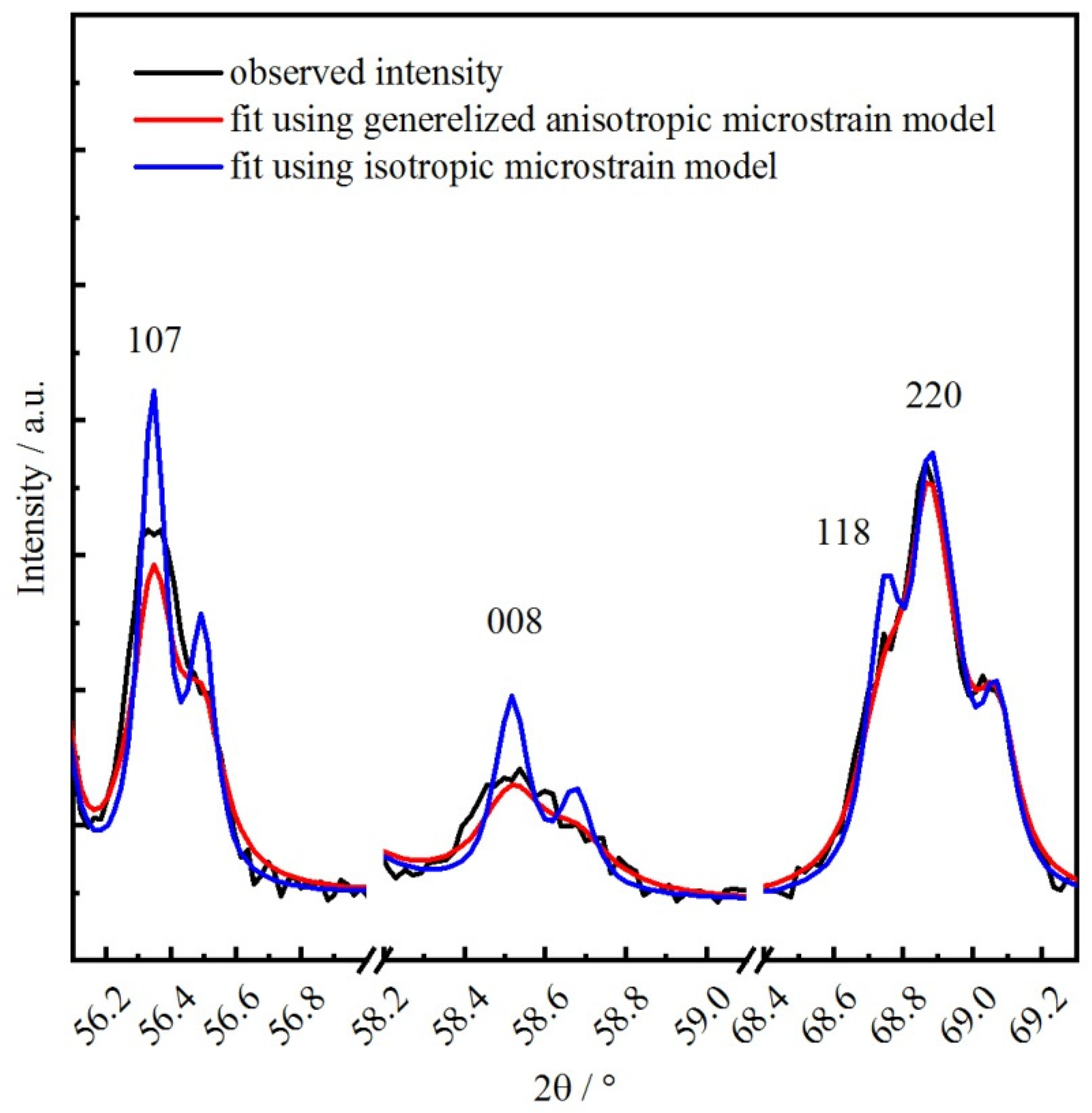
| microstrain, Δd/d 10−6 | 907 (76) | 889 (32) | S 4002 34,500 (1200) S 004 1400 S 220 39,100 (1600) S 022 −200 | 692 (27) |
| δ | 0.18 (4) | 0.14 (2) | 0.12 | 0.16 (2) |
| wR, % | 11.72 | 3.73 | 4.94 | 3.88 |
| GoF | 2.6 | 1.28 | 1.58 | 1.53 |
Publisher’s Note: MDPI stays neutral with regard to jurisdictional claims in published maps and institutional affiliations. |
© 2022 by the authors. Licensee MDPI, Basel, Switzerland. This article is an open access article distributed under the terms and conditions of the Creative Commons Attribution (CC BY) license (https://creativecommons.org/licenses/by/4.0/).
Share and Cite
Mishchenko, D.; Vinokurov, Z.; Gerasimov, E.; Filonova, E.; Shmakov, A.; Pikalova, E. Unusual Lattice Parameters Behavior for La1.9Ca0.1NiO4+δ at the Temperatures below Oxygen Loss. Crystals 2022, 12, 344. https://doi.org/10.3390/cryst12030344
Mishchenko D, Vinokurov Z, Gerasimov E, Filonova E, Shmakov A, Pikalova E. Unusual Lattice Parameters Behavior for La1.9Ca0.1NiO4+δ at the Temperatures below Oxygen Loss. Crystals. 2022; 12(3):344. https://doi.org/10.3390/cryst12030344
Chicago/Turabian StyleMishchenko, Denis, Zakhar Vinokurov, Evgeny Gerasimov, Elena Filonova, Alexander Shmakov, and Elena Pikalova. 2022. "Unusual Lattice Parameters Behavior for La1.9Ca0.1NiO4+δ at the Temperatures below Oxygen Loss" Crystals 12, no. 3: 344. https://doi.org/10.3390/cryst12030344