Article

Hydrothermal Synthesis, Crystal Structure, and Spectroscopic Properties of Pure and Eu3+-Doped NaY[SO4]2 ∙ H2O and Its Anhydrate NaY[SO4]2

1
Institute for Inorganic Chemistry, University of Stuttgart, D-70569 Stuttgart, Germany
2
Department of Chemical Engineering, FH Münster University of Applied Sciences, D-48565 Steinfurt, Germany
*
Author to whom correspondence should be addressed.
Crystals 2021, 11(6), 575; https://doi.org/10.3390/cryst11060575
Submission received: 27 April 2021 / Revised: 17 May 2021 / Accepted: 17 May 2021 / Published: 21 May 2021

Abstract

:
The water-soluble colorless compound NaY[SO4]2 ∙ H2O was synthesized with wet methods in a Teflon autoclave by adding a mixture of Na2[SO4] and Y2[SO4]3 ∙ 8 H2O to a small amount of water and heating it up to 190 °C. By slow cooling, single crystals could be obtained and the trigonal crystal structure of NaY[SO4]2 ∙ H2O was refined based on X-ray diffraction data in space group P3221 (a = 682.24(5) pm, c = 1270.65(9) pm, Z = 3). After its thermal decomposition starting at 180 °C, the anhydrate NaY[SO4]2 can be obtained with a monoclinic crystal structure refined from powder X-ray diffraction data in space group P21/m (a = 467.697(5) pm, b = 686.380(6) pm, c = 956.597(9) pm, β = 96.8079(5), Z = 2). Both compounds display unique Y3+-cation sites with eightfold oxygen coordination (d(Y–Os = 220–277 pm)) from tetrahedral [SO4]2− anions (d(S–O = 141–151 pm)) and a ninth oxygen ligand from an H2O molecule (d(Y–Ow = 238 pm) in the hydrate case. In both compounds, the Na+ cations are atoms (d(Na–Os = 224–290 pm) from six independent [SO4]2− tetrahedra each. Thermogravimetry and temperature-dependent PXRD experiments were performed as well as IR and Raman spectroscopic studies. Eu3+-doped samples were investigated for their photoluminescence properties in both cases. The quantum yield of the red luminescence for the anhydrate NaY[SO4]2:Eu3+ was found to be almost 20 times higher than the one of the hydrate NaY[SO4]2 ∙ H2O:Eu3+. The anhydrate NaY[SO4]2:Eu3+ exhibits a decay time of about τ1/e = 2.3 µm almost independent of the temperature between 100 and 500 K, while the CIE1931 color coordinates at x = 0.65 and y = 0.35 are very temperature-consistent too. Due to these findings, the anhydrate is suitable as a red emitter in lighting for emissive displays.

1. Introduction

Eu3+-doped luminescence materials based on complex oxides are very important in application [1,2] and show a red emission with typical 5D07FJ transitions between 610 and 620 nm [3]. They could be prepared on “classic” solid-state routes at high temperatures, as has been done for the examples of Y2[MoO4]3:Eu3+ and Y2[MoO4]2[Mo2O7]:Eu3+ [4], GdSb2O4Br:Eu3+ [5], as well as YNbO4:Eu3+ and YTaO4:Eu3+ [6]. Another energy-saving synthesis route to get Eu3+-doped luminescence materials without heating uses wet synthesis strategies. For example the Eu3+-doped xenotime-type yttrium oxoarsenate Y[AsO4]:Eu3+ [7], the oxophosphate Y[PO4]:Eu3+ [8], and the oxocarbonate Y2[CO3]3:Eu3+ · n H2O [9] were synthesized following a wet route.
With NaCe[SO4]2 ∙ H2O (trigonal, P3121), Lindgren reported for the first time in 1977 the crystal structure of a sodium rare-earth metal oxosulfate monohydrate yielded from a wet synthesis by adding Ce[OH]3 and Na2[SO4] to aqueous sulfuric acid (H2SO4) and heating it to 230 °C for 7 days [10]. In later works of other groups, the crystal structure of the sodium rare-earth (RE) metal oxosulfate monohydrates NaRE[SO4]2 ∙ H2O for the elements RE = La—Nd and Sm—Dy was solved either in space group P3121 (no. 152) or its enantiomorphic analog P3221 (no. 154). The samples were produced by different syntheses routes [11,12,13,14,15,16,17,18] and an Indian paper from 1989 deals with these double sulfates of trivalent plutonium, as well as the rare-earth metals RE = La—Nd, Sm—Yb, and Y, but does not give some detailed crystallographic information except for the space group derived from powder X-ray diffraction data [19].
By changing the alkali metal sodium to the next bigger one, potassium namely, KLa[SO4]2 ∙ H2O emerges as the only known alkali-metal rare-earth metal oxosulfate monohydrate with the mentioned trigonal structure [20]. For the smaller rare-earth metals (RE = Ce—Nd, Sm—Dy), the potassium-containing oxosulfate monohydrates KRE[SO4]2 ∙ H2O crystallize monoclinically in space group P21/c [20,21,22,23] in analogy to the isotypic rubidium compounds RbRE[SO4]2 ∙ H2O with RE = Ce, Gd, Ho and Yb [24,25,26]. For silver instead of an alkali-metal cation also AgRE[SO4]2 ∙ H2O representatives with the crystal structure of NaCe[SO4]2 ∙ H2O were found [27] and by the exchange of the rare-earth metal cation with trivalent bismuth, its oxosulfate monohydrate NaBi[SO4]2 ∙ H2O [28] shows the same trigonal structure as the related rare-earth metal compounds NaRE[SO4]2 ∙ H2O.
In 2006, the photoluminescence spectrum of NaEu[SO4]2 ∙ H2O (excited at λ = 393 nm) [11] and in 2016 analogous spectra of NaTb[SO4]2 ∙ H2O (excited at λ = 320 nm) and NaDy[SO4]2 ∙ H2O (excited at λ = 387 nm) were measured at room temperature [12]. In 2011, Ce3+- and Tb3+-doped samples of NaY[SO4]2 ∙ H2O were the subject of a luminescence investigation [29]. Moreover, in 2015, the sodium rare-earth metal oxosulfate monohydrates NaRE[SO4]2 ∙ H2O with RE = La, Nd, and Gd could be successfully tested as heterogeneous redox catalysts for the selective oxidation of organic sulfides [13]. It is worth mentioning that NaY[SO4]2 · H2O even occurs as a mineral with the name chinleite-(Y) [30], naturally containing all the lanthanoids with roughly the same size as yttrium. The crystallographic data from a structure refinement in space group P3221 have never been deposited at a common database, however.
For the anhydrous sodium rare-earth metal oxosulfates NaRE[SO4]2, their monoclinic crystal structure was solved in space group P21/m for RE = Er [31] and Tm [32] and the triclinic one in space group P 1 ¯ for RE = La [33] and Nd [31]. For trivalent gold instead of RE3+ cations, the monoclinic crystal structure of NaAu[SO4]2 was described in space group P21/n [34], but Au3+ in square planar oxygen coordination causes marked topological differences. Not so different from the Na+ analogs, for triclinic AgEu[SO4]2 (space group: P 1 ¯ ) with Ag+ in eightfold oxygen coordination, its Eu3+ bulk luminescence was also measured very recently [35].
In the following contribution, we report on the preparation of NaY[SO4]2 · H2O via wet synthesis, its trigonal crystal structure, and the red luminescence of Eu3+-doped samples. After thermal decomposition, we obtained its monoclinic anhydrate NaY[SO4]2, which shows an even stronger red luminescence, when Eu3+-doped.

2. Materials and Methods

2.1. Synthesis

Sodium yttrium oxosulfate monohydrate NaY[SO4]2 ∙ H2O was obtained from a wet synthesis by adding 6.6 mmol Na2[SO4] (ChemPur, 99.9%) and 5.5 mmol Y2[SO4]3 ∙ 8 H2O, which means an excess of Na2[SO4], to about 4 ml demineralized water and heated the obtained wet powder to 190 °C in a 25 ml Teflon autoclave overnight, with a yield only limited by the solubility of the monohydrate. Thus, the yield was about 2/3 of the theoretical possible quantity. It could be increased by evaporating the water, but the change of contamination with Y2[SO4]3 ∙ 8 H2O becomes higher then. By slowly cooling the solution down (5 °C per 1 h), single crystals in a size up to 0.3 mm edge length (Figure 1) of the water-soluble colorless compound NaY[SO4]2 ∙ H2O could be isolated (Equation (1)) and washed with ethanol (Brüggemann, denaturized with petrol ether). The starting material Y2[SO4]3 ∙ 8 H2O was synthesized by evaporating a solution of Y2O3 (ChemPur, 99.9%) in 96% sulfuric acid H2SO4 (Scharr, pure) according to Equation (2).
The anhydrous oxosulfate NaY[SO4]2 can be obtained by heating NaY[SO4]2 ∙ H2O in air at a temperature of 180 °C or higher (Equation (3)). The powder, which was used for the crystal structure refinement, was drained at 550 °C. For the luminescence measurements, a Eu3+-doped sample of NaY[SO4]2 ∙ H2O (0.5% Eu instead of Y) was produced by adding Eu2[SO4]3 ∙ 8 H2O (synthesis analogous to Y2[SO4]3 ∙ 8 H2O with Eu2O3 (ChemPur, 99.9%) instead of Y2O3) to the process, which is described in Equation (1), and NaY[SO4]2:Eu3+ has been prepared by draining the doped sample at 550 °C in air.
Na2[SO4] + Y2[SO4]3 ∙ 8 H2O → 2 NaY[SO4]2 ∙ H2O + 7 H2O
RE2O3 + 3 H2SO4 + 5 H2O → RE2[SO4]3 ∙ 8 H2O (RE = Y and Eu)
NaY[SO4]2 ∙ H2O → NaY[SO4]2 + H2O ↑

2.2. X-ray Experiments and Crystal-Structure Solution

For single-crystal X-ray diffraction experiments, a suitable crystal was selected under a light microscope and fixed inside of a glass capillary with an outer diameter of 0.1 mm and a length of about 15 mm. The crystal was measured with a κ-CCD four-circle X-ray diffractometer (Bruker Nonius, Karlsruhe, Germany) with Mo-Kα radiation (λ = 71.07 pm) at 293 K (room temperature). Crystal-structure solution and refinement for NaY[SO4]2 ∙ H2O (CSD-2016596) in the trigonal space group P3221 were carried out with the program package SHELX-97 [36,37] by Sheldrick, and the program HABITUS by Bärninghausen and Herrendorf was applied [38] for a numerical absorption correction.
For X-ray powder diffraction (PXRD), part of the sample was fixed on a STADI-P diffractometer (Stoe & Cie, Darmstadt, Germany) and measured with Cu-Kα radiation (λ = 154.06 pm) in transmission setting. The monohydrate was measured from 2ϴ = 10–90° for checking phase purity and the anhydrate NaY[SO4]2 (CSD-2072719) was measured from 2ϴ = 8–110° for solving its crystal structure in the NaEr[SO4]2-type arrangement [31] with the program FULLPROF [39,40]. The measured powder X-ray diffraction pattern of NaY[SO4]2 ∙ H2O can be seen in Figure 2 (top) and the measured PXRD pattern together with the difference plot of the Rietveld refinement for NaY[SO4]2 is shown in Figure 2 (bottom).
Temperature-depending powder X-ray diffraction data were measured in the interval 2ϴ = 10–90° with a RIGAKU SmartLab diffractometer (Neu-Isenburg, Germany) using Cu-Kα radiation (λ = 154.06 pm) in reflection setting from 25 up to 900 °C.
While all the atomic displacement parameters of NaY[SO4]2 ∙ H2O could be refined anisotropically based on single-crystal X-ray diffraction data, the atomic displacement parameters of NaY[SO4]2 were only treated isotropically with Rietveld refinement based on PXRD data.

2.3. Thermal Analysis

Thermal analysis (thermogravimetry) was performed with about 36 mg of a NaY[SO4]2 ∙ H2O samples with a Netzsch device of the type STA-449C (Selb, Germany) in a corundum crucible under argon atmosphere. The sample was heated with 5 K/min from 25 to 1400 °C.

2.4. Luminescence Spectroscopy

Excitation and emission spectra were collected using a fluorescence spectrometer FLS920 (Edinburgh Instruments, Livingston, UK) equipped with a 450 W ozone-free xenon discharge lamp (Osram, München, Germany) and a cryostat “MicrostatN” from Oxford Instruments (Abingdon, UK) as the sample chamber. Additionally, a mirror optic for powder samples was applied. For detection, an R2658P single-photon-counting photomultiplier tube (Hamamatsu, Hamamatsu, Japan) was used. All photoluminescence spectra were recorded with a spectral resolution of 0.5 nm and a dwell time of 0.5 s in 0.5 nm steps.
The photoluminescence decay times were measured on an FLS920 spectrometer (Edinburgh Instruments, Livingston, UK). A Xe μ-flash lamp μF920 was used as an excitation source. For detection, an R2658P single-photon-counting photomultiplier tube (Hamamatsu Photonics, Hamamatsu, Japan) found application.
For the reflection spectra, the investigated samples were placed into an integrating sphere, and FLS920 spectrometer (Edinburgh Instruments, Livingston, UK) equipped with a 450 W Xe lamp, and a cooled (−20 °C) single-photon-counting photomultiplier (Hamamatsu R928) was used. Ba[SO4] was applied as the reflectance standard. The excitation and emission bandwidths were 10.00 and 0.06 nm, respectively. Step width was 0.5 nm and integration time 0.5 s.
Quantum yields were determined according to the method published by Kawamura et al. [41] upon excitation at 395 nm using a 7 nm excitation and 0.5 nm emission slit. The scan steps were 0.5 nm, while the respective emission intensity from 370 to 750 nm was recorded.
The CIE1931 color coordinates and luminous efficacy (LE) values were calculated from the temperature-dependent emission spectra of NaY[SO4]2:Eu3+ using the Color Calculator 6.75 software from Osram (Osram, München, Germany) [42].
The LE value (unit: lm/W) is a parameter describing, how bright the radiation is perceived by an average human observer at a photopic illumination situation. It scales with the photopic human eye sensitivity curve V(λ) and can be calculated from the normalized emission spectrum I(λ) of the sample as follows [43]:
L E ( lm / W ) = 683   ( lm / W ) 380 nm 780 nm I ( λ ) V ( λ ) d λ 380 nm 780 nm I ( λ ) d λ

2.5. IR and Raman Spectra

Infrared spectra for powder samples of NaY[SO4]2 ∙ H2O and NaY[SO4]2 was measured from 700 to 4000 cm−1 with a NICOLET iS5 device from Thermo Scientific (Karlsruhe, Germany). Raman spectroscopy was performed with a DXR SmartRaman spectrometer from Thermo Scientific (Karlsruhe, Germany) with a red laser (λ = 780 nm) and a laser power of 10 mW from 200 to 1800 cm−1.

3. Results and Discussion

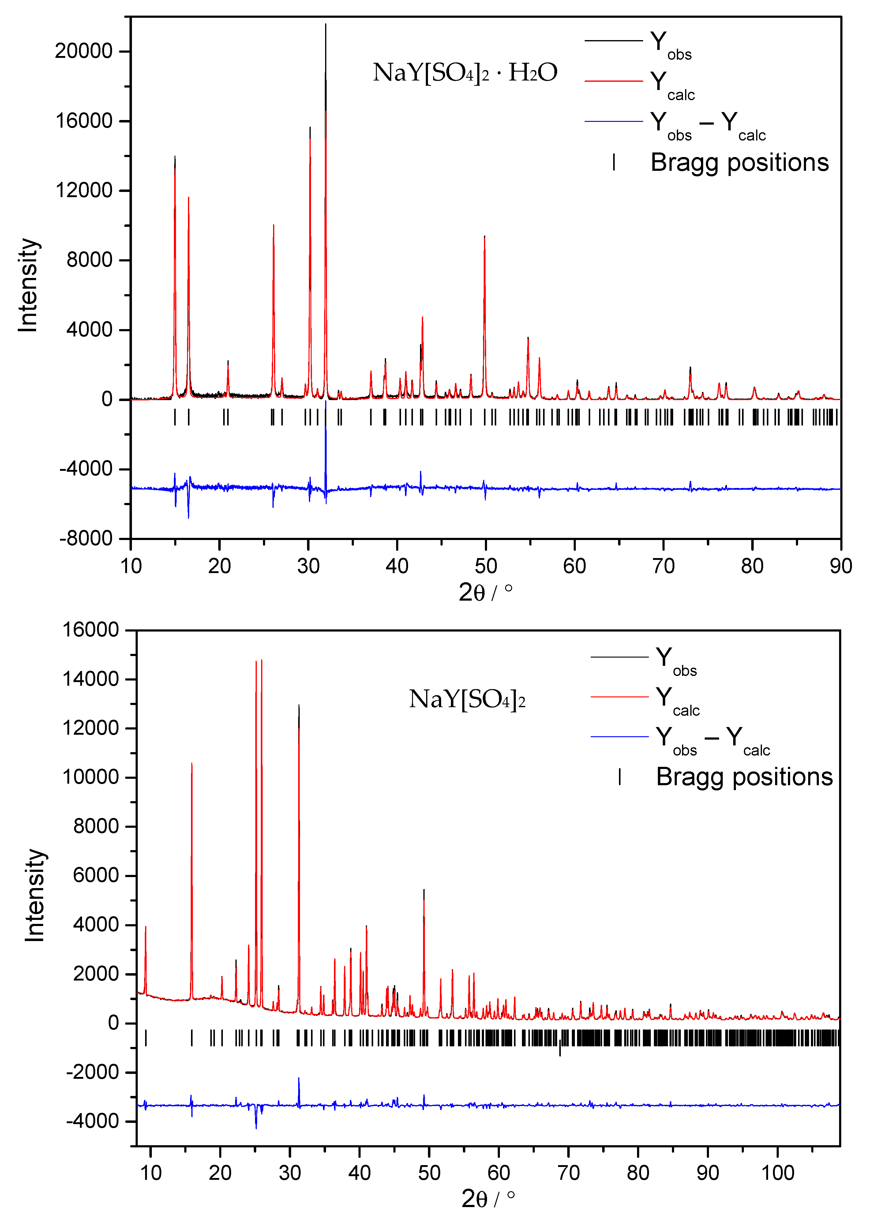
3.1. Structure Refinement and Description of NaY[SO4]2 ∙ H2O and NaY[SO4]2

The most relevant crystallographic data of the wet synthesized NaY[SO4]2 ∙ H2O compared to its anhydrate NaY[SO4]2 are shown in Table 1. The given lattice parameters of NaY[SO4]2 ∙ H2O stems from single-crystal data, while its lattice parameters from PXRD experiments amount to a = 682.82(3) pm and c = 1270.77(6) pm (c/a = 1.861).
Table 2 shows the fractional atomic coordinates with the site symmetry for all atoms and Ueq or Uiso values of NaY[SO4]2 ∙ H2O and NaY[SO4]2.
While NaY[SO4]2 ∙ H2O crystallizes in the trigonal space group P3221 (no. 154) with a = 682.24(5) pm, and c = 1270.65(9) pm (c/a = 1.862) for Z = 3, NaY[SO4]2 adopts the monoclinic space group P21/m (no. 11) with a = 467.697(5) pm, b = 686.380(6) pm, c = 956.597(10) pm, and β = 96.8079(5)° for Z = 2. The b-axes of both compounds differ by only 0.6% and the c-axis of the monohydrate is about 4/3 of the one of the anhydrate. While in the hydrate monolayers of Na+ and Y3+ cations take turns along [001], in the anhydrate double layers of each Na+ and Y3+ alternate along [001]. Extended unit cells of NaY[SO4]2 ∙ H2O and NaY[SO4]2 can be seen in Figure 3.
In NaY[SO4]2 ∙ H2O, the Y3+ cations are coordinated by nine oxygen atoms (eight from oxosulfate anions (d(Y–O) = 237–248 pm) and one from a water molecule (d(Y–O5w) = 238 pm). Only eight oxygen atoms covalently bonded to sulfur in [SO4]2–, and units occur as Y3+ coordination sphere (d(Y–O) = 220–277 pm) in NaY[SO4]2.
Y3+ in NaY[SO4]2 ∙ H2O resides on the Wyckoff site 3a with C2 symmetry (Figure 4, left), whereas Y3+ in NaY[SO4]2 occupies the 2e position on a mirror plane (Figure 4, right).
In Y2[SO4]3 ∙ 8 H2O [45], the unique Y3+ cations are also surrounded by eight oxygen atoms (four from water molecules and four more from oxosulfate anions) with distances between 230 and 247 pm, while in the anhydrous oxosulfate Y2[SO4]3, Y3+ is surrounded octahedrally by only six oxygen atoms from oxosulfate groups with distances between 220 and 224 pm [46]. While Y3+ is coordinated by just one oxygen atom per [SO4]2– anion in both Y2[SO4]3 ∙ 8 H2O [45] and Y2[SO4]3 [46], the same is observed in NaY[SO4]2 ∙ H2O and NaY[SO4]2, but now with two oxygen atoms of the same oxosulfate unit. In Y2[SO4]3 [46], NaY[SO4]2 ∙ H2O and NaY[SO4]2 six [SO4]2– anions coordinate the Y3+ cations, while in Y2[SO4]3 ∙ 8 H2O [45] there are only four of them. Compounds of the type ARE[SO4]2 ∙ H2O with A = Na crystallize trigonally in space group P3221 (or P3121) [10,11,12,13,14,15,16,17,18], but monoclinically in space group P21/c with A = K for RE = Ce—Nd, Sm—Dy [20,21,22,23]. The water molecule in NaY[SO4]2 ∙ H2O is only coordinated to yttrium, whereas in the KRE[SO4]2 ∙ H2O examples [20,21,22,23], it further coordinates the alkali-metal cation. The crystal structure of the anhydrous potassium rare-earth metal oxosulfates are described triclinically in space group P 1 ¯ for RE = Pr [47] and Nd [48], but monoclinically in space group P21/c for RE = Nd [49] and Er [50]. In the triclinic structure, the coordination number of RE3+ is eight, while in the monoclinic one, it surprisingly increases to nine. Two oxosulfate anions coordinate with two oxygen atoms each in the monoclinic KRE[SO4]2 representatives, while in the triclinic cases only one [SO4]2– group has two contacts to the rare-earth metal cations. The coordination environments of those in the two title compounds are compared to the other alkali-metal rare-earth metal oxosulfates in Figure 5.
The sodium cations in both title compounds are surrounded by eight oxygen atoms from six different oxosulfate units as a bicapped octahedron. While Na+ in NaY[SO4]2 ∙ H2O is only connected with [SO4]2− anions and no water molecules, in the related potassium compound the K+ cation has contact with six of them and one water molecule [20,21,22,23]. The anhydrous potassium rare-earth metal oxosulfates show a coordination sphere around the alkali-metal cation erected by ten oxygen atoms from six oxosulfate anions in case of the triclinic examples [47,48] and seven terminal [SO4]2– units in the monoclinic cases [49,50]. In the orthorhombic salt Na2[SO4], the sodium cations show six oxygen atoms from five oxosulfate groups as next neighbors [51], while in its decahydrate Na2[SO4] ∙ 10 H2O, Na+ is only surrounded by six water molecules octahedrally [52] (Figure 6).
While NaY[SO4]2 ∙ H2O exhibits only one singular crystallographic [SO4]2− anion, its anhydrate has two different ones of them (Figure 7). All oxygen atoms in NaY[SO4]2 ∙ H2O are surrounded approximately in a plane triangular fashion by Y3+, Na+, and S6+, while in NaY[SO4]2 O2 and O6 differ from this scheme since O2 is coordinated by one S6+ and two Y3+ and O6 by one S6+ and two Na+ cations. Even O5w has one Y3+ and two H+ cations, three neighbors. The triangular environments of the oxygen atoms in NaY[SO4]2 ∙ H2O and NaY[SO4]2 can be seen in Figure 8. Selected interatomic distances (d/pm) are summarized in Table 3.
For confirmation of the Na+ and Y3+ sites in NaY[SO4]2 ∙ H2O and NaY[SO4]2, bond-valence calculations were carried out with the parameters used by Brese and O’Keeffe [53]. With calculated charges of 3.06 in NaY[SO4]2 ∙ H2O and 3.19 in NaY[SO4]2 for the Y3+ sites next to 1.20 in NaY[SO4]2 ∙ H2O and 1.23 in NaY[SO4]2 for the Na+ sites, their positions can just be confirmed. More details of these calculations can be seen in Table 4. The bond-valence equation for the calculation of the charge given by Brese and O’Keeffe [53] is vij = exp[(Rijdij)/b] with the valence vij, the universal constant b = 0.37 Å, the bond-valence parameter Rij, and the Ångström distance of the considered atoms dij between the atoms i and j. The sum ∑(vij) represents the charge of the regarded ion.
The motifs of mutual adjunction for the atoms in both title compounds NaY[SO4]2 ∙ H2O and NaY[SO4]2 can be seen in Table 5.
We became aware of a competing structure refinement for trigonal NaY[SO4]2 · H2O (a = 681.91(3) pm, c = 1270.35(11) pm, c/a = 1.863) in space group P3121 that was already in the progress of publication [54], simultaneous to our activities writing this article. The lower CSD deposition number (ours for NaY[SO4]2 · H2O in space group P3221: 2016596 versus the Chinese competitor one for NaY[SO4]2 · H2O in space group P3121: 2058909) should grant us a priority, despite the almost identical results in both papers from the year 2021.

3.2. Thermal Analysis

A thermogravimetrical curve for the decomposition of NaY[SO4]2 ∙ H2O between 25 and 1400 °C is depicted in Figure 9.
The first mass-loss at about 180 °C with 5.6% represents the release of water and the transformation from NaY[SO4]2 ∙ H2O (100% mass; M = 322.036 g/mol) to NaY[SO4]2 (94.4% mass; M = 304.021 g/mol). The second decomposition leads to a mixture of Y2O2[SO4] [55] with the crystal structure of monoclinic La2O2[SO4] [56] and Eu2O2[SO4] [57] or orthorhombic Nd2O2[SO4] [58] together with Na2[SO4] [51,59], confirmed by powder X-ray diffraction experiments (Figures S1 and S2 in the Supplementary Information). For Y2O2[SO4], there are no known or other good crystal-structure data available, so there are differences in intensity and position, but the final decomposition step leads to a mixture of cubic Y2O3 with a bixbyite-type structure [60] and orthorhombic Na2[SO4] [51,59] (Figure S3). The TG curve (Figure 9) appears to be similar to that of NaRE[SO4]2 ∙ H2O with RE = La, Ce, Nd, and Sm, which have been measured in 1994 by Kolcu and Zümreoǧlu-Karan [18]. The dehydration temperature of the lanthanum compound is 297 °C and gets lower with decreasing RE3+-cation radius along with the lanthanoid contraction [61]. With a dehydration temperature of 265 °C for the samarium compound, this trend is further confirmed with our yttrium analog at 180 °C.
Additional to the thermogravimetry, temperature-dependent X-ray diffraction experiments were performed (Figure 10).
While the TG curve (Figure 9) shows a phase transformation from NaY[SO4]2 ∙ H2O to NaY[SO4]2 at 180 °C, the temperature-depending PXRD indicates the anhydrous compound for the first time at 350 °C. At 550 °C the water-containing compound could not be detected anymore. The XRD intensities became lower again with rising temperatures and suggest a starting decomposition of NaY[SO4]2 to Y2O2[SO4] and Na2[SO4]. The reflection at 12.8° resulted from the X-ray powder-diffractometer setting.

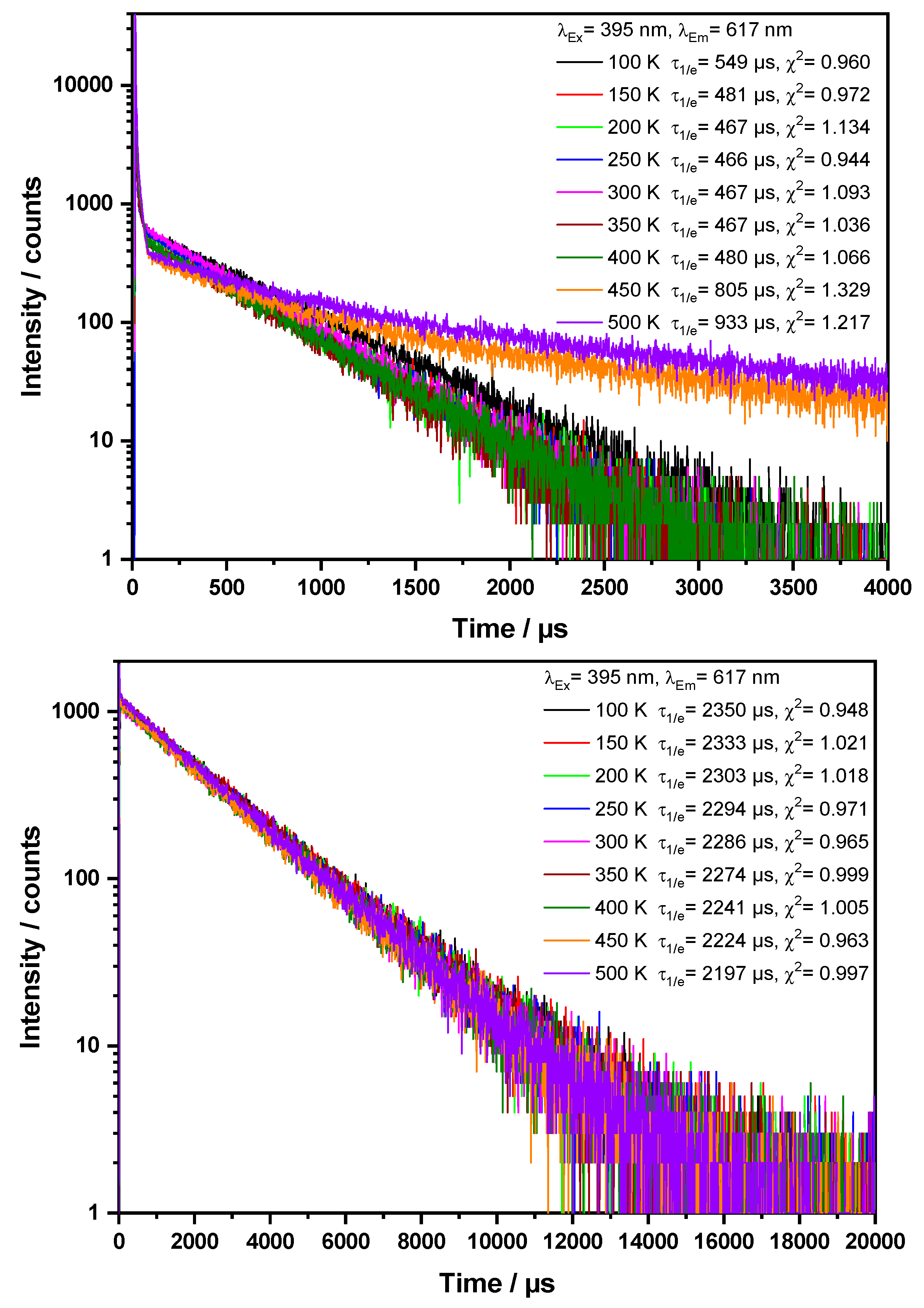
3.3. Luminescence-Spectroscopic Properties

Eu3+-doped samples of NaY[SO4]2 · H2O and NaY[SO4]2 under UV irradiation (λ = 254 nm) can be seen in Figure 11.
Both compounds display a reflection spectrum, which is in line with plain white powders of good optical quality and high crystallinity, due to the lack of greying or defect bands. The absorption edge of the anhydrous compound at about 270 nm is assigned to the LMCT (ligand-to-metal charge-transfer) absorption band of Eu3+, which is a typical energetic position of the LMCT process of Eu3+ in an oxidic environment [62]. The reflectance values at longer wavelengths were close to unity, pointing to a high optical quality of the prepared materials. In both reflection spectra (Figure 12), the typical Eu3+ absorption lines originating from the 7F05L6 and 7F05D2 transitions could be observed in the ranges of 395–397 nm and 450–470 nm, respectively [63].
Temperature-dependent excitation spectra of NaY[SO4]2 · H2O:Eu3+ and NaY[SO4]2:Eu3+ reveal the typical intraconfigurational 4f–4f transitions of Eu3+ between 280 and 550 nm [63,64] and the position of the LMCT of Eu3+ in the anhydrous compound. The LMCT band was located at 270 nm and was in good agreement with the position derived from the reflection spectrum. The excitation spectra of both compounds are plotted in Figure 13.
Noteworthy was the temperature-dependent excitation spectra of NaY[SO4]2 ∙ H2O:Eu3+, since a closer look at the UV-A range revealed a distinct change of the pattern of 7F05L6 (390–405 nm) and 7F05L8 + 5GJ + 5L9 + 5L10 (J = 2–6) (373–387 nm) transitions [65]. The thermal population of the 7F1 level could explain some changes in the excitation line pattern. However, the shift and broadening of the most intense line of the 7F05L6 multiplet at 394 nm from 400 K onwards pointed to a phase transition. This finding could be explained by the loss of water and the transformation of NaY[SO4]2 ∙ H2O:Eu3+ to NaY[SO4]2:Eu3+ in good accordance with the results from thermal gravimetry (Figure 9). Temperature-dependent emission spectra of NaY[SO4]2 ∙ H2O:Eu3+ and NaY[SO4]2:Eu3+ are shown in Figure 14.
The emission spectra of Eu3+-comprising materials consisted of the orange allowed magnetic-dipole (MD) transition 5D07F1, the red parity-forbidden electric-dipole (ED) transition 5D07F2, and further line multiplets in the deep red spectral range around 650 and 695 nm due to the ED transitions 5D07F3 and 5D07F4. For light sources and emissive displays, the emission spectrum should consist mainly of emission lines resulting from the 5D07F2 transitions [66,67]. This means that the Eu3+ cation has to occupy a crystallographic site without inversion symmetry (see Figure 4 for symmetry examination). This also induces the deep red emission lines. Fortunately, the 5D07F2 transition is hypersensitive and small deviations of the inversion symmetry strongly enhance the probability of the 5D07F2 transitions. The intensity of the strongly forbidden transition 5D07F0 is known to correlate with the linear terms of the crystal-field parameter and polarizability of the Eu3+ cation [67].
However, the emission spectrum of NaY[SO4]2 ∙ H2O:Eu3+ upon 395 nm excitation revealed the typical emission line pattern between 580 and 720 nm due to the 5D07FJ (J = 0–4) transitions of Eu3+ [62,63,68]. Unfortunately, the signal-to-noise ratio is rather low, which points to a low quantum yield. Indeed, the determination of the quantum efficiency according to Kawamura [41] yielded a value of solely about 1%. Such a low quantum yield can be explained by the presence of crystal water since the high phonon frequency of the O–H vibration of water quenches efficiently the Eu3+ luminescence [69].
As already observed for the excitation spectra, the temperature-dependent emission spectra of NaY[SO4]2 ∙ H2O:Eu3+ showed a distinct change once the temperature exceeded 400 K, resulting in the increase of intensity and the width of the 5D0 → F1, 5D07F2, and 5D07F4 transitions, as well as the appearance of the 5D07F0 transition, which was absent at room temperature. This change again points to a phase transition, i.e., the transformation of NaY[SO4]2 ∙ H2O:Eu3+ to NaY[SO4]2:Eu3+, which goes along with an increase of the crystal-field strength causing a larger energetic spread of the Stark components of the above mentioned 5D07FJ, transitions. This finding was in good agreement with the decline of the coordination number from 9 to 8 and a shorter average Y–O distance. However, even though the emission spectra of the anhydrous sample obtained after the phase transition resembled that of the as-prepared anhydrous sample, the emission spectra were not completely the same. We assumed that after the phase transition a higher defect density remained, which resulted in line-broadening and a lower signal-to-noise ratio since, without further high-temperature treatment, defects caused by the water removal cannot be healed. In contrast, the as-prepared samples of anhydrous NaY[SO4]2:Eu3+ showed a much higher quantum yield. This value was determined to be almost around 20%, which also explained the much better signal-to-noise ratio of the respective emission spectra as a function of temperature (Figure 14, bottom).
The CIE1931 color coordinates of NaY[SO4]2:Eu3+ are at x = 0.65 and y = 0.35, while the temperature impact is rather low, branding the substance as a stable color-consistent material for application in displays or fluorescent light sources [1]. However, the magnification of the color space in Figure 15 demonstrates that the color point shifts slightly to the orange range, which can be caused by the reduction of the asymmetry ratio 5D07F2 /5D07F1 [63] or by the reduction of the covalency related to the 5D07F4/5D07FJ ratio [3]. However, both effects are in line with a thermal expansion of the crystals and the Eu3+ site causes a decrease of the covalent interaction between Eu3+ and oxygen and an increase of the local symmetry.
Noteworthy were the intensities of the temperature-dependent emission spectra of NaY[SO4]2 ∙ H2O:Eu3+ as depicted in Figure 16. While the intensity decreased between 100 and 300 K due to typical thermal quenching, it increased again between 300 and 500 K. This effect was caused by the phase transition towards the formation of the more efficiently luminescent NaY[SO4]2:Eu3+ upon increasing the temperature.
In contrast, the temperature-dependent emission spectra of NaY[SO4]2:Eu3+ itself show a typical decrease of the intensity or quantum yield of Eu3+ phosphors with increasing temperature [63].
Finally, we investigated the time-dependent luminescence (Figure 17) of the 5D07F2 transition of Eu3+ at 617 nm upon 395 nm excitation of NaY[SO4]2 ∙ H2O:Eu3+ and NaY[SO4]2:Eu3+. As discussed before, NaY[SO4]2 ∙ H2O:Eu3+ shows a peculiar behavior due to the phase transition between 400 and 500 K, which means that the decay time increases from 550 µs at 100 K to about 930 µs at 500 K. At the same time, the decay curves become bi-exponential, which points to the formation of a novel phase with a prolonged decay time and enhanced internal quantum efficiency. The decay curves of NaY[SO4]2:Eu3+ between 100 and 500 K were almost perfectly mono-exponential over three orders of magnitude, while the derived decay times remained rather constant, as proven by the just slight decline from 2.35 ms to 2.20 ms. This finding meant that the internal quantum yield stayed quite stable, and thus, thermal quenching of the Eu3+ photoluminescence is a minor issue.

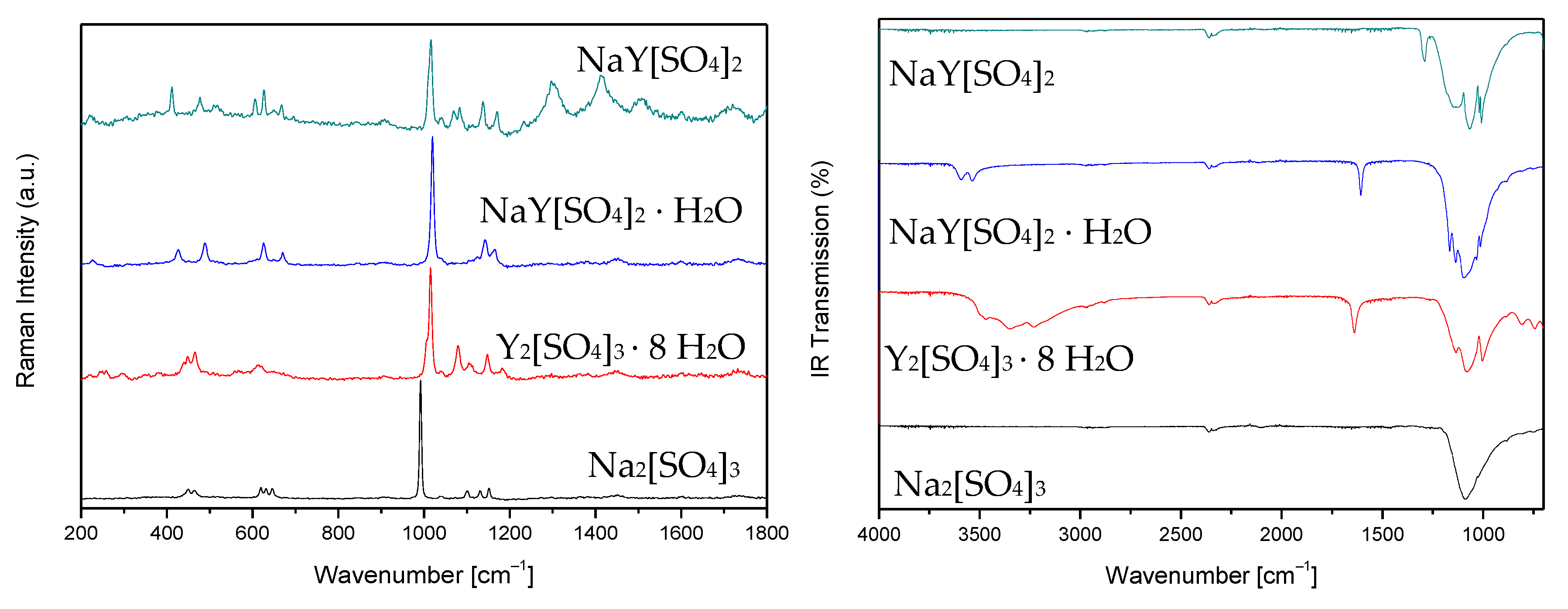
3.4. IR and Raman Studies of NaY[SO4]2 ∙ H2O and NaY[SO4]2

The Raman and IR spectra of NaY[SO4]2 ∙ H2O and NaY[SO4]2 are shown together with those of Y2[SO4]3 ∙ 8 H2O and Na2[SO4] in Figure 18 and the values are given in Table 6 compared to the literature data for Y2[SO4]3 [69] and Na2[SO4] (thenardite) [70]. The vibration at about 2300 cm–1 represents CO2 in the laboratory environment. While the ideal [SO4]2– anion with Td symmetry should only have four visible vibration bands (νas and δas: IR and Raman active, νs and δs: only Raman active), in the measured solid-state samples there were more bands measured. This was because of the no longer ideal [SO4]2– units and their considerable symmetry reduction.

4. Conclusions

Phase-pure white powder and even colorless single crystals of sodium yttrium oxosulfate monohydrate NaY[SO4]2 ∙ H2O could be synthesized hydrothermally from a mixture of Na2[SO4] and Y2[SO4]3 ∙ 8 H2O in demineralized water. The anhydrate NaY[SO4]2 was obtained by thermal decomposition at temperatures above 180 °C and is stable up to 800 °C. While the trigonal crystal structure of NaY[SO4]2 ∙ H2O was solved from single-crystal X-ray diffraction data in space group P3221, the monoclinic crystal structure of NaY[SO4]2 was refined with Rietveld methods from powder X-ray diffraction data in space group P21/m. The Na+ cations are coordinated by eight oxygen atoms from six tetrahedral [SO4]2− anions in both compounds and the coordination numbers of the Y3+ cations in the hydrate amount to nine (eight oxygen atoms from six [SO4]2− units plus one from a water molecule) and eight again in the anhydrate (eight oxygen atoms from six [SO4]2— anions). Both compounds suit as red-emitting luminescent materials, if doped with 0.5 % Eu3+, as shown by luminescence spectroscopy, but the anhydrate NaY[SO4]2:Eu3+ exhibits an almost twenty times higher quantum efficiency than the monohydrate NaY[SO4]2 ∙ H2O:Eu3+ owing to the water of hydration, which works as a vibrational quencher. The almost perfect monoexponential decay curves of the anhydrate NaY[SO4]2:Eu3+ and thus the lack of afterglow also prove the presence of a material with high quality, i.e., a low defect density.

Supplementary Materials

The Supplementary Material contains PXRD data from a sample after thermal treatment from 1000 °C (Figure S1 and S2) and 1400 °C (Figure S3) after the thermogravimetry experiment. They are available online at https://www.mdpi.com/article/10.3390/cryst11060575/s1.

Author Contributions

C.B. synthesized the pure and the Eu3+-doped compounds, which are described here, and measured their IR and Raman spectra. C.B. and T.S. solved the crystal structures of both compounds. T.S. contributed the reagents, the materials, the scientific equipment, and the infrastructure for synthesis, IR, and Raman spectroscopy. D.E. and T.J. measured and interpreted the Eu3+ luminescence of both doped compounds. The paper was written by all authors. All authors have read and agreed to the published version of the manuscript.

Funding

This research was funded by the State of Baden-Württemberg (Stuttgart).

Data Availability Statement

Crystallographic data are available in the ICSD-Database with the CSD-numbers 2016596 for NaY[SO4]2 ∙ H2O and 2072719 for NaY[SO4]2.

Acknowledgments

We thank Falk Lissner for the single-crystal X-ray diffraction measurements, Patrik Djendjur for the thermal analyses, and Jean-Louis Hoslauer for the temperature-dependent PXRD experiments.

Conflicts of Interest

The authors declare no conflict of interest.

References

  1. Jüstel, T.; Nikol, H.; Ronda, C. New Developments in the Field of Luminescent Materials for Lighting and Displays. Angew. Chem. Int. Ed. 1998, 37, 3084–3103. [Google Scholar] [CrossRef]
  2. Feldmann, C.; Jüstel, T.; Ronda, C.R.; Schmidt, P.J. Inorganic Luminescent Materials: 100 Years of Research and Application. Adv. Funct. Mater. 2003, 13, 511–516. [Google Scholar] [CrossRef]
  3. Skaudzius, R.; Katelnikovas, A.; Enseling, D.; Kareiva, A.; Jüstel, T. Dependence of the 5D07F4 transitions of Eu3+ on the local environment in phosphates and garnets. J. Lumin. 2014, 147, 290–294. [Google Scholar] [CrossRef]
  4. Laufer, S.; Strobel, S.; Schleid, T.; Cybinska, J.; Mudring, A.-V.; Hartenbach, I. Yttrium(III) Oxomolybdates(VI) as Potential Host Materials for Luminescence Applications: An Investigation of Eu3+-Doped Y2[MoO4]3 and Y2[MoO4]2[Mo2O7]. New J. Chem. 2013, 37, 1919–1926. [Google Scholar] [CrossRef]
  5. Goerigk, F.C.; Paterlini, V.; Dorn, K.V.; Mudring, A.-V.; Schleid, T. Synthesis and Crystal Structure of the Short LnSb2O4Br Series (Ln = Eu–Tb) and Luminescence Properties of Eu3+-Doped Samples. Crystals 2020, 10, 1089. [Google Scholar] [CrossRef]
  6. Popovic, E.J.; Imre-Lucaci, F.; Muresan, L.; Stefan, M.; Bica, E.; Grecu, R.; Indrea, E. Spectral investigations on niobium and rare earth activated yttrium tantalate powders. J. Optoelectron. Adv. Mater. 2008, 10, 2334–2337. [Google Scholar]
  7. Ledderboge, F.; Nowak, J.; Massonne, H.-J.; Förg, K.; Höppe, H.A.; Schleid, T. High-Pressure Investigations of Yttrium(III) Oxoarsenate(V): Crystal Structure and Luminescence Properties of Eu3+-Doped Scheelite-Type Y[AsO4] from Xenotime-Type Precursors. J. Solid State Chem. 2018, 263, 65–71. [Google Scholar] [CrossRef]
  8. Cybińska, J. Temperature dependent morphology variation of red emitting microcrystalline YPO4:Eu3+ fabricated by hydrothermal method. Opt. Mater. 2017, 65, 88–94. [Google Scholar] [CrossRef]
  9. Chang, H.-Y.; Chen, F.-S.; Lu, C.-H. Preparation and luminescence characterization of new carbonate (Y2(CO3)3 · n H2O:Eu3+) phosphors via the hydrothermal route. J. Alloys Compd. 2011, 509, 10014–10019. [Google Scholar] [CrossRef]
  10. Lindgren, O. The Crystal Structure of Sodium Cerium(III) Sulfate Hydrate, NaCe(SO4)2 ∙ H2O. Acta Chem. Scand. 1977, 31, 591–594. [Google Scholar] [CrossRef]
  11. Wu, C.-D.; Liu, Z.-Y. Hydrothermal synthesis of a luminescent europium(III) sulfate with three-dimensional chiral framework structure. J. Solid State Chem. 2006, 179, 3500–3504. [Google Scholar] [CrossRef]
  12. Zhai, B.; Li, Z.; Zhang, C.; Zhang, F.; Zhang, X.; Zhang, F.; Cao, G.; Li, S.; Yang, X. Three rare Ln–Na heterometallic 3D polymers based on sulfate anion: Syntheses, structures, and luminescence properties. Inorg. Chem. Commun. 2016, 63, 16–19. [Google Scholar] [CrossRef]
  13. Perles, J.; Fortes-Revilla, C.; Gutiérrez-Puebla, E.; Iglesias, M.; Monge, M.Á.; Ruiz-Valero, C.; Snejko, N. Synthesis, Structure, and Catalytic Properties of Rare-Earth Ternary Sulfates. Chem. Mater. 2005, 17, 2701–2706. [Google Scholar] [CrossRef]
  14. Paul, A.K.; Kanagaraj, R. Synthesis, Characterization, and Crystal Structure Analysis of New Mixed Metal Sulfate NaPr(SO4)2(H2O). J. Struct. Chem. 2019, 60, 477–484. [Google Scholar] [CrossRef]
  15. Blackburn, A.C.; Gerkin, R.E. Sodium lanthanum(III) sulfate monohydrate, NaLa(SO4)2 ∙ H2O. Acta Crystallogr. 1994, 50, 835–838. [Google Scholar] [CrossRef] [Green Version]
  16. Blackburn, A.C.; Gerkin, R.E. Redetermination of sodium cerium(III) sulfate monohydrate, NaCe(SO4)2 ∙ H2O. Acta Crystallogr. 1995, 51, 2215–2218. [Google Scholar] [CrossRef] [PubMed] [Green Version]
  17. Kolcu, Ö.; Zümreoğlu-Karan, B. Nonisothermal Dehydration Kinetics of Sodium-Light Lanthanoid Double Sulfate Monohydrates. Thermochim. Acta 1997, 296, 135–139. [Google Scholar] [CrossRef]
  18. Kolcu, Ö.; Zümreoǧlu-Karan, B. Thermal Properties of Sodium-Light-Lanthanoid Double Sulfate Monohydrates. Thermochim. Acta 1994, 240, 185–198. [Google Scholar] [CrossRef]
  19. Iyer, P.N.; Natarajan, P.R. Double Sulphates of Plutonium(III) and lanthanides with sodium. J. Less Common Met. 1989, 146, 161–166. [Google Scholar] [CrossRef]
  20. Kazmierczak, K.; Höppe, H.A. Syntheses, crystal structures and vibrational spectra of KLn(SO4)2 · H2O (Ln = La, Nd, Sm, Eu, Gd, Dy). J. Solid State Chem. 2010, 183, 2087–2094. [Google Scholar] [CrossRef]
  21. Jemmali, M.; Walha, S.; Ben Hassen, R.; Vaclac, P. Potassium cerium(III) bis(sulfate) monohydrate, KCe(SO4)2 ∙ H2O. Acta Crystallogr. 2005, 61, i73–i75. [Google Scholar] [CrossRef]
  22. Iskhakova, L.D.; Sarukhanyan, N.L.; Shchegoleva, T.M.; Trunov, V.K. The crystal structure of KPr(SO4)2 ∙ H2O. Kristallografiya 1985, 30, 474–479. [Google Scholar]
  23. Lyutin, V.I.; Safyanov, Y.N.; Kuzmin, E.A.; Ilyukhin, V.V.; Belov, N.V. Rhomb evaluation of the crystal structure of K-Tb double sulfate. Kristallografiya 1974, 19, 376–378. [Google Scholar]
  24. Robinson, P.D.; Jasty, S. Rubidium Cerium Sulfate Monohydrate, RbCe(SO4 )2 ∙ H2O. Acta Crystallogr. 1998, C54, IUC9800021. [Google Scholar] [CrossRef]
  25. Prokofev, M.V. Crystal structure of the double sulfate of rubidium and holmium. Kristallografiya 1981, 26, 598–600. [Google Scholar]
  26. Sarukhanyan, N.L.; Iskhakova, L.D.; Trunov, V.K.; Ilyukhin, V.V. Crystal structure of RbLn(SO4)2(H2O) (Ln = Gd, Ho, Yb). Koord. Khim. 1984, 10, 981–987. [Google Scholar]
  27. Audebrand, N.; Auffrédic, J.-P.; Louër, D. Crystal structure of silver cerium sulfate hydrate, AgCe(SO4)2 · H2O. Z. Kristallogr. 1998, 213, 481. [Google Scholar] [CrossRef]
  28. Cheng, S.; Wu, Y.; Mei, D.; Wen, S.; Doert, T.H. Synthesis, Crystal Structures, Spectroscopic Characterization, and Thermal Analyses of the New Bismuth Sulfates NaBi(SO4)2 · H2O and ABi(SO4)2 (A = K, Rb, Cs). Z. Anorg. Allg. Chem. 2020, 646, 1688–1695. [Google Scholar] [CrossRef]
  29. Song, Y.; Zou, H.; Sheng, Y.; Zheng, K.; You, H. 3D Hierarchical Architectures of Sodium Lanthanide Sulfates: Hydrothermal Synthesis, Formation Mechanisms, and Luminescence Properties. J. Phys. Chem. C 2011, 115, 19463–19469. [Google Scholar] [CrossRef]
  30. Kampf, A.R.; Nash, B.P.; Marty, J. Chinleite-(Y), NaY(SO4)2 · H2O, a new rare-earth sulfate mineral structurally related to bassanite. Mineral. Mag. 2017, 81, 909–916. [Google Scholar] [CrossRef]
  31. Sirotinkine, S.P.; Tchijov, S.M.; Pokrovskii, A.N.; Kovba, L.M. Structure cristalline de sulfates doubles de sodium et de terres rares. J. Less Common Met. 1978, 58, 101–105. [Google Scholar] [CrossRef]
  32. Chizhov, S.M.; Pokrovskii, A.N.; Kovba, L.M. The crystal structure of alpha-NaTm(SO4)2. Kristallografiya 1982, 27, 997–998. [Google Scholar]
  33. Chizhov, S.M.; Pokrovskii, A.N.; Kovba, L.M. The crystal structure of NaLa(SO4)2. Kristallografiya 1981, 26, 834–836. [Google Scholar]
  34. Wickleder, M.S.; Büchner, O. The Gold Sulfates MAu(SO4)2 (M = Na, K, Rb). Z. Naturforsch. 2001, 56, 1340–1343. [Google Scholar] [CrossRef]
  35. Denisenko, Y.G.; Atuchin, V.V.; Molokeev, M.S.; Aleksandrovsky, A.S.; Krylov, A.S.; Oreshonkov, A.S.; Volkova, S.S.; Andreev, O.V. Structure, Thermal Stability, and Spectroscopic Properties of Triclinic Double Sulfate AgEu(SO4)2 with Isolated SO4 Groups. Inorg. Chem. 2018, 57, 13279–13288. [Google Scholar] [CrossRef]
  36. Sheldrick, G.M. A short history of SHELX. Acta Crystallogr. 2008, 64, 112–122. [Google Scholar] [CrossRef] [PubMed] [Green Version]
  37. Sheldrick, G.M. Program. Suite for the Solution and Refinement of Crystal Structures; Univeristy of Göttingen: Göttingen, Germany, 1997. [Google Scholar]
  38. Bärnighausen, W.; Herrendorf, H. Habitus; Program for the Optimization of the Crystal Shape for Numerical Absorption Correction in X-SHAPE; Karlsruhe: Gießen, Germany, 1993. [Google Scholar]
  39. Rodríguez-Carvajal, J. Recent advances in magnetic structure determination by neutron powder diffraction. Phys. B Condens. Matter 1993, 192, 55–69. [Google Scholar] [CrossRef]
  40. Roisnel, T.; Rodríguez-Carvajal, J. WinPLOTR: A Windows tool for powder diffraction analysis. In Materials Science Forum. Proceedings of the European Powder Diffraction Conference; CiteSeerX: State College, PA, USA, 2001. [Google Scholar]
  41. Kawamura, Y.; Sasabe, H.; Adachi, C. Simple Accurate System for Measuring Absolute Photoluminescence Quantum Efficiency in Organic Solid-State Thin Films. Jpn. J. Appl. Phys. 2004, 43, 7729–7730. [Google Scholar] [CrossRef]
  42. Downloaded at 10 April 2021. Available online: https://www.osram.us/cb/tools-and-resources/applications/led-colorcalculator/index.jsp (accessed on 27 April 2021).
  43. Smet, P.F.; Parmentier, A.B.; Poelman, D. Selecting Conversion Phosphors for White Light-Emitting Diodes. J. Electrochem. Soc. 2011, 158, 37. [Google Scholar] [CrossRef] [Green Version]
  44. Fischer, R.X.; Tillmanns, E. The equivalent isotropic displacement factor. Acta Crystallogr. 1988, 44, 775–776. [Google Scholar] [CrossRef] [Green Version]
  45. Held, P.; Wickleder, M.S. Yttrium(III) sulfate octahydrate. Acta Crystallogr. 2003, 59, i98–i100. [Google Scholar] [CrossRef]
  46. Wickleder, M.S. Wasserfreie Sulfate der Selten-Erd-Elemente: Synthese und Kristallstruktur von Y2(SO4)3 und Sc2(SO4)3. Z. Anorg. Allg. Chem. 2000, 626, 1468–1472. [Google Scholar] [CrossRef]
  47. Degtyarev, P.A.; Pokrovskii, A.N.; Kovba, L.M. Crystal structure of the anhydrous double sulfate KPr(SO4)2. Kristallografiya 1978, 23, 840–843. [Google Scholar]
  48. Degtyarev, P.A.; Korytnaya, F.M.; Pokrovskii, A.N.; Kovba, L.M. Crystal structure of the anhydrous double sulfate of potassium and neodymium KNd(SO4)2. Vestn. Mosk. Univ. Seriya 2 Khimiya 1997, 16, 705–708. [Google Scholar]
  49. Iskhakova, L.D.; Gasanov, Y.M.; Trunov, V.K. Crystal structure of the monoclinic modification of KNd(SO4)2. J. Struct. Chem. 1988, 29, 242–246. [Google Scholar] [CrossRef]
  50. Sarukhanyan, N.L.; Iskhakova, L.D.; Trunov, V.K. The crystal structure of KEr(SO4)2. Kristallografiya 1985, 30, 274–278. [Google Scholar]
  51. Zachariasen, W.H.; Ziegler, G.E. The crystal structure of anhydrous sodium sulfate Na2SO4. Z. Kristallogr. 1932, 81, 92–101. [Google Scholar] [CrossRef]
  52. Ruben, H.W.; Templeton, D.H.; Rosenstein, R.D.; Olovsson, I. Crystal Structure and Entropy of Sodium Sulfate Decahydrate. J. Am. Chem. Soc. 1961, 83, 820–824. [Google Scholar] [CrossRef] [Green Version]
  53. Brese, N.E.; O’Keeffe, M. Bond-valence parameters for solids. Acta Crystallogr. 1991, 47, 192. [Google Scholar] [CrossRef]
  54. Wu, C.; Lin, L.; Wu, T.; Huang, Z.; Zhang, C. Deep-ultraviolet transparent alkali metal-rare earth metal sulfate NaY(SO4)2 · H2O as a nonlinear optical crystal: Synthesis and characterization. CrystEngComm 2021, 23, 2945–2951. [Google Scholar] [CrossRef]
  55. Kijima, T.; Shinbori, T.; Sekita, M.; Uota, M.; Sakai, G. Abnormally enhanced Eu3+ emission in Y2O2SO4:Eu3+ inherited from their precursory dodecylsulfate-templated concentric-layered nanostructure. J. Lumin. 2008, 128, 311–316. [Google Scholar] [CrossRef]
  56. Zhukov, S.; Yatsenko, A.; Chernyshev, V.; Trunov, V.; Tserkovnaya, E.; Antson, O.; Hölsä, J.; Baulés, P.; Schenk, H. Structural study of lanthanum oxysulfate (LaO)2SO4. Mater. Res. Bull. 1997, 32, 43–50. [Google Scholar] [CrossRef]
  57. Hartenbach, I.; Schleid, T. Serendipitous Formation of Single-Crystalline Eu2O2[SO4]. Z. Anorg. Allg. Chem. 2002, 628, 2171. [Google Scholar] [CrossRef]
  58. Golovnev, N.N.; Molokeev, M.S.; Vereshchagin, S.N.; Atuchin, V.V. Synthesis and thermal transformation of a neodymium(III) complex [Nd(HTBA)2(C2H3O2)(H2O)2]· 2 H2O to non-centrosymmetric oxosulfate Nd2O2SO4. J. Coord. Chem. 2015, 68, 1865–1877. [Google Scholar] [CrossRef]
  59. Niggli, A. Die Raumgruppe von Na2CrO4. Acta Crystallogr. 1954, 7, 776. [Google Scholar] [CrossRef]
  60. Brauer, G.; Gradinger, H. Über heterotype Mischphasen bei Seltenerdoxyden. Z. Anorg. Allg. Chem. 1954, 276, 209–226. [Google Scholar] [CrossRef]
  61. Shannon, R.D. Revised effective ionic radii and systematic studies of interatomic distances in halides and chalcogenides. Acta Crystallogr. 1976, 32, 751. [Google Scholar] [CrossRef]
  62. Blasse, B.; Grabmaier, C. Luminescent Materials; Springer: Berlin/Heidelberg, Germany; New York, NY, USA, 1994; ISBN 3-540-58019-0. [Google Scholar]
  63. Binnemans, K. Interpretation of europium(III) spectra. Coord. Chem. Rev. 2015, 295, 1–45. [Google Scholar] [CrossRef] [Green Version]
  64. Dorenbos, P. The Eu3+ charge transfer energy and the relation with the band gap of compounds. J. Lumin. 2005, 111, 89–104. [Google Scholar] [CrossRef]
  65. Baur, F.; Jüstel, T. New Red-Emitting Phosphor La2Zr3(MoO4)9:Eu3+ and the Influence of Host Absorption on its Luminescence Efficiency. Aust. J. Chem. 2015, 68, 1727–1734. [Google Scholar] [CrossRef]
  66. Baur, F.; Jüstel, T. Eu3+ activated molybdates—Structure property relations. Opt. Mater. X 2019, 1, 100015. [Google Scholar] [CrossRef]
  67. Blasse, G. Reminiscencies of a quenched luminescence investigatory. J. Lumin. 2002, 100, 65–67. [Google Scholar] [CrossRef]
  68. Frech, R.; Cole, R.; Dharmasena, G. Raman Spectroscopic Studies of Y2(SO4)3 Substitution in LiNaSO4 and LiKSO4. J. Solid State Chem. 1993, 105, 151–160. [Google Scholar] [CrossRef]
  69. Supkowski, R.M.; Horrocks, W.D.W. On the determination of the number of water molecules, q, coordinated to europium(III) ions in solution from luminescence decay lifetimes. Inorg. Chim. Acta 2002, 340, 44–48. [Google Scholar] [CrossRef]
  70. Prieto-Taboada, N.; Fdez-Ortiz de Vallejuelo, S.; Veneranda, M.; Lama, E.; Castro, K.; Arana, G.; Larrañaga, A.; Madariaga, J.M. The Raman spectra of the Na2SO4–K2SO4 system: Applicability to soluble salts studies in built heritage. J. Raman Spectrosc. 2019, 50, 175–183. [Google Scholar] [CrossRef]
Figure 1. Colorless single crystals of NaY[SO4]2 ∙ H2O.
Crystals 11 00575 g001
Figure 2. Rietveld refinement based on PXRD data of NaY[SO4]2 ∙ H2O (top) for checking its phase purity and NaY[SO4]2 (bottom) for crystal-structure determination and refinement.
Crystals 11 00575 g002
Figure 3. Extended unit cells of NaY[SO4]2 ∙ H2O (left) and NaY[SO4]2 (right). While in NaY[SO4]2 ∙ H2O monolayers of Na+ and Y3+ alternate along [001], in NaY[SO4]2 double layers of each Na+ and Y3+ do so.
Crystals 11 00575 g003
Figure 4. Y3+ is coordinated by nine oxygen atoms in NaY[SO4]2 ∙ H2O with C2 symmetry (left) and by eight oxygen atoms in NaY[SO4]2 residing in a mirror plane (right).
Crystals 11 00575 g004
Figure 5. Coordination spheres of the RE3+ cations in different rare-earth metal oxosulfates. The blue box contains the title compounds NaY[SO4]2 ∙ H2O and NaY[SO4]2.
Crystals 11 00575 g005
Figure 6. Coordination spheres of the alkali-metal cations (A = Na and K) in different oxosulfates. The blue box contains the title compounds NaY[SO4]2 ∙ H2O and NaY[SO4]2.
Crystals 11 00575 g006
Figure 7. Coordination spheres of the tetrahedral oxosulfate anions [SO4]2– in NaY[SO4]2 ∙ H2O (left) and NaY[SO4]2 (right).
Crystals 11 00575 g007
Figure 8. Coordination spheres of the oxygen atoms in NaY[SO4]2 ∙ H2O (left) and NaY[SO4]2 (right).
Crystals 11 00575 g008
Figure 9. Thermogravimetrical curve of NaY[SO4]2 ∙ H2O between 25 and 1400 °C.
Crystals 11 00575 g009
Figure 10. Temperature-dependent PXRD data of NaY[SO4]2 ∙ H2O in the range from 25 to 900 °C measured with Cu-Kα radiation (λ = 154.06 pm) in a reflection setting.
Crystals 11 00575 g010
Figure 11. NaY[SO4]2 · H2O:Eu3+ (left) and NaY[SO4]2:Eu3+ (right) under UV irradiation (λexc = 254 nm).
Crystals 11 00575 g011
Figure 12. Reflection spectra of NaY[SO4]2 · H2O:Eu3+ (black curve) and NaY[SO4]2:Eu3+ (red curve).
Crystals 11 00575 g012
Figure 13. Excitation spectra of NaY[SO4]2 ∙ H2O:Eu3+ (top) and NaY[SO4]2:Eu3+ (bottom) as a function of temperature between 100 and 500 K.
Crystals 11 00575 g013
Figure 14. Temperature-dependent emission spectra of NaY[SO4]2 ∙ H2O:Eu3+ (top) and NaY[SO4]2:Eu3+ (bottom) between 100 and 500 K upon 395 nm excitation.
Crystals 11 00575 g014
Figure 15. Temperature-dependent CIE1931 color points of the anhydrous NaY[SO4]2:Eu3+ between 100 and 500 K upon 395 nm excitation (left) and zoom for the magnification of the red area of the color triangle (right).
Crystals 11 00575 g015
Figure 16. Temperature-dependent emission integrals of NaY[SO4]2 ∙ H2O:Eu3+ (left) and NaY[SO4]2:Eu3+ (right) between 100 and 500 K upon 395 nm excitation.
Crystals 11 00575 g016
Figure 17. Temperature-dependent decay curves of NaY[SO4]2 ∙H2O:Eu3+ (top) and NaY[SO4]2:Eu3+ (bottom) between 100 and 500 K upon 395 nm excitation.
Crystals 11 00575 g017
Figure 18. Raman (left) and IR spectra (right) of NaY[SO4]2, NaY[SO4]2 ∙ H2O, Y2[SO4]3 ∙ 8 H2O, and Na2[SO4] (from top to bottom).
Crystals 11 00575 g018
Table 1. Crystallographic data of NaY[SO4]2 ∙ H2O (left) and NaY[SO4]2 (right).
CompoundNaY[SO4]2 ∙ H2O NaY[SO4]2
Crystal systemtrigonal monoclinic
Space groupP3221 (no. 154) P21/m (no. 11)
Lattice parameters,
a [pm]682.24(5) 467.697(5)
b [pm]= a 686.380(6)
c [pm]1270.65(9) 956.597(9)
β [°]90 96.8079(5)
Number of formula units, Z3 2
Unit-cell volume, Vuc [nm³]0.51219(4) 0.304919(5)
Molar volume, Vm [cm3 ∙ mol−1]102.81 91.81
Calculated density, Dx [g ∙ cm−3]3.132 3.311
Diffraction methodsingle crystal powder
Instrumentκ-CCD Stadi-P (transmission)
RadiationMo-Kα, λ = 71.07 pm Cu-Kα, λ = 154.06 pm
Structure resolution and refinementSHELX-97 FULLPROF
Range in ±h, ±k, ±l8, 8, 16 4, 7, 10
Range of 2ϴ [°]3–55 8–110
Absorption coefficient, μ [mm−1]19.25
Extinction coefficient, g0.0174(15)
Reflections collected8159 438
and unique786
Rint / Rσ0.080/0.036
R1 / wR2 for all reflections0.031/0.070
Goodness of Fit (GooF)1.074
Residual e density (max. / min.)0.60 and −0.48
Flack-x parameter−0.021(9)
Rp 4.67
Rwp 7.52
Rexp 4.33
χ2 3.02
CSD number2016596 2072719
Table 2. Fractional atomic coordinates, site symmetry and U values * of NaY[SO4]2 ∙ H2O (top) and NaY[SO4]2 (bottom).
AtomWyckoff SiteSymmetryx/ay/bz/cU/pm2
Na3b.2.0.5299(3)01/6211(5)
Y3a.2.00.56341(8)1/3145(2)
S6c10.9864(2)0.5437(2)0.09243(6)134(2)
O16c10.1273(5)0.5055(5)0.0180(2)217(7)
O26c10.8273(5)0.5829(5)0.0316(2)210(7)
O36c10.8677(5)0.3517(5)0.1655(2)192(7)
O46c10.1249(5)0.7408(5)0.1610(2)196(7)
O5w3a.2.00.9123(8)1/3369(14)
H6c10.063(11)0.957(11)0.042(4)554(36)
Na2em0.6289(11)1/40.3506(4)195(12)
Y2em0.6536(3)1/40.82110(12)167(3)
S12em0.1619(7)1/40.5875(3)163(9)
S22em0.1407(6)1/40.0715(3)183(9)
O12em0.8254(13)1/40.0738(6)114(18)
O22em0.2317(13)1/40.9259(6)105(17)
O34f10.3075(10)0.0730(6)0.6574(4)177(14)
O44f10.2628(10)0.0699(6)0.1470(4)176(13)
O52em0.8757(14)1/40.6311(6)119(18)
O62em0.1881(13)1/40.4401(6)106(18)
* U values for NaY[SO4]2 ∙ H2O: Ueq = 1/3 [U33 + 4/3 (U11 + U22 − U12)] [44], but for all atoms of NaY[SO4]2 and H of NaY[SO4]2 ∙ H2O: Uiso.
Table 3. Selected interatomic distances (d/pm) in the crystal structures of NaY[SO4]2 ∙ H2O (left) and NaY[SO4]2 (right).
NaY[SO4]2 ∙ H2O NaY[SO4]2
d(Y–O2)(1×)236.7(3)d(Y–O5)(1×)219.8(7)
d(Y–O2)(1×)239.7(4)d(Y–O4)(2×)224.5(4)
d(Y–O5W)(1×)238.0(3)d(Y–O2)(1×)231.7(6)
d(Y–O1)(2×)239.1(3)d(Y–O3)(2×)243.9(4)
d(Y–O4)(2×)224.0(3)d(Y–O1)(1×)245.5(6)
d(Y–O3)(2×)247.9(3)d(Y–O2)(1×)277.0(6)
d ¯ (Y–O)(C.N. = 9)241.5 d ¯ (Y–O)(C.N. = 8)238.8
d(Na–O3)(2×)235.4(4)d(Na–O3)(2×)223.9(4)
d(Na–O4)(2×)242.5(4)d(Na–O6)(1×)232.4(8)
d(Na–O1)(2×)253.6(3)d(Na–O6)(1×)265.4(8)
d(Na–O2)(2×)287.9(3)d(Na–O4)(2×)273.2(5)
d ¯ (Na–O)(C.N. = 8)254.9d(Na–O5)(1×)279.3(7)
d(Na–O1)(1×)290.4(7)
d ¯ (Na–O)(C.N. = 8)257.7
d(S–O1)(1×)146.2(3)
d(S–O2)(1×)146.2(4)d(S1–O6)(1×)141.0(7)
d(S–O3)(1×)147.4(3)d(S1–O5)(1×)144.9(8)
d(S–O4)(1×)148.0(3)d(S1–O3)(2×)151.0(4)
d ¯ (S–O)(C.N. = 4)147.2 d ¯ (S1–O)(C.N. = 4)147.4
d(S2–O1)(1×)147.7(7)
d(S2–O2)(1×)150.4(7)
d(S2–O4)(2×)150.9(4)
d ¯ (S2–O)(C.N. = 4)150.0
Table 4. Results of the bond-valence calculations for NaY[SO4]2 ∙ H2O and NaY[SO4]2.
NaY[SO4]2 ∙ H2O
for YO2O2’O5O1O1’O4O4’O3O3’
d(Y–O) [pm]236.68236.70238.03239.12239.13243.98244.06247.86247.93∑(vij)
vij0.3850.3850.3720.3610.3610.3160.3160.2850.2843.065
for NaO3O3’O4O4’O1O1’O2O2’
d(Na–O) [pm]235.35235.35242.50242.50253.58253.66287.92287.99 ∑(vij)
vij0.2240.2240.1850.1850.1370.1370.0540.054 1.199
for SO1O2O3O4 for H O5w
d(S–O) [pm]101.46101.46101.47101.48∑(vij) d(H–O) [pm]97.86
vij1.5501.5491.5001.4776.076 vij0.926
NaY[SO4]2
for YO5O4O4O2O3O3O1O2
d(Y–O) [pm]219.77224.48224.48231.68243.87243.87245.53277.00 ∑(vij)
vij0.6090.5360.5360.4410.3170.3170.3030.130 3.189
for NaO3O3’O6O6’O4O4’O5O1
d(Na–O) [pm]223.94223.94232.42265.37272.18273.18279.25290.39 ∑(vij)
vij0.3050.3050.2420.1000.0830.0810.0680.051 1.234
for S1O6O5O3 (2×) for S2 O1O2O4 (2×)
d(S1–O) [pm]142.97144.85150.96∑(vij)d(S2–O) [pm]147.74150.39150.93∑(vij)
vij1.6911.6071.3626.022 vij1.4861.3831.3635.597
Rij constant from [53] forYNaSH
distance to O2.0141.801.6240.95Å
Table 5. Motifs of mutual adjunction for NaY[SO4]2 ∙ H2O (top) and NaY[SO4]2 (bottom).
NaY[SO4]2 ∙ H2OO1O2O3O4O5wC.N.
Y2/12/12/12/11/19
Na2/12/12/12/10/08
S1/11/11/11/10/04
H0/00/00/00/01/21
C.N.33333
NaY[SO4]2O1O2O3O4O5O6C.N.
Y1/12/22/12/11/10/08
Na1/10/02/12/11/12/28
S10/00/02/10/01/11/14
S21/11/10/02/10/00/04
C.N.333333
Table 6. Raman (black) and IR vibration values (blue) of NaY[SO4]2 and NaY[SO4]2 ∙ H2O as compared to those of Y2[SO4]3 ∙ 8 H2O, Y2[SO4]3 [69], and Na2[SO4] (thenardite) [70].
ν ˜   [ cm 1 ] NaY[SO4]2NaY[SO4]2 ∙ H2OY2[SO4]3 ∙ 8 H2OY2[SO4]3 *Na2[SO4] (thenardite) *
[SO4]2–
δs
Crystals 11 00575 i001
413, 479429, 492442, 450, 467452, 484, 504451, 466
δas
Crystals 11 00575 i002
608, 629, 669
 
667, 685
628, 669
 
600, 626, 660
618
 
638, 652, 688, 743
609, 654
 
  
621, 632, 647
 
609, 634, 668
νs
Crystals 11 00575 i003
1015, 1046
1007, 1010, 1068
1020
1012, 1032, 1092
1015
1002, 1080, 1032
1013
  
933
  
νas
Crystals 11 00575 i004
1080, 1085,
1140, 1172
1120, 1140, 1289
1145, 1168
 
1135, 1165
1080, 1088,
1112, 1146
1132
1122, 1145, 1184
 
 
1102, 1129, 1152
 
1090
H2O
νas,s
Crystals 11 00575 i005
3536, 35923229, 3348, 3467
δ
Crystals 11 00575 i006
16061640
* Raman data of Y2[SO4]3 and Na2[SO4] from literature [69,70], IR data measured in this work.
Publisher’s Note: MDPI stays neutral with regard to jurisdictional claims in published maps and institutional affiliations.

