Article

Synthesis, Spectroscopic, and Biological Assessments on Some New Rare Earth Metal Adrenaline Adducts

by
Sulaiman A. Al Yousef
1,
Asma S. Al-Wasidi
2,
Ibtisam I. S. AlZahrani
2,
Hotoun I. Thawibaraka
2,
Ahmed M. Naglah
3,4,
Shaima A. El-Mowafi
4,
Omar B. Ibrahim
5,
Moamen S. Refat
5,* and
Ahmed Gaber
6
1
Department of Clinical Laboratory Sciences, College of Applied Medical Science, University of Hafr Al Batin, P.O. Box 1803, Hafar Al Batin 31991, Saudi Arabia
2
Department of Chemistry, College of Science, Princess Nourah Bint Abdulrahman University, Riyadh 11671, Saudi Arabia
3
Department of Pharmaceutical Chemistry, Drug Exploration & Development Chair (DEDC), College of Pharmacy, King Saud University, Riyadh 11451, Saudi Arabia
4
Peptide Chemistry Department, Chemical Industries Research Division, National Research Centre, Dokki, Cairo 12622, Egypt
5
Department of Chemistry, College of Science, Taif University, P.O. Box 11099, Taif 21944, Saudi Arabia
6
Department of Biology, College of Science, Taif University, P.O. Box 11099, Taif 21944, Saudi Arabia
*
Author to whom correspondence should be addressed.
Crystals 2021, 11(12), 1536; https://doi.org/10.3390/cryst11121536
Submission received: 20 October 2021 / Revised: 15 November 2021 / Accepted: 6 December 2021 / Published: 9 December 2021
(This article belongs to the Special Issue Research about Vital Organic Chelates and Metal Ion Complexes)

Abstract

:
Adrenaline (Adr) reacts with chlorides of Y3+, Ce3+, Nd3+ and Sm3+ in methanol at 60 °C to yield metal ion adducts of definite composition. These compounds are characterized by elemental analyses, molar conductivity, UV-Vis., 1H–NMR, Raman laser, scanning electron microscopy, energy-dispersive X-ray spectroscopy (EDX), and mid infrared spectral measurement investigations. The adducts are found to have the formulae [Y2(Adr)2(H2O)8]Cl3.8H2O, [Ce(Adr)2(H2O)2]Cl3.10H2O, [Nd(Adr)2(H2O)2]Cl3.6H2O, and [Sm(Adr)2(H2O)2]Cl3.12H2O, respectively. The two phenolic groups of the catechol moiety are linked to central metal ions based on the infrared and Raman laser spectra. The new compounds were tested against five gram-positive and two-gram negative bacteria, in addition to two Aspergillus strains. Metal adducts were shown to have stronger antibacterial and antifungal properties than free adrenaline compounds.

1. Introduction

In addition to being a neurotransmitter, adrenaline (C9H13NO3) is a hormone [1]. An increase in sympathetic nervous system activity is associated with increased heart rate, blood vessel constriction, and airway dilation [2]. Adrenaline was the first hormone discovered, and it was manufactured effectively in 1904. It belongs to the biogenic amines family, which contains serotonin and histamine. It belongs to the catecholamine family of compounds, which also contains norepinephrine and dopamine. Chronic stress is indicated by persistently elevated levels of catecholamines in the blood. The adrenal gland in the body of many animals produces the hormone adrenaline. It raises the heart rate, dilates blood vessels and air channels, and has a number of other modest effects when created in the body.
Adrenaline is a powerful vasoconstrictor and stimulant of the heart [3]. Adrenaline is a catecholamine that, like other catecholamines like dopamine and noradrenaline, plays a vital role in physiology as a neurotransmitter in the central nervous system. This makes adrenaline crucial for keeping homeostasis in the body and responding to acute and chronic stress by orchestrating cardiovascular, metabolic, and visceral functions [3]. Although adrenaline makes up just 5% to 10% of the total catecholamines in the central nervous system, it is thought to play a role in the central control of blood pressure [4], respiration [5], and pituitary hormone production [5]. In addition, adrenaline is the most common treatment for anaphylactic shock, and it should be given as soon as a person begins to have severe allergic responses.
The literature on adrenaline and its derivative adducts is extensive [6,7,8,9,10,11,12,13,14,15]. The complexes of the first-row transition-metal adrenaline complexes were formed, and the stabilities were discussed [6]. The chemical structures of the complexes of adrenaline and related compounds with Ni2+, Cu2+, Zn2+, Cd2+ and Pb2+ were studied using spectroscopic and physicochemical analyses [7]. The magnesium, adenosine triphosphate, and catecholamine complexes have been synthesized and the application of new means of identifying solute–solute interactions of biological significance were achieved using NMR chemical shift data [8]. The role of metals in the enzymatic and nonenzymatic oxidation of epinephrine as well as the physics and chemistry of rare earths elements have been investigated [9,10]. The biological activity screening of metal copper(II) and nickel(II) tungstate complexes with ethylenediamine and propylenediamine were performed [11]. The stability of metal complexes with dopamine and adrenaline ligands of biological interest [12], as well as the stability and luminescence properties of Tb(III) complexes with adrenaline [13] were assigned. A thermoanalytical study of unusual adrenaline coordination compounds with the divalent transition-metal ions Co(II), Ni(II) and Cu(II) was reported [14,15].
However, only a small amount of spectroscopical, mid infrared and Raman laser spectra data are provided, and without biotic evaluation directly connected to the organization of rare earth ions, as described in the literature. Adrenaline compounds containing rare earth metal ions, such as Y3+, Ce3+, Nd3 and Sm3+, might be excellent models for learning about catecholamine adducting behavior and discovering antimicrobial characteristics of these compounds in solid form.
We evaluate the UV spectroscopic and biotic properties of adduct adrenaline precipitated from CH3OH solutions utilizing metal chlorides and adrenaline. With succeeding binding moiety particles being coupled to Y3+ adrenaline adduct (1:1) and Ce3+, Nd3+, and Sm3+ metal ions (1:2), the resulting compounds displayed molar proportions of 1:1 and 1:2. The characteristics of the obtained adducts were discovered by scanning electron microscopy, energy-dispersive X-ray spectroscopy (EDX), UV-Vis., 1H–NMR, Raman laser, conductivity measurements and mid infrared.

2. Experimental

2.1. Materials and Synthesis of Adrenaline Adduct

The compounds YCl3·6H2O, CeCl3·7H2O, NdCl3·6H2O, SmCl3.6H2O, and CH3OH were bought from the Aldrich Company (Laramie, WY, USA). Adrenaline was purchased from the Fluka chemical company (Buchs, Switzerland). All compounds utilized in this investigation were pure and no more purification was undertaken. A 1:1 (MIII)/Adr ratio was used to precipitate all adducts.
The [Y2(Adr)2(H2O)8]Cl3·8H2O, [Ce(Adr)2(H2O)2]Cl3·10H2O, [Nd(Adr)2(H2O)2]Cl3·6H2O and [Sm(Adr)2(H2O)2]Cl3·12H2O adducts were obtained by heating (60 °C) and stirring a 25 mL 99.9% CH3OH solution of YCl3·6H2O (0.303 g; 1 mmol), CeCl3·7H2O (0.373 g; 1 mmol), NdCl3·6H2O (0.359 g; 1 mmol), or SmCl3·6H2O (0.365 g; 1 mmol) rare earth metal chlorides to the added solution of adrenaline (0.183 g; 1 mmol) in 25 mL 99.9% CH3OH. These mixtures were refluxed for 3 h, after which the produced white solid was washed with 99.9% CH3OH, then dried in a vacuum over anhydrous CaCl2.

2.2. Measurements

The atom investigations of C and H components were achieved via the microanalysis unit at Cairo University, Egypt, using a Perkin Elmer CHN 2400 instrument. The soluble adrenaline adducts were measured in freshly produced 1.0 × 10⁻² g/5 cm3 dimethyl formamide (DMF) solutions using a Jenway 4510 conductivity meter (Jenway company, Staffordshire, UK). The 4510 conductivity meter supplied with a glass conductivity probe with ATC (K = 1) (027 013), electrode holder and UK power supply, at a reference temperature of 25 °C and with the calibration at cell constant or standard solutions. On a Bruker FT-IR Spectrophotometer (4000–400 cm−1), infrared spectra with KBr discs were recorded, and Raman laser spectra of materials were analyzed on a Bruker FT-Raman (Bruker company, Billerica, MA, USA), with laser of 50 mW. The 1H-NMR spectra were documented on Varian Mercury VX-300 NMR spectrometer (Bruker company, Billerica, MA, USA). The 1H spectra were performed by 300 MHz spectra in deuterated dimethylsulphoxide (DMSO-d6). Chemical shifts are mentioned in δ and were correlated to that of the solvents. An UV2Unicam UV/Vis Spectrophotometer coupled with a quartz cell of 1.0 cm path length was used to measure the spectra of electron absorption of epinephrineadducts in DMSO solvent throughout the 900–200 nm region. Scanning electron microscopy (SEM) images and Energy Dispersive X-ray Detection (EDX) were recorded in Joel JSM-6390 equipment (Jeol Company, Peabody, MA, USA), with an accelerating voltage of 20 KV.

2.3. Antimicrobial Properties

A modified Kirby-Bauer disc diffusion technique [16] was used to evaluate the antimicrobial activity of the tested materials. In a nutshell, 100 μL of the finest bacteria/fungus were cultured in 10 mL of a new medium tilla cell count of around 108 cells/mL for bacteria and 105 cells/mL for fungi [17]. Of this, 100 μL of the microbial suspension were distributed on agar plates that corresponded to the broth in which they were kept. Isolated colonies of each organism that might be detrimental were chosen from the main agar plates and their vulnerability was evaluated using the disc dispersion technique [18,19,20,21,22,23,24,25,26,27,28]. There are many research papers that deal with the disc dispersion technique in conducting the biological evaluation of the prepared chemical compounds, and the findings of these can be listed as follows: the use of Ruthenium complexes with 2-pyridin-2-yl-1H-benzimidazole as potential antimicrobial agents has been discussed according to the correlation between chemical properties and anti-biofilm effects [18]. The possibility of redesigning the nature of ruthenium flavonoid complexes with antitumor, antimicrobial and cardioprotective activities was studied [19]. The synthesis, spectral characterization and biological activities of Co(II) and Ni(II) mixed ligand complexes were investigated [20]. The antimicrobial efficiency against planktonic and adherent microbes and in vitro cytotoxicity features of the new cobalt (II) complexes with imidazole derivatives were screened [21]. Providing insight into their analytical applications and biological activity, the quinolone complexes with lanthanide ions [22], Cu(II), Zn(II) complexes with theophylline [23], Mn(II), Co(II), Ni(II), Cr(III) and Fe(III) melatonin drug complexes [24] were discussed. The antimicrobial and antifungal screening of the 3-benzenedicarbonyl-bis-(amino acid) and dipeptide candidates [25], as well as the pentafluorophenyl platinum(II) complexes of N-allyl and N-benzyl derivatives, were reported [26].
Mueller-Hinton agar is recommended by the National Committee for Clinical Laboratory Standards (NCCLS) because it has high batch-to-batch repeatability. The NCCLS [27,28,29] established an authorized standard technique M38-A for assessing the susceptibility of filament-like mold to antifungal drugs, which was used to evaluate the disc dispersion technique for filament-like fungi. The NCCLS [30] created a standard yeast diffusion technique (M44-P). Plates injected by filament-like fungi, Aspergillus niger, were maintained at an ambient temperature for two days; gram (+) bacteria, Bacillus subtilis, Streptococcus pneumonia, Staphylococcus aureus; gram (−) bacteria, Escherichia Coli, Pseudomonas aeruginosa, were kept at 35–37 °C for one to two days; similarly yeast Candida Albicans was kept at 30 °C for one to two days and the inhibition regions’ sizes were then evaluated in millimeters [16]. Positive controls for antimicrobial activity were Tetracycline (Antibacterial agent) and Amphotericin B (Antifungal agent), whereas a negative control was a filter disc coated with 10 μL of solvent distilled water, chloroform, DMSO.
Meuller-Hinton agar was utilized, which has been thoroughly evaluated for composition and pH. In the disc dispersion technique, the agar deepness in the plate is also a factor to consider. This approach is well-studied, and ordinary zones of inhibition for vulnerable values have been established. An amount of 10 μL of the measured concentration of the stock solutions were saturated onto blank paper discs Schleicher & Schuell (Sigma-Aldrich, Laramie, WY, USA), with a diameter of 8.0 mm. When a chemical is saturated, the filter paper disc is positioned on agar and the chemical disperses from the disc into the agar. As a result of this dispersion, the substance will only be available in the agar surrounding the disc. The substance’s solubility and size will define the substance’s penetration area surrounding the disc. If a microorganism is chemically sensitive, no growth will be observed surrounding the disc within the agar. A zone of inhibition or clear zone is the region around the disc where no development occurs. The zone diameters were evaluated using sliding calipers from the National for Clinical Laboratory Standards [27] for disc dispersion. Because agar-based techniques, such as Etest disc diffusion, are simpler and quicker than broth methods [31,32], they can be good alternatives.

3. Results and Discussion

The molar conductance measurements, elemental investigates and physical of Y3+, Ce3+, Nd3+, Sm3+, adrenaline adducts are provided in Table 1.
The atomic investigation data of the produced adducts displaying (1:1) or (1:2) of the molar proportion (M: Adr), which are similar to the universal formulae [Y2(Adr)2(H2O)8]Cl3·8H2O, [Ce(Adr)2(H2O)2]Cl3·10H2O, and [Nd(Adr)2(H2O)2]Cl3·6H2O, [Sm(Adr)2(H2O)2]Cl3·12H2O (Figure 1). The adrenaline compounds were produced at 60 °C, and the reactions may be described by the succeeding stoichiometric Equations (1)–(4):
YCl 3 + Adr   60 °C [ Y 2 ( Adr ) 2 ( H 2 O ) 8 ] Cl 3 · 8 H 2 O
CeCl 3 + Adr   60 °C [ Ce ( Adr ) 2 ( H 2 O ) 2 ] Cl 3 · 10 H 2 O
NdCl 3 + Adr   60 °C [ Nd ( Adr ) 2 ( H 2 O ) 2 ] Cl 3 · 6 H 2 O
SmCl 3 + Adr   60 °C [ Sm ( Adr ) 2 ( H 2 O ) 2 ] Cl 3 · 12 H 2 O
The compounds are air-stable, moisture absorbing, have a high freezing-point, and are DMF and DMSO soluble. The 10–2 g/5 mL solutions of the synthesized compounds’ conductivities in DMF (Table 1) suggest that they are electrolytic with data disparity.

3.1. Conductivity Measurements

In the DMF solvent (10–2 g/5 mL solution), the conductivity values for adrenaline compounds are 449, 347, 558, and 709 Ω−1 cm2 mol−1 for [Y2(Adr)2(H2O)8]Cl3·8H2O, [Ce(Adr)2(H2O)2]Cl3·10H2O, [Nd(Adr)2(H2O)2]Cl3·6H2O, and [Sm(Adr)2(H2O)2]Cl3·12H2O adducts, respectively, implying that they are electrolytes (Table 1). Within the limitations of their solubility, conductive evaluations have regularly been utilized in the structure of metal adducts (coordination mode). They provide a way for determining an adduct’s ionization degree; the more liberation of ions in a molar adduct, the greater the molar conductivity of the adduct, and vice versa. The conductivity results clearly show that all the adducts are electrolytes. Furthermore, the molar conductance values show that all adrenaline adducts have anions present beyond the coordination sphere. The elemental analysis findings supported this conclusion. When the AgNO3 solution was added, however, Cl anions precipitated, indicating the existence of Cl anions. These findings demonstrate the adducts’ stoichiometry, which agrees with the general formulae proposed in Figure 1a,b.

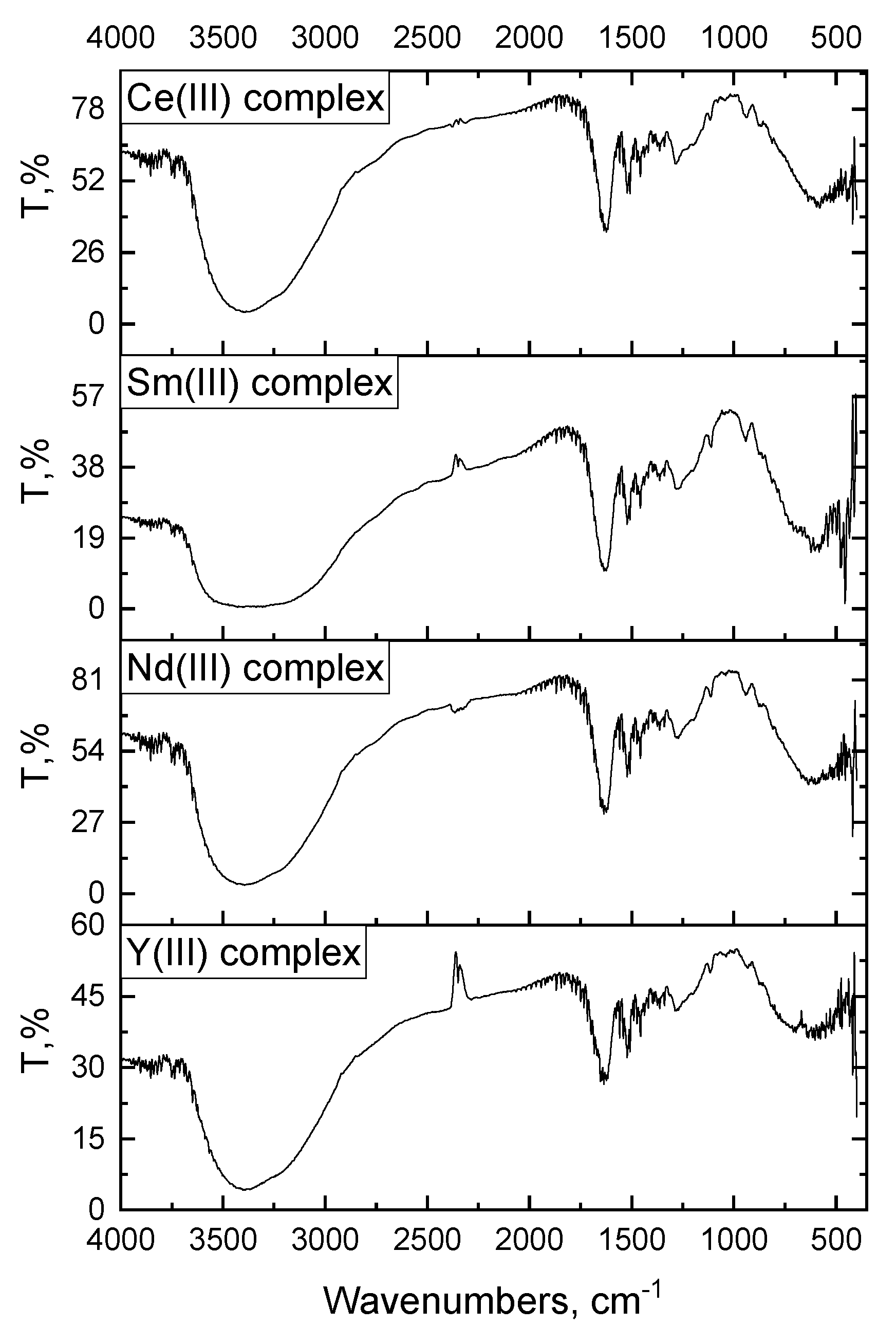
3.2. Infrared and Raman Spectra

The adrenaline free binding chemical structure belongs to the Cs point group [33]. The N-methyl secondary amino group of adrenaline has been classified as point mass CH2NHCH3. It contains 48 normal vibration modes, spread as Γvib = 33A′ + 15A of the Cs point group. All modes are active in both “infrared and visible Raman laser spectra”. Figure 2 and Figure 3 show the infrared and Raman spectra. Because of their poor symmetry and great complexity, the spectrum of the adrenaline molecule is difficult to understand, and because of our research goals, only 29 vibrations are investigated, which are dispersed as 19A′ vibrations and 10A′ vibrations.
The vibrations allocated to the basic frequencies for adrenaline were established based on the size and comparative strengths of the observable bands in similarity with the associated substances [33] and are summarized in Table S1a,b.
The infrared spectra show two absorption bands for the aromatic C–C vibrations of the benzene ring at 1525 and 1496 cm−1 and just one band at 1504 cm−1. The infrared spectra of a substituted benzene ring show a stretching vibration motion of aromatic C–H vibrations at 3035 and 3023 cm−1, while Raman spectra show it at 3024 and 3047 cm−1. The absorption region for the alcohol C–O group, due to its extending vibration, is 1200–1000 cm−1 which is absorbed in the infrared spectrum at 1157, 1175 and 1278 cm−1, while in the Raman spectrum is absorbed at 1177 and 1288 cm−1. The O–H vibrations infrared spectrum was at 3351, 3368 and 3386 cm−1. For the aliphatic moiety, the C–H stretching vibrations occur in the IR spectrum at 3000 cm−1 and in the Raman spectrum at 2995 cm−1. The stretching vibrations (C–C) motion of ν(C–C) of the aliphatic moiety and CH–CH2–NH–CH3 absorbances are at 1205 and 1141 cm−1, respectively. There are various bending deformation vibrations recorded at changed frequencies in the adrenaline-free moiety:
  • δ(C–C–C): (IR = 612, 633, 649 and 689 cm−1; Raman = 655 cm−1)
  • δ(C–O–H): (IR = 1256 and 1350 cm−1; Raman = 1346 cm−1)
  • δ(C–C–H): (IR = 1029, 1061, 1082 and 1105 cm−1; Raman = 1035, 1087 and 1109 cm−1)
  • δ(C–C–O): (IR = 483, 504, 512, 535, 583 and 598 cm−1; Raman = 491, 537 and 605 cm−1)
Using infrared and Raman laser spectra to compare free adrenaline ligand and its Y3+, Ce3+, Nd3+, and Sm3+ adrenaline adducts, we discovered that the distinctive bands ascribed to ν(O–H), ν(C–O) and δ(C–O–H) are shifted to lower wavenumbers, and their intensities are decreasing. These findings support the hypothesis that the adrenaline compounds of heavy metal ions are mediated by the two phenolic families of the monoamine moiety, as indicated in Figure 1a,b.
The Raman spectra of Y3+, Ce3+, Nd3+, and Sm3+ adrenaline adducts (Figure 3) show a spike enlargement with distortion in the extending vibration bands. This can be explained in light of the fact that the Raman analysis of fluorescent materials and structures is difficult to study due to the fluorescence intersection, which can overcome the integrally fragile Raman scattering simplification. The adrenaline bidentate ligand is thought to coordinate through the oxygens of phenolic groups, producing a steady compound [34,35].

3.3. Electronic Spectra

Two important bands of absorbances were detected at 299 and 376 nm and credited to the transitions π→π*, n→π*, correspondingly, for the benzene ring, phenolic, alcoholic, —NH, and methyl groups in the electronic spectra of adrenaline (Adr) free hormone moities (Figure 4). These transitions were shifted either to a lower or higher wavenumber of the adrenaline adducts spectra of (Figure 4); [Y2(Adr)2(H2O)8]Cl3·8H2O (297 and 308 nm), [Ce(Adr)2(H2O)2]Cl3·10H2O(295 and 306 nm), [Nd(Adr)2(H2O)2]Cl3·6H2O (295 and 308 nm) and [Sm(Adr)2(H2O)2]Cl3·12H2O (297–308 nm).
The coordination of the adrenaline ligand with the trivalent rare earth metal ions has been confirmed. At 299 nm, the UV-visible peak matching to the ligand’s π→π* transitions were detected [36]. As a result of coordination when binding with metal, the peaks of the π→π* transitions are shifted to a lower wavelength, indicating the production of M–Adr metal adducts.

3.4. 1Hnmr Spectra

The typical 1H–NMR spectrum of adrenaline [37] consisted of 10 distinct signals resulting from ten different proton environments. The signals in the 7.0–8.0 ppm range and at 9.10, 9.04, 8.69, 6.804, 6.749, 6.619, 5.96, 4.775, 2.94, and 2.555 ppm are common for protons coupled to an aromatic or benzene ring. The H of the aromatic phenolic family emits signals at 9.10 and 8.69 ppm, the alcoholic –OH group emits signals at 5.96 ppm, and the H+s of the secondary amine –NH group emits signals at 9.04 ppm. The ligational behavior of the ligand was shown by comparing the 1H–NMR spectra of adrenaline free moiety with the 1H–NMR spectrum of the [Al(Adr)3](NO3)3 adduct [38]. The presence of 2OH phenolic groups in the complexation behavior was suggested by the up–field of peaks at 9.10 and 8.69 ppm, which were attributed to H+s of the aromatic phenolic family to 8.363 and 8.913 ppm. The unshared secondary amine in the coordination process is ascribed to the downfield of –NH H+ from 9.04 ppm (Adr free ligand) to 9.264 ppm.

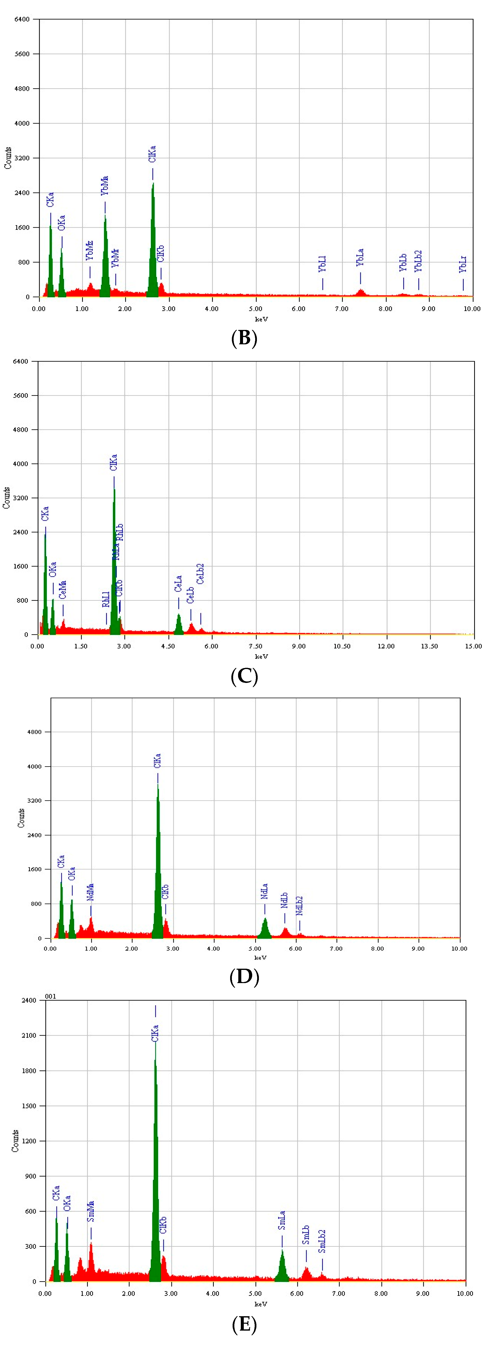
3.5. SEM and EDX Studies

SEM is a basic method that may be utilized to obtain a sense of the micro-features of the physical behavior of adrenaline adducts (Figure S1A–E). Even though this approach is not a reliable way to validate adduct formation, it may be used to check for the existence of a single component in synthetic adducts. The produced adducts exhibit a tiny particle dimension with nano-properties which result in the images. The generated adducts’ chemical examination by EDX shows a homogeneous distribution of the metal cation and adrenaline moiety. SEM studies of the shape of the surfaces of the adducts revealed tiny particles with a proclivity for forming aggregates of various forms in contrast to the starting materials. The peaks of these adducts’ EDX profiles (Figure 5A–E) relate to all components that make up the molecules of adrenaline adducts [Y2(Adr)2(H2O)8]Cl3·8H2O, [Ce(Adr)2(H2O)2]Cl3·10H2O, [Nd(Adr)2(H2O)2]Cl3·6H2O, and [Sm(Adr)2(H2O)2]Cl3·12H2O that were clearly identified, confirming the atoms ratio (molecular formulas).

3.6. Biological Evaluation

The antibacterial property of goal adducts against gram-positive bacteria, Bacillus subtilis, Streptococcus penumonia, and Staphylococcus aureus and gram-negative bacteria, Escherichia coli and Pseudomonas aeruginosa, were tested in biological assessments. Additionally, two strains of fungus, Aspergillus niger and Penicillium, were also tested. Table 2 shows the results of the agar disc dispersion tests for the antibacterial activity of the target compounds, which are also shown in Figure S2. The diameters of the zone of inhibition (in mm) of the standard drug, tetracycline, against the gram-positive bacteria, B. subtilis and S. aureus, in addition to the gram-negative bacteria, E. coli and P. aeruginosa, were 36, 30, 31 and 35 mm, correspondingly, while the standard drug, amphotericin B, against Aspergillus flavus and Candida albicans were 18 and 19. Under matching circumstances, Table 2 displays that the adrenaline free binding moiety, [Y2(Adr)2(H2O)8]Cl3·8H2O, [Ce(Adr)2(H2O)2]Cl3·10H2O, [Nd(Adr)2(H2O)2]Cl3·6H2O, and [Sm(Adr)2(H2O)2]Cl3·12H2O adducts (10, 5, 5, 15, 5, 5 and 5), (5, 15, 15, 5, 5,15 and 5), (15, 5, 5, 0, 5, 10 and 10), (10, 5, 15, 10, 5, 15 and 10), respectively, relate to Bacillus subtilis, Streptococcus pneumonia, Staphylococcus aureus, Escherichia coli, Pseudomonas aeruginosa, Aspergillus niger and penicillium.

4. Conclusions

Four rare earth metal adrenaline complexes were formed from the reaction of adrenaline with Y3+, Ce3+, Nd3+ and Sm3+ ions in a methanolic solvent at 60 °C. The final reaction products have been isolated and characterized using elemental analyses (% of carbon, hydrogen and nitrogen), conductivity measurements, mid infrared, Raman laser, UV—Vis spectra, scanning electron microscopy and energy-dispersive X-ray spectroscopy (EDX). Upon the spectroscopic, conductivity and elemental analyses, the stoichiometric reactions indicated that the data obtained refer to 1:1 (M:L) for the Y3+, complex [Y2(Adr)2(H2O)8]Cl3·8H2O, while the molar ratio was 1:2 (M:L) for Ce3+, Nd3+ and Sm3+ with formulas [Ce(Adr)2(H2O)2]Cl3·10H2O, [Nd(Adr)2(H2O)2]Cl3·6H2O, and [Sm(Adr)2(H2O)2]Cl3·12H2O. The infrared and Raman laser spectra interpreted the mode of interactions which associated through the two phenolic groups of the catechol moiety. The adrenaline compounds have been screened for their in vitro antibacterial activity against four bacteria, both gram-positive and gram-negative, and two strains of fungus. The metal complexes were shown to possess more antibacterial and antifungal activities than the free adrenaline compound.

Supplementary Materials

The following are available online at https://www.mdpi.com/article/10.3390/cryst11121536/s1, Figure S1A: SEM photo of adrenaline free binding moiety, Figure S1B: SEM photo of [Y2(Adr)2(H2O)8]Cl3.8H2Oadduct, Figure S1C: SEM photo of [Nd(Adr)2(H2O)2]Cl3.6H2O adduct, Figure S1D: SEM photo of [Ce(Adr)2(H2O)2]Cl3.10H2O adduct, Figure S1E: SEM photo of [Sm(Adr)2(H2O)2]Cl3.12H2Oadduct, Figure S2: The width of the inhibition zone of adrenaline and its adducts against bacteria and fungus, Table S1a: Infrared frequency categorization (cm–1) and preliminary designations of adrenaline and its adducts, Table S1b: Categorization Raman frequencies (cm–1) and tentative adrenaline.

Author Contributions

Conceptualization, S.A.A.Y., A.S.A.-W. and M.S.R.; methodology, I.I.S.A., H.I.T., A.M.N., S.A.E.-M. and M.S.R.; software, O.B.I. and A.M.N.; validation, A.G., S.A.A.Y. and M.S.R.; formal analysis, A.G., A.S.A.-W., I.I.S.A., S.A.E.-M. and A.S.A.-W.; investigation, A.G., H.I.T., O.B.I. and M.S.R.; resources, S.A.A.Y., A.M.N. and S.A.E.-M.; data curation, S.A.A.Y., A.S.A.-W. and M.S.R.; writing—original draft preparation, A.G., A.M.N., A.S.A.-W. and M.S.R.; writing—review and editing, A.G., A.M.N. and M.S.R.; visualization, O.B.I., A.S.A.-W. and S.A.E.-M.; supervision, M.S.R.; project administration, A.G.; funding acquisition, S.A.A.Y. and A.M.N. All authors have read and agreed to the published version of the manuscript.

Funding

This work was funded by the Deanship of Scientific Research at Princess Nourah Bint Abdulrahman University, through the Research Groups Program Grant No. (RGP-1440-0003) (3).

Institutional Review Board Statement

The experiment did not require ethical approval.

Informed Consent Statement

Not applicable.

Data Availability Statement

The data used to support the findings of this study are included within the article.

Acknowledgments

This work was funded by the Deanship of Scientific Research at Prin-cess Nourah Bint Abdulrahman University, through the Research Groups Program Grant No. (RGP-1440-0003) (3).

Conflicts of Interest

The authors declare no conflict of interest.

References

  1. Berecek, K.H.; Brody, M.J. Evidence for a neurotransmitter role for epinephrine derived from the adrenal medulla. Am. J. Physiol. 1982, 242, H593–H601. [Google Scholar] [CrossRef]
  2. Cannon, W.B.; Newton, H.F.; Bright, E.M.; Menkin, V.; Moore, R.M. Some Aspects of the Physiology of Animals Surviving Complete Exclusion of Sympathetic Nerve Impulses. Am. J. Physiol. 1929, 89, 84–107. [Google Scholar] [CrossRef]
  3. Hoffmann, B.B. Adrenoceptor-activating and other sympathminetic drugs. In Basic and Clinical Pharmacology, 6th ed.; Katzung, B.G., Ed.; Appleton and Lange: New York, NY, USA, 1995; pp. 115–131. [Google Scholar]
  4. Burtis, C.A.; Ashwood, E.R. (Eds.) Tietz Textbook of Clinical Chemistry, 3rd ed.; W.B. Saunders: Philadelphia, PA, USA, 1998; pp. 1570–1572. [Google Scholar]
  5. Mefford, I.N.; Lister, R.G.; Ota, M.; Linnoila, M. Antagonism of ethanol intoxication in rats by inhibitors of phenylethanolamine N-methyltransferase. Alcohol. Clin. Exp. Res. 1990, 14, 53–57. [Google Scholar] [CrossRef] [PubMed] [Green Version]
  6. Jameson, R.F.; Neillie, W.F.S. Complexes formed by adrenaline and related compounds with transition-metal ions—III: The stabilities of some first-row transition-metal complexes. J. Inorg. Nucl. Chem. 1966, 28, 2667–2675. [Google Scholar] [CrossRef]
  7. Grgas-Kuznar, B.; Simeon, V.L.; Weber, O.A. Complexes of adrenaline and related compounds with Ni2+, Cu2+, Zn2+, Cd2+ and Pb2+. J. Inorg. Nucl. Chem. 1974, 36, 2151–2154. [Google Scholar] [CrossRef]
  8. Hughes, M.S.; Crow, D.R.; Birch, N.J. Magnesium, adenosine triphosphate, and catecholamine complexes: Application of new means of identifying solute-solute interactions of biological significance using NMR chemical shift data. Magnes. Res. 1992, 5, 89–96. [Google Scholar]
  9. Ryan, T.P.; Miller, D.M.; Aust, S.D. The role of metals in the enzymatic and nonenzymatic oxidation of epinephrine. J. Biochem. Toxicol. 1993, 8, 33–39. [Google Scholar] [CrossRef] [PubMed]
  10. Shen, Z.Q.; Ouyang, J. Handbook on the Physics and Chemistry of Rare Earths; Elsevier Science Publisher: North Holand, The Netherlands, 1987; p. 61. [Google Scholar]
  11. Narain, G.; Jain, J.K. Screening of metal complexes for biological activity. III: Copper(II) and nickel(II) tungstate complexes with ethylenediamine and propylenediamine. J. Indian Chem. Soc. 1994, 71, 101. [Google Scholar]
  12. Gerard, C.; Chehhal, H. Stability of metal complexes with ligands of biological interest: Dopamine and adrenaline. Bull. Soc. Chim. Fr. 1997, 134, 1069–1074. [Google Scholar]
  13. Gao, F.; Han, J.F.; Wu, Z.J.; Wang, S.Y.; Niu, C.J.; Ni, J.Z. Stability and luminescence properties of Tb (III) complexes with adrenaline. Chin. Chem. Lett. 1998, 9, 1033. [Google Scholar]
  14. Materazzi, S.; Nugnes, C.; Gentili, A.; Curini, R. Complexes of adrenaline with some divalent transition-metal ions. Thermochim. Acta 2001, 369, 167–173. [Google Scholar] [CrossRef]
  15. Materazzi, S.; Vasca, E.; Tentolini, U.; Aquilig, S.; Curini, R. A thermoanalytical study of unusual adrenaline complexes. Thermochim. Acta 2002, 389, 179–184. [Google Scholar] [CrossRef]
  16. Bauer, A.W.; Kirby, W.M.; Sherris, C.; Turck, M. Antibiotic susceptibility testing by a standardized single disc method. Am. J. Clin. Pathol. 1966, 45, 493. [Google Scholar] [CrossRef]
  17. Pfaller, M.A.; Burmeister, L.; Bartlett, M.A.; Rinaldi, M.G. Multicenter evaluation of four methods of yeast inoculum preparation. J. Clin. Microbiol. 1988, 26, 1437. [Google Scholar] [CrossRef] [Green Version]
  18. Jabłońska-Wawrzycka, A.; Rogala, P.; Czerwonka, G.; Gałczyńska, K.; Drabik, M.; Dańczuk, M. Ruthenium Complexes with 2-Pyridin-2-yl-1H-benzimidazole as Potential Antimicrobial Agents: Correlation between Chemical Properties and Anti-Biofilm Effects. Int. J. Mol. Sci. 2021, 22, 10113. [Google Scholar] [CrossRef] [PubMed]
  19. Santos, N.E.; Braga, S.S. Redesigning Nature: Ruthenium Flavonoid Complexes with Antitumour, Antimicrobial and Cardioprotective Activities. Molecules 2021, 26, 4544. [Google Scholar] [CrossRef]
  20. Manimaran, P.; Balasubramaniyan, S.; Azam, M.; Rajadurai, D.; Al-Resayes, S.I.; Mathubala, G.; Manikandan, A.; Muthupandi, S.; Tabassum, Z.; Khan, I. Synthesis, Spectral Characterization and Biological Activities of Co(II) and Ni(II) Mixed Ligand Complexes. Molecules 2021, 26, 823. [Google Scholar] [CrossRef] [PubMed]
  21. Fudulu, A.; Olar, R.; Maxim, C.; Scăeţeanu, G.V.; Bleotu, C.; Matei, L.; Chifiriuc, M.C.; Badea, M. New Cobalt (II) Complexes with Imidazole Derivatives: Antimicrobial Efficiency against Planktonic and Adherent Microbes and In Vitro Cytotoxicity Features. Molecules 2021, 26, 55. [Google Scholar] [CrossRef]
  22. Măciucă, A.-M.; Munteanu, A.-C.; Uivarosi, V. Quinolone Complexes with Lanthanide Ions: An Insight into their Analytical Applications and Biological Activity. Molecules 2020, 25, 1347. [Google Scholar] [CrossRef] [Green Version]
  23. Gacki, M.; Kafarska, K.; Pietrzak, A.; Korona-Głowniak, I.; Wolf, W.M. Double Palindrome Water Chain in Cu(II) Theophylline Complex. Synthesis, Characterization, Biological Activity of Cu(II), Zn(II) Complexes with Theophylline. Crystals 2020, 10, 97. [Google Scholar] [CrossRef] [Green Version]
  24. Al-Wasidi, A.S.; Naglah, A.M.; Refat, M.S.; El-Megharbel, S.M.; Kalmouch, A.; Moustafa, G.O. Synthesis, spectroscopic characterization and antimicrobial studies of Mn(II), Co(II), Ni(II), Cr(III) and Fe(III) melatonin drug complexes. Egypt. J. Chem. 2020, 63, 1469–1481. [Google Scholar]
  25. Naglah, A.M.; Moustafa, G.O.; Elhenawy, A.A.; Mounier, M.M.; El-Sayed, H.; Al-Omar, M.A.; Almehizia, A.A.; Bhat, M.A. Nα-1, 3-Benzenedicarbonyl-bis-(Amino Acid) and Dipeptide Candidates: Synthesis, Cytotoxic, Antimicrobial, Antifungal and Molecular Docking Investigation. Drug Des. Dev. Ther. 2021, 15, 1315–1332. [Google Scholar] [CrossRef] [PubMed]
  26. Sgarbossa, P.; Śliwińska-Hill, U.; Guedes da Silva, M.F.C.; Bażanów, B.; Pawlak, A.; Jackulak, N.; Poradowski, D.; Pombeiro, A.J.L.; Smoleński, P. Pentafluorophenyl Platinum(II) Complexes of PTA and Its N-Allyl and N-Benzyl Derivatives: Synthesis, Characterization and Biological Activity. Materials 2019, 12, 3907. [Google Scholar] [CrossRef] [Green Version]
  27. Chang, J.C.; Hsueh, P.R.; Wu, J.J.; Ho, S.W.; Hsieh, W.C.; Luh, K.T. Antimicrobial Susceptibility of Flavobacteria as Determined by Agar Dilution and Disk Diffusion Methods. Antimicrob. Agents Chemother. 1997, 41, 1301. [Google Scholar] [CrossRef] [PubMed] [Green Version]
  28. National Committee for Clinical Laboratory Standards. Methods for Dilution Antimicrobial Susceptibility Tests for Bacteria That Grow Aerobically; Approved standard M7-A3; National Committee for Clinical Laboratory Standards: Villanova, PA, USA, 1993. [Google Scholar]
  29. National Committee for Clinical Laboratory Standards. Reference Method for Broth Dilution Antifungal Susceptibility Testing of Conidium-Forming Filamentous Fungi: Proposed Standard M38-A; NCCLS: Wayne, PA, USA, 2002. [Google Scholar]
  30. National Committee for Clinical Laboratory Standards. Methods for Antifungal Disk Diffusion Susceptibility Testing of Yeast: Proposed Guideline M44-P; NCCLS: Wayne, PA, USA, 2003. [Google Scholar]
  31. Liebowitz, L.D.; Ashbee, H.R.; Evans, E.G.V.; Chong, Y.; Mallatova, N.; Zaidi, M.; Gibbs, D.; Global Antifungal Surveillance Group. A two year global evaluation of the susceptibility of Candida species to fluconazole by disk diffusion. Diagn. Microbiol. Infect. Dis. 2001, 4, 27–33. [Google Scholar] [CrossRef]
  32. Matar, M.J.; Ostrosky-Zeichner, L.; Paetznick, V.L.; Rodriguez, J.R.; Chen, E.; Rex, J.H. Correlation between E-Test, Disk Diffusion, and Microdilution Methods for Antifungal Susceptibility Testing of Fluconazole and Voriconazole. Antimicrob. Agents Chemother. 2003, 47, 1647–1651. [Google Scholar] [CrossRef] [Green Version]
  33. Gunasekaran, S.; Kumar, R.T.; Ponnusamy, S. Vibrational spectra and normal coordinate analysis of adrenaline and dopamine. Indian J. Pure Appl. Phys. 2007, 45, 884–892. [Google Scholar]
  34. Bell, S.E.J.; Sirimuthu, N.M.S. Quantitative surface-enhanced Raman spectroscopy. Chem. Soc. Rev. 2008, 37, 1012–1024. [Google Scholar] [CrossRef] [PubMed]
  35. Kagan, M.R.; McCreery, R.L. Reduction of Fluorescence Interference in Raman Spectroscopy via Analyte Adsorption on Graphitic Carbon. Anal. Chem. 1994, 66, 4159–4165. [Google Scholar] [CrossRef]
  36. Oztürk, O.F.; Şekerci, M.; Ozdemir, E. Synthesis of 5, 6-O-cyclohexylidene-1-amino-3-azahexane and its Co(II), Ni(II), Cu(II) complexes. Russ. J. Coord. Chem. 2005, 31, 687. [Google Scholar] [CrossRef]
  37. Available online: https://www.chemicalbook.com/SpectrumEN_329-63-5_1HNMR.htm (accessed on 1 May 2021).
  38. Ibrahim, O.B.; Mohamed, M.A.; Refat, M.S. Study the chemical composition and biological outcomes resulting from the interaction of the hormone adrenaline with heavy elements: Infrared, Raman, electronic, 1H NMR, XRD and SEM studies. J. Mol. Struct. 2014, 1056–1057, 13–24. [Google Scholar] [CrossRef]
Figure 1. (a): Suggested structures of [Y2(Adr)2(H2O)8]Cl3·8H2O adduct. (b): Suggested structures of [Ce(Adr)2(H2O)2]Cl3·10H2O, [Nd(Adr)2(H2O)2]Cl3·6H2O and [Sm(Adr)2(H2O)2]Cl3·12H2O adducts.
Crystals 11 01536 g001
Figure 2. Infrared spectra of [Y2(Adr)2(H2O)8]Cl3·8H2O, [Ce(Adr)2(H2O)2]Cl3·10H2O, [Nd(Adr)2(H2O)2]Cl3·6H2O, and [Sm(Adr)2(H2O)2]Cl3·12H2O adducts.
Crystals 11 01536 g002
Figure 3. Raman spectra of [Y2(Adr)2(H2O)8]Cl3·8H2O, [Ce(Adr)2(H2O)2]Cl3·10H2O, [Nd(Adr)2(H2O)2]Cl3·6H2O, and [Sm(Adr)2(H2O)2]Cl3·12H2O adducts.
Crystals 11 01536 g003
Figure 4. Electronic spectrum of adrenaline free and their complexes [Y2(Adr)2(H2O)8]Cl3·8H2O, and [Ce(Adr)2(H2O)2]Cl3·10H2O, [Nd(Adr)2(H2O)2]Cl3·6H2O, Sm(Adr)2(H2O)2]Cl3·12H2O.
Crystals 11 01536 g004
Figure 5. (A). EDX free adrenaline spectrum. (B). EDX spectrum of [Y2(Adr)2(H2O)8]Cl3·8H2O adduct. (C). EDX spectrum of [Ce(Adr)2(H2O)2]Cl3·10H2O adduct. (D). EDX spectrum of [Nd(Adr)2(H2O)2]Cl3·6H2O adduct. (E). EDX spectrum of [Sm(Adr)2(H2O)2]Cl3·12H2O adduct.
Crystals 11 01536 g005aCrystals 11 01536 g005b
Table 1. Adrenaline adducts: physical characteristics, microanalytical data, and molar conductance data.
ComplexesMp, °CM.Wt, g/molColorElemental Analysis (%)
Found (Calcd.)
Λm
−1 cm2 mol−1)
Yields, %
CHN
[Y2(Adr)2(H2O)8]Cl3·8H2O>250938.579White(23.01)
23.20
(4.03)
4.18
(2.98)
2.76
44974
[Ce(Adr)2(H2O)2]Cl3·10H2O>250828.887White(26.06)
26.29
(6.03)
6.92
(3.38)
3.36
34779
[Nd(Adr)2(H2O)2]Cl3·6H2O>250761.007White(28.38)
28.12
(5.52)
5.96
(3.68)
3.59
55872
[Sm(Adr)2(H2O)2]Cl3·12H2O>250875.127White(24.68)
24.54
(6.17)
6.11
(3.20)
3.16
70975
Table 2. Adrenaline and its adducts’ inhibition zone diameter (mm/mg sample).
SampleInhibition Zone Diameter (mm/mg Sample)
Bac. sub.
(G+)
Srep. pen.
(G+)
Stap. aure.
(G+)
E. coli
(G)
Pseu. spp.
(G)
Asper. niger
(Fungus)
Penici. spp.
(Fungus)
Control: DMSO0.00.0 0.00.00.00.0
Standard Tetracycline
Antibacterial agent
3631-3530--
Amphotericin B
Antifungal agent
-----1819
Adr86-1050.00.0
Y(III)105515555
Ce(III)5151555155
Nd(III)1555-51010
Sm(III)105151051510
Publisher’s Note: MDPI stays neutral with regard to jurisdictional claims in published maps and institutional affiliations.

