
Cocrystals versus Salts of Fluorescein


Inorganic Chemistry Department, Faculty of Chemistry, University of Bucharest, 23 Dumbrava Rosie, 020464 Bucharest, Romania
*
Author to whom correspondence should be addressed.
Crystals 2021, 11(10), 1217; https://doi.org/10.3390/cryst11101217
Submission received: 22 September 2021
/
Revised: 2 October 2021
/
Accepted: 6 October 2021
/
Published: 9 October 2021
(This article belongs to the Special Issue Feature Papers in Organic Crystalline Materials)
Abstract
:A series of nitrogen-containing organic molecules (4,4’-bipyridyl; trans-1,2-bis(4-pyridyl)ethylene; 1,2-bis(4-pyridyl)ethane; 4-aminopyridine and trans-1,4-diaminocyclohexane) was envisaged for cocrystallization experiments together with fluorescein. These compounds, containing pyridyl or/and amino nitrogen atoms, can act either as hydrogen bond acceptors for the phenol groups of fluorescein-generating cocrystals or as proton acceptors forming organic salts. Five cocrystals were obtained with the partners containing only pyridyl groups: {(H2Fl)2(bipy)} (1); {(H2Fl)2(bipy)(MeOH)2} (2); {(H2Fl)2(bpete)(EtOH)2} (3); {(H2Fl)(bpete)} (4); {(H2Fl)(bpeta)} (5). The compounds bearing amino groups deprotonate fluorescein producing salts: [(HFl)(Hampy)]∙2H2O (6); [(HFl)(Hampy)] (7); [(Fl)(H2diach)]∙3H2O (8); [(HFl)2(H2diach)]∙2H2O∙EtOH (9); and [(HFl)2(Fl)2(H2diach)3]∙4H2O (10). Optical properties of the cocrystals and salts were investigated.
1. Introduction
Fluorescein is a xanthene derivative widely used as platform for designing various chromogenic or fluorogenic probes [1]. Fluorescein derivatives can serve as fluorescent chelators for Zn(II) [2,3], Cu(II) [4], Co(II), Ni(II) [5], Hg(II) or Ag(I) [6] metal ions. As biological applications of the fluorescein derivatives we can also mention detection of nitric oxide production in some tissues [7], detection of homocysteine and cysteine [8], detection of hypochlorite [9], study of serotonin receptors [10], enzymes [11] and live bacteria detection [12].
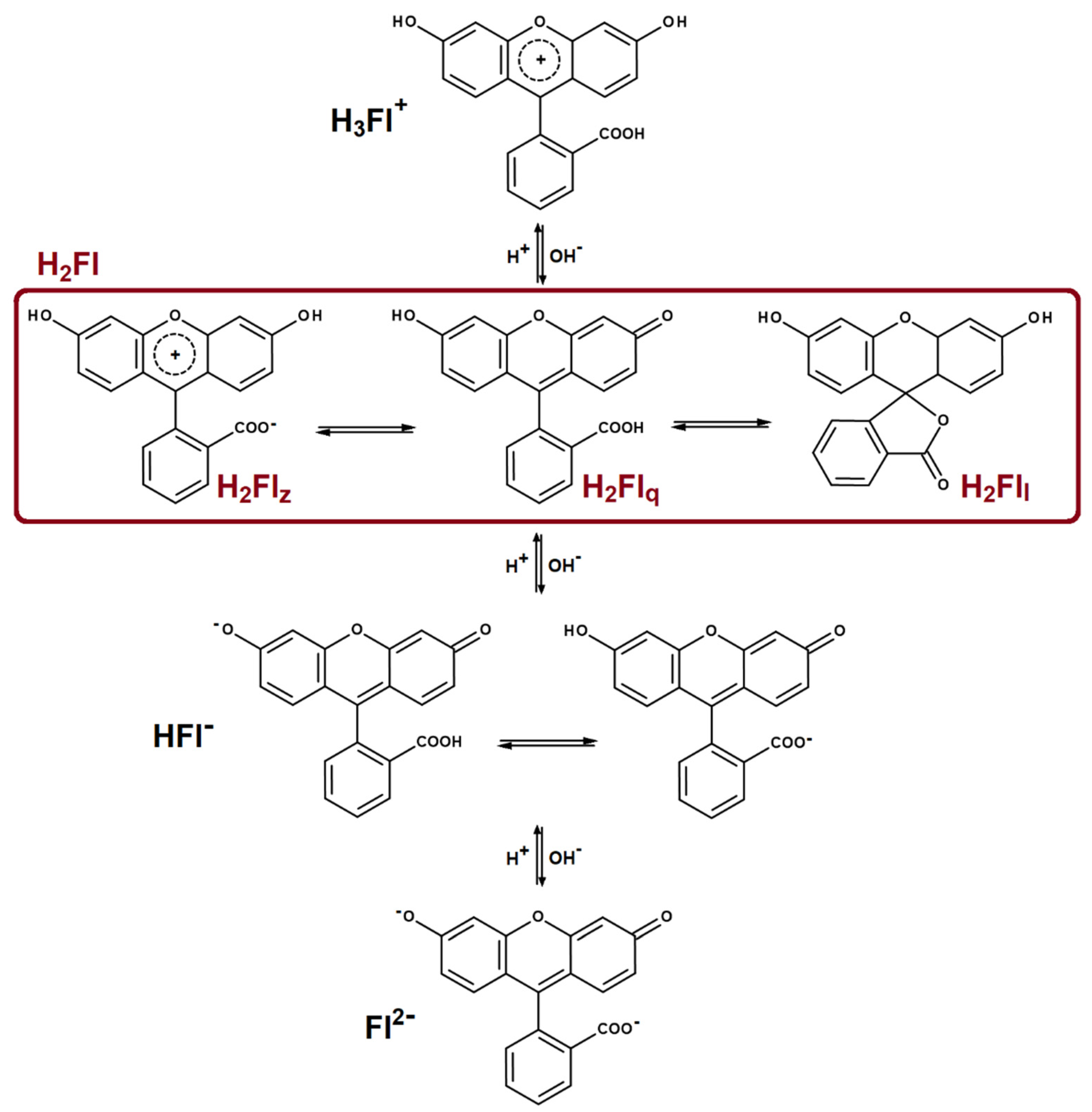
The chromogenic mechanism of fluorescein is based on protonation–deprotonation reactions. Due to the biological applications of the fluorescein dyes, solution studies have attracted particular attention. The ionization equilibria of fluorescein are presented in Scheme 1. Depending on pH, in solution can be identified cationic (H3Fl+), neutral (H2Fl) or anionic species (HFl− and Fl2−). The neutral form (H2Fl) presents in solution three tautomers: zwitterion (H2Flz), quinonoid (H2Flq) and lactone (H2Fll). For the monoanionic form (HFl−), the phenolate tautomer appears in small quantities only in pure solvents such as DMSO, acetonitrile or acetone [13].
In solid state, the three tautomers of the neutral form are characterized by different colors: the zwitterionic form is yellow, the quinonoid form is red and the lactonoid form of fluorescein is colorless. The crystal structures of H2Flq and H2Flz have been determined by powder X-ray diffraction [14,15]. The crystal structure of the pure lactonoid form of fluorescein has not been reported. The lactonoid form crystallizes with solvent molecules, and the methanol [16], acetone [15,17] and 1,4-dioxane [15,18] solvates were structurally characterized by X-ray diffraction on a single crystal.
The lactone (H2Fll) can also cocrystallize with nitrogen-containing heteroaromatic molecules: acridine, phenanthiridine and pyrazine [19]. The use of basic nitrogen-containing molecules as partners for cocrystallization offers the theoretical possibility of proton transfer with the formation of organic salts. Such salts were reported for eosin (2′,4′,5′,7′-tetrabromofluorescein) with 4-aza-1-azoniabicyclo[2.2.2]octane [20] and guanidium cations [21]. In Mannich derivatives of fluorescein, the presence of basic nitrogen atoms attached to the xanthene core allows the intramolecular transfer of the protons with the formation of zwitterions [22].
In this paper we report the synthesis and structural characterization of organic cocrystals and salts of fluorescein with different nitrogen-containing partners: 4,4’-bipyridyl (bipy); trans-1,2-bis(4-pyridyl)ethylene (bpete); 1,2-bis(4-pyridyl)ethane (bpeta); 4-aminopyridine (ampy) or trans-1,4-diaminocyclohexane (diach). Determination of the specific solid form (cocrystal or salt) is of particular interest especially for active pharmaceutical ingredients [23].
2. Materials and Methods
2.1. Synthesis
The chemicals used as well as all the solvents were of reagent grade and were purchased from commercial sources.
2.1.1. Synthesis of {(H2Fl)2(bipy)} (1)
Fluorescein (0.0664 g, 0.2 mmol) and 4,4’-bipyridyl (0.0156 g, 0.1 mmol) were dissolved in 20 mL ethanol and 20 mL acetonitrile. The mixture was stirred for 15 min and then filtered. The light-yellow crystals formed after several days. FT-IR (cm−1): 3373s, 3053w, 2891w, 2800w, 2166–2700br, 1758vs, 1602vs, 1501s, 1449s, 1338s, 1257s, 1175vs, 1098s, 816s, 680s.
2.1.2. Synthesis of {(H2Fl)2(bipy)(MeOH)2} (2)
Fluorescein (0.1328 g, 0.4 mmol) and 4,4’-bipyridyl (0.0312 g, 0.2 mmol) were dissolved in 80 mL methanol. The mixture was stirred for 15 min and then filtered. The light-yellow crystals formed after several days. FT-IR (cm−1): 3580s, 3478vs, 3035s, 2913s, 2810s, 2692s, 2000-2589br, 1735vs, 1595vs, 1460vs, 1331s, 1279vs, 1176vs, 995s, 833s, 753s, 614m.
2.1.3. Synthesis of {(H2Fl)2(bpete)(EtOH)2} (3)
Fluorescein (0.0664 g, 0.2 mmol) and trans-1,2-bis(4-pyridyl)ethene (0.0182 g, 0.1 mmol) were dissolved in 20 mL ethanol and 20 mL acetonitrile. The mixture was stirred for 15 min and then filtered. After several days yellow prismatic crystals formed. FT-IR (cm−1): 3400br, 3039m, 28886m, 2801m, 2676s, 2582s, 2113–2468br, 1919m, 1750vs, 1595vs, 1503s, 1420m, 1333m, 1284s, 1246s, 1178s, 1000m, 825s, 543s.
2.1.4. Synthesis of {(H2Fl)(bpete)} (4)
Fluorescein (0.0997 g, 0.3 mmol) and trans-1,2-bis(4-pyridyl)ethene (0.0546 g, 0.3 mmol) were dissolved in 40 mL ethanol and 20 mL water. The mixture was stirred for about 15 min and then filtered. After the slow evaporation of the solvent yellow crystals formed. FT-IR (cm−1): 3039w, 2891w, 2803w, 2747w, 2681w, 2580m, 2108-2477br, 1751vs, 1594vs, 1465s, 1417s, 1384s, 1247vs, 1183s, 1070s, 962s, 827s, 587s.
2.1.5. Synthesis of {(H2Fl)(bpeta)} (5)
Fluorescein (0.0664 g, 0.2 mmol) and 1,2-bis(4-pyridyl)ethane (0.0364 g, 0.2 mmol) were dissolved in 40 mL methanol followed by 15 min of stirring and then the mixture was filtered. After one-week yellow crystals were obtained. FT-IR (cm−1): 3496w, 3037m, 2950m, 2895m, 2804m, 2751m, 2680s, 2580s, 2480–2126br, 1834w, 1752vs, 1598vs, 1465s, 1418s, 1332s, 1246vs, 1185vs, 1098vs, 1001s, 895s, 819s, 686s.
2.1.6. Synthesis of [(HFl)(Hampy)]∙2H2O (6) and [(HFl)(Hampy)] (7)
Compound 6 was obtained by dissolving fluorescein (0.0664 g, 0.2 mmol) and 4-aminopyridine (0.0188 g, 0.2 mmol) in 20 mL ethanol (96%) and 20 mL acetonitrile. The mixture was stirred for 15 min and then filtered, and it was left for slow evaporation of the solvent. After several days prismatic orange-red crystals of 6 appeared on the wall of the beaker. The precipitate obtained on the bottom of the beaker was dissolved in 20 mL ethanol and 20 mL water, then it was stirred for 15 min and filtered. After several days red needle crystals of 7 were obtained. FT-IR (cm−1) (6): 3625w, 3166s, 3098s, 2910s, 2669s, 2178–2582br, 2045w, 1873br, 1645s, 1571vs, 1507vs, 1460vs, 1366vs, 1313vs, 1275vs, 1194s, 1112s, 829s, 656m; (7): 3640w, 3065m, 2024–2601br, 1764w, 1600vs, 1565vs, 1447vs, 1282vs, 1180vs, 1095vs, 912m, 837s, 752s, 645m.
2.1.7. Synthesis of [(Fl)(H2diach)]∙3H2O (8)
Fluorescein (0.0664 g, 0.2 mmol) and trans-1,4-diaminocyclohexane (0.0228 g, 0.2 mmol) were dissolved in 40 mL ethanol and 20 mL water. The mixture was stirred for 15 min and then filtered. Red crystals formed after several days. FT-IR (cm−1): 3639w, 3427w, 2861s, 2570s, 1690–2000br, 1627w, 1568vs, 1456vs, 1380vs, 1325vs, 1271vs, 1208vs, 1158s, 1090s, 908m, 845m, 582w.
2.1.8. Synthesis of [(HFl)2(H2diach)]∙2H2O∙EtOH (9)
Fluorescein (0.1328 g, 0.4 mmol) and trans-1,4-diaminocyclohexane (0.0228 g, 0.2 mmol) were dissolved in 40 mL ethanol and 20 mL water. The mixture was stirred for 15 min and then filtered. After a week red crystals formed. FT-IR (cm−1): 3600–3047br, 2931s, 2861s, 2611s, 2111–2554br, 1907w, 1621vs, 1564vs, 1457vs, 1380vs, 1324vs, 1204s, 1159s, 1051s, 836s, 665m.
2.1.9. Synthesis of [(HFl)2(Fl)2(H2diach)3]∙4H2O (10)
Fluorescein (0.1328 g, 0.4 mmol) and trans-1,4-diaminocyclohexane (0.0342 g, 0.3 mmol) were dissolved in 50 mL ethanol and 20 mL water. The mixture was stirred for 15 min and then filtered. Red crystals formed after a week. FT-IR (cm−1): 3503w, 3431w, 1628s, 1567vs, 1455s, 1377vs, 1321vs, 1203s, 1092s, 833s, 589s.
2.2. Physical Measurements
2.2.1. X-ray Structure Determination
X-ray diffraction measurements for the crystals 1–3, 5–7, 9 and 10 were performed on a STOE IPDS II diffractometer, operating with a Mo-Kα (λ = 0.71073 Å) X-ray tube with a graphite monochromator. X-ray diffraction measurements for crystals 4 and 8 were performed on a Rigaku XtaLAB Synergy-S diffractometer operating with a Mo-Kα (λ = 0.71073 Å) micro-focus sealed X-ray tube. The structures were solved by direct methods and refined by full-matrix least-squares techniques based on F2. The non-H atoms were refined with anisotropic displacement parameters. Calculations were performed using SHELX-2014 or SHELX-2018 crystallographic software packages. A summary of the crystallographic data and the structure refinement for crystals 1–10 are given in Table S1. CCDC reference numbers: 2111108–2111117.
The X-ray powder diffraction measurements (XRPD) were carried out on a Proto AXRD Benchtop using Cu-Kα radiation with a wavelength of 1.54059 Å in the 2θ range of 5–35°.
2.2.2. Spectroscopy
IR spectra were recorded on an FT-IR Bruker Vertex 70 spectrometer in the 4500–400 cm−1 range using the ATR technique. The following abbreviations were used: w = weak, m = medium, s = strong, v = very, br = broad. Absorption spectra on powder (diffuse reflectance technique) were measured with a JASCO V-670 spectrophotometer. The fluorescence spectra were collected on powder using a JASCO FP-6500 spectrofluorometer.
3. Results
A series of nitrogen-containing organic molecules, namely 4,4’-bipyridyl; trans-1,2-bis(4-pyridyl)ethylene; 1,2-bis(4-pyridyl)ethane; 4-aminopyridine and trans-1,4-diaminocyclohexane, was envisaged for cocrystallization experiments together with fluorescein. These compounds contain pyridyl or/and amino nitrogen atoms and, theoretically, these groups can act either as hydrogen bond acceptors for the phenol groups of fluorescein or as proton acceptors. The following ten compounds were obtained using solution-based crystallization methods (slow evaporation at room temperature) and different stoichiometric ratios between the components: {(H2Fl)2(bipy)} (1); {(H2Fl)2(bipy)(MeOH)2} (2); {(H2Fl)2(bpete)(EtOH)2} (3); {(H2Fl)(bpete)} (4); {(H2Fl)(bpeta)} (5); [(HFl)(Hampy)]∙2H2O (6); [(HFl)(Hampy)] (7); [(Fl)(H2diach)]∙3H2O (8); [(HFl)2(H2diach)]∙2H2O∙EtOH (9); and [(HFl)2(Fl)2(H2diach)3]∙4H2O (10). The cocrystals are indicated by braces (1–5) whereas the salts are indicated by square brackets (6–10). All ten systems were structurally characterized by X-ray diffraction on a single crystal.
3.1. Description of the Crystal Structures
For a molar ratio between fluorescein and 4,4’-bipyridyl of 2:1, two types of cocrystals were obtained depending on the solvents used in the crystallization experiments: {(H2Fl)2(bipy)} (1) crystallizes from an ethanol-acetonitrile mixture, while {(H2Fl)2(bipy)(MeOH)2} (2) crystallizes from methanol.
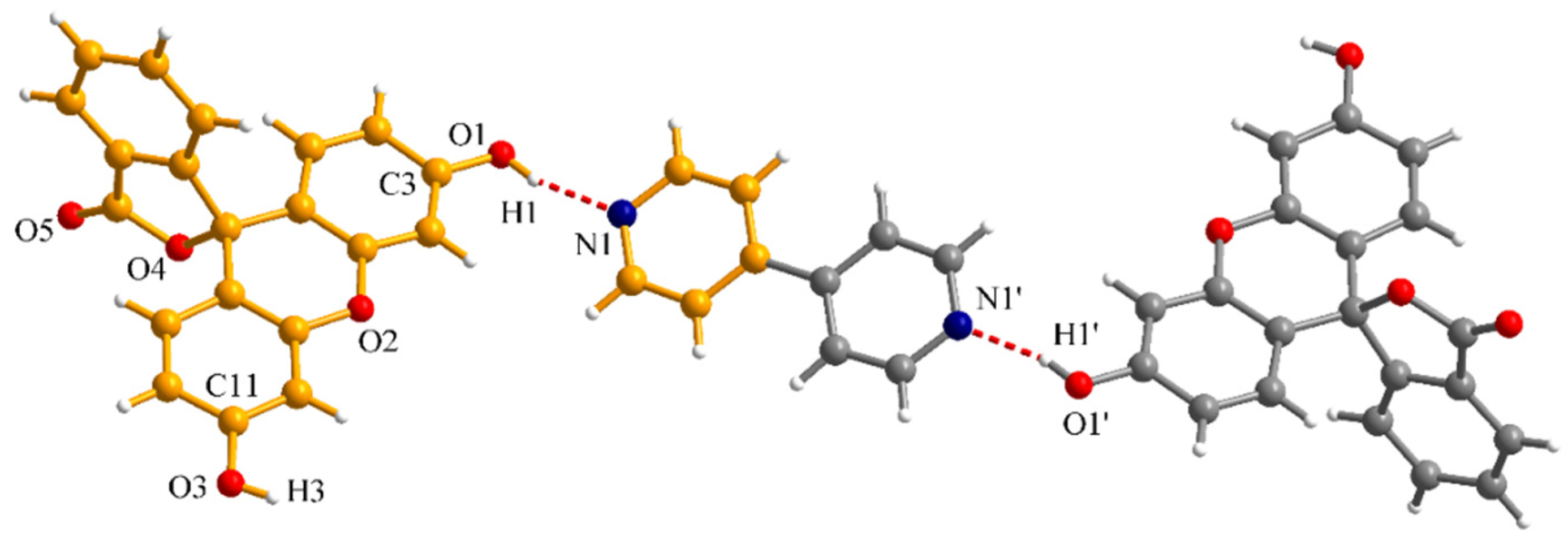
Crystal 1 contains only fluorescein and bipy in a 2:1 molar ratio. The asymmetric unit consists of one fluorescein molecule and half a bipy molecule (Figure 1). The fluorescein molecules are in the lactonoid form and the C-O bond lengths for the phenol groups are C3-O1 = 1.350(3) and C11-O3 = 1.355(3) Å. The bipy molecules are hydrogen bonded through the nitrogen atoms to two OH groups belonging to two different fluorescein molecules. The (O1-)H1···N1 distance for the intermolecular hydrogen interaction is 1.828 Å and the corresponding O1-H1···N1 angle is 165.0°.
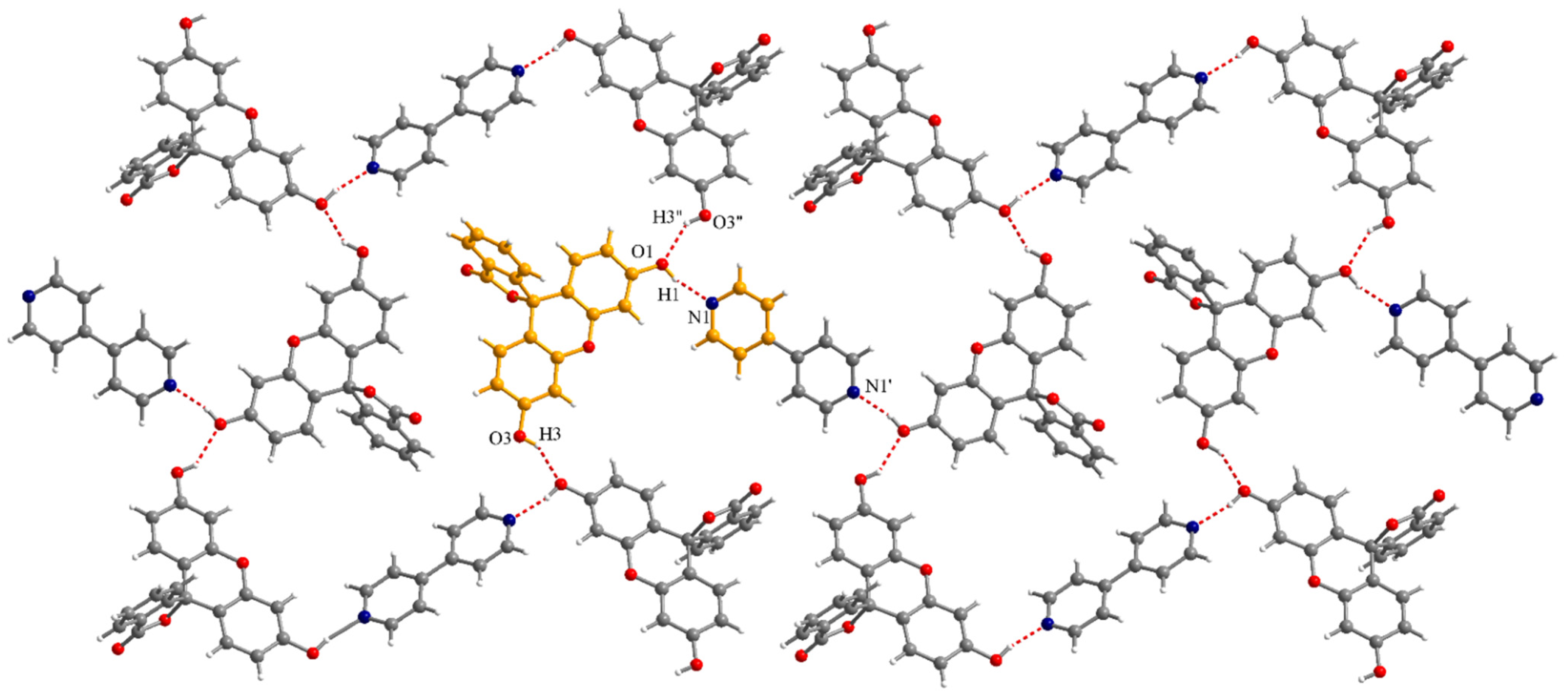
The O1 oxygen atom also acts as a hydrogen bond acceptor for an O3-H3 phenol group of neighboring fluorescein molecules. The (O3”-)H3”···O1 distance for this hydrogen interaction is 1.984 Å and the corresponding O3”-H3”···O1 angle is 144.8° (symmetry code: ” = 0.5−x, −0.5 + y, −z). The interplay between the two types of hydrogen interactions generates a supramolecular 2D architecture (Figure 2).
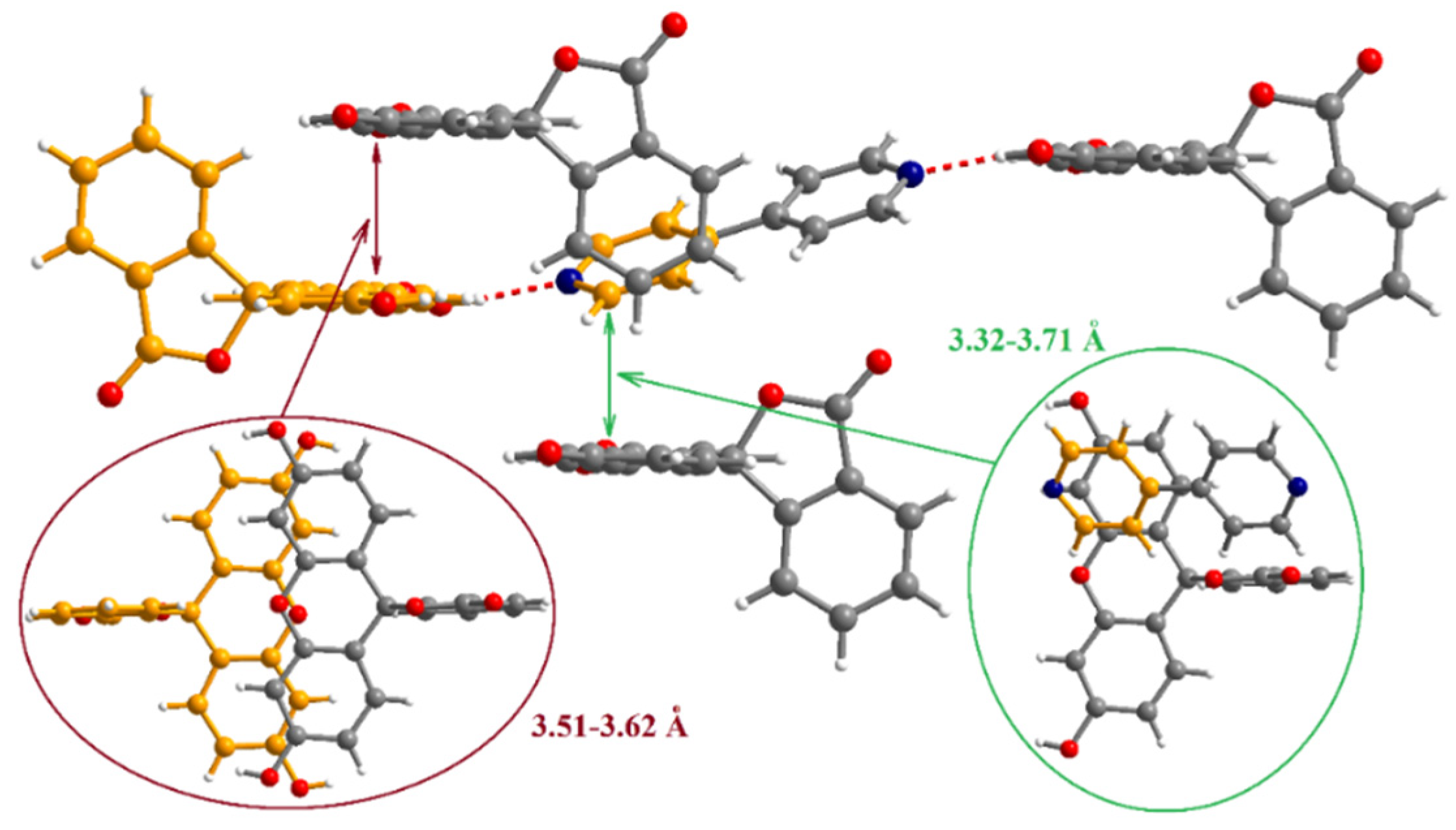
The supramolecular interactions are further extended to the third dimension through π-π interactions established between the layers. Both organic molecules contain aromatic systems and two patterns of π-π interactions can be described (Figure 3). The first one involves the xanthene cores of the fluorescein molecules with a separation of 3.51–3.62 Å between the fragments. The mean planes of interacting xanthene fragments are parallel. The second type of π-π interaction implicates one pyridine ring of a bipy molecule and one phenol moiety of a fluorescein molecule. The dihedral angle between the mean planes of pyridine and xanthene fragments is 31.1° and the shortest contacts between these fragments range between 3.32 and 3.71 Å.
Cocrystal 2 is a pseudopolymorph of 1 and contains fluorescein, bipy and methanol in a 2:1:2 stoichiometry. It crystalizes in the triclinic P-1 space group and the asymmetric unit comprises one fluorescein molecule, half a bipy molecule and one methanol molecule (Figure 4).
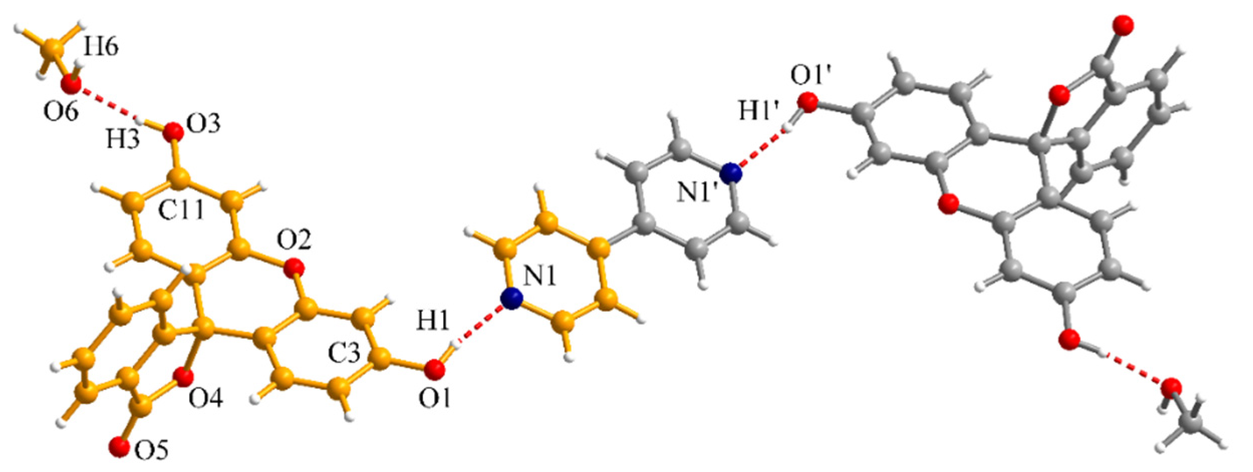
The fluorescein is also in the lactonoid form with the bond lengths for the phenol groups of 1.357(4) (C3-O1) and 1.358(4) Å (C11-O3). The two phenol groups of fluorescein act as hydrogen bond donors towards one bipy molecule and one methanol molecule, with the (O1-)H1···N1 and (O3-)H3···O6 distances of 1.890 and 1.858 Å, respectively. The corresponding O1-H1···N1 and O3-H3···O6 angles are 169.4 and 171.3°.
The methanol molecule is also a hydrogen donor for another phenol group of a different fluorescein molecule generating supramolecular units formed by two fluorescein molecules and two methanol molecules (Figure 5). The (O6-)H6···O1” distance is 2.264 Å, while the O6-H6···O1 angle is 135.5°. These supramolecular units are connected by the bipy molecules in a 1D array. In crystal 2, the presence of the solvent molecules prevents the extension of the hydrogen networking to a 2D system. Within the supramolecular dimers the xanthene fragments also establish π-π interactions (3.38–3.55 Å).
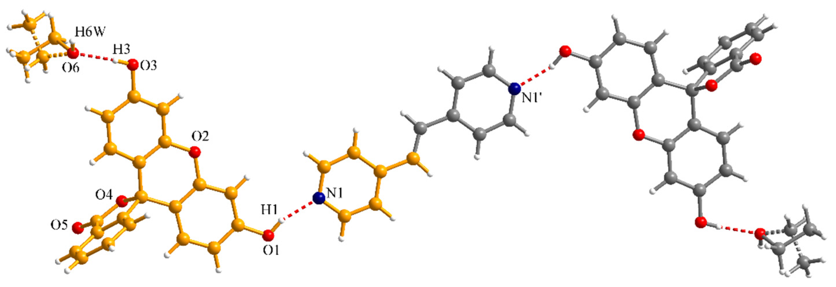
For the cocrystallization experiments of fluorescein with trans-1,2-bis(4-pyridyl)ethylene two different molar ratios were used: 2:1 and 1:1. The corresponding cocrystals obtained are {(H2Fl)2(bpete)(EtOH)2} (3) and {(H2Fl)(bpete)} (4). Cocrystal 3 contains, similarly with crystal 2, fluorescein, bpete and solvent in a 2:1:2 stoichiometry (Figure 6). In this case, the solvent is ethanol and the CH3-CH2- fragment is disordered on two independent crystallographic positions with site occupancy factors of 0.5 each. Similar to compound 2, the two phenol groups of fluorescein act as hydrogen bond donors towards one bpete molecule and one ethanol molecule. The (O1-)H1···N1 distance is 1.823 Å and the corresponding O1-H1···N1 angle is 161.0°.
In crystal 3, extension of the hydrogen bonding follows a different pattern with formation of a railroad-like arrangement. The alternating fluorescein and ethanol molecules connected by hydrogen interactions, with the (O3-)H3···O6 and (O6-)H6W···O1” distances of 1.896 and 1.957 Å respectively, are generating the rails while the sleepers are formed by the bpete molecules (Figure 7).
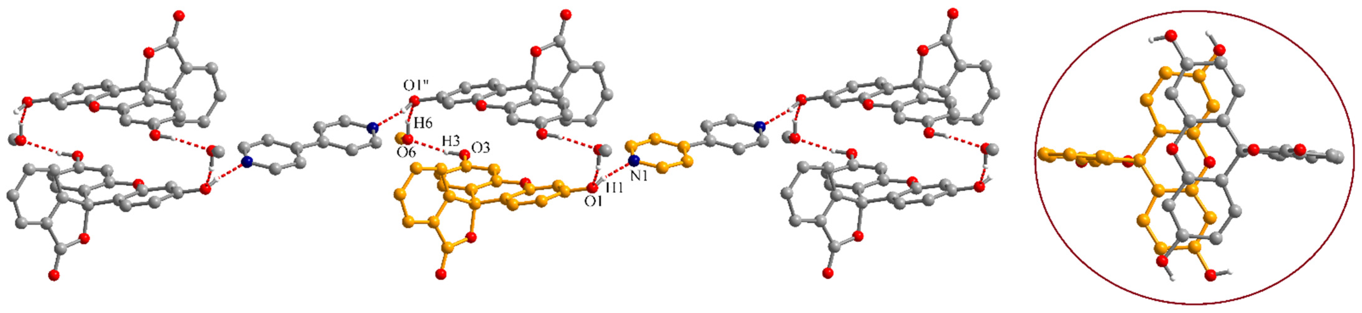
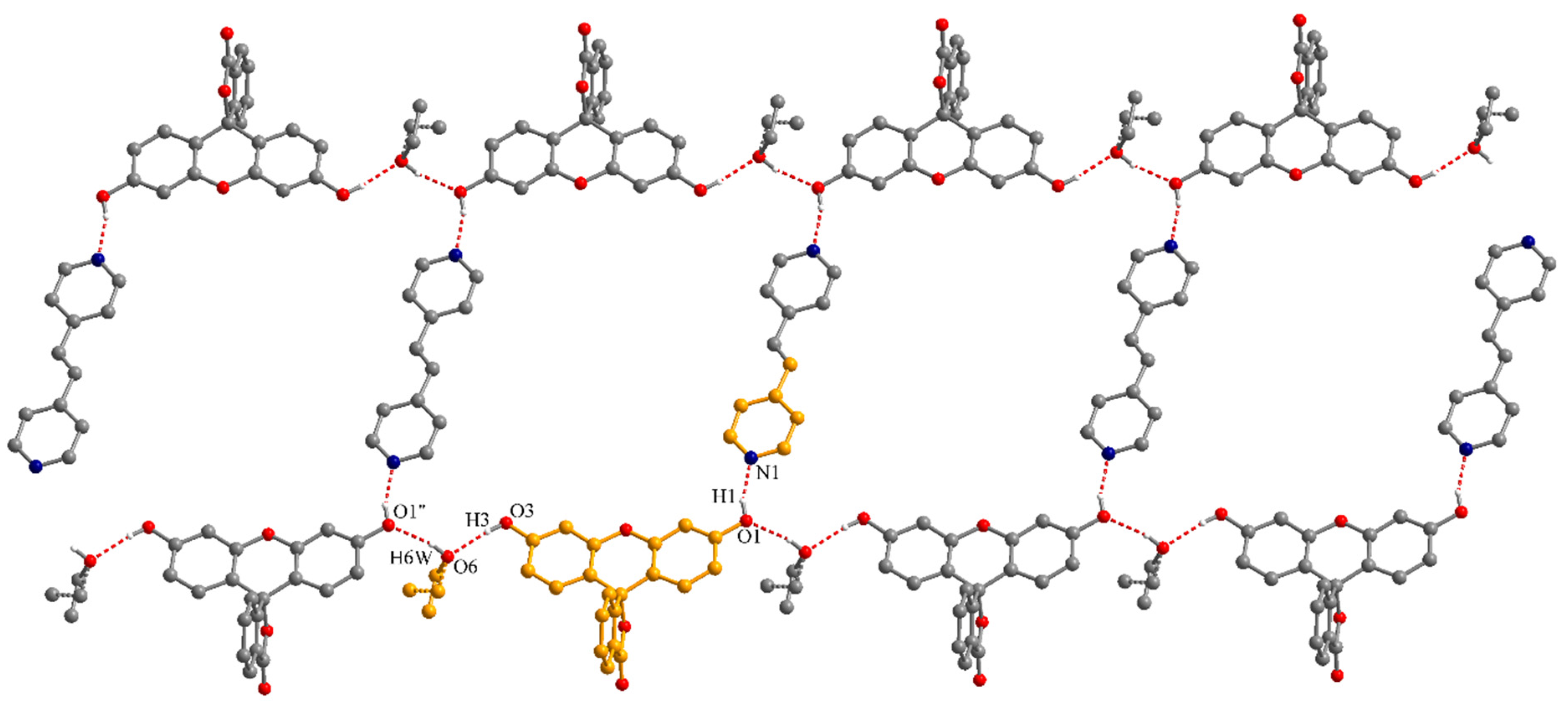
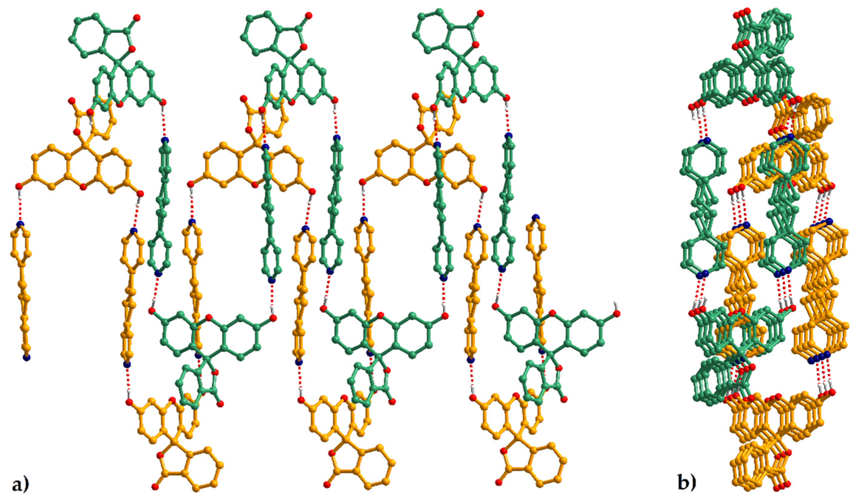
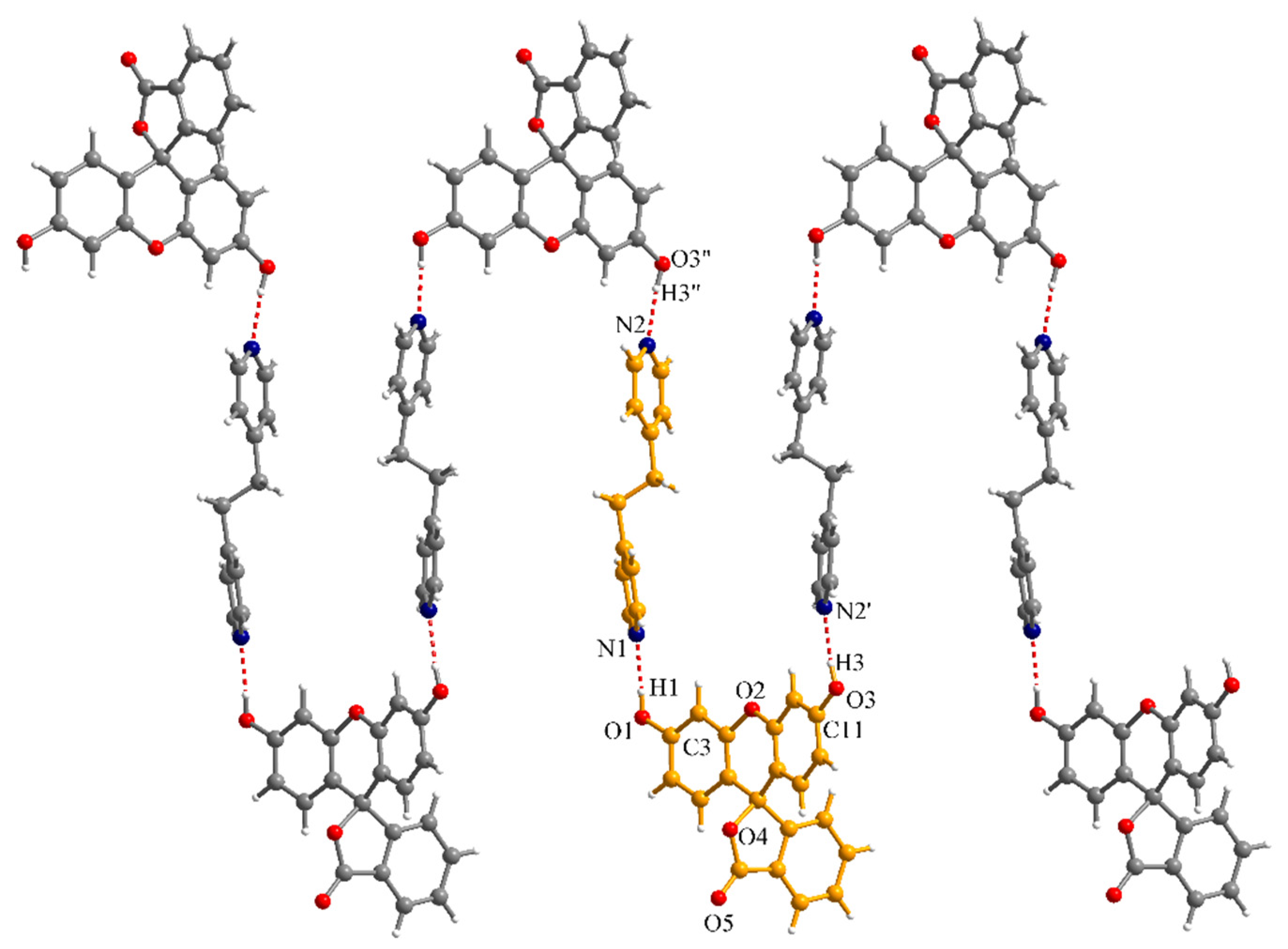
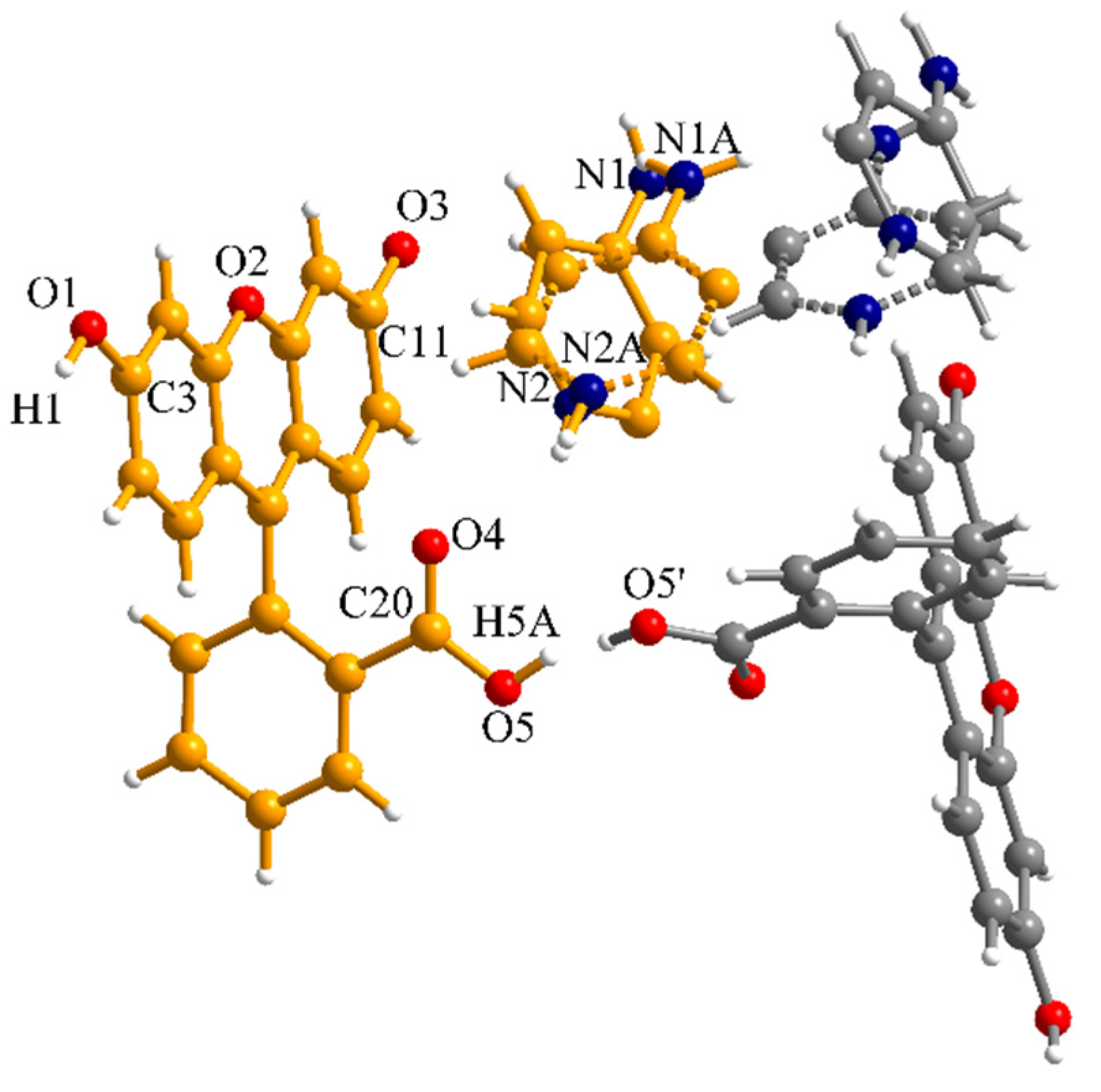
In crystal 4 the molar ratio between the fluorescein and bpete is 1:1. The fluorescein is in the neutral lactonoid form and both phenol groups are involved in hydrogen interactions with bpete molecules generating a crenel-like supramolecular chain (Figure 8). The C-O bond lengths for the phenol groups are C3-O1 = 1.358(5) and C11-O3 = 1.355(5) Å. The distances for the intermolecular hydrogen interactions are (O1-)H1···N1 = 1.894 and (O3-)H3···N2’ = 1.905 Å, with the corresponding angles of 171.9 and 174.6°, respectively (symmetry code: ’ = −x, −0.5 + y, −0.5 − z). The ethene fragment of the bpete molecule is disordered on two crystallographic positions with site occupancy factors of 0.75 and 0.25.
The supramolecular chains formed by hydrogen interactions intercalate and the bpete molecules from neighboring chains establish π-π interactions ranging between 3.27 and 3.64 Å (Figure 9).
We have to mention here that compound 4 is not crystallizing as a pure phase and the bulk sample probably contains a small amount of compound 3 (Figures S3–S5).
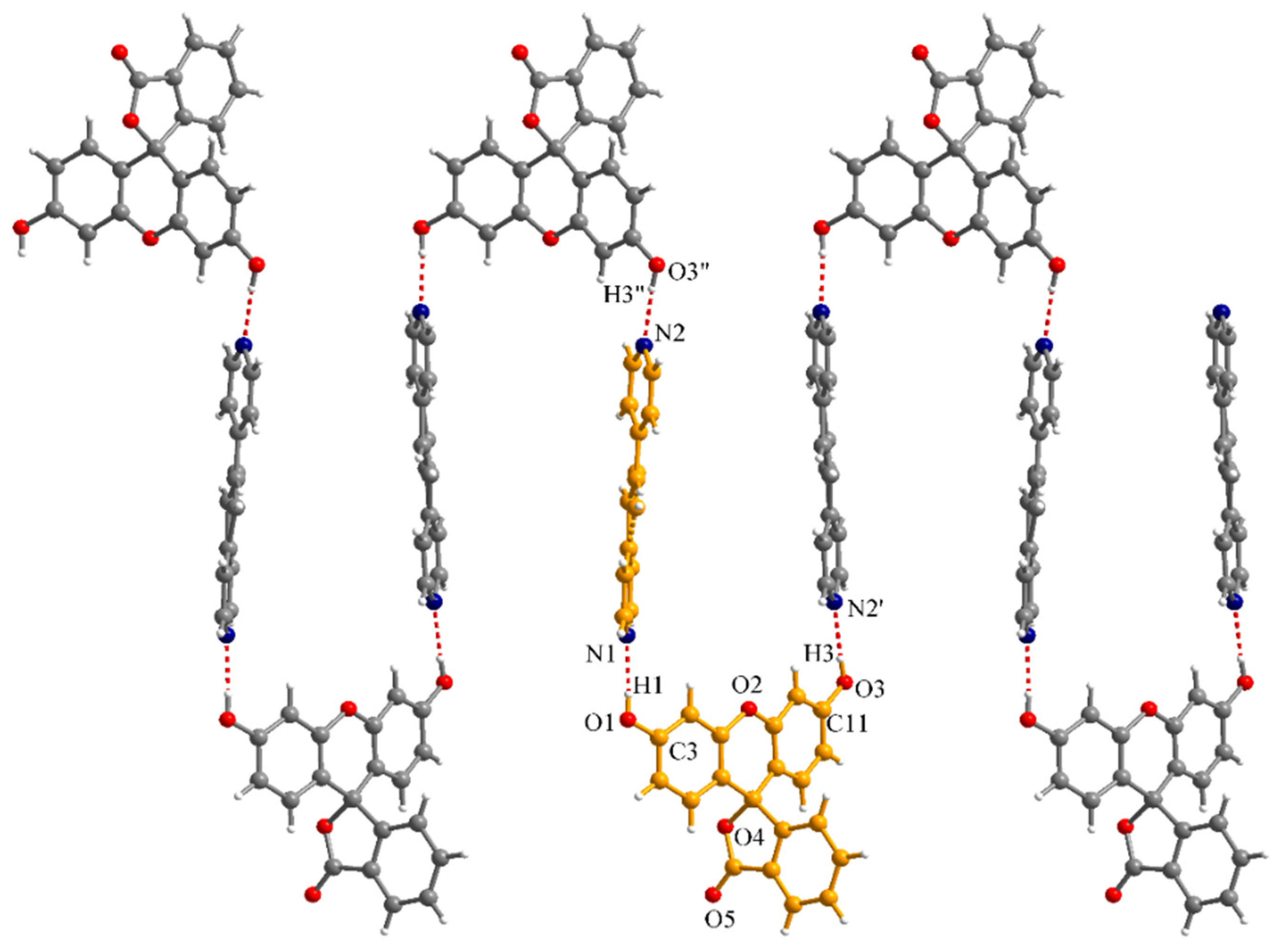
Cocrystal 5, {(H2Fl)(bpeta)}, was obtained by slow evaporation of a methanolic solution containing fluorescein and 1,2-bis(4-pyridyl)ethane in a 1:1 stoichiometry. In this case, the powder X-ray diffraction shows that the sample contains only one crystalline phase (Figure S6). Structural characterization of compound 5 by X-ray diffraction on a single crystal reveals formation of crenel-like supramolecular chains, similarly to crystal 4, by hydrogen interactions established between phenol and pyridyl groups (Figure 10). The C-O bond lengths for the phenol groups are: C3-O1 = 1.361(5) and C11-O3 = 1.356(6) Å. The distances for the hydrogen interactions are (O1-)H1···N1 = 1.867 and (O3-)H3···N2’ = 1.894 Å, with the corresponding angles of 170.8 and 175.1°, respectively (symmetry code: ’ = 1.5 − x, −0.5 + y, 1 − z).
The second type of nitrogen-containing organic molecules used as partners for fluorescein to obtain binary systems were the amino derivatives 4-aminopyridine and trans-1,4-diaminocyclohexane. The first notable observation is the color change induced by the amino derivatives. The solid products obtained in the presence of 4-aminopyridine and trans-1,4-diaminocyclohexane are red, while the compounds 1–5 are pale yellow or colorless. Since the chromogenic mechanism of fluorescein is based on protonation–deprotonation reactions, the color change is most probably an indication of the proton transfer between the fluorescein and the amino partner.
For a 1:1 stoichiometry between fluorescein and 4-aminopyridine, two types of crystals were obtained depending on the solvents used for crystallization: [(HFl)(Hampy)]∙2H2O (6), obtained in small amounts as orange-red prismatic crystals on the wall of the beaker from a mixture of ethanol (96%)-acetonitrile, and [(HFl)(Hampy)] (7) as red needle-like crystals resulted from the evaporation of an ethanol–water solution.
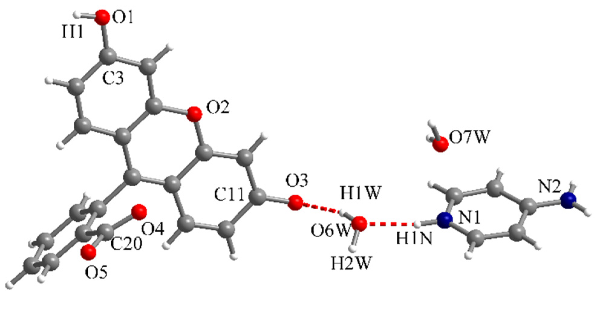
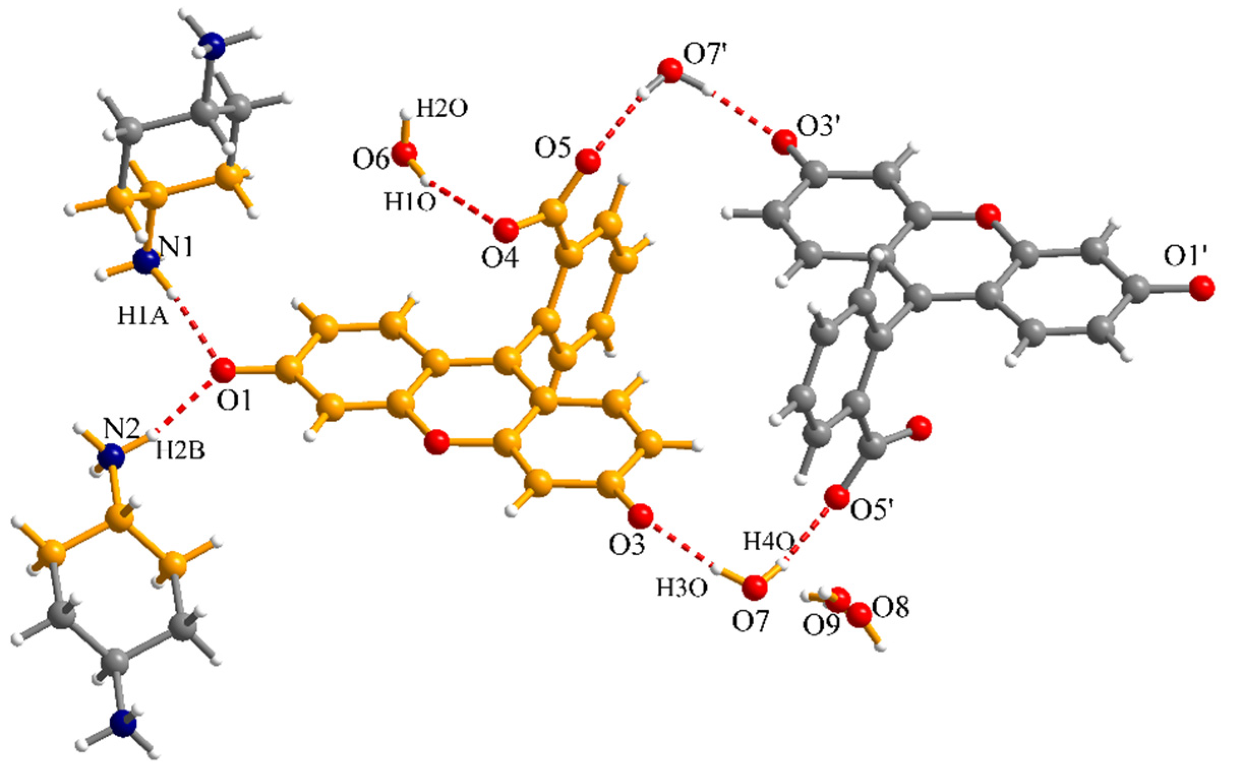
X-ray diffraction on a single crystal shows for compound 6 the presence of fluorescein monoanions (HFl−), Hampy+ cations and crystallization water molecules (Figure 11). The lactone spirocycle of fluorescein is open and the carboxylate bond lengths are C20-O4 = 1.249(5) and C20-O5 = 1.270(6) Å. For the phenol group, the C3-O1 bond length is 1.340(5) Å, while the quinonic C11-O3 bond length is 1.271(5) Å. The ampy molecule is protonated on a pyridine nitrogen atom (N1). The quinonic O3 atom acts as a hydrogen bond acceptor for a crystallization water molecule, which is also a hydrogen bond acceptor for the pyridinium fragment (Figure 11). The (O6W-)H1W···O3 and (N1-)H1N···O6W distances are 1.996 and 1.836 Å, respectively, while the corresponding O6W-H1W···O3 and N1-H1N···O6W angles are 163.1 and 167.9°.
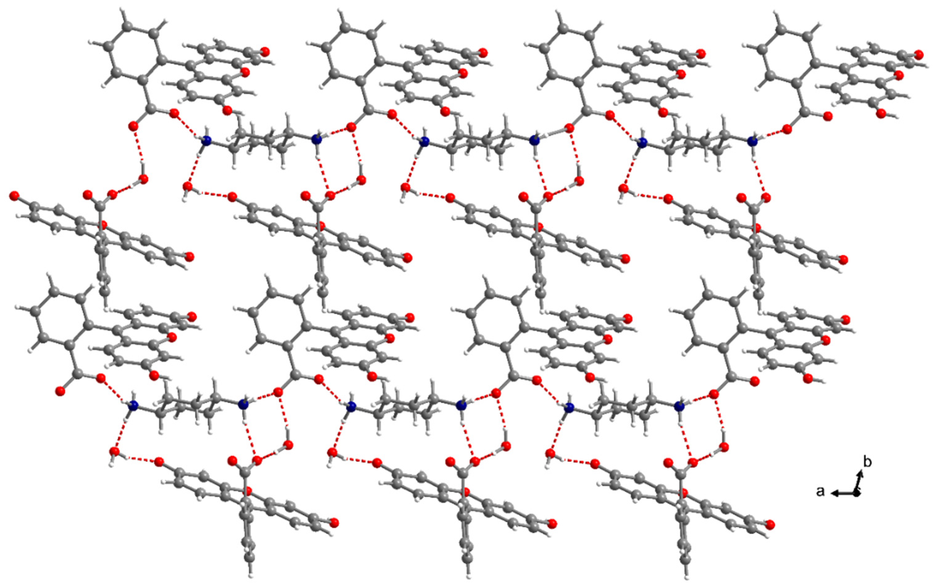
The xanthene fragments establish π-π interactions (3.57–3.65 Å) forming supramolecular dimers. The fluorescein supramolecular dimers are connected into an extended supramolecular network through hydrogen interactions with crystallization water molecules and 4-aminopyridinium cations (Figure 12). Except the xanthene O2 atom, all the other oxygen atoms of the fluorescein are involved in hydrogen interactions.
In crystal 7, the lactone spirocycle of fluorescein is also open but only half of the molecules are deprotonated. The proton from one phenol group is transferred on a pyridine nitrogen atom or on a carboxylate O5 atom (Figure 13). The ampy molecules are disordered on two crystallographic positions with site occupancy factors of 0.5. Due to the disorder of ampy molecules we could not refine the percentages of proton transfer on pyridine nitrogen atoms. We estimated that half of the protons are transferred on the carboxylate due to the short O5···O5’ distance of 2.506 Å between the carboxylate groups of neighboring fluorescein molecules, which can be explained by hydrogen interactions (the occupancy of the H5A atom was fixed at 0.5). The C3-O1 bond length is 1.336(7) Å, while the C11-O3 bond length is 1.300(6) Å. The carboxylate bond lengths are C20-O4 = 1.218(7) and C20-O5 = 1.275(7) Å.
In the reactions of fluorescein with trans-1,4-diaminocyclohexane, we initially attempted two stoichiometric ratios, 1:1 and 2:1. In the 1:1 reaction we obtained the compounds [(Fl)(H2diach)]∙3H2O (8) and [(HFl)2(Fl)2(H2diach)3]∙4H2O (10), while for the 2:1 ratio one type of crystal resulted: [(HFl)2(H2diach)]∙2H2O∙EtOH (9). Compound 10 was obtained subsequently as pure phase using a 4:3 molar ratio between fluorescein and diach.
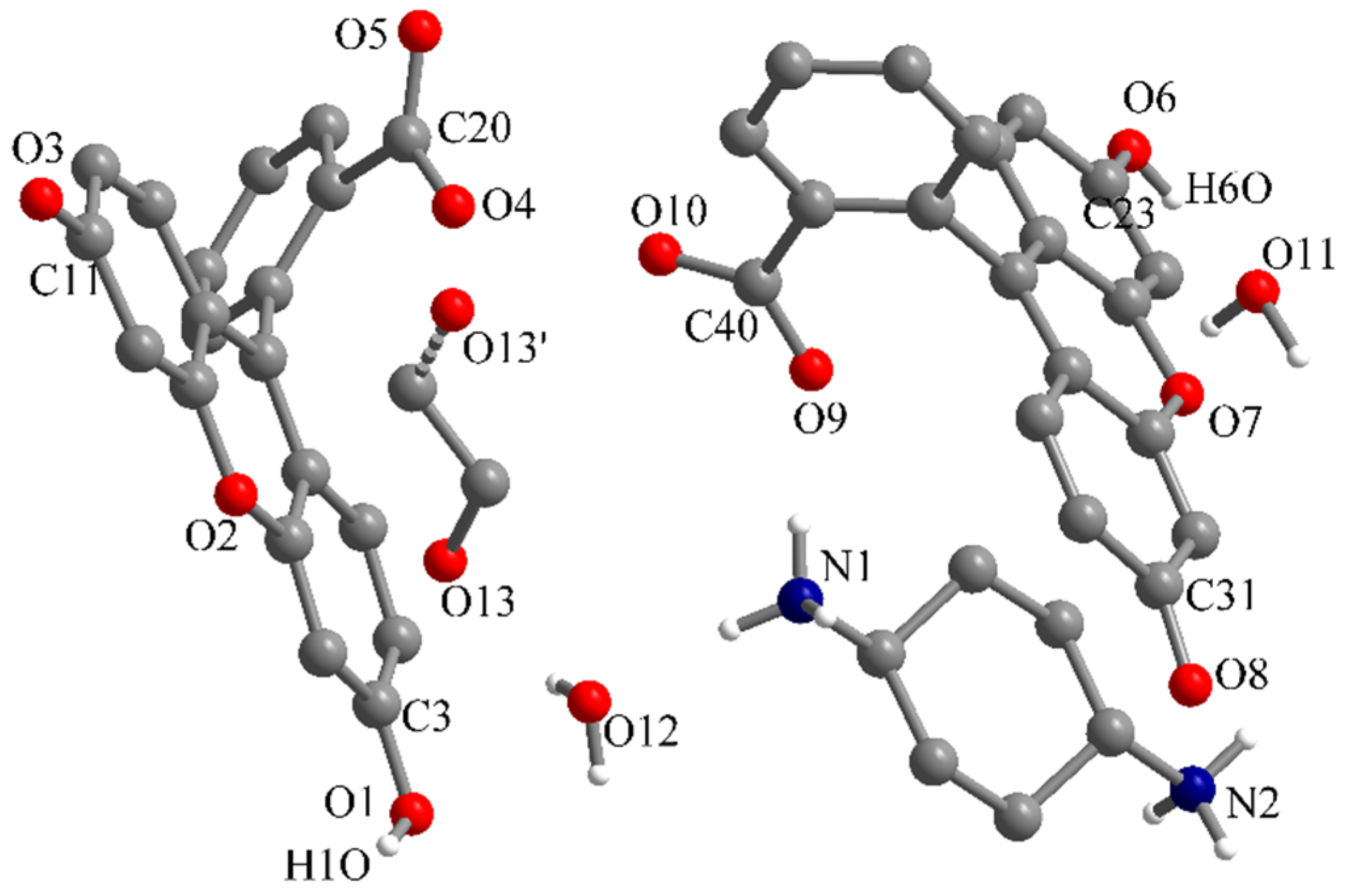
Compound 8 contains fluorescein dianions (Fl2−), H2diach2+ dications and crystallization water molecules. The asymmetric unit is formed by one Fl2- dianion, two halves of H2diach2+ dications and three water molecules (Figure 14). One water molecule is disordered on two crystallographic positions with site occupancy factors of 0.6 (O8) and 0.4 (O9). The C3-O1 and C11-O3 bond lengths are 1.288(3) and 1.278(3) Å, respectively. In the carboxylate group the bond lengths are C20-O4 = 1.225(3) and C20-O5 = 1.231(4) Å. The protonated amino groups are hydrogen bonded to the O1 atom with the (N1-)H1A···O1 and (N2-)H2B···O1 distances of 1.831 and 1.881 Å, respectively. The distances for the hydrogen interactions involving the O6 and O7 water molecules are (O6-)H1O···O4 = 2.169, (O7-)H3O···O3 = 1.814 and (O7-)H4O···O5’ = 1.918 Å (symmetry code: ’ = 2−x, 2−y, 2−z).
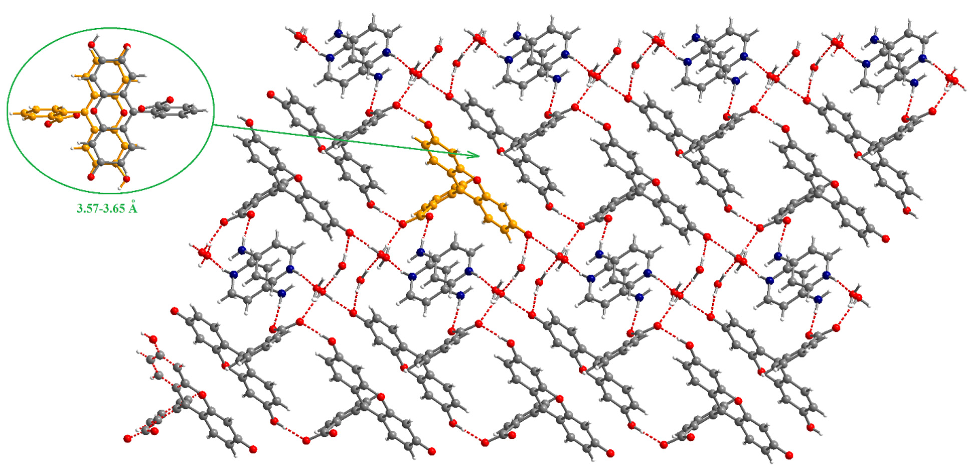
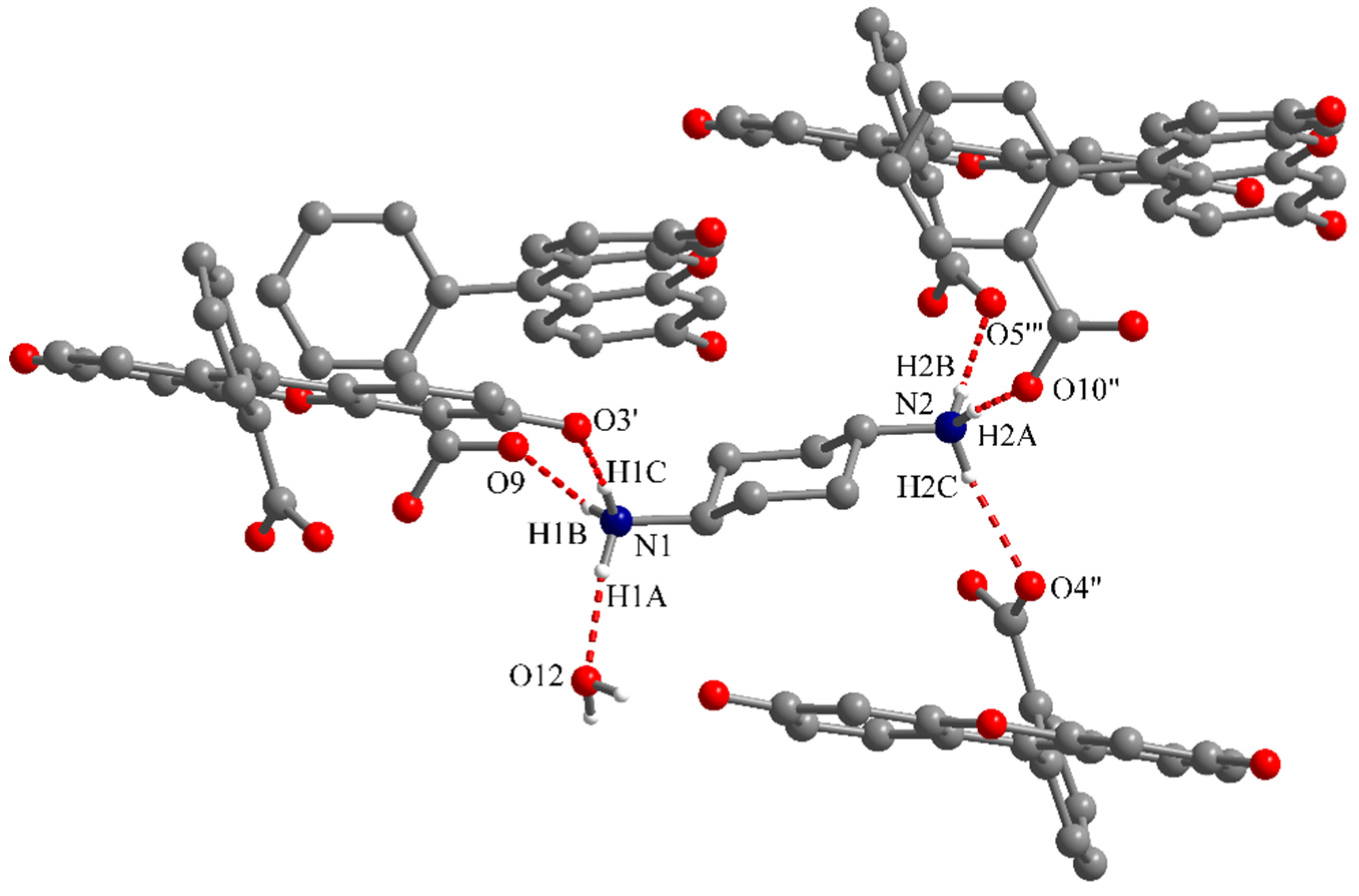
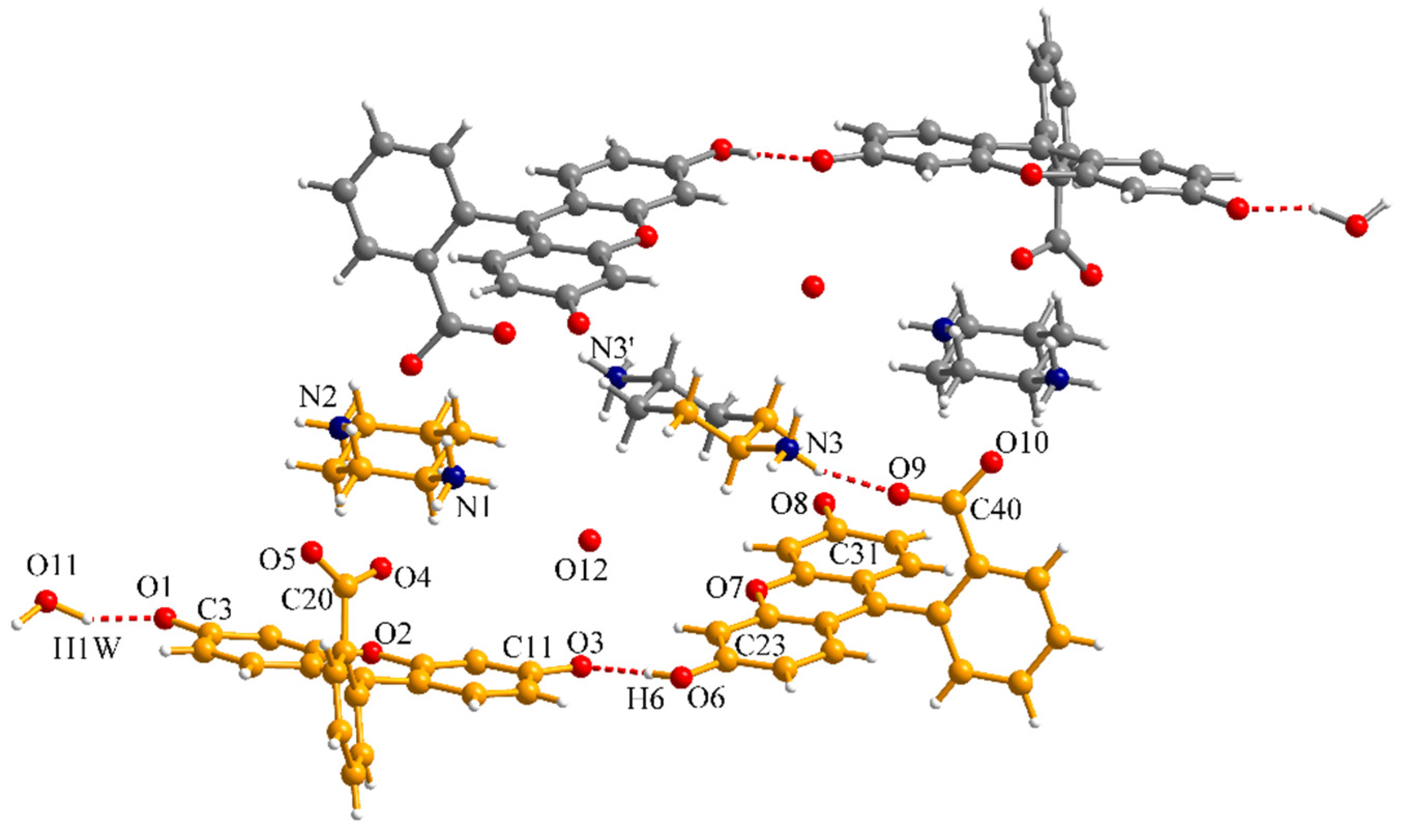
In compound 9, the stoichiometry used in the reaction was recovered in the crystal structure. The asymmetric unit contains two fluorescein monoanions (HFl−), one H2diach2+ dication and crystallization solvent molecules, two water molecules and one ethanol molecule (Figure 15). The ethanol molecule is disordered on two crystallographic positions. The two HFl− monoanions are in the quinonoid open-ring form. The bond lengths for the phenol and carbonyl groups are C3-O1 = 1.333(2), C23-O6 = 1.338(2) Å, C11-O3 = 1.270(2) and C31-O8 = 1.261(2) Å. The bond lengths in the two carboxylate groups are C20-O4 = 1.240(2), C20-O5 = 1.254(2), C40-O9 = 1.234(2) and C40-O10 = 1.255(2) Å.
The two protonated amino groups of the H2diach2+ dication are hydrogen bond donors towards four carboxylate groups, one carbonyl group and one water molecule (Figure 16 and Figure 17). The distances for these hydrogen interactions are (N1)H1A···O12 = 1.885, (N1)H1B···O9 = 1.885, (N1)H1C···O3’ = 1.975, (N2)H2A···O10” = 1.852, (N2)H2B···O5’” = 1.861 and (N2)H2C···O4” = 2.085 Å. The corresponding angles are N1-H1A···O12 = 172.8, N1-H1B···O9 = 155.0, N1-H1C···O3’ = 169.7, N2-H2A···O10” = 174.5, N2-H2B···O5’” = 166.6 and N2-H2C···O4” = 156.2°.
The compound 10, [(HFl)2(Fl)2(H2diach)3]∙4H2O, was obtained initially as by-product in the synthesis of compound 8 but subsequently we managed to obtain it as pure phase using a 4:3 molar ratio between fluorescein and diach (Figures S10–S13). Compound 10 is also a salt containing two types of fluorescein anions, mono- (HFl−) and dianions (Fl2−), H2diach2+ dications and crystallization water molecules (Figure 18). Within monoanions the bond lengths for the phenol and carbonyl groups are C23-O6 = 1.320(5) and C31-O8 = 1.283(5) Å, while for the dianions the C3-O1 and C11-O3 bond lengths are 1.285(5) and 1.296(5) Å, respectively. The phenol group of the monoanion is involved in hydrogen interaction with a carbonyl group of the dianion. The second carbonyl group of the dianion is hydrogen bonded to a crystallization water molecule. The distances for these hydrogen interactions are (O6)H6···O3 = 1.700 and (O11)H1W···O1 = 1.739 Å. The bond lengths in the two carboxylate groups are C20-O4 = 1.221(5), C20-O5 = 1.263(5), C40-O9 = 1.239(5) and C40-O10 = 1.245(5) Å. The simultaneous presence of both mono- and dianions of fluorescein was previously reported for lanthanide coordination polymers [24].
3.2. Spectral Properties
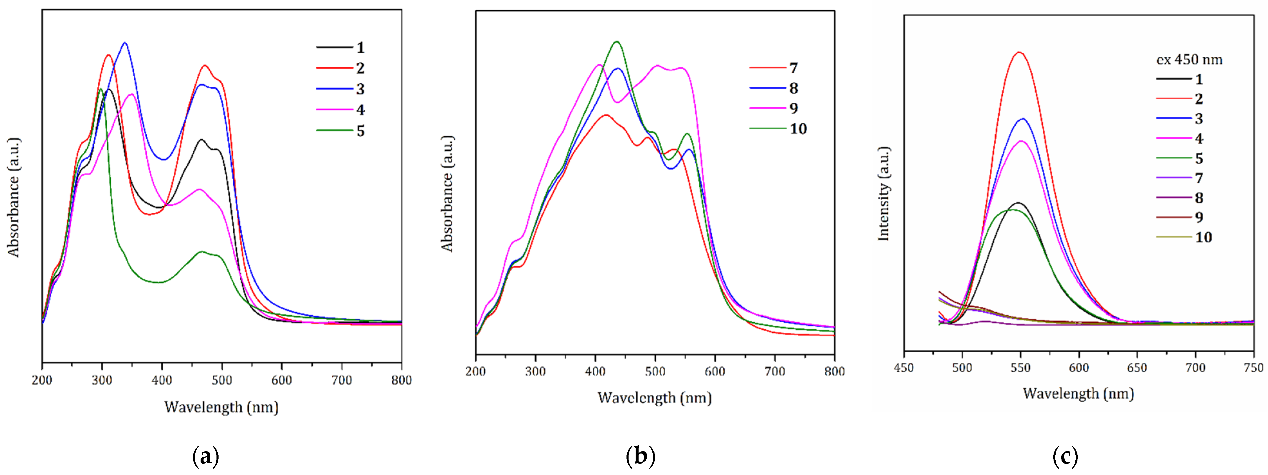
The absorption spectra of compounds 1–5 and 7–10 have been acquired over a wavelength range from 200 to 1000 nm on solid samples (using the diffuse reflectance technique). Compound 6 was obtained only in small amounts on the wall of the beaker and we were not able to investigate the optical properties for this compound. As we already mentioned, there is significant color difference between the cocrystals of fluorescein (compounds 1–5) and the organic salts of fluorescein (compounds 6–10). The cocrystals are pale yellow or colorless and the salts are red. The cocrystals present clearly separated absorption bands in the UV (with maxima around 300–350 nm) and visible regions (with maxima around 460 and 490 nm). The salts present broad bands covering the UV and visible regions with bathochromic shifts in the absorptions. The solid-state absorption spectra of compounds 1–5 and 7–10 are presented in Figure 19a,b and in Table 1 the wavelengths for the absorption maxima are gathered.
The room temperature photoluminescence of compounds 1–5 and 7–10 was investigated using different wavelengths for excitation in the 430–460 nm range. The resulting emission spectra using λex = 450 nm are presented in Figure 19c. First of all, we have to notice that no emission was observed in solid-state for the salts. The emission spectra of compounds 1–4 display symmetric bands with maxima at 550 nm. Cocrystal 5 presents a broader emission band with a maximum at 542 nm.
4. Conclusions
Fluorescein proved to be a versatile molecule in the binary systems formed with different nitrogen-containing organic molecules. The phenol groups of the fluorescein can act either as hydrogen bond donors generating cocrystals or as proton donors producing organic salts. For molecules containing only pyridine rings such as 4,4’-bipyridyl; trans-1,2-bis(4-pyridyl)ethylene; and 1,2-bis(4-pyridyl)ethane, we obtained cocrystals. The presence of more basic amino groups on the nitrogen-containing organic partner conducted syntheses to salts and the deprotonation degree of the fluorescein depended on the molar ratio used. With trans-1,4-diaminocyclohexane, by modifying the stoichiometry, we obtained salts containing only monoanions of fluorescein, only dianions of fluorescein or both mono- and dianions. This versatility of fluorescein allows modulation of the optical properties of the binary systems by choosing a suitable partner.
Supplementary Materials
The following are available online at https://www.mdpi.com/article/10.3390/cryst11101217/s1, Figures S1–S13: Powder X-ray diffraction patterns, Table S1: Crystallographic data, details of data collection and structure refinement parameters for compounds 1–10.
Author Contributions
Conceptualization, A.M.M.; methodology, A.M.M.; synthesis, M.R.; formal analysis, M.R. and A.M.M.; investigation, M.R. and A.M.M.; writing—original draft preparation, A.M.M.; writing—review and editing, M.R. and A.M.M.; funding acquisition, A.M.M. All authors have read and agreed to the published version of the manuscript.
Funding
This research was funded by UEFISCDI (Project PN-III-P4-ID-PCE-2016-0442 nr. 89/2017).
Institutional Review Board Statement
Not applicable.
Informed Consent Statement
Not applicable.
Data Availability Statement
Not applicable.
Acknowledgments
M.R. is grateful to Maria Maganu for recording the IR spectra.
Conflicts of Interest
The authors declare no conflict of interest.
References
- Li, X.; Gao, X.; Shi, W.; Ma, H. Design Strategies for Water-Soluble Small Molecular Chromogenic and Fluorogenic Probes. Chem. Rev. 2014, 114, 590–659. [Google Scholar] [CrossRef]
- Zhang, X.; Hayes, D.; Smith, S.J.; Friedle, S.; Lippard, S.J. New Strategy for Quantifying Biological Zinc by a Modified Zinpyr Fluorescence Sensor. J. Am. Chem. Soc. 2008, 130, 15788–15789. [Google Scholar] [CrossRef] [PubMed] [Green Version]
- Wong, B.A.; Friedle, S.; Lippard, S.J. Solution and Fluorescence Properties of Symmetric Dipicolylamine-Containing Dichlorofluorescein-Based Zn2+ Sensors. J. Am. Chem. Soc. 2009, 131, 7142–7152. [Google Scholar] [CrossRef] [PubMed] [Green Version]
- Hyman, L.M.; Stephenson, C.J.; Dickens, M.G.; Shimizu, K.D.; Franz, K.J. Toward the development of prochelators as fluorescent probes of copper-mediated oxidative stress. Dalton Trans. 2010, 39, 568–576. [Google Scholar] [CrossRef] [PubMed]
- Abebe, F.A.; Eribal, C.S.; Ramakrishna, G.; Sinn, E. A ‘turn-on’ fluorescent sensor for the selective detection of cobalt and nickel ions in aqueous media. Tetrahedron Lett. 2011, 52, 5554–5558. [Google Scholar] [CrossRef]
- Shen, W.; Wang, L.; Wu, M.; Bao, X. A fluorescein derivative FLTC as a chemosensor for Hg2+ and Ag+ and its application in living-cell imaging. Inorg. Chem. Commun. 2016, 70, 107–110. [Google Scholar] [CrossRef] [Green Version]
- McQuade, L.E.; Ma, J.; Lowe, G.; Ghatpande, A.; Gelperin, A.; Lippard, S.J. Visualization of nitric oxide production in the mouse main olfactory bulb by a cell-trappable copper(II) fluorescent probe. Proc. Natl. Acad. Sci. USA 2010, 107, 8525–8530. [Google Scholar] [CrossRef] [Green Version]
- Wang, W.; Rusin, O.; Xu, X.; Kim, K.K.; Escobedo, J.O.; Fakayode, S.O.; Fletcher, K.A.; Lowry, M.; Schowalter, C.M.; Lawrence, C.M.; et al. Detection of Homocysteine and Cysteine. J. Am. Chem. Soc. 2005, 127, 15949–15958. [Google Scholar] [CrossRef] [PubMed] [Green Version]
- Yin, W.; Zhu, H.; Wang, R. A sensitive and selective fluorescence probe based fluorescein for detection of hypochlorous acid and its application for biological imaging. Dye. Pigment. 2014, 107, 127–132. [Google Scholar] [CrossRef]
- Jack, T.; Simonin, J.; Ruepp, M.-D.; Thompson, A.J.; Gertsch, J.; Lochner, M. Characterizing new fluorescent tools for studying 5-HT3 receptor pharmacology. Neuropharmacology 2015, 90, 63–73. [Google Scholar] [CrossRef]
- Wang, T.; Hong, T.; Huang, Y.; Su, H.; Wu, F.; Chen, Y.; Wei, L.; Huang, W.; Hua, X.; Xia, Y.; et al. Fluorescein Derivatives as Bifunctional Molecules for the Simultaneous Inhibiting and Labeling of FTO Protein. J. Am. Chem. Soc. 2017, 137, 13736–13739. [Google Scholar] [CrossRef]
- Guilini, C.; Baehr, C.; Schaeffer, E.; Gizzi, P.; Rufi, F.; Haiech, J.; Weiss, E.; Bonnet, D.; Galzi, J.-L. New Fluorescein Precursors for Live Bacteria Detection. Anal. Chem. 2015, 87, 8858–8866. [Google Scholar] [CrossRef] [PubMed]
- Mchedlov-Petrossyan, N.O.; Vodolazkaya, N.A.; Salamanova, N.V.; Roshal, A.D.; Filatov, D.Y. In Search for the “Phenolate” Monoanion of Fluorescein in Solution. Chem. Lett. 2010, 39, 30–31. [Google Scholar] [CrossRef]
- Tremayne, M.; Kariuki, B.M.; Harris, K.D.M. Structure Determination of a Complex Organic Solid from X-ray Powder Diffraction Data by a Generalized Monte Carlo Method: The Crystal Structure of Red Fluorescein. Angew. Chem. Int. Ed. Engl. 1997, 36, 770–772. [Google Scholar] [CrossRef]
- Arhangelskis, M.; Eddleston, M.D.; Reid, D.G.; Day, G.M.; Bučar, D.-K.; Morris, A.J.; Jones, W. Rationalization of the Color Properties of Fluorescein in the Solid State: A Combined Computational and Experimental Study. Chem. Eur. J. 2016, 22, 10065–10073. [Google Scholar] [CrossRef] [Green Version]
- Polyakova, I.N.; Starikova, Z.A.; Parusnikov, B.V.; Krasavin, I.A.; Dobryakova, G.M.; Zhdanov, B.V. The lactone form of fluorescein: Crystal structure of the 1:1 molecular complex of fluorescein with methanol. J. Struct. Chem. 1984, 25, 752–757. [Google Scholar] [CrossRef]
- Osborn, R.S.; Rogers, D. The crystal and molecular structure of the 1:1 complex of acetone with the lactoid form of fluorescein. Acta Crystallogr. Sect. B 1975, 31, 359–364. [Google Scholar] [CrossRef]
- Anthoni, U.; Christophersen, C.; Nielsen, P.H.; Püschl, A.; Schaumburg, K. Structure of red and orange fluorescein. Struct. Chem. 1995, 6, 161–165. [Google Scholar] [CrossRef]
- Bučar, D.-K.; Filip, S.; Arhangelskis, M.; Lloyd, G.O.; Jones, W. Advantages of mechanochemical cocrystallisation in the solid-state chemistry of pigments: Colour-tuned fluorescein cocrystals. CrystEngComm 2013, 15, 6289–6291. [Google Scholar] [CrossRef] [Green Version]
- Harrison, W.T.A.; Ramadevi, P.; Seethalakshmi, P.G.; Kumaresan, S. 4-Aza-1-azoniabicyclo[2.2.2]octane eosinide. Acta Cryst. 2007, 63, o3911. [Google Scholar] [CrossRef]
- Ramasubramanian, R.; Indrani, M.; Stephen, A.D.; Kumaradhas, P.; Sridhar, B.; Kumaresan, S. Crystal Structure of a Proton-Transfer Self-associated Compound Tetrakis(guanidinium)bis(eosinate) hexahydrate. Anal. Sci. X-Ray Struct. Anal. Online 2008, 24, x243–x244. [Google Scholar] [CrossRef] [Green Version]
- Răducă, M.; Mădălan, A.M. Synthesis, crystal structures and solid-state optical properties of two Mannich derivatives of 2’,7’-dichlorofluorescein. Rev. Roum. Chim. 2020, 65, 617–622. [Google Scholar] [CrossRef]
- Aitimpamula, S.; Banerjee, R.; Bansal, A.K.; Biradha, K.; Cheney, M.L.; Choudhury, A.R.; Desiraju, G.R.; Dikundwar, A.G.; Dubey, R.; Duggirala, N.; et al. Polymorphs, Salts, and Cocrystals: What’s in a Name? Cryst. Growth Des. 2012, 12, 2147–2152. [Google Scholar] [CrossRef]
- Thomas, J.; Ambili, K.S. Synthesis, crystal structure and luminescent properties of a new samarium-fluorescein metal-organic framework. J. Mol. Struct. 2015, 1098, 167–174. [Google Scholar] [CrossRef]
Scheme 1.
The protonation−deprotonation equilibria of fluorescein.

Figure 1.
Crystal structure of 1. The asymmetric unit is depicted in orange. Symmetry code: ’ = 1 − x, 1 − y, −1 − z.
Figure 1.
Crystal structure of 1. The asymmetric unit is depicted in orange. Symmetry code: ’ = 1 − x, 1 − y, −1 − z.

Figure 2.
View of the 2D supramolecular array formed through hydrogen interactions in crystal 1. Symmetry codes: ’ = 1 − x, 1 − y, −1 − z; ” = 0.5 − x, −0.5 + y, −z.
Figure 2.
View of the 2D supramolecular array formed through hydrogen interactions in crystal 1. Symmetry codes: ’ = 1 − x, 1 − y, −1 − z; ” = 0.5 − x, −0.5 + y, −z.

Figure 3.
Details of the π-π interactions established in crystal 1.

Figure 4.
Crystal structure of 2. The asymmetric unit is depicted in orange. Symmetry code: ’ = 3 − x, 3 − y, 2 − z.
Figure 4.
Crystal structure of 2. The asymmetric unit is depicted in orange. Symmetry code: ’ = 3 − x, 3 − y, 2 − z.

Figure 5.
Perspective view of the 1D supramolecular array formed through hydrogen interactions in crystal 2. The inset shows a detail of the π-π interactions established within the supramolecular dimers. Symmetry code: “= 1 − x, 2 − y, 1 − z.
Figure 5.
Perspective view of the 1D supramolecular array formed through hydrogen interactions in crystal 2. The inset shows a detail of the π-π interactions established within the supramolecular dimers. Symmetry code: “= 1 − x, 2 − y, 1 − z.

Figure 6.
Crystal structure of 3. The asymmetric unit is depicted in orange. Symmetry code: ’ = 2 − x, 3 − y, 2 − z.
Figure 6.
Crystal structure of 3. The asymmetric unit is depicted in orange. Symmetry code: ’ = 2 − x, 3 − y, 2 − z.

Figure 7.
Perspective view of the 1D supramolecular array formed through hydrogen interactions in crystal 3 (the hydrogen atoms not involved in hydrogen interactions were omitted for clarity). Symmetry code: “= −1 + x, y, −1 + z.
Figure 7.
Perspective view of the 1D supramolecular array formed through hydrogen interactions in crystal 3 (the hydrogen atoms not involved in hydrogen interactions were omitted for clarity). Symmetry code: “= −1 + x, y, −1 + z.

Figure 8.
Perspective view of the supramolecular chain formed through hydrogen interactions in crystal 4. The asymmetric unit is depicted in orange. Symmetry codes: ’ = −x, −0.5 + y, −0.5 − z; “ = −x, 0.5 + y, −0.5 − z.
Figure 8.
Perspective view of the supramolecular chain formed through hydrogen interactions in crystal 4. The asymmetric unit is depicted in orange. Symmetry codes: ’ = −x, −0.5 + y, −0.5 − z; “ = −x, 0.5 + y, −0.5 − z.

Figure 9.
Perspective (a) and side views (b) of two neighboring supramolecular chains in crystal 4.

Figure 10.
Perspective view of the supramolecular chain formed through hydrogen interactions in crystal 5. The asymmetric unit is depicted in orange. Symmetry codes: ’ = 1.5 − x, −0.5 + y, 1 − z; “ = 1.5 − x, 0.5 + y, 1 − z.
Figure 10.
Perspective view of the supramolecular chain formed through hydrogen interactions in crystal 5. The asymmetric unit is depicted in orange. Symmetry codes: ’ = 1.5 − x, −0.5 + y, 1 − z; “ = 1.5 − x, 0.5 + y, 1 − z.

Figure 11.
Perspective view of the asymmetric unit in crystal 6.

Figure 12.
View of a packing diagram in crystal 6. The inset shows a detail of the π-π interactions established within the supramolecular dimers formed by the fluorescein monoanions.
Figure 12.
View of a packing diagram in crystal 6. The inset shows a detail of the π-π interactions established within the supramolecular dimers formed by the fluorescein monoanions.

Figure 13.
Crystal structure of 7. The asymmetric unit is depicted in orange. Symmetry code: ’ = −x, y, 1.5 − z.
Figure 13.
Crystal structure of 7. The asymmetric unit is depicted in orange. Symmetry code: ’ = −x, y, 1.5 − z.

Figure 14.
Crystal structure of 8. The asymmetric unit is depicted in orange. Symmetry code: ’ = 2 − x, 2 − y, 2 − z.
Figure 14.
Crystal structure of 8. The asymmetric unit is depicted in orange. Symmetry code: ’ = 2 − x, 2 − y, 2 − z.

Figure 15.
View of the asymmetric unit in crystal 9 (part of the hydrogen atoms was omitted for clarity). Symmetry code: ’ = −x, 1 − y, −z.
Figure 15.
View of the asymmetric unit in crystal 9 (part of the hydrogen atoms was omitted for clarity). Symmetry code: ’ = −x, 1 − y, −z.

Figure 16.
Detail of the hydrogen interactions established by H2diach2+ dications in crystal 9 (part of the hydrogen atoms was omitted for clarity). Symmetry codes: ’ = −x, 1 − y, −z; “ = 1 + x, y, z; ’” = 1 − x, 1− y, 1 − z.
Figure 16.
Detail of the hydrogen interactions established by H2diach2+ dications in crystal 9 (part of the hydrogen atoms was omitted for clarity). Symmetry codes: ’ = −x, 1 − y, −z; “ = 1 + x, y, z; ’” = 1 − x, 1− y, 1 − z.

Figure 17.
View along the crystallographic c axis of a packing diagram in crystal 9.

Figure 18.
Crystal structure of compound 10. The asymmetric unit is depicted in orange. Symmetry code: ’ = 1 − x, 1 − y, 1 − z.
Figure 18.
Crystal structure of compound 10. The asymmetric unit is depicted in orange. Symmetry code: ’ = 1 − x, 1 − y, 1 − z.

Figure 19.
The solid-state absorption spectra of cocrystals 1–5 (a) and salts 7–10 (b). Emission spectra of compounds 1–5 and 7–10 (c).
Figure 19.
The solid-state absorption spectra of cocrystals 1–5 (a) and salts 7–10 (b). Emission spectra of compounds 1–5 and 7–10 (c).


Table 1.
Absorption maxima for compounds 1–5 and 7–10.
| Compound | Wavelength (nm) | ||
|---|---|---|---|
| 1 | 311 | 465 | 490 |
| 2 | 311 | 471 | 494 |
| 3 | 337 | 465 | 485 |
| 4 | 350 | 463 | |
| 5 | 298 | 466 | 490 |
| 7 | 418 | 487 | 531 |
| 8 | 435 | 497 | 553 |
| 9 | 438 | 497 | 555 |
| 10 | 406 | 504 | 542 |
Publisher’s Note: MDPI stays neutral with regard to jurisdictional claims in published maps and institutional affiliations. |
© 2021 by the authors. Licensee MDPI, Basel, Switzerland. This article is an open access article distributed under the terms and conditions of the Creative Commons Attribution (CC BY) license (https://creativecommons.org/licenses/by/4.0/).
Share and Cite
MDPI and ACS Style
Răducă, M.; Mădălan, A.M. Cocrystals versus Salts of Fluorescein. Crystals 2021, 11, 1217. https://doi.org/10.3390/cryst11101217
AMA Style
Răducă M, Mădălan AM. Cocrystals versus Salts of Fluorescein. Crystals. 2021; 11(10):1217. https://doi.org/10.3390/cryst11101217
Chicago/Turabian StyleRăducă, Mihai, and Augustin M. Mădălan. 2021. "Cocrystals versus Salts of Fluorescein" Crystals 11, no. 10: 1217. https://doi.org/10.3390/cryst11101217
Note that from the first issue of 2016, this journal uses article numbers instead of page numbers. See further details here.