Article

Novel Pb(II) Complexes: X-Ray Structures, Hirshfeld Surface Analysis and DFT Calculations

by
Ghodrat Mahmoudi
1,*,
Saikat Kumar Seth
2,
Antonio Bauza Riera
3,*,
Fedor Ivanovich Zubkov
4 and
Antonio Frontera
3
1
Department of Chemistry, Faculty of Science, University of Maragheh, 55181-83111 Maragheh, Iran
2
Department of Physics, Jadavpur University, Kolkata 700032, India
3
Departament de Química, Universitat de les Illes Balears, Crta. de Valldemossa km 7.5, 07122 Palma de Mallorca (Baleares), Spain
4
Organic Chemistry Department, Faculty of Science, Peoples’ Friendship University of Russia (RUDN University), Miklukho-Maklaya Str. 6, Moscow 117198, Russian
*
Authors to whom correspondence should be addressed.
Crystals 2020, 10(7), 568; https://doi.org/10.3390/cryst10070568
Submission received: 5 June 2020 / Revised: 26 June 2020 / Accepted: 29 June 2020 / Published: 2 July 2020

Abstract

:
Herein we report the synthesis and detailed structural characterization of two new centrosymmetric dinuclear coordination compounds of Pb(II) [Pb2L2(NCS)4] (1) and [Pb2L2(NO3)4]∙2MeOH (2), using the organic ligand 1,2-diphenyl-1,2-bis((methyl(pyridin-2-yl)methylene)hydrazono)ethane (L). In both complexes, each subunit [PbLX2 (X = NO3 or NCS)] adopts a quasi-aromatic Möbius metal chelate structure. Each ligand L is coordinated in a tetradentate coordination mode to Pb(II), yielding the 12π electron chelate ring via two pyridyl-imine units. In compound (1), the coordination sphere is completed by one disordered N,S-coordinated thiocyanate anion and two μ1,1-bridging N-coordinated thiocyanate anions. In compound (2), the coordination sphere of Pb(II) is completed by two monodentate and two bidentate nitrato ligands (two of them acting as bridging ligands). Crystal packing of both compounds is stabilized by intermolecular hydrogen bonds, intra- and intermolecular C–H∙∙∙π interactions. The Hirshfeld molecular surfaces of (1) and (2) demonstrate that their packing is dominated by C–H∙∙∙O/N/S interactions as well as by far less favored H∙∙∙H contacts.

Graphical Abstract

1. Introduction

The development of new ligands capable of trapping Pb(II) is of continuous interest [1]. The rich and versatile coordination chemistry of Pb(II) is related to its large size in combination with the presence of a 6s2 electron pair, which can remain inert or stereoactive [2]. In fact, the coordination number usually ranges from four to nine. This extraordinary versatility has been exploited for the preparation of a variety of hybrid inorganic-organic polymers and complexes [3,4,5]. In spite of the negative implications of lead related to health and environmental issues, it is used for the production of materials with interesting properties like semiconductors, NLO (non-linear optical) and ferroelectric materials [6].
In supramolecular chemistry, hemi-directionally coordinated complexes of Pb(II) have been utilized to construct a variety of supramolecular assemblies based on tetrel bonding interactions [7,8,9] and metal organic frameworks (MOFs) [10].
In the present work, the tetradentate 1,2-diphenyl-1,2-bis((methyl(pyridin-2-yl)methylene)- hydrazono) ethane N4-ligand has been used to prepare and X-ray which characterizes two new dinuclear lead(II) complexes (see Scheme 1): [Pb2(L)2(SCN)4] (1) and [Pb2(L)2(NO3)2]2MeOH (2). Moreover, supramolecular assemblies have been studied using Hirshfeld surface analyses and rationalized using molecular electrostatic potential (MEP) surface calculations. In these complexes the conventional coordination number is n ≥ 7 and consequently holodirected coordination mode is observed.
In both structures each PbLX2 subunit adopts a quasi-aromatic Möbius metal chelate structure which has been discussed in the literature [11,12,13,14,15,16]. Particularly, the complexation ability of L and other related N6–tetradentate ligands to yield complexes with a quasi-aromatic Möbius topology has been extensively analyzed [17,18,19,20,21,22,23,24,25]. The complexes reported herein further demonstrate that quasi-aromatic structures with a Möbius topology are feasible for the Pb(II) metal center. The 12π electron chelate ring of the PbL fragment (see Scheme 1) is a Möbius object and therefore aromatic, as previously demonstrated in several works [19,21,26].

2. Materials and Methods

2.1. Reagents

All reactants were used as received without further purification. The ligand was synthesized following the protocol available in the literature [26].

2.2. Crystallography

Single crystal X-ray diffraction data was collected at 293(2)K by using a Bruker APEX-II CCD diffractometer (Bruker, Karlsruhe, Germany) with MoKα radiation (λ = 0.71073 Å). The Bruker SAINT [27] program was used for data reduction and absorption correction was performed through the multi-scan method [28]. The title structures were solved by direct method through the program SHELXS-14 [29] and then refined by SHELXL-18 [30]. The calculations were performed using the programs WinGX V2014.1 [31] and PLATON [32]. The details of crystal data and structure refinement factors are included in Table 1. CCDC 2005501–2005502 contain the crystallographic data.

2.3. Other Physical Measurements

Microanalyses were performed using a Heraeus CHN-O-Rapid analyser (Heraeus, Hanau, Germany). The FTIR spectra were recorded on a Bruker Tensor 27 FTIR spectrometer (Bruker, Karlsruhe, Germany).

2.4. Synthesis

Complexes were synthesized using a methodology consisting of the utilization of a branched tube as described in the literature [26]. A suspension of L (0.222 g) and Pb(NCS)2 or Pb(NO3)2 (0.162 and 0.166 g, respectively; 0.5 mmol) was placed in the main arm of a branched tube. EtOH or MeOH (15 mL) was carefully added to fill the arms. The tube was sealed and immersed in an oil bath at 60 °C while the branched arm was kept at ambient temperature. After about one week, X-ray suitable single crystals of the corresponding complex were formed in the cooler arm of the tube. Crystals were isolated by filtration.

2.4.1. [Pb2L2(NCS)4)] (1)

Yield: 0.245 g (64% based on Pb). FTIR, ν: 1590 (C=N), 2061 (NCS), 2922 (CH) cm−1. Anal. Calc. for C30H24N8PbS2 (768.13): C 46.92, H 3.15 and N 14.95; found: C 47.11, H 3.28 and N 15.02%.

2.4.2. [Pb2L2(NO3)4)]·2MeOH (2)

Yield: 0.310 g (80% based on Pb). FTIR, ν: 1589 (C=N), 1384 (N–O), 2982 (CH) cm−1. Anal. Calc. for C28H24N8O6Pb (776.16): C 43.35, H 3.12 and N 14.44; found: C 43.11, H 3.21 and N 14.32%.

2.5. Hirshfeld Surface Analysis

The molecular Hirshfeld surface [33,34,35] depends on the molecular electron distribution. By using Equation (1), the normalized contact distance (dnorm) is calculated by identifying both internal and external intermolecular contacts simultaneously [36]. di and de are the distances from the point to the nearest nucleus internal and external to the surface respectively. rivdw and revdw are the internal and external van der Waals (vdW) radii. The 2D fingerprint plot [3,4,5,6,7,8,9,10,11,12,13,14,15,16,17,18,19,20,21,22,23,24,25,26,27,28,29,30,31,32,33,34,35,36,37,38,39,40] provides all intermolecular contacts that are involved in the crystal structures. The Hirshfeld surfaces were calculated by using the program CrystalExplorer [41].
d n o r m = d i r i v d w r i v d w + d e r e v d w r e v d w   eq

2.6. Theoretical Methods

Gaussian-16 program [42] was used to carry out the DFT calculations. The PBE1PBE-D3/def2-TZVP level of theory [43,44,45,46] was employed for computing the molecular electrostatic potential (MEP) using the crystallographic coordinates. The 0.001 a.u. isosurface was used for plotting the MEP.

3. Results and Discussion

3.1. Synthetic Considerations

The reaction of a suspension of either Pb(NCS)2 or Pb(NO3)2with L in a branched tube apparatus at 60 °C in MeOH yields dinuclear discrete complexes (1) and (2) in moderate to good yields, as represented in Scheme 1.

3.2. Structural Description

Complex (1) crystallizes in the triclinic space group P-1 with half of the dinuclear complex in the asymmetric unit, as represented in Figure 1. Each metal center is seven-coordinated by two pyridyl-imine units of the parent ligand L, one disordered terminal thiocyanate anion and two bridging N-coordinated thiocyanate anions. The S(1), C(30) and N(8) atoms of the terminal thiocyanate anion are disordered over two sets of sites with refined occupancies 0.880(7) and 0.120(7). Each L exhibits a µ4-coordination mode towards Pb(II). The Pb–NPy distances are slightly longer than the Pb–Nimine ones within the same pyridyl-imine fragment (see Table 2).
Compound (1) exhibits C–H···N and C–H···S unconventional hydrogen-bonding contacts (see Table 3) and C–H∙∙∙π interactions (see Table 4). The unconventional H-bonds involve (see Figure 2) the thiocianate group as H-bond acceptor and C–H donors belonging to the methyl or pyridine groups. The acidity of the H-atoms that participate in the H-bonding interactions is enhanced due to the coordination of the ligand (L) to the Pb(II) metal center, as further analyzed below. The C–H···π interactions are represented in Figure 3, where one carbon atom C(2) of the pyridine ring is in contact with the centroid of another pyridine ring (N6/C24–C28) at (1 + x, y, z) by leading the molecules to propagate along the [100] direction. Moreover, one methyl hydrogen atom (C23–H23) interacts with the centroid of the aryl ring (C16–C21) of the partner molecule at (−x, 2 − y, 1 − z). This interconnection of the parallel chains leads to the formation of a supramolecular assembly in the (101) plane as depicted in Figure 3.
Compound (1): Cg(2) and Cg(4) are the centroids of the rings generated through the atoms (N6/C24–C28) and (C16–C21), respectively. Compound (2): Cg(18) is the centroid of the ring generated through the atoms (N1/C1–C5).
Complex (2) crystallizes in the triclinic space group P-1 with half of the complex and one lattice MeOH molecule in the asymmetric unit (Figure 4). Each metal center is nine-coordinated by two pyridyl-imine units of the parent ligand L, two types of bidentate nitrate anions (one terminal and one bridging ligand) and one monodentate nitrato bridging ligand. The ORTEP view of complex (2) is included in Figure 4 and, as noticed, the bond lengths and bond angles are in normal range (Table 2) and are comparable with related structures reported earlier [2,9]. The carbon atoms (C11–C15) of one phenyl ring are disordered over two sets of sites and refined with half-occupancies.
In the solid-state, compound (2) is stabilized through weak C–H∙∙∙O and C–H∙∙∙N unconventional hydrogen bonds and C–H∙∙∙π interactions (Table 3 and Table 4). In one substructure, the aryl ring carbon atom C21 interacts with the nitrate oxygen atom O4 at (–1 – x, –y, –z); therefore, forming a centrosymmetric dimeric ring centered at (1/2, 0, 0) (see Figure 5). These dimeric rings lead the molecules to propagate along the [010] direction. These parallel chains are further interconnected through the C7–H7B···N3 hydrogen bonds by generating another centrosymmetric dimer centered at (0, ½, ½). The interconnection of these two centrosymmetric dimeric rings generates a supramolecular framework in the (011) plane (see Figure 5).
In another substructure, compound (2) generates a network in the (110) plane through weak C–H∙∙∙O bonds where C3–H3 and C12–H12 groups acts as donors to the O2 and O1 atoms at (−1 + x, y, z) and (x, -1 + y, z), respectively (Figure 6 and Table 3). The pyridine ring via the C28–H28 group acts as a donor to the centroid of another pyridine ring of the partner molecule at (1 + x, y, z) to build another network in (2) (see Figure 7 and Table 4).

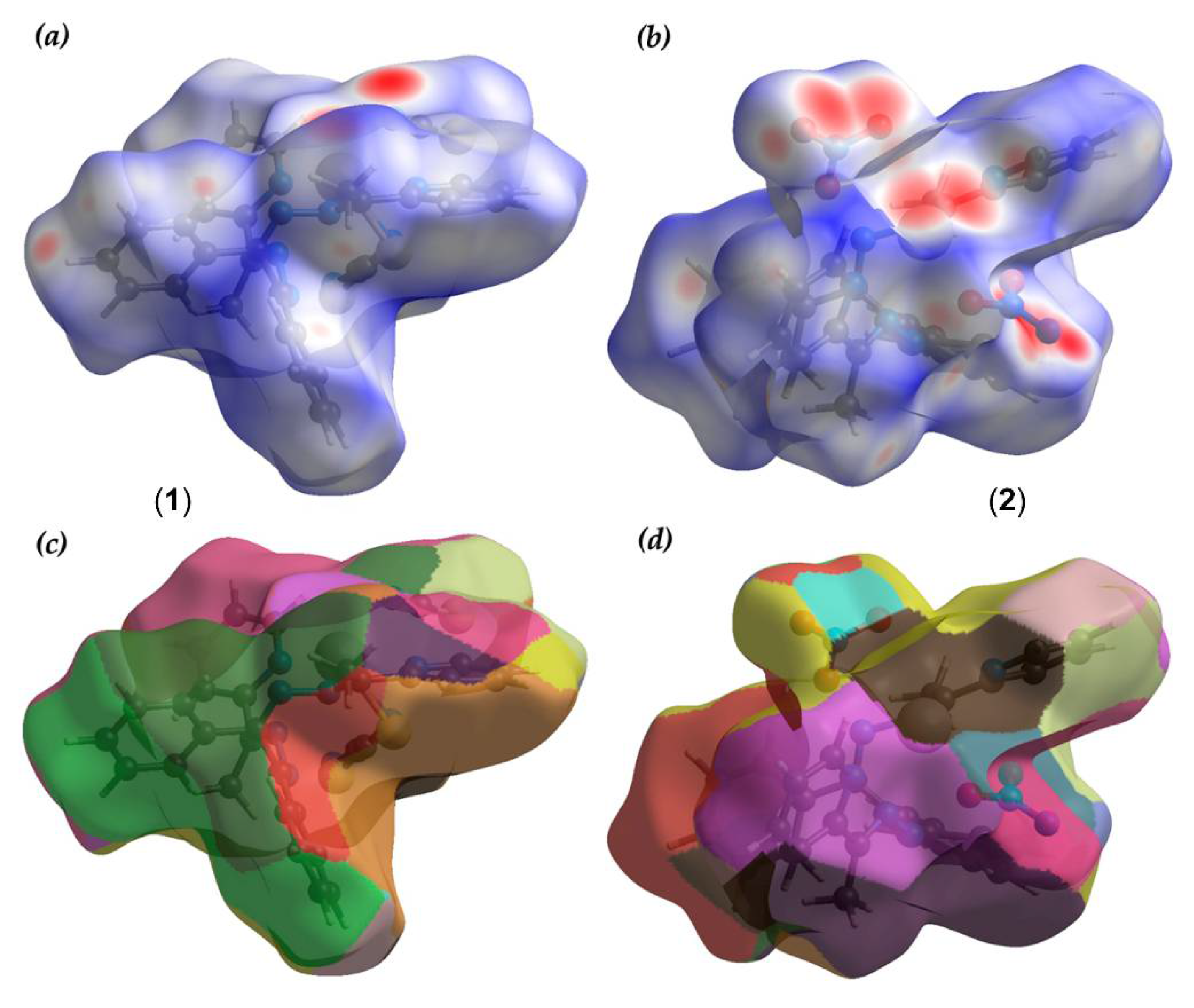
3.3. Hirshfeld Surface

We have examined the Hirshfeld surfaces to quantify the contribution of noncovalent interactions in the crystal packing. In the present study, we have evaluated the contacts that are responsible in building the supramolecular assemblies. The symmetry-generated counterparts of the structures were not included in the Hirshfeld surface calculations. The Hirshfeld surface of compounds (1) and (2) are mapped over dnorm in Figure 8 (in the ranges (−0.3775 to 1.6794 Å) in (1) and (−0.5624 to 1.3287 Å) in (2)) and fragment patches (0.000 to 25.000 Å in (1) and 0.000 to 29.000 Å in (2), respectively). The dnorm surfaces (Figure 8a,b) have been plotted based on the normalized function di and de using a white, red and blue color scheme. The circular deep red spots that are noticeable on the dnorm surface of both compounds are due to the coordination bonds, since the symmetrically generated counterparts (see Figure 1 and Figure 4) have not been included in the calculation. The nearest neighbor coordination environment of the molecules is identified from the color patches on the Hirshfeld surface based on their closeness to adjacent molecules. Therefore, the fragment patches show the appropriate approach for the identification of the nearest neighboring coordination environment of compounds (1) and (2) (see Figure 8c,d).
The scattered points of the fingerprint plots (Figure 9 and Figure 10) evidence all interactions involved within the structures. The long coordination bonds (Pb···N/N···Pb) and (Pb···O/O···Pb) are characterized in the fingerprint plots of (1) and (2), respectively. In (2), the (Pb···O/O···Pb) contacts contributed 1.7% to the total surface area of (2), whereas compound (1) contributed 1.9% due to (Pb···N/N···Pb) interaction (See Figure 9 and Figure 10). In (2), (O···H/H···O) interaction comprises 28.5%, whereas compound (1) contributed 17.8% (S···H/H···S) interactions. The N···H/H···N interactions for both compounds are verified by two distinct spikes on the fingerprint plot (see Figure 9 and Figure 10). The contribution of C···H/H···C contacts is higher in compound (1) compared to compound (2), and for both structures the C···H contact contributed more than its H···C counterpart. Moreover, the wings on the fingerprint plot of both structures designate the presence of C‒H···π interaction. The total C···H/H···C contacts comprise 29.2% and 22.1% in compounds (1) and (2), respectively. All other contacts that are involved within the structures of the title compounds are quantified accordingly and are included in Figure 9 and Figure 10.

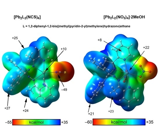
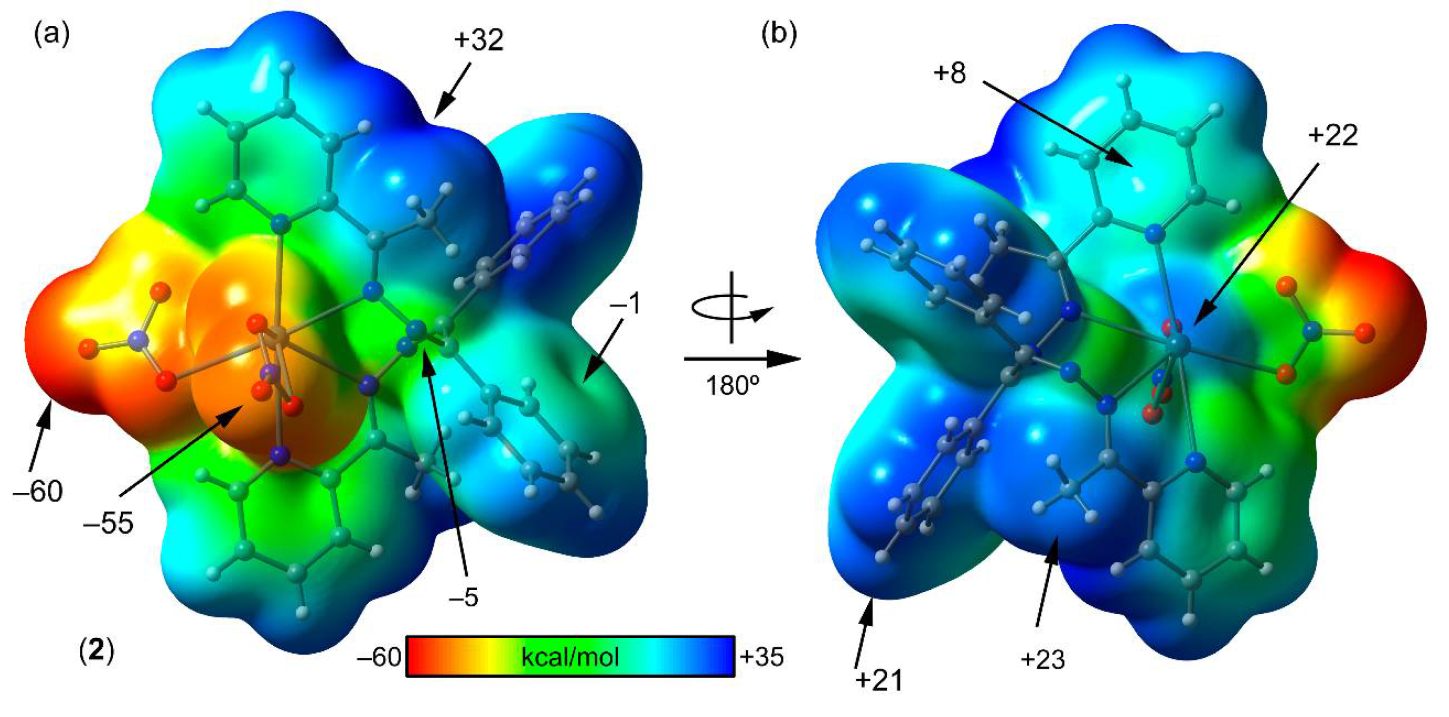
3.4. Mep Surface Analysis

In order to rationalize the noncovalent interactions described above in the crystal packing of compounds (1) and (2), the molecular electrostatic potential (MEP) surfaces have been computed for both compounds using the asymmetric unit. The MEP surface of compound (1) is shown in Figure 11, and it shows that the most negative MEP is located at the negative belt around the S-atom of the N-coordinated thiocyanate ligand (see Figure 11a). The MEP at the extension of the C=S bond is significantly smaller (in absolute value) than that at the belt. The MEP value at the N-atom of the S-coordinated thiocyanate ligand is slightly smaller (–52 kcal/mol). The most positive MEP value is located at the aromatic H-atoms of the Pb(II)-coordinated pyridine ring (+35 kcal/mol) due to inductive effects. The MEP value over the aromatic phenyl ring is slightly negative (–4 kcal/mol). The MEP values are also positive at the H-atoms of the methyl group and the phenyl rings. The formation of dinuclear centrosymmetric dimers, as observed in the X-ray structure, is favored electrostatically since the MEP value at the Pb(II) atom is positive (+10 kcal/mol) and negative at the N-atom of the thiocyanate ligand (–49 kcal/mol). In addition, the formation of the supramolecular H-bonded self-assembled dimers in (1) shown in Figure 2 strongly agrees with the MEP surface analysis, since the H-bond acceptors (S and N-atoms of the anionic ligands) correspond to the atoms bearing the most negative MEP values. Moreover, the H-bond donors are either an aromatic H-atom of the coordinated pyridine ring (in the C–H···S H-bond) or an H-atom of the methyl group (in the C–H···N H-bond) that correspond to the regions where the MEP value is maximum.
The MEP surface of compound (2) is shown in Figure 12, showing that the most negative MEP is located at the equatorial nitrato ligand (−60 kcal/mol). The MEP at the O-atom of the axial nitrato ligand is slightly smaller in absolute value (−55 kcal/mol). Similarly to compound (1), the most positive MEP value is located at the aromatic H-atoms of the Pb(II)-coordinated pyridine ring (+32 kcal/mol), due to the enhancement of their acidity upon complexation. The MEP value over the aromatic phenyl ring is slightly negative (−1 kcal/mol), and positive over the coordinated pyridine ligand (+8 kcal/mol). The MEP values are also positive at the H-atoms of the methyl group (+23 kcal/mol) and the phenyl rings (+21 kcal/mol). The formation of the dinuclear centrosymmetric dimer, as observed in the X-ray structure, is more favored electrostatically than that in (1) since the MEP value at the Pb(II) atom is more positive (+22 kcal/mol), and at the O-atom of the nitrato ligand (−49 kcal/mol) more negative than those of compound (1) (see Figure 11). Moreover, the formation of the two-dimensional network in compound (2) generated through C‒H···O hydrogen bonds in the (011) plane (see Figure 5) also agrees well with the MEP surface of (2), since the H-bond acceptor is the most negative O-atom. The C–H···N bonds that are also formed in the 2D layer are expected to be weaker, since the MEP value at the non-coordinated N-atom of the ligand is only −5 kcal/mol (see Figure 11a). This is also in good agreement with the Hirshfeld surface analysis of (2), since the quantification of the contribution of the O···H/H···O contacts is 28.5% and the contribution of the N–H/H–N contacts is only 7.4%.

4. Concluding Remarks

The protocol to synthesize two new dinuclear Pb(II) coordination compounds involving 1,2-diphenyl-1,2-bis((methyl(pyridin-2-yl)methylene)hydrazono)ethane ligand and SCN and NO3 counter anions is reported in this manuscript. Both complexes were characterized in the solid state by X-ray diffraction. The crystal packing of both compounds is dominated by C–H···O,N,S noncovalent interactions, which is further evidenced by the Hirshfeld surface analysis and rationalized using the MEP surface analysis. The latter demonstrates that the most electron rich regions of the complexes that correspond to the anionic coligands are the most electron poor to the aromatic H-atoms, thus favoring the C–H···O,N,S H-bonds. Moreover, although the ligand presents an extended aromatic surface (composed by four aromatic rings) π–π interactions are not observed. Interestingly, C–H···π interactions seem to dominate the crystal packing in both compounds (1) and (2).

Author Contributions

Conceptualization, A.F. and G.M.; methodology, F.I.Z.; software, A.F. and S.K.S.; investigation, all authors; writing—original draft preparation, A.B.R., A.F. and S.K.S. writing—review and editing, all authors; visualization, A.F. and S.K.S.; project administration, A.F. and F.I.Z.; funding acquisition, A.F. and F.I.Z. All authors have read and agreed to the published version of the manuscript.

Funding

The publication has been prepared with the support of the “RUDN University Program 5-100” (recipient F. I. Zubkov, synthesis of the ligands). This research was funded by the MICIU/AEI of Spain (project CTQ2017-85821-R FEDER funds).

Acknowledgments

We thank the “Centre de Tecnologies de la Informació” (CTI), Universitat de les Illes Balears for computational facilities. We also thank all projects for financial support.

Conflicts of Interest

The authors declare no conflict of interest.

References

  1. Casas, J.S.; Sordo, J. Lead: Chemistry, Analytical Aspects, Environmental Impact and Health Effects; Elsevier: Amsterdam, The Netherlands, 2006. [Google Scholar]
  2. Bauzá, A.; Seth, S.K.; Frontera, A. Tetrel bonding interactions at work: Impact on tin and lead coordination compounds. Coord. Chem. Rev. 2019, 384, 107–125. [Google Scholar] [CrossRef]
  3. Reger, D.L.; Wright, T.D.; Little, C.A.; Lamba, J.J.S.; Smith, M.D. Control of the Stereochemical Impact of the Lone Pair in Lead(II) Tris(pyrazolyl)methane Complexes. Improved Preparation of Na{B[3,5-(CF3)2C6H3]4}. Inorg. Chem. 2001, 40, 3810–3814. [Google Scholar] [CrossRef] [PubMed]
  4. Fleischer, H.; Schollmeyer, D. Synthesis of and Structural Studies on Lead(II) Cysteamin Complexes. Inorg. Chem. 2004, 43, 5529–5536. [Google Scholar] [CrossRef] [PubMed]
  5. Mahmudov, K.T.; Gurbanov, A.V.; Guseinov, F.I.; Guedes da Silva, M.F.C. Noncovalent interactions in metal complex catalysis. Coord. Chem. Rev. 2019, 387, 32–46. [Google Scholar] [CrossRef]
  6. Olvera, A.; Shi, G.; Djieutedjeu, H.; Page, A.; Uher, C.; Kioupakis, E.; Poudeu, P.F.P. Pb7Bi4Se13: A Lillianite Homologue with Promising Thermoelectric Properties. Inorg. Chem. 2015, 54, 746–755. [Google Scholar] [CrossRef]
  7. Mahmoudi, G.; Bauzá, A.; Frontera, A. Concurrent agostic and tetrel bonding interactions in lead(ii) complexes with an isonicotinohydrazide based ligand and several anions. Dalton Trans. 2016, 45, 4965–4969. [Google Scholar] [CrossRef]
  8. Roy, S.; Drew, M.G.B.; Bauzá, A.; Frontera, A.; Chattopadhyay, S. Non-covalent tetrel bonding interactions in hemidirectional lead(II) complexes with nickel(II)-salen type metallo-ligands. New J. Chem. 2018, 42, 6062–6076. [Google Scholar] [CrossRef]
  9. Mahmoudi, G.; Seth, S.K.; Zubkov, F.I.; López-Torres, E.; Bacchi, A.; Stilinović, V.; Frontera, A. Supramolecular Assemblies in Pb(II) Complexes with Hydrazido-Based Ligands. Crystals 2019, 9, 323. [Google Scholar] [CrossRef] [Green Version]
  10. Servati, G.M.; Stilinović, V.; Bauzá, A.; Frontera, A.; McArdle, P.; Van Derveer, D.; Ng, S.W.; Mahmoudi, G. Design of Lead(II) Metal-Organic Frameworks Based on Covalent and Tetrel Bonding. Chem. Eur. J. 2015, 21, 17951–17958. [Google Scholar] [CrossRef] [PubMed]
  11. John, R.P.; Park, M.; Moon, D.; Lee, K.; Hong, S.; Zou, Y.; Hong, C.S.; Lah, M.S.A. A chiral pentadecanuclear metallamacrocycle with a sextuple twisted möbius topology. J. Am. Chem. Soc. 2007, 129, 14142–14143. [Google Scholar] [CrossRef]
  12. De, S.; Drew, M.G.B.; Rzepa, H.S.; Datta, D. Linking number analysis of a self-assembled lemniscular Möbius-metallamacrocycle. New J. Chem. 2008, 32, 1831–1834. [Google Scholar] [CrossRef]
  13. Rzepa, H.S. Linking number analysis of a pentadecanuclear metallamacrocycle: A möbius-craig system revealed. Inorg. Chem. 2008, 47, 8932–8934. [Google Scholar] [CrossRef] [PubMed]
  14. Sankar, J.; Mori, S.; Saito, S.; Rath, H.; Suzuki, M.; Inokuma, Y.; Shinokubo, H.; Kim, K.S.; Yoon, Z.S.; Shin, J.Y.; et al. Unambiguous identification of Möbius aromaticity for meso-aryl-substituted [28]hexaphyrins(1.1.1.1.1.1). J. Am. Chem. Soc. 2008, 130, 13568–13579. [Google Scholar] [CrossRef] [PubMed]
  15. Sung, Y.M.; Yoon, M.C.; Lim, J.M.; Rath, H.; Naoda, K.; Osuka, A.; Kim, D. Reversal of Hückel (anti)aromaticity in the lowest triplet states of hexaphyrins and spectroscopic evidence for Baird’s rule. Nat. Chem. 2015, 7, 418–422. [Google Scholar] [CrossRef]
  16. Masoumi, A.; Gargari, M.S.; Mahmoudi, G.; Machura, B.; Lynch, V.; Giester, G.; Abedi, M.; Hazendonk, P. Structural diversity in manganese(II) complexes with multidentate N-donor imino pyridyl ligands. Z. Anorg. Allg. Chem. 2015, 641, 1176–1181. [Google Scholar] [CrossRef]
  17. Mahmoudi, G.; Stilinović, V.; Gargari, M.S.; Bauzá, A.; Zaragoza, G.; Kaminsky, W.; Lynch, V.; Choquesillo-Lazarte, D.; Sivakumar, K.; Khandari, A.A.; et al. From monomers to polymers: Steric and supramolecular effects on dimensionality of coordination architectures of heteroleptic mercury(ii) halogenide-tetradentate Schiff base complexes. CrystEngComm 2015, 17, 3493–3502. [Google Scholar] [CrossRef] [Green Version]
  18. Mahmoudi, G.; Zaręba, J.K.; Gurbanov, A.V.; Bauzá, A.; Zubkov, F.I.; Kubicki, M.; Stilinović, V.; Kinzhybalo, V.; Frontera, A. Benzyl Dihydrazone versus Thiosemicarbazone Schiff Base: Effects on the Supramolecular Arrangement of Cobalt Thiocyanate Complexes and the Generation of CoN6 and CoN4S2 Coordination Spheres. Eur. J. Inorg Chem. 2017, 4763–4772. [Google Scholar] [CrossRef]
  19. Afkhami, F.A.; Mahmoudi, G.; Gurbanov, A.V.; Zubkov, F.I.; Qu, F.; Gupta, A.; Safin, D.A. Solvent-driven azide-induced mononuclear discrete versus one-dimensional polymeric aromatic Möbius cadmium(ii) complexes of an N6 tetradentate helical ligand. Dalton Trans. 2017, 46, 14888–14896. [Google Scholar] [CrossRef] [Green Version]
  20. Seth, S.K.; Bauzá, A.; Mahmoudi, G.; Stilinović, V.; López-Torres, E.; Zaragoza, G.; Keramidas, A.D.; Frontera, A. On the importance of Pb⋯X (X = O, N, S, Br) tetrel bonding interactions in a series of tetra- and hexa-coordinated Pb(ii) compounds. CrystEngComm 2018, 20, 5033–5044. [Google Scholar] [CrossRef]
  21. Mahmoudi, G.; Afkhami, F.A.; Castiñeiras, A.; García-Santos, I.; Gurbanov, A.; Zubkov, F.I.; Mitoraj, M.P.; Kukułka, M.; Sagan, F.; Szczepanik, D.W.; et al. Quasi-aromatic Möbius Metal Chelates. Inorg. Chem. 2018, 57, 4395–4408. [Google Scholar] [CrossRef]
  22. Mitoraj, M.P.; Mahmoudi, G.; Afkhami, F.A.; Castiñeiras, A.; Giester, G.; Konyaeva, I.A.; Khandar, A.A.; Qu, F.; Gupta, A.; Sagan, F.; et al. Effect of Solvent on the Structural Diversity of Quasi-Aromatic Möbius Cadmium(II) Complexes Fabricated from the Bulky N 6 Tetradentate Helical Ligand. Cryst. Growth Des. 2019, 19, 1649–1659. [Google Scholar] [CrossRef]
  23. Afkhami, F.A.; Mahmoudi, G.; White, J.M.; Lipkowski, J.; Konyaeva, I.A.; Safin, D.A. Möbius-like metal chelates constructed from CdHal2 (Hal = Cl, Br, I) and benzilbis(pyridin-2-yl)methylidenehydrazone. Inorg. Chim. Acta 2019, 484, 481–490. [Google Scholar] [CrossRef]
  24. Afkhami, F.A.; Mahmoudi, G.; Khandar, A.A.; White, J.M.; Konyaeva, I.A.; Safin, D.A. Metal chelates constructed from CdHal2 (Hal = Cl, Br, I) and 1,2-diphenyl-1,2-bis((phenyl(pyridin-2-yl)methylene)hydrazono)ethane. J. Molec. Struct. 2019, 1176, 743–750. [Google Scholar] [CrossRef]
  25. Mitoraj, M.P.; Afkhami, F.A.; Mahmoudi, G.; Khandar, A.A.; Gurbanov, A.V.; Zubkov, F.I.; Waterman, R.; Babashkina, M.G.; Szczepanik, D.W.; Jena, H.S.; et al. Structural versatility of the quasi-aromatic Möbius type zinc(ii)-pseudohalide complexes-experimental and theoretical investigations. RSC Adv. 2019, 9, 23764–23773. [Google Scholar] [CrossRef] [Green Version]
  26. Afkhami, F.A.; Mahmoudi, G.; Qu, F.; Gupta, A.; Zangrando, E.; Frontera, A.; Safin, D.A. Complexes of BiCl3 with hydrazone derived ligands: Möbius-like discrete metal chelate versus salt-like porous polymeric structure. Inorg. Chim. Acta 2020, 502, 119350. [Google Scholar] [CrossRef]
  27. Bruker. SAINT, version 6.36a; Bruker-AXS Inc.: Madison, WI, USA, 2002. [Google Scholar]
  28. Bruker. SMART, Version 5.625 and SADABS, Version 2.03a; Bruker AXS Inc.: Madison, WI, USA, 2001. [Google Scholar]
  29. Sheldrick, G.M. A short history of SHELX. ActaCryst. 2008, A64, 112–122. [Google Scholar]
  30. Sheldrick, G.M. Crystal structure refinement with SHELXL. ActaCryst. 2015, C71, 3–8. [Google Scholar]
  31. Farrugia, L.J. WinGX suit for small-molecule single-crystal crystallography. J. Appl. Crystallogr. 1999, 32, 837–838. [Google Scholar] [CrossRef]
  32. Spek, A.L. Single-crystal structure validation with the program PLATON. J. Appl. Crystallogr. 2003, 36, 7–13. [Google Scholar] [CrossRef] [Green Version]
  33. Spackman, M.A.; McKinnon, J.J. Fingerprinting intermolecular interactions in molecular crystals. CrystEngComm 2002, 4, 378–392. [Google Scholar] [CrossRef]
  34. Seth, S.K.; Saha, I.; Estarellas, C.; Frontera, A.; Kar, T.; Mukhopadhyay, S. Supramolecular Self-Assembly of M-IDA Complexes Involving Lone-Pair⋯π Interactions: Crystal Structures, Hirshfeld Surface Analysis, and DFT Calculations [H2IDA = iminodiacetic acid, M = Cu(II), Ni(II)]. Cryst. Growth Des. 2011, 11, 3250–3265. [Google Scholar] [CrossRef]
  35. McKinnon, J.J.; Spackman, M.A.; Mitchell, A.S. Novel tools for visualizing and exploring intermolecular interactions in molecular crystals. Acta Cryst. 2004, B60, 627–668. [Google Scholar] [CrossRef]
  36. Rohl, A.L.; Moret, M.; Kaminsky, W.; Claborn, K.; Mckinnon, J.J.; Kahr, B. Hirshfeld Surfaces Identify Inadequacies in Computations of Intermolecular Interactions in Crystals: Pentamorphic 1,8-Dihydroxyanthraquinone. Cryst. Growth Des. 2008, 8, 4517–4525. [Google Scholar] [CrossRef]
  37. Seth, S.K. Tuning the formation of MOFs by pH influence: X-ray structural variations and Hirshfeld surface analyses of 2-amino-5-nitropyridine with cadmium chloride. Acta Cryst. 2013, 15, 1772–1781. [Google Scholar] [CrossRef]
  38. Seth, S.K.; Bauzá, A.; Frontera, A. Screening polymorphism in a Ni(II) metal–organic framework: Experimental observations, Hirshfeld surface analyses and DFT studies. CrystEngComm 2018, 20, 746–754. [Google Scholar] [CrossRef]
  39. Seth, S.K. Structural characterization and Hirshfeld surface analysis of a CoII complex with imidazo[1,2-a]-pyridine. Acta Cryst. 2018, E74, 600–606. [Google Scholar] [CrossRef] [Green Version]
  40. Maity, T.; Mandal, H.; Bauza, A.; Samanta, B.C.; Frontera, A.; Seth, S.K. Quantifying conventional C–H⋯π(aryl) and unconventional C–H⋯π(chelate) interactions in dinuclear Cu(II) complexes: Experimental observations, Hirshfeld surface and theoretical DFT study. New J. Chem. 2018, 42, 10202–10213. [Google Scholar] [CrossRef]
  41. Wolff, S.K.; Grimwood, D.J.; McKinnon, J.J.; Jayatilaka, D.; Spackman, M.A. CrystalExplorer 3.1; University of Western Australia: Perth, Australia, 2007. [Google Scholar]
  42. Frisch, M.J.; Trucks, G.W.; Schlegel, H.B.; Scuseria, G.E.; Robb, M.A.; Cheeseman, J.R.; Scalmani, G.; Barone, V.; Petersson, G.A.; Nakatsuji, H.; et al. Gaussian 16, Revision A.03; Gaussian, Inc.: Wallingford, CT, USA, 2016. [Google Scholar]
  43. Adamo, C.; Barone, V. Toward reliable density functional methods without adjustable parameters: The PBE0 model. J. Chem. Phys. 1999, 110, 6158–6169. [Google Scholar] [CrossRef]
  44. Grimme, S.; Antony, J.; Ehrlich, S.; Krieg, H. A dispersion correction for density functionals, Hartree-Fock and semi-empirical quantum chemical methods DFT-D3. J. Chem. Phys. 2010, 132, 154104. [Google Scholar] [CrossRef] [Green Version]
  45. Weigend, F.; Ahlrichs, R. Balanced basis sets of split valence, triple zeta valence and quadruple zeta valence quality for H to Rn: Design and assessment of accuracy. Phys. Chem. Chem. Phys. 2005, 7, 3297–3305. [Google Scholar] [CrossRef]
  46. Weigend, F. Accurate Coulomb-fitting basis sets for H to Rn. Phys. Chem. Chem. Phys. 2006, 8, 1057–1065. [Google Scholar] [CrossRef] [PubMed]
Scheme 1. Synthesis of complexes [Pb2L2(NCS)4] (1) and [Pb2L2(NO3)4] (2).
Crystals 10 00568 sch001
Figure 1. View of the asymmetric unit of compound (1) with a partial atom numbering scheme. Hydrogen atoms are omitted for clarity. The unlabeled counterpart is generated through the symmetry operation. Thermal ellipsoids are drawn at the 30% probability level.
Crystals 10 00568 g001
Figure 2. Partial view of the X-ray structure of (1) with indication of C–H···N and C–H···N H-bonds.
Crystals 10 00568 g002
Figure 3. Self-assembled supramolecular framework in (1) in (101) plane through C‒H···π interactions.
Crystals 10 00568 g003
Figure 4. View of the asymmetric unit of compound (2) with partial atom numbering scheme. Hydrogen atoms are omitted for clarity. The unlabeled counterpart is generated through the symmetry operation. Thermal ellipsoids are drawn at the 30% probability level.
Crystals 10 00568 g004
Figure 5. Two-dimensional framework generated through C‒H···O and C‒H···N hydrogen bonds in the (011) plane.
Crystals 10 00568 g005
Figure 6. Supramolecular self-assembly generated through weak C‒H···O hydrogen bonds in the (110) plane.
Crystals 10 00568 g006
Figure 7. Perspective view of the supramolecular network through C‒H···π interactions in (2).
Crystals 10 00568 g007
Figure 8. Hirshfeld surfaces mapped with dnorm ((a) and (b)) and Fragment patches ((c) and (d)) for compounds (1) and (2), respectively.
Crystals 10 00568 g008
Figure 9. Full and decomposed fingerprint plots corresponding to various contacts involved within the structure of compound (1).
Crystals 10 00568 g009
Figure 10. Full and decomposed fingerprint plots corresponding to various contacts involved within the structure of compound (2).
Crystals 10 00568 g010
Figure 11. Molecular electrostatic potential (MEP) surface (isosurface 0.001 a.u.) of the two sides (a) and (b) of compound (1) at the PBE1PBE-D3/def2-TZVP. The MEP values at selected points of the surface are given in kcal/mol.
Crystals 10 00568 g011
Figure 12. MEP surface (isosurface 0.001 a.u.) of the two sides (a) and (b) of compound (2) at the PBE1PBE-D3/def2-TZVP. The MEP values at selected points of the surface are given in kcal/mol.
Crystals 10 00568 g012
Table 1. Crystal data and structure refinement parameters for compounds (1) and (2).
Structure(1)(2)
Empirical formula C60H48N16Pb2S4C57H52N16O13Pb2
Formula Weight1535.761583.52
Temperature (K)100(2)100(2)
Wavelength (Å)0.710730.71073
Crystal systemTriclinicTriclinic
space groupP‒1 P‒1
a, b, c (Å)9.2815(7), 10.4330(8), 16.3791(11)10.0664(6),11.0250(7),14.1786(10)
α, β, γ (°)81.444(3), 74.809(3), 78.168(4)105.444(3), 95.245(3), 104.866(3)
Volume (Å3)1490.51(19)1444.27(16)
Z / Density (calc.) (Mg/m3)1/1.7111/1.821
Absorption coefficient (mm−1)5.8345.901
F(000)748774
Crystal size (mm3)0.45 × 0.40 × 0.200.45 × 0.25 × 0.11
θ range for data collection2.274 to 25.3421.512 to 26.242
Completeness to θ (%)99.898.9
Absorption correctionSemi-empirical from equivalentsSemi-empirical from equivalents
Max. and min. transmission0.362 and 0.1540.552 and 0.165
Refinement methodFull-matrix least-squares on F2Full-matrix least-squares on F2
Data/parameters5442/3825905/416
Goodness-of - fit on F21.0281.051
Final R indices [I > 2σ(I)]R1 = 0.0283, wR2 = 0.0581R1 = 0.0265, wR2 = 0.0529
R indices (all data)R1 = 0.0337, wR2 = 0.0600R1 = 0.0323, wR2 = 0.0548
Largest diff. peak and hole (e.Å−3)1.279 and −1.8141.127 and −1.132
R1 = ∑||Fo|–|Fc||/∑|Fo|, wR2 = [∑{(Fo2–Fc2)2}/∑{w(Fo2)2}]1/2, w = 1/{σ2(Fo2) + (aP)2 + bP}, where a = 0.0212 and b = 2.5138 for (1) and a = 0.0238 and b = 0.8790 for (2). P = (Fo2 + 2Fc2)/3 for both structures.
Table 2. Selected (Å) and bond angles (°) for compounds (1) and (2) as determined by X-ray diffraction.
Complex-(1)
Pb(1)-N(1)2.629(4)Pb(1)-N(6)2.657(3)
Pb(1)-N(2)2.645(3)Pb(1)-N(7)2.615(4)
Pb(1)-N(5)2.745(3)Pb(1)-S(1)2.756(2)
O(1)-Pb(1)-N(3)60.39(9)O(1)-Pb(1)-O(7)142.78(7)
O(1)-Pb(1)-N(4)115.35(9)N(3)-Pb(1)-O(7)117.30(7)
N(3)-Pb(1)-N(4)57.86(9)N(4)-Pb(1)-O(7)84.17(6)
Complex-(2)
Pb(1)-O(1)2.614(3)Pb(1)-N(1)2.720(3)
Pb(1)-O(3)2.621(3)Pb(1)-N(5)2.763(3)
Pb(1)-N(2)2.670(3)
O(1)-Pb(1)-O(3)48.61(8)N(2)-Pb(1)-N(1)59.79(9)
O(1)-Pb(1)-N(2)75.78(9)O(1)-Pb(1)-N(5)130.88(9)
O(3)-Pb(1)-N(2)81.63(9)O(3)-Pb(1)-N(5)90.89(9)
O(1)-Pb(1)-N(1)70.93(9)N(2)-Pb(1)-N(5)71.13(8)
O(3)-Pb(1)-N(1)114.86(9)N(1)-Pb(1)-N(5)118.16(8)
Table 3. Relevant hydrogen bonding parameters of compounds (1) and (2).
D–H···AD–HH···AD···AD–H···ASymmetry
Compound (1)
C27–H27···S20.952.943.716(5)1391 − x, 2 − y, −z
C7–H7C···N80.982.643.56(2)158x, 1 + y, z
Compound (2)
O7–H71···O30.842.042.849(8)162----
C1–H1···O50.952.263.041(5)139−x, 1 − y, −z
C3–H3···O20.952.573.451(5)153−1 + x, y, z
C4–H4···O70.952.403.218(9)144−1 + x, y, z
C7–H7B···N30.982.553.373(5)141−x, 1 − y, 1 − z
C7–H7C···N30.982.302.755(5)107----
C12–H12···O10.952.473.102(6)124x, −1 + y, z
C15–H15B···O71.402.403.574(9)139----
C21–H21···O40.952.573.243(5)128−1 − x, −y, −z
C23–H23C···N40.982.392.789(5)104----
C29–H29···O40.952.413.161(5)136−x, 1 − y, −z
Table 4. Geometrical parameters (Å, °) for C–H∙∙∙π interactions.
Y–X∙∙∙Cg(I)X∙∙∙CgY∙∙∙CgY–X∙∙∙CgX-PerpSymmetry
Compound (1)
C(2)–H(2)∙∙∙Cg(2)2.963.570(5)1232.891 + x, y, z
C(23)–H(23C)∙∙∙Cg(4)2.983.560(5)1192.73−x, 2 − y, 1 − z
Compound (2)
C(28)–H(28)∙∙∙Cg(18)2.963.413(4)1102.801 + x, y, z

