Review

Peroxisome Proliferator-Activated Receptor and Vitamin D Receptor Signaling Pathways in Cancer Cells

by
Satoru Matsuda
*,† and
Yasuko Kitagishi
Department of Food Science and Nutrition, Nara Women's University, Kita-Uoya Nishimachi, Nara 630-8506, Japan
*
Author to whom correspondence should be addressed.
These authors contributed equally to this work.
Cancers 2013, 5(4), 1261-1270; https://doi.org/10.3390/cancers5041261
Submission received: 2 September 2013 / Revised: 1 October 2013 / Accepted: 10 October 2013 / Published: 21 October 2013
(This article belongs to the Special Issue Vitamin D: Role in Cancer Causation, Progression and Therapy)

Abstract

:
Peroxisome proliferator-activated receptors (PPARs) are members of the superfamily of nuclear hormone receptors, which respond to specific ligands such as polyunsaturated fatty acids by altering gene expression. Three subtypes of this receptor have been discovered, each evolving to achieve different biological functions. Like other nuclear receptors, the transcriptional activity of PPARs is affected not only by ligand-stimulation, but also by cross-talk with other molecules. For example, both PPARs and the RXRs are ligand-activated transcription factors that coordinately regulate gene expression. In addition, PPARs and vitamin D receptor (VDR) signaling pathways regulate a multitude of genes that are of importance for cellular functions including cell proliferation and cell differentiation. Interaction of the PPARs and VDR signaling pathways has been shown at the level of molecular cross-regulation of their transcription factor. A variety of ligands influencing the PPARs and VDR signaling pathways have been shown to reveal chemopreventive potential by mediating tumor suppressive activities in human cancers. Use of these compounds may represent a potential novel strategy to prevent cancers. This review summarizes the roles of the PPARs and the VDR in pathogenesis and progression of cancer.

1. Introduction

Nuclear receptor family is divided into two subfamilies. The first group includes the estrogen, androgen, progesterone and mineralocorticoid receptors [1], and the second group includes vitamin D receptor (VDR), the thyroid receptor (TR), retinoic acid receptor (RAR), retinoid X receptor (RXR), and peroxisome proliferator—activated receptors (PPARs) [2]. The second group of receptors can form heterodimers with each other, and can function through interacting with appropriate ligands at genetic level [1,2]. In particular, the PPARs and/or like the VDR represent a major research target for the understanding and treatment of many diseases [3]. After activation through a ligand, the PPARs and VDR form a heterodimer with the RXR and induce antitumor effects in a variety of human carcinomas [4]. Therefore, these pathways may play an important role in cancer treatment and prevention. Furthermore, modulating PPAR signaling pathways would represent a potential novel strategy for inhibiting carcinogenesis and progression. This review paper will focus on the evidence of the functions of PPARs and VDR in cancer.

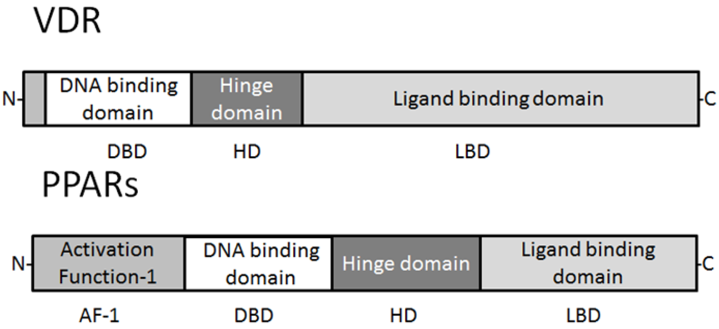
2. Expression and Characteristics of PPARs and Vitamin D Receptor

PPARs are ligand-activated transcription factors that are involved in the genetic regulation of mammalian metabolism, including fatty acid oxidation, transport and metabolism mediating proteins through the formation of a DNA binding heterodimer complex [5,6,7]. These receptors have also been shown to be implicated in cellular proliferation, differentiation, tumor promotion, apoptosis and immune reaction/inflammation. Three genetically and functionally distinct PPAR isoforms, PPARs (PPARα, PPARβ/δ, and PPARγ), have been described. PPARα is expressed at high levels in tissues that catabolize fatty acids [8], as in the adult liver, heart, kidney, large intestine and skeletal muscle. PPARβ/δ mRNA is ubiquitously distributed with a higher expression in digestive tract and placenta [9]. PPARγ is mostly expressed in the adipose tissue [10] and immune system, and is an important regulator of their differentiation and metabolism. All distinct PPARs subtypes exhibit distinct patterns of tissue distribution and share a high degree of structural homology with other members of the superfamily, particularly in the DNA-binding domain and ligand-binding domain [5,7,11] (Figure 1). Each isotype is a product of a separate gene. Retinoic X receptor (RXR) is a functional partner of PPAR. RXRα and PPARγ function potently in metabolic diseases, and are both important targets for anti-diabetic drugs. Coactivation of RXRα and PPARγ is believed to synergize their effects on glucose and lipid metabolism [12]. The transcriptional regulation by PPARs requires heterodimerization with the retinoid X receptor (RXR) (Figure 2). PPARs bind to a variety of PPAR response elements (PPREs) present in the promoter regions of the responsive genes [13]. Thus, selective action of PPARs in vivo results from the interplay at a time point of each of the cofactors available. The RXRs are able to influence the transcription of a wide variety of genes, because they can activate gene transcription by binding to specific sites on DNA as homodimers and/or as the heterodimers with other related nuclear receptors including the PPARs, VDR, and TR, so forth [14,15,16]. A variety of compounds have been identified as PPARs ligands. Among the synthetic ligands, fibrates and thiazolidinediones are PPARα and PPARγ agonists, respectively [17]. A PPARα specific ligand, 8S-HETE, and a PPARγ specific ligand, PGJ, 15-deoxy-Δ12,14-prostaglandin J2, and a peroxisome proliferator, clofibrate, all are able to induce expression of both PPARα and PPARγ [18,19,20]. Subsequent work has led to the identification of various PPAR ligands that include eicosanoids, hypolipidemic agents, and antidiabetic drugs [21,22]. PPARγ is also activated by prostaglandins and leukotrienes [23]. Besides natural ligands such as polyunsaturated fatty acids (including linoleic acid, linolenic acid and arachidonic acid), a large number of synthetic PPAR ligands have been identified. Clinically used drugs like the thiazolidinediones (troglitazone and pioglitazone), a class of insulinsensitizing agents and the fibrates (bezafibrate and clofibrate), which are used as hypolipidaemic drugs, are also binding to the PPARs [24]. In the presence of ligands, conformational changes of the ligand binding domain result in the recruitment of co-activator proteins or release of co-repressor proteins, and following association of a protein complex that enhances transcription activity of the target genes [25,26].
Figure 1. Schematic structure of VDR and PPAR protein. The predicted consensual important domain structures for VDR and PPAR are depicted, which are common in some species. AF-1 = activation function-1, DBD = DNA binding domain, HD = hinge domain linking DBD and LBD, LBD = C-terminal ligand binding domain. Note that the sizes of protein are modified for clarity.
Cancers 05 01261 g001
The nutritional forms of vitamin D had been identified as 1,25(OH)2D3 (calcitriol), an active form of vitamin D (aVitD) [27]. The majority of the aVitD effect is thought to be carried out by VDR localized in the nucleus. The ligand of VDR was thus discovered before the cloning of the receptor. VDR is a high-affinity hormone receptor and a nonpermissive partner of RXR [28]. Ligand-bound VDR can regulate gene expression via various molecular mechanisms, including direct activation where VDR-RXR most effectively binds DR-3 (direct repeat with 3-bp spacer) response element [29]. The function of VDR is essential for promoting calcium absorption in the gut and maintaining adequate serum calcium and phosphate concentrations to enable normal mineralization of bone [30]. It is also required for bone growth and bone remodeling by osteoblasts and osteoclasts. VDR is expressed in most tissues in the human body, and vitamin D plays an important role in decreasing the risk of many chronic illnesses, cancers, autoimmune diseases, infectious diseases, and cardiovascular disease [31]. Vitamin D2 (ergocalciferol) has been shown to contribute to the vitamin D status in humans, and metabolized in a similar fashion as vitamin D3. Vitamin D3 can be synthesized in the skin when ultraviolet B (UVB) penetrates the skin [32]. In various cell types, including normal and cancer cells, the effects of aVitD and VDR mediated genomic pathways include the regulation of cell growth and differentiation. VDRE have been reported in the proximal promoter of a number of vitamin D-responding genes including the human vitamin D 24-hydroxylase (CYP24) [33,34]. The inactivation of vitamin D metabolites is carried out by the CYP24 that is a key enzyme in 24-hydroxylation.
Figure 2. Schematic depiction of the model of mechanism of VDR and PPARs action. Similar to other nuclear hormone receptors, VDR and PPARs act as a ligand activated transcription factor. Both VDR and PPARs in response to their ligand binding, hetero-dimerize with RXR, and bind VDRE and PPRE DNA sequences in the promoters of target genes, respectively. Note that some critical molecules have been omitted for clarity.
Cancers 05 01261 g002

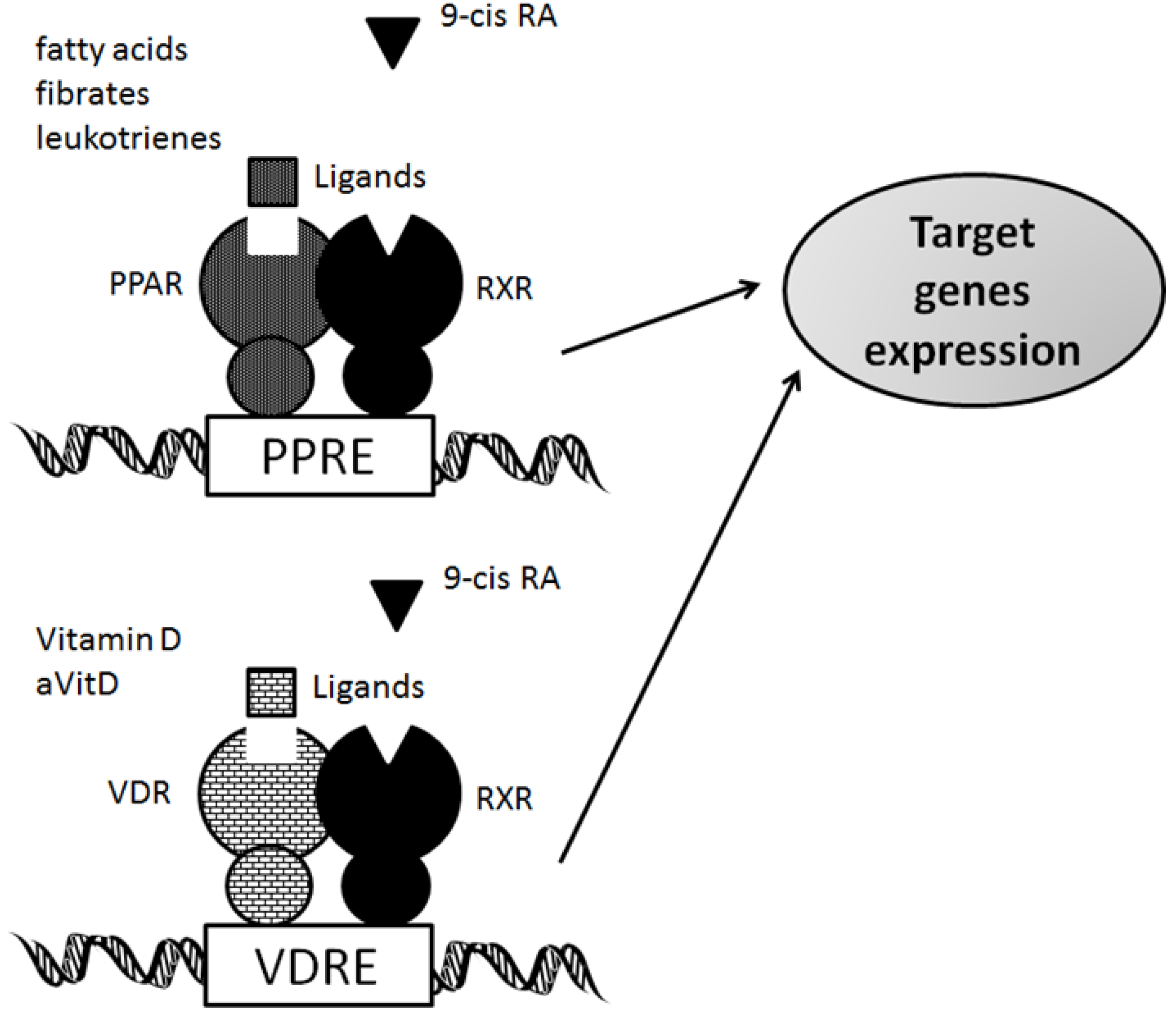
3. Functional Interplay of Vitamin D Receptor with PPARs

Both PPARs and VDR could form heterodimers with the retinoid-X receptor (RXR). Both the VDR and PPAR compete for their predominant heterodimerisation partner, RXR, complex transcriptional regulation of target genes might be expected [35,36]. VDR associates with vitamin D and forms heterodimers with RXR and exerts its activity through binding to vitamin D response elements (VDREs) of target genes. In adipocyte, vitamin D and VDR inhibit both PPARγ activity and adipogenesis [35,37]. There is a potent VDRE in human PPAR promoter [38]. So, PPAR is a primary aVitD responding gene and that VDR and PPAR signaling pathways are interconnected at the level of cross-regulation of their respective transcription factor mRNA levels. This cross-talk may involve a competition for the same heterodimerisation partner, RXR and the presence of VDREs and peroxisome proliferator response elements (PPREs). Both PPAR ligands and vitamin D analogs have been shown to be implicated in tumor progression and cellular differentiation. After activation through a ligand binding, the conformation of PPARs and VDR is altered and stabilized, resulting in the creation of a binding fissure and recruitment of transcriptional coactivators. The provided link between the PPARs and the VDR is bidirectional with either side being able to influence the other’s activity. It has been shown that PPARγ binds to VDR and inhibits vitamin D mediated transactivation [39]. The cross-talk is also important for the ability of gene expression and regulates a multitude of genes that are of importance for various cellular functions including cell proliferation, cell differentiation, immune responses and apoptosis. The signaling pathways of the VDR and the PPARs are interconnected in a large number of cancer cell lines [40,41]. However, the complete mechanisms of this cross-talk between the VDR and PPAR signaling pathways are not yet known. Further investigations are required to evaluate the physiological and pathophysiological relevance of this cross-talk. If so, activation of PPAR signaling pathways by aVitD or other vitamin D ligands may open new perspectives for treatment or prevention of cancer cells.

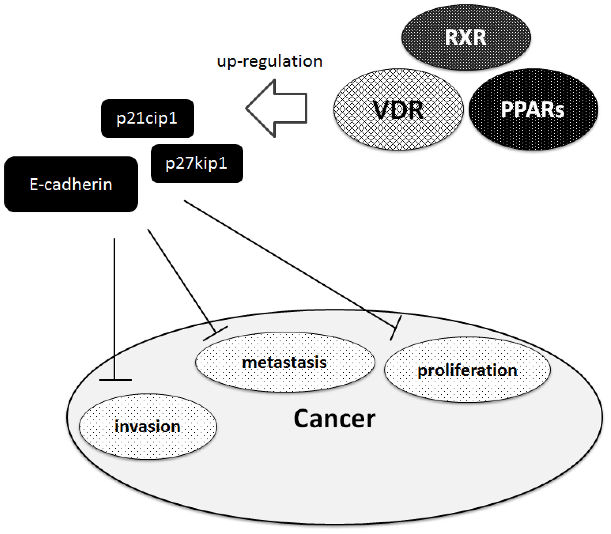
4. PPARs and Vitamin D Receptor in Cancer

Generally, non‑steroidal nuclear receptors play a major role in cancer development. Antiproliferative effects of PPARs ligands could be demonstrated in different cell lines. Thiazolidinedione, a PPAR ligand, currently used to treat diabetes, inhibits the proliferation of cancer cells [42,43]. In addition, PPAR activation by corresponding ligands (ex. fenofibrate) decreases the metastatic potential via down-regulation of Akt signaling [44]. These antiproliferative effects are mediated by cell-cycle arrest through a PPAR dependent pathway. Therefore modulating PPAR signaling pathways represents a potential novel strategy for inhibiting carcinogenesis and its progression. Vitamin D also elicits antiproliferative effects in a variety of cancer cell types including cell lines derived from prostate. The anticancer mechanisms include induction of cell cycle arrest, promotion of differentiation, inhibition of proliferation and angiogenesis, as well as inhibition of invasive and migratory potential of cancer cells. The aVitD exerts a significant inhibitory effect on the G1/S checkpoint of the cell cycle by upregulating the cyclin dependent kinase inhibitors such as p27kip1 and p21cip1 [45]. Beside the growth regulation of cells, aVitD also has an effect on tumor invasion, angiogenesis and metastastic behavior in various malignancies [46,47]. Vitamin D and AKT inhibitors synergistically inhibit prostate cancer growth through induction of cell cycle [48]. Mechanisms involve in the aVitD-induced inhibition of tumor invasion and metastasis include inhibition of serine proteinases and metalloproteinases as well as the up-regulation of E-cadherin [49]. The VDR gene is a target of epithelial to mesenchymal transition (EMT) promoters [50]. Considering a number of target genes of VDR and PPARs, these nuclear factors may modulate proliferation and differentiation of normal and cancer cells via various mechanisms (Figure 3). Ligands and other agents influencing the PPAR and VDR signaling pathways have been shown to reveal chemopreventive potential by mediating tumor suppressive activities in a variety of human cancers [41]. Further studies have to show if PPARs and VDR open new perspectives as agents inhibiting malignant potential of cancer. The loss of anti-proliferative responsiveness in cancer cells toward ligands for VDR, RXRs, and PPARs may require underlying epigenetic events [51]. Actually, function of VDR can be modulated epigenetically by histone acetylation, and it cooperates with other nuclear receptors which are influenced by histone acetyl transferases as well as histone deacetylases [52].
Figure 3. Implication of VDR, PPARs, and RXR in inhibition of cancer. VDR, PPARs, and RXR may be involved in inhibition of several aspect including proliferation, metastasis and invasion in cancer. Hammerheads mean inhibition. Note that some critical pathways have been omitted for clarity.
Cancers 05 01261 g003

5. Perspectives

The signaling pathways of PPARs and VDR regulate a multitude of genes that are of importance for various cellular functions. The findings of this study may therefore open new perspectives for treatment and/or prevention of cancers. Clearly, more work is needed to develop a comprehensive understanding of the cellular and molecular mechanisms in regulating PPARγ expression by vitamin D. Further study of PPARs, RXRs, and VDR functions in cancer cells may indicate pathways that are common to critical carcinogenic processes, providing additional focus for research in important cancers. In parallel, defining more specific mode of action by identifying the endogenous co-activators and modulators of these transcription factors in animal models will help to build more efficient therapeutic strategy. Future studies using functional genomic approaches will be required to more clearly establish the complicated mechanisms by which PPARs and VDR exert their actions. The link between the PPAR and VDR signaling pathways may help guide molecular-based treatment strategies and allow the synthesis of new agents for cancer treatment. Further investigations also have to show the benefit of PPAR ligands and aVitD compared to conventional chemotherapeutic regimens.

6. Conclusions

Interaction of the PPARs and VDR signaling pathways has been shown at the level of molecular cross-regulation of their transcription factor in pathogenesis and progression of cancer. The link between the PPARs and VDR signaling pathways may guide molecular-based treatment strategies and allow the creation of new tools for cancer treatment.

Abbreviations

aVitD
active form of vitamin D: 1,25(OH)2D3
AP-1
activator protein-1
EMT
mesenchymal transition
mRNA
messenger RNA
PPAR
peroxisome proliferator-activated receptor
PPRE
PPAR response element
RA
retinoic acid
RARs
retinoic acid receptors
RE
response element
RXR
retinoid X receptor
TR
thyroid receptor
VDR
vitamin D receptor
VDREs
vitamin D response elements

Acknowledgments

This work was supported by grants-in-aid from the Ministry of Education, Culture, Sports, Science and Technology in Japan. In addition, this work was supported in part by the grant from Nakagawa Masashichi Shoten Co., Ltd.

Conflicts of Interest

The authors declare that they have no competing financial interests.

References

  1. Hu, X.; Funder, J.W. The evolution of mineralocorticoid receptors. Mol. Endocrinol. 2006, 20, 1471–1478. [Google Scholar]
  2. Pérez, E.; Bourguet, W.; Gronemeyer, H.; de Lera, A.R. Modulation of RXR function through ligand design. Biochim. Biophys. Acta 2012, 1821, 57–69. [Google Scholar] [CrossRef]
  3. Carlberg, C.; Dunlop, T.W. An integrated biological approach to nuclear receptor signaling in physiological control and disease. Crit. Rev. Eukaryot. Gene Expr. 2006, 16, 1–22. [Google Scholar] [CrossRef]
  4. Ditsch, N.; Mayr, D.; Lenhard, M.; Strauss, C.; Vodermaier, A.; Gallwas, J.; Stoeckl, D.; Graeser, M.; Weissenbacher, T.; Friese, K.; Jeschke, U. Correlation of thyroid hormone, retinoid X, peroxisome proliferator-activated, vitamin D and oestrogen/progesterone receptors in breast carcinoma. Oncol. Lett. 2012, 4, 665–671. [Google Scholar]
  5. Schulman, I.G. Nuclear receptors as drug targets for metabolic disease. Adv. Drug Deliv. Rev. 2010, 62, 1307–1315. [Google Scholar] [CrossRef]
  6. Wahli, W.; Michalik, L. PPARs at the crossroads of lipid signaling and inflammation. Trends Endocrinol. Metab. 2012, 23, 351–363. [Google Scholar] [CrossRef]
  7. Becker, J.; Delayre-Orthez, C.; Frossard, N.; Pons, F. Regulation of inflammation by PPARs: A future approach to treat lung inflammatory diseases? Fundam. Clin. Pharmacol. 2006, 20, 429–447. [Google Scholar] [CrossRef]
  8. Ringseis, R.; Eder, K. Influence of pharmacological PPARalpha activators on carnitine homeostasis in proliferating and non-proliferating species. Pharmacol. Res. 2009, 60, 179–184. [Google Scholar] [CrossRef]
  9. Wagner, K.D.; Wagner, N. Peroxisome proliferator-activated receptor beta/delta (PPARbeta/delta) acts as regulator of metabolism linked to multiple cellular functions. Pharmacol. Ther. 2010, 125, 423–435. [Google Scholar] [CrossRef]
  10. Cipolletta, D.; Feuerer, M.; Li, A.; Kamei, N.; Lee, J.; Shoelson, S.E.; Benoist, C.; Mathis, D. PPAR-γ is a major driver of the accumulation and phenotype of adipose tissue Treg cells. Nature 2012, 486, 549–553. [Google Scholar]
  11. Giaginis, C.; Tsantili-Kakoulidou, A.; Theocharis, S. Peroxisome proliferator-activated receptors (PPARs) in the control of bone metabolism. Fundam. Clin. Pharmacol. 2007, 21, 231–244. [Google Scholar] [CrossRef]
  12. Zhang, H.; Xu, X.; Chen, L.; Chen, J.; Hu, L.; Jiang, H.; Shen, X. Molecular determinants of magnolol targeting both RXRα and PPARγ. PLoS One 2011, 6, e28253. [Google Scholar]
  13. Temple, K.A.; Cohen, R.N.; Wondisford, S.R.; Yu, C.; Deplewski, D.; Wondisford, F.E. An intact DNA-binding domain is not required for peroxisome proliferator-activated receptor gamma (PPARgamma) binding and activation on some PPAR response elements. J. Biol. Chem. 2005, 280, 3529–3540. [Google Scholar]
  14. Clarke, S.D.; Thuillier, P.; Baillie, R.A.; Sha, X. Peroxisome proliferator-activated receptors: A family of lipid-activated transcription factors. Am. J. Clin. Nutr. 1999, 70, 566–571. [Google Scholar]
  15. Nezbedova, P.; Brtko, J. 1alpha,25-dihydroxyvitamin D3 inducible transcription factor and its role in the vitamin D action. Endocr. Regul. 2004, 38, 29–38. [Google Scholar]
  16. Wolf, G. Is 9-cis-retinoic acid the endogenous ligand for the retinoic acid-X receptor? Nutr. Rev. 2006, 64, 532–538. [Google Scholar]
  17. Benz, V.; Kintscher, U.; Foryst-Ludwig, A. Sex-specific differences in type 2 diabetes mellitus and dyslipidemia therapy: PPAR agonists. Handb. Exp. Pharmacol. 2012, 214, 387–410. [Google Scholar]
  18. Forman, B.M.; Tontonoz, P.; Chen, J.; Brun, R.P.; Spiegelman, B.M.; Evans, R.M. 15-Deoxy-delta 12, 14-prostaglandin J2 is a ligand for the adipocyte determination factor PPAR gamma. Cell 1995, 83, 803–812. [Google Scholar] [CrossRef]
  19. Morosetti, R.; Servidei, T.; Mirabella, M.; Rutella, S.; Mangiola, A.; Maira, G.; Mastrangelo, R.; Koeffler, H.P. The PPARgamma ligands PGJ2 and rosiglitazone show a differential ability to inhibit proliferation and to induce apoptosis and differentiation of human glioblastoma cell lines. Int. J. Oncol. 2004, 25, 493–502. [Google Scholar]
  20. Yu, Z.; Schneider, C.; Boeglin, W.E.; Brash, A.R. Epidermal lipoxygenase products of the hepoxilin pathway selectively activate the nuclear receptor PPARalpha. Lipids 2007, 42, 491–497. [Google Scholar] [CrossRef]
  21. Kouroumichakis, I.; Papanas, N.; Zarogoulidis, P.; Liakopoulos, V.; Maltezos, E.; Mikhailidis, D.P. Fibrates: Therapeutic potential for diabetic nephropathy? Eur. J. Intern. Med. 2012, 23, 309–316. [Google Scholar] [CrossRef]
  22. Friedland, S.N.; Leong, A.; Filion, K.B.; Genest, J.; Lega, I.C.; Mottillo, S.; Poirier, P.; Reoch, J.; Eisenberg, M.J. The cardiovascular effects of peroxisome proliferator-activated receptor agonists. Am. J. Med. 2012, 125, 126–133. [Google Scholar] [CrossRef]
  23. Ibabe, A.; Herrero, A.; Cajaraville, M.P. Modulation of peroxisome proliferator-activated receptors (PPARs) by PPAR(alpha)- and PPAR(gamma)-specific ligands and by 17beta-estradiol in isolated zebrafish hepatocytes. Toxicol. In Vitro 2005, 19, 725–735. [Google Scholar] [CrossRef]
  24. Baker, P.R.; Lin, Y.; Schopfer, F.J.; Woodcock, S.R.; Groeger, A.L.; Batthyany, C.; Sweeney, S.; Long, M.H.; Iles, K.E.; Baker, L.M.; et al. Fatty acid transduction of nitric oxide signaling: multiple nitrated unsaturated fatty acid derivatives exist in human blood and urine and serve as endogenous peroxisome proliferator-activated receptor ligands. Biol. Chem. 2005, 280, 42464–42475. [Google Scholar] [CrossRef]
  25. Yu, S.; Reddy, J.K. Transcription coactivators for peroxisome proliferator-activated receptors. Biochim. Biophys. Acta 2007, 1771, 936–951. [Google Scholar]
  26. Waku, T.; Shiraki, T.; Oyama, T.; Morikawa, K. Atomic structure of mutant PPARgamma LBD complexed with 15d-PGJ2: Novel modulation mechanism of PPARgamma/RXRalpha function by covalently bound ligands. FEBS Lett. 2009, 583, 320–324. [Google Scholar] [CrossRef]
  27. Hansen, C.M.; Binderup, L.; Hamberg, K.J.; Carlberg, C. Vitamin D and cancer: Effects of 1,25(OH)2D3 and its analogs on growth control and tumorigenesis. Front. Biosci. 2001, 6, D820–D848. [Google Scholar] [CrossRef]
  28. Ebert, R.; Schütze, N.; Adamski, J.; Jakob, F. Vitamin D signaling is modulated on multiple levels in health and disease. Mol. Cell Endocrinol. 2006, 248, 149–159. [Google Scholar] [CrossRef]
  29. Jensen, T.J.; Henriksen, L.O.; Sølvsten, H.; Kragballe, K. Inhibition of the 1,25-dihydroxyvitamin D3-induced increase in vitamin D receptor (VDR) levels and binding of VDR-retinoid X receptor (RXR) to a direct repeat (DR)-3 type response element by an RXR-specific ligand in human keratinocyte cultures. Biochem. Pharmacol. 1998, 55, 767–773. [Google Scholar] [CrossRef]
  30. Shirazi, L.; Almquist, M.; Malm, J.; Wirfält, E.; Manjer, J. Determinants of serum levels of vitamin D: a study of life-style, menopausal status, dietary intake, serum calcium, and PTH. BMC Womens Health 2013. [Google Scholar] [CrossRef]
  31. Wacker, M.; Holick, M.F. Vitamin D—Effects on skeletal and extraskeletal health and the need for supplementation. Nutrients 2013, 5, 111–148. [Google Scholar] [CrossRef]
  32. Malley, R.C.; Muller, H.K.; Norval, M.; Woods, G.M. Dietary vitamin D alters the response of the skin to UVB-irradiation depending on the genetic background of the mice. Photochem. Photobiol. Sci. 2013, 12, 536–545. [Google Scholar]
  33. Luo, W.; Hershberger, P.A.; Trump, D.L.; Johnson, C.S. 24-Hydroxylase in cancer: Impact on vitamin D-based anticancer therapeutics. J. Steroid Biochem. Mol. Biol. 2013, 136, 252–257. [Google Scholar]
  34. Deeb, K.K.; Trump, D.L.; Johnson, C.S. Vitamin D signalling pathways in cancer: Potential for anticancer therapeutics. Nat. Rev. Cancer 2007, 7, 684–700. [Google Scholar] [CrossRef]
  35. Wood, R.J. Vitamin D and adipogenesis: New molecular insights. Nutr. Rev. 2008, 66, 40–46. [Google Scholar] [CrossRef]
  36. Mulholland, D.J.; Dedhar, S.; Coetzee, G.A.; Nelson, C.C. Interaction of nuclear receptors with the Wnt/beta-catenin/Tcf signaling axis: Wnt you like to know? Endocr. Rev. 2005, 26, 898–915. [Google Scholar] [CrossRef]
  37. Narvaez, C.J.; Simmons, K.M.; Brunton, J.; Salinero, A.; Chittur, S.V.; Welsh, J.E. Induction of STEAP4 correlates with 1,25-dihydroxyvitamin D3 stimulation of adipogenesis in mesenchymal progenitor cells derived from human adipose tissue. J. Cell Physiol. 2013, 228, 2024–2036. [Google Scholar] [CrossRef]
  38. Dunlop, T.W.; Väisänen, S.; Frank, C.; Molnár, F.; Sinkkonen, L.; Carlberg, C. The human peroxisome proliferator-activated receptor delta gene is a primary target of 1alpha,25-dihydroxyvitamin D3 and its nuclear receptor. J. Mol. Biol. 2005, 349, 248–260. [Google Scholar] [CrossRef]
  39. Alimirah, F.; Peng, X.; Yuan, L.; Mehta, R.R.; von Knethen, A.; Choubey, D.; Mehta, R.G. Crosstalk between the peroxisome proliferator-activated receptor γ (PPARγ) and the vitamin D receptor (VDR) in human breast cancer cells: PPARγ binds to VDR and inhibits 1α,25-dihydroxyvitamin D3 mediated transactivation. Exp. Cell. Res. 2012, 318, 2490–2497. [Google Scholar]
  40. Sertznig, P.; Seifert, M.; Tilgen, W.; Reichrath, J. Activation of vitamin D receptor (VDR)- and peroxisome proliferator-activated receptor (PPAR)-signaling pathways through 1,25(OH)(2)D(3) in melanoma cell lines and other skin-derived cell lines. Dermato-endocrinology 2009, 1, 232–238. [Google Scholar]
  41. Sertznig, P.; Seifert, M.; Tilgen, W.; Reichrath, J. Peroxisome proliferator-activated receptor (PPAR) and vitamin D receptor (VDR) signaling pathways in melanoma cells: Promising new therapeutic targets? J. Steroid Biochem. Mol. Biol. 2010, 121, 383–386. [Google Scholar]
  42. Bambury, R.M.; Iyer, G.; Rosenberg, J.E. Specific PPAR gamma agonists may have different effects on cancer incidence. Ann. Oncol. 2013. [Google Scholar] [CrossRef]
  43. Terrasi, M.; Bazan, V.; Caruso, S.; Insalaco, L.; Amodeo, V.; Fanale, D.; Corsini, L.R.; Contaldo, C.; Mercanti, A.; Fiorio, E.; et al. Effects of PPARγ agonists on the expression of leptin and vascular endothelial growth factor in breast cancer cells. J. Cell Physiol. 2013, 228, 1368–1374. [Google Scholar] [CrossRef] [Green Version]
  44. Grabacka, M.; Plonka, P.M.; Urbanska, K.; Reiss, K. Peroxisome proliferator-activated receptor alpha activation decreases metastatic potential of melanoma cells in vitro via down-regulation of Akt. Clin. Cancer Res. 2006, 12, 3028–3036. [Google Scholar]
  45. Wang, Q.M.; Jones, J.B.; Studzinski, G.P. Cyclin-dependent kinase inhibitor p27 as a mediator of the G1-S phase block induced by 1,25-dihydroxyvitamin D3 in HL60 cells. Cancer Res. 1996, 56, 264–267. [Google Scholar]
  46. Tosetti, F.; Ferrari, N.; De Flora, S.; Albini, A. Angioprevention: Angiogenesis is a common and key target for cancer chemopreventive agents. FASEB J. 2002, 16, 2–14. [Google Scholar] [CrossRef]
  47. Lee, H.J.; Liu, H.; Goodman, C.; Ji, Y.; Maehr, H.; Uskokovic, M.; Notterman, D.; Reiss, M.; Suh, N. Gene expression profiling changes induced by a novel Gemini Vitamin D derivative during the progression of breast cancer. Biochem. Pharmacol. 2006, 72, 332–343. [Google Scholar]
  48. Axanova, L.S.; Chen, Y.Q.; McCoy, T.; Sui, G.; Cramer, S.D. 1,25-dihydroxyvitamin D(3) and PI3K/AKT inhibitors synergistically inhibit growth and induce senescence in prostate cancer cells. Prostate 2010, 70, 1658–1671. [Google Scholar] [CrossRef]
  49. Kouchi, Z.; Fujiwara, Y.; Yamaguchi, H.; Nakamura, Y.; Fukami, K. Phosphatidylinositol 5-phosphate 4-kinase type II beta is required for vitamin D receptor-dependent E-cadherin expression in SW480 cells. Biochem. Biophys. Res. Commun. 2011, 408, 523–529. [Google Scholar]
  50. Xiong, M.; Gong, J.; Liu, Y.; Xiang, R.; Tan, X. Loss of vitamin D receptor in chronic kidney disease: A potential mechanism linking inflammation to epithelial-to-mesenchymal transition. Am. J. Physiol. Renal Physiol. 2012, 303, F1107–F1115. [Google Scholar]
  51. Battaglia, S.; Maguire, O.; Thorne, J.L.; Hornung, L.B.; Doig, C.L.; Liu, S.; Sucheston, L.E.; Bianchi, A.; Khanim, F.L.; Gommersall, L.M.; et al. Elevated NCOR1 disrupts PPARalpha/gamma signaling in prostate cancer and forms a targetable epigenetic lesion. Carcinogenesis 2010, 31, 1650–1660. [Google Scholar]
  52. Karlic, H.; Varga, F. Impact of vitamin D metabolism on clinical epigenetics. Clin. Epigenet. 2011, 2, 55–61. [Google Scholar]

