Review

Infrequent Presentations of Chronic NPM1-Mutated Myeloid Neoplasms: Clinicopathological Features of Eight Cases from a Single Institution and Review of the Literature

by
Sandra Castaño-Díez
1,2,3,
Francesca Guijarro
3,4,
Mònica López-Guerra
3,4,5,
Amanda Isabel Pérez-Valencia
1,3,
Marta Gómez-Núñez
6,
Dolors Colomer
3,4,5,
Marina Díaz-Beyá
1,3,7,
Jordi Esteve
1,2,3,7 and
María Rozman
3,4,*
1
Hematology Department, Hospital Clínic Barcelona, 08036 Barcelona, Spain
2
Medical School, University of Barcelona, 08036 Barcelona, Spain
3
Institut d’Investigacions Biomèdiques August Pi i Sunyer (IDIBAPS), 08036 Barcelona, Spain
4
Hematopathology Section, Servei d’Anatomia Patològica, CDB, Hospital Clínic Barcelona, 08036 Barcelona, Spain
5
Centro de Investigación Biomédica en Red de Cáncer (CIBERONC), 28029 Madrid, Spain
6
Hospital Parc Tauli Sabadell, 08208 Barcelona, Spain
7
Josep Carreras Leukemia Research Institute, 08916 Badalona, Spain
*
Author to whom correspondence should be addressed.
Cancers 2024, 16(4), 705; https://doi.org/10.3390/cancers16040705
Submission received: 13 December 2023 / Revised: 18 January 2024 / Accepted: 3 February 2024 / Published: 7 February 2024
(This article belongs to the Special Issue Diagnosis of Hematologic Malignancies)

Abstract

:

Simple Summary

We describe the clinicopathologic features of eight patients with atypical presentation of NPM1-mutated myeloid neoplasms (MN) and review the literature. Initially, we extensively describe the rarest case, a patient diagnosed with chronic eosinophilic leukemia (CEL) with less than 1% bone marrow blasts associated with an NPM1 mutation who progressed to an acute myeloid leukemia (AML). Secondly, we summarize the clinicopathologic features of seven additional cases with infrequent presentation of NPM1-mutated MN, five of them corresponding to CMML and the other two to MDS. Thirdly, we review the literature.

Abstract

Non-acute myeloid neoplasms (MNs) with NPM1 mutations (NPM1mut-MNs) pose a diagnostic and therapeutic dilemma, primarily manifesting as chronic myelomonocytic leukemia (CMML) and myelodysplastic syndromes (MDS). The classification and treatment approach for these conditions as acute myeloid leukemia (AML) are debated. We describe eight cases of atypical NPM1mut-MNs from our institution and review the literature. We include a rare case of concurrent prostate carcinoma and MN consistent with chronic eosinophilic leukemia, progressing to myeloid sarcoma of the skin. Of the remaining seven cases, five were CMML and two were MDS. NPM1 mutations occur in 3–5% of CMML and 1–6% of MDS, with an increased likelihood of rapid evolution to AML. Their influence on disease progression varies, and their prognostic significance in non-acute MNs is less established than in AML. Non-acute MNs with NPM1 mutations may display an aggressive clinical course, emphasizing the need for a comprehensive diagnosis integrating clinical and biological data. Tailoring patient management on an individualized basis, favoring intensive treatment aligned with AML protocols, is crucial, regardless of blast percentage. Research on the impact of NPM1 mutations in non-acute myeloid neoplasms is ongoing, requiring challenging prospective studies with substantial patient cohorts and extended follow-up periods for validation.

1. Introduction

Acute myeloid leukemia (AML) with nucleophosmin (NPM1) mutation was fully accepted as a specific AML category in the 2017 World Health Organization (WHO) classification of hematologic neoplasms, with the requirement of a blast cell count ≥ 20% for its diagnosis. Nevertheless, NPM1 gene mutations rarely occur in other myeloid neoplasms (MNs) with <20% blasts. Current classifications of myeloid neoplasms [1,2] emphasize the importance of genetic work-up, but present overlapping criteria with some differences [1,2,3,4,5]. Notably, the 2022 World Health Organization (WHO) [2] classification has eliminated the blast percentage prerequisite for the diagnosis of NPM1-mutated AML though emphasizes the necessity of an integrated diagnosis based on clinicopathologic correlation. In contrast, the 2022 International Consensus Classification (ICC) [1] maintains a 10% blast threshold, as in other AMLs with recurrent genetic alterations.
Nowadays, there is no agreement in the literature on whether non-acute NPM1-mutated MNs (NPM1mut-MNs) such as chronic myelomonocytic leukemia (CMML) and myelodysplastic syndromes (MDS) with NPM1mut should be diagnosed and treated as AML. Recent studies show an aggressive clinical evolution of most cases of non-acute NPM1mut-MNs and suggest managing them with intensive AML-type therapeutic protocols whenever possible, including targeted therapy and stem cell transplant [5,6,7,8,9,10,11,12,13,14]. Nevertheless, there are still scarce non-acute NPM1mut-MN patients showing prolonged survival under non-intensive management [15].
Furthermore, there are proposals to redefine the blast boundary [14] and create a new MDS/AML category (with 10–30% blasts) [13]. This redefinition aims to provide patients with access to better treatment options, including those in clinical trials, making them eligible for both MDS and AML protocols [13]. These proposals are based on clinical and biological data, suggesting that the distinction between high-risk MDS and AML has no significant impact on clinical outcome in terms of overall survival and event-free survival [13,14]. Nevertheless, these studies also suggest that this new blast boundary (10–30%) is relatively arbitrary too and could be refined based on the mutational profile [13,14]. DiNardo et al. [14] propose considering fitter and younger patients with ≥10% blasts for intensive AML-like treatment, especially when they harbor NPM1 mutations and/or normal cytogenetics. Conversely, they suggest that older unfit patients with unfavorable genetic risk (complex karyotype, rearranged MECOM, TP53 mutations) should be treated with hypomethylating agents in combination with novel and promising agents such as venetoclax [14]. As the understanding of NPM1 mutations and their consequences has recently become clearer, various targeting approaches such as menin inhibitors are under investigation [16], particularly for refractory and relapsed AML patients.
The goal of this review is to better understand the significance of NPM1 mutation in non-acute MN. We herein describe eight patients with infrequent presentation of NPM1mut MNs and review the literature. These uncommon cases highlight the requirement for an integrated approach to MN, including clinical, morphological, and molecular data to find the genetic drivers of the disease, which is crucial for a correct diagnosis and risk-adapted treatment decisions.

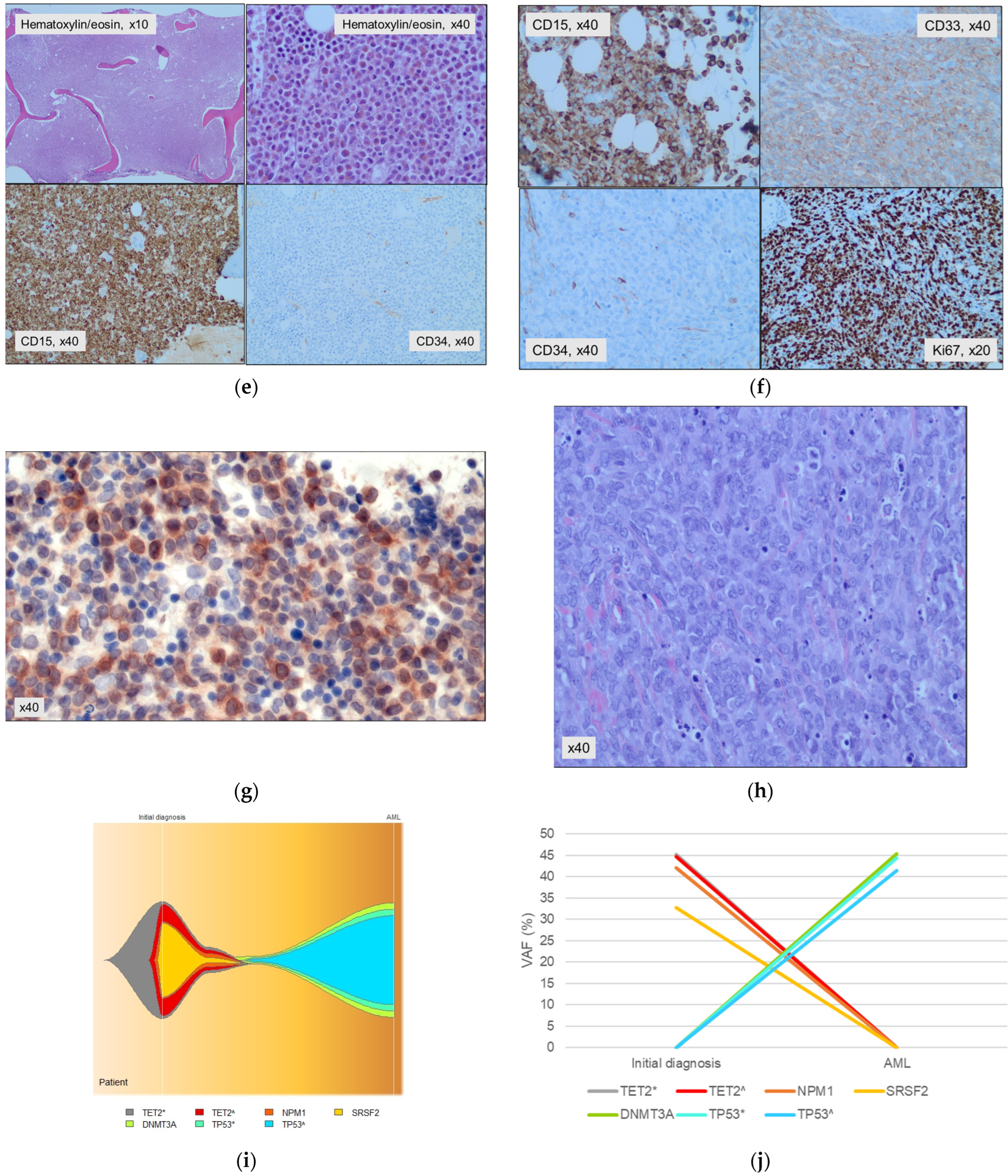
2. An Atypical Presentation of NPM1mut MN

A 63-year-old man was diagnosed simultaneously with prostate cancer and a NPM1mut myeloid neoplasm in June 2021. He consulted with symptoms of prostatitis and the work-up revealed a prostatic adenocarcinoma together with alterations in the hemogram. Examination was normal. Complete blood count showed macrocytic anemia (MCV 102.6 fL, hemoglobin 106 g/L), with normal platelets (163 × 109/L) and leukocytes (8.24 × 10⁹/L), 39% segmented neutrophils, 1% segmented bands, 19% eosinophils (0.3 × 10⁹/L), 0% basophils, 35% lymphocytes, and 6% monocytes. Marked dysplasia was seen in the eosinophilic elements consisting of nuclear segmentation alterations and wide cytoplasm degranulated spaces, as well as immature granulocytes with eosinophilic granulation (Figure 1a).
These findings led us to make several additional studies, including a bone marrow aspirate and biopsy. The aspirate (Figure 1b,c) was hypercellular and revealed a massive eosinophilic infiltration composed of eosinophilic myelocytes and metamyelocytes with marked dysplasia (sparse granulation or degranulation, abnormal nuclear segmentation, occasional pre-eosinophilic granulation). The red precursors and megakaryocytic elements were slightly decreased without dysplasia and had a normal distribution. Numerous sea blue histiocytes were also observed. Blasts accounted for <1%. Flow cytometry showed that 90% of the bone marrow cells were eosinophils (CD13+, CD33+, CD34−, CD117−, HLA-DR+, CD15+, CD11b+, CD36−, CD66−, NG2−, CD38−, and negative for lymphoid antigens) (Figure 1d). The core biopsy specimen was hypercellular (>90%), with a striking increase in granulocytic cells (positive for CD15 and lysozyme), most of them with eosinophilic differentiation (Figure 1e,f). Megakaryocytes, erythroblasts, mastocytes, and lymphocytes had a normal distribution. Immature neutrophil granulocytic precursors (MPO+) were scarce and located only in the paratrabecular areas. There were <5% of CD34-positive cells. A diffuse loose network of reticulin (MF-1) was observed. Fluorescence in situ hybridization (FISH) excluded both FIP1L1-PDGFRA and PDGFRB rearrangements. Targeted Next Generation Sequencing (NGS) showed three pathogenic mutations: TET2 with two variants (c.2671C > T VAF 45.15%, and c.3435delT 44.76%); NPM1 (c.863_864insTCTG VAF 42.33%); and SRSF2 (c.284C > T VAF 32.33%). Bone marrow biopsy staining with an anti-NPM1 antibody showed a cytoplasmic positivity in most of the cells consistent with a delocalization of the protein due to the mutation (courtesy of Dr. Falini) (Figure 1g).
Our diagnosis at that moment was chronic eosinophilic leukemia (CEL) NPM1mut vs. acute myeloid leukemia (AML) NPM1mut with eosinophilic differentiation. The patient then received radiotherapy over the involved field together with hormone blocking therapy for the prostate neoplasm and was scheduled to start chemotherapy for the MN when he presented subcutaneous nodular lesions on his face. The biopsy of the skin lesions showed a dense infiltration by large cells of blastic appearance that were CD34−, CD15+, CD33+ and MPO−, consistent with a cutaneous sarcoma (Figure 1h). Intensive therapy for the MN was then started and consisted of induction chemotherapy according to the CETLAM-2012 protocol (idarubicin and cytarabine), and one consolidation course with high-dose cytarabine. Therefore, he achieved a morphological complete remission (CR) with a positive molecular measurable residual disease (mutNPM1/ABL1 ratio of 0.0041). Afterwards, he received an allogeneic stem cell transplant (alloHCT) of a matched unrelated donor, with a non-myeloablative conditioning regimen and post-transplant graft-versus-host disease prophylaxis based on cyclophosphamide and tacrolimus. He achieved a complete molecular remission (NPM1 mut undetectable by qRT-PCR). Thirteen months after alloHCT, he presented with 60% bone marrow blasts with megakaryocytic differentiation (CD36+, CD42a/CD61+, CD42b+, CD41+, CD56+). At that moment, new mutations in DNMT3A and TP53 (two variants) were detected, while original mutations in TET2, SRSF2 were not identified and NPM1 remained undetectable by qPCR (Figure 1i,j). A chimerism analysis confirmed the recipient origin of this otherwise clonally unrelated second AML. Immunosuppressive therapy was reduced and treatment with Azacytidine and Magrolimab was started, without any response. He died one and a half years after alloHCT.

3. Seven Additional Cases of Unusual Presentation of NPM1mut MN

Here, we present seven cases of patients diagnosed through a comprehensive evaluation of clinical, morphological, and molecular data, and their treatment was tailored based on risk assessment (Table 1). Five cases were consistent with chronic myelomonocytic leukemia and the other two corresponded to myelodysplastic syndromes. Four of the five CMML cases were included in a multicentric work presented in [17], submitted elsewhere.
Patients with CMML carrying the NPM1 mutation presented at a relatively young age, with a median of 60 years (range, 55–80). This subgroup exhibited distinctive characteristics, including a median bone marrow blast count of 13% (10–19), leukocytes of 15.7 × 10⁹ (12.24–30.6), a platelet count of 98 × 109 (35–110), and a median hemoglobin concentration of 85 g/L (49–138). Among these five patients, three were eligible for intensive chemotherapy and attained a complete response with a significant reduction in NPM1 ratio. The same occurred with the patient with an MDS and the case with eosinophilic presentation, both eligible for intensive chemotherapy. All five cases achieved a molecular complete response after allogeneic hematopoietic cell transplantation (n = 3) or venetoclax and azacytidine treatment (n = 2). In contrast, another patient treated upfront with venetoclax + azacytidine remained with stable disease. Two other patients were older and constrained by comorbidities and could only benefit from cytoreduction with hydroxyurea (n = 1) and best supportive care.

4. Review

4.1. NPM1 in Leukemogenesis and Its Clinical Impact

The nucleophosmin (NPM1) gene encodes a multifunctional chaperone protein predominantly situated in the nucleolus. This protein shuttles between the nucleus and the cytoplasm [19,20]. NPM1 plays pivotal roles in preserving genomic stability, participating in p53-dependent stress responses, facilitating ribosome genesis, and modulating growth suppressive pathways through interactions with Alternate Reading Frame protein (Arf) [21].
Mutations in the NPM1 gene are found in exon 12 in most cases, consisting of a 4 base pair insertion that causes the loss of a few C-terminal amino acids of the protein [22]. This results in a stronger nuclear export signal, leading to the cytoplasmic localization of the protein, in contrast to the predominantly nucleolus-located wild-type [23]. Aberrant cytoplasmic localization is a shared characteristic of all NPM1 mutants and is crucial for its role in leukemogenesis [24]. However, the mechanisms governing leukemogenesis remain a subject of debate. It has been proposed that NPM1 mutations drive leukemia through a combination of the gain and loss of functions in distinct cellular processes at both the nuclear and cytoplasmic levels [25,26,27,28]. In the cytoplasm, mutant NPM1 may function through two mechanisms: (1) cytoplasmic misplacement of nuclear proteins (ARF, CTCF, FBW7, HEXIM1, MIZ1, and PU.1), although the significance of this process is not yet fully understood; and (2) binding to cytoplasmic proteins caspase-6 and -8, subsequently inhibiting their myeloid differentiation abilities [25,26,27,28]. Recent findings suggest that mutant NPM1 exerts its nuclear-level role by binding to chromatin at the HOXA/B and MEIS1 loci in cooperation with the MLL complex, leading to an increase in the expression of these oncogenic stem cell programs and blocking the normal differentiation of hematopoietic progenitors [29,30]. Aberrant cytoplasmic localization can be easily assessed by immunohistochemistry [11,31,32], and through this method, other rare NPM1 mutations out of exon 12 and fusion genes involving NPM1 have been identified, as well as NPM1 mutations in non-acute MN.
NPM1 mutations are AML-driving events responsible for around one-third of all AML cases [20], though they have also been identified in non-acute myeloid neoplasms with an initial blast count lower than 20%. It is widely accepted that NPM1-mutated AML develops from preexisting clonal hematopoiesis [24], which explains its frequent co-occurrence with DNMT3A, TET2, and IDH1/2 mutations [10,20].
Even when NPM1mut is tightly associated with de novo AML, on rare occasions, NPM1 mutations can emerge in other myeloid neoplasms as secondary mutations, ultimately contributing to the development of AML, as suggested by clinical studies [4,27] and mouse models [28]. These non-acute MNs have also been regarded as early stage AML [12], due to a mutational profile with fewer myelodysplasia-related gene mutations [5,6], their sensitivity to chemotherapy, the eradication of NPM1 following intensive treatment and its reappearance at relapse [5,6].
NPM1 mutations have a strong prognostic significance in the context of de novo AML, defining a favorable risk stratification in the absence of co-occurring FLT3-ITD mutations. In these patients, if MRD is cleared after intensive treatment, consolidation with alloHCT is not recommended. However, the prognostic significance of NPM1 in other myeloid neoplasms remains unknown as well as the best therapy for these patients.
Finally, various NPM1-targeted approaches such as menin inhibitors and FLT3 kinase inhibitors are under investigation with promising results [10,33], which warrants the identification of this potential druggable candidate in all patients with a myeloid malignancy [16].

4.2. NPM1 Mutation in CMML

CMML is a clonal hematopoietic malignancy characterized by features of both myeloproliferative neoplasms and myelodysplastic syndromes. In this neoplasm the most prevalent mutations include TET2 (approximately 60%), followed by SRSF2 (around 50%), ASXL1 (about 40%), and those related to the oncogenic RAS pathway (approximately 30%) [34,35,36]. Significantly, ASXL1 and NRAS mutations have been identified as indicators of a poorer prognosis [37], while the prognostic implications of RUNX1 and SETBP1 remain a subject of ongoing debate [38,39,40,41,42]. While NPM1 mutation is not a typical hallmark of CMML, it has been infrequently observed in this entity, accounting for 3–5% according to various series [32,43,44,45,46,47,48,49]. The classical type A mutation (c.860_863dup) is found in most cases [48,49]. Studies with paired samples have reported clonal evolution from CMML to AML, revealing the emergence of new mutations and an increase in variant allele frequency (VAF), including NPM1 [50]. Remarkably, in one of these studies, two out of five evaluable patients revealed no additional cytogenetic or molecular alterations at the moment of AML diagnosis, highlighting the exclusive appearance of the NPM1 mutation [48]. In the same study, a positive correlation between DNMT3A, FLT3-ITD, and NPM1 in CMML was found [43,48], while a negative association existed with TET2 and ASXL1 [48]. Another study [43] reported varying prevalences of NPM1 and ASXL1 according to the 2017 WHO classification, with more ASXL1 mutations in CMML-0 and more NPM1 mutations in CMML-2.
Previous studies have suggested that NPM1mut CMML patients have a higher likelihood of AML transformation, while others have considered them to be in an “early stage” of AML based on shared clinical and molecular characteristics [32,46,48]. In this context, NPM1mut CMML patients exhibited lower hemoglobin levels, an increased median leukocyte count (median of 20 × 109/L) [47], elevated bone marrow monocyte and blast percentages, an increased probability of AML evolution, and inferior overall survival compared to NPM1wt CMML [44,47,48]. Blast transformation occurred in 62% of cases, with a median time of 5 months after the initial CMML diagnosis. Additionally, AML in NPM1mut progressed patients did not show the favorable prognosis associated with the de novo NPM1mut AML [46,48,49,50].
The 2022 ICC classification of MNs [4,8] acknowledges the presence of the NPM1 mutation in rare CMML cases but notes that these may not be diagnosed with de novo AML, even if the blast count is 10–19% [4]. In contrast, the 2022 WHO classifies these cases as AML with mutated NPM1 [2]. The management of these CMML cases is challenging, due to the low number of cases there is with a lack of large series [5,9,44,45,48]. Earlier studies suggested a higher probability of AML transformation and poorer overall survival when patients were treated with CMML protocols [47]. In vitro investigations have also provided evidence for the heightened chemosensitivity of leukemic blasts with NPM1 mutations compared to those with NPM1 wild-type [22], which has even been demonstrated in NPM1mut CMML [5,6,48]. In this sense, Vallapureddy et al. [48] presented a cohort of 373 patients diagnosed with CMML, among whom 8 (2%) had NPM1 mutation. In this study, five out of eight NPM1mut CMML patients progressed to AML at a median of 5 months. Among these, four out of five patients with blast transformation received AML-like induction chemotherapy, and two of them subsequently underwent alloHCT. The median OS after transformation was 3 months, with only one patient who had undergone alloHCT still alive at the last follow-up. Montalban-Bravo et al. [5] demonstrated that individuals with NPM1-mutated MDS or MDS/MPN undergoing intensive chemotherapy exhibited elevated overall response rates (100%) and complete response rates (90%). Additionally, they experienced more favorable progression-free survival and overall survival compared to outcomes achieved with hypomethylating agents (HMAs). Nevertheless, the patients treated with chemotherapy were notably younger than those who received HMAs. This age difference could introduce selection bias since these individuals were perceived as more suitable candidates for this treatment, and as a result, better outcomes were anticipated. However, this study was unable to assert that chemotherapy alone represents the optimal strategy due to the short follow-up for the non-transplanted patients. In individuals initially treated with HMAs, consolidation followed by alloHCT demonstrated better survival outcomes, although the transplanted patients were younger. The study by Patel et al. [6] reported unfavorable overall outcomes in their cohort of NPM1-mutated myeloid neoplasms (including CMML and MDS), with the majority of patients undergoing initial HMA therapy. Notably, three patients received upfront induction chemotherapy, and none of them developed an AML. These findings led the authors to propose that upfront HMA therapy might have been insufficient for certain patients. These results suggest that suitable patients with NPM1mut non-acute MNs with <20% blasts may benefit from AML-type chemotherapy more than the standard CMML approach. Furthermore, alloHCT remains the sole potential curative approach for CMML.

4.3. NPM1 Mutation in MDS

The occurrence of NPM1 mutations in MDS patients is reported to be in the range of 1–6% [7,15,51,52]. Zhang and collaborators [51] were the pioneers in identifying NPM1 mutations in two MDS patients (5.2%), both of which were type A. Subsequently, Bains et al. [52] reported NPM1 mutations in seven MDS patients (4.4%), all falling under the refractory anemia with excess blasts category, and with a normal karyotype (p < 0.001). They highlighted a significant association between FLT3 and NPM1 mutations, with FLT3 being nearly four times more prevalent in NPM1mut MDS patients compared to NPM1wt (p < 0.001). Although there were no notable differences in terms of progression to AML between NPM1mut and NPM1wt MDS (p = 0.133), the combination of FLT3mut and NPM1mut adversely impacted progression-free survival (p = 0.026). Moreover, the presence of NPM1mut in MDS patients has been linked to multilineage dysplasia [12], normal cytogenetics, an excess of blasts [7], CD34 negativity [5], and aggressive clinical evolution with an elevated risk of AML progression [5,52]. Based on these findings, it was suggested that NPM1 mutation plays a pivotal role in myelodysplasia development [52]. Supporting this claim, the role of NPM1 heterozygosity in the development of a hematological syndrome resembling human MDS was demonstrated in a mouse model [53,54].
Wu et al. [15] identified 12 high-risk MDS patients (6.2%) carrying the L287fs NPM1 mutation who received decitabine-based treatment. These patients achieved a higher complete response compared to NPM1wt MDS patients (50% vs. 29.1%, p = 0.191) and the results were even better for DNMT3Awt cases [15]. They also found a good correlation between the mutational burden and the response depth. In patients achieving complete response, NPM1 mutation was not detected; those with hematological improvement showed a reduction in NPM1 VAF and non-responsive patients retained an unaltered NPM1 level [15].
Nonetheless, other studies [5,6] have pointed out that NPM1mut MDS patients treated with intensive chemotherapy with or without consolidation with alloHCT (“AML-like” treatment) presented better outcomes, including higher remission rates and better progression-free and overall survival.

4.4. NPM1 Mutation in Other Non-Acute Myeloid Neoplasms

The pathogenic involvement of NPM1mut in myeloproliferative neoplasms (MPNs) has scarcely been explored so far and remains unclear. Animal models (knock-in mouse model [55,56] and zebrafish [57]) with artificially induced human NPM1 mutant expression exhibited an increase in hematopoietic cells and the development of myeloproliferation, but this seems not to correlate with patients, as large cohorts of Philadelphia-negative MPNs have failed to find mutations affecting NPM1. Nevertheless, cases of NPM1mut AML progressing from MPNs have been described, though they are so scarce that no conclusions can be derived about their particular features and outcome [58]. Schittger et al. identified NPM1 mutations in 6 out of 67 patients with AML (sAML) who had a previous history of MPN. They concluded that the NPM1 mutation is not solely a crucial factor in the onset of the novo AML but may also play a role in the development of AML subsequent to MPN [59]. Another study proposed that the candidacy of NPM1 mutation in the development of human MPNs is uncertain and questionable [60]. This uncertainty arises from the absence of NPM1 hotspot mutation in a relatively larger cohort of MNs, which included 120 classic MPN cases and 9 cases of MDS/MPN. This could be attributed to the possible presence of activating mutations in other non-examined regions of this selected gene A relatively old study [45] with a limited cohort of classic MPNs, including 14 cases of polycythemia vera, 7 cases of essential thrombocythemia, and 9 cases of primary myelofibrosis, was not able to find NPM1 mutations in these patients. Nevertheless, additional investigations are necessary to elucidate their specific roles in hematopoiesis and their involvement in the pathogenesis of MPN, and to assess their prognostic and therapeutic implications. In the interest of our case with eosinophilic presentation, we have also explored the presence of NPM1mut in CEL. CEL is a rare condition characterized by persistent blood eosinophilia (>1.5 × 10⁹/L), without class-defining rearrangements. It was first included as a distinct entity by the 2008 4th edition of the WHO Classification [61]. Information about this condition is limited to case reports [62]. Typically, it presents as an aggressive disorder with unfavorable prognosis, resistance to conventional therapy, and high rates of AML transformation. The most comprehensive case series collected 10 patients [63], and in this study, the reported median overall survival was 22 months. Five patients underwent transformation to acute leukemia, of both myeloid and lymphoid types, with a median time from acute transformation to death of two months [63].
As far as we know, there is only one previous report of CEL with NPM1mut [64]. It describes a 70-year-old female who presented with leukocytosis and marked eosinophilia and did not respond to Imatinib. AML transformation occurred five months after initial diagnosis, with the identification of an NPM1 type A mutation (c.860_863dup) together with mutations affecting TET2 and FLT3-TKD. The retrospective analysis of the NPM1 mutation showed that it was already present at the initial CEL diagnosis, with a high variant allele frequency. The patient did not respond to decitabine and passed away because of a subdural hematoma.

5. Conclusions and Future

Non-acute NPM1-mutated MNs (NPM1mut-MNs) represent both a diagnostic and therapeutic challenge. Most of them correspond to chronic myelomonocytic leukemia (CMML) and myelodysplastic syndromes (MDS), and it is still controversial as to whether they should be diagnosed and/or treated as AML.
Moreover, exploring the role of NPM1 mutation in non-acute myeloid neoplasms is an evolving area of research, with the possibility of new insights emerging over time. The impact of this mutation on disease progression and patient outcomes varies depending on the specific type of myeloid disorder. Moreover, the prognostic significance of NPM1 mutations in non-acute MNs is not as firmly established as it is in AML. The notably aggressive clinical course observed in non-acute MNs with NPM1 mutation, leading to a frequent and rapid evolution to AML, highlights the necessity for a comprehensive diagnosis that incorporates both clinical and biological data. This reinforces the clinical rationale to customize the management of these patients on an individualized basis, opting for intensive treatment more aligned with AML protocols, regardless of blast percentage. Therefore, it is essential and challenging to validate these hypotheses and findings through prospective studies that include a substantial number of patients with a long follow-up period.

Author Contributions

Conceptualization, S.C.-D., F.G. and M.R.; methodology, S.C.-D., F.G. and M.R.; investigation, S.C.-D., F.G. and M.R.; resources, S.C.-D., F.G., M.L.-G., A.I.P.-V., M.G.-N., D.C., M.D.-B., J.E. and M.R.; data curation, S.C.-D., F.G., M.L.-G., D.C., M.D.-B., J.E. and M.R.; writing—original draft preparation, S.C.-D., F.G. and M.R.; writing—review and editing, S.C.-D., F.G., M.L.-G., D.C., M.D.-B., J.E. and M.R.; visualization, S.C.-D., F.G. and M.R.; supervision F.G. and M.R.; project administration, M.R.; funding acquisition, S.C.-D., F.G., M.D.-B., J.E. and M.R. All authors have read and agreed to the published version of the manuscript.

Funding

This research was supported by grants from the resident award Contractes Clínic de Recerca Emili Letang-Josep Font 2021, granted by Hospital Clínic de Barcelona, and by the Convocatòria d’Intensificació Interna per als professionals de l’HCB 2023 (MDB). This study was funded by Instituto de Salud Carlos III (ISCIII) through the project “FIS PI19/01476” and “FIS PI22/01660” and co-funded by the European Union. It was also supported by grants from Fundació la Marató TV3 sobre Càncer 201 201930-31.

Institutional Review Board Statement

This study was conducted in accordance with the Declaration of Helsinki, and approved by the Institutional Review Board (or Ethics Committee) of Hospital Clínic Barcelona (2021-1230; on 20 July 2022).

Informed Consent Statement

Written informed consent was obtained from all subjects involved in the study.

Data Availability Statement

Due to privacy and ethical concerns, the data that support the findings of this study are available on request from the corresponding author.

Conflicts of Interest

J.E.: consultancy-honoraria (Abbvie, Novartis, Astellas, Jazz Pharmaceuticals, BMS-Celgene, Pfizer, Daiichi-Sankyo), research grants (Novartis, Jazz Pharmaceuticals); M.D.B.: consultant or advisory role, travel grants or speaker (Bristol Myers Squibb, Abbvie, Astellas, JazzPharma, Takeda, Novartis). The other authors have no conflicts of interest.

References

  1. Arber, D.A.; Orazi, A.; Hasserjian, R.P.; Borowitz, M.J.; Calvo, K.R.; Kvasnicka, H.M.; Wang, S.A.; Bagg, A.; Barbui, T.; Branford, S.; et al. International Consensus Classification of Myeloid Neoplasms and Acute Leukemias: Integrating morphologic, clinical, and genomic data. Blood 2022, 140, 1200–1228. [Google Scholar] [CrossRef] [PubMed]
  2. Khoury, J.D.; Solary, E.; Abla, O.; Akkari, Y.; Alaggio, R.; Apperley, J.F.; Bejar, R.; Berti, E.; Busque, L.; Chan, J.K.C.; et al. The 5th edition of the World Health Organization Classification of Haematolymphoid Tumours: Myeloid and Histiocytic/Dendritic Neoplasms. Leukemia 2022, 36, 1703–1719. [Google Scholar] [CrossRef] [PubMed]
  3. Patwardhan, P.P.; Aarabi, M.; Aggarwal, N. Genomics of myelodysplastic/myeloproliferative neoplasm. Semin. Diagn. Pathol. 2023, 40, 195–201. [Google Scholar] [CrossRef] [PubMed]
  4. Hasserjian, R.P.; Orazi, A.; Orfao, A.; Rozman, M.; Wang, S.A. The International Consensus Classification of myelodysplastic syndromes and related entities. Virchows Arch. 2023, 482, 39–51. [Google Scholar] [CrossRef] [PubMed]
  5. Montalban-Bravo, G.; Kanagal-Shamanna, R.; Sasaki, K.; Patel, K.; Ganan-Gomez, I.; Jabbour, E.; Kadia, T.; Ravandi, F.; DiNardo, C.; Borthakur, G.; et al. NPM1 mutations define a specific subgroup of MDS and MDS/MPN patients with favorable outcomes with intensive chemotherapy. Blood Adv. 2019, 3, 922–933. [Google Scholar] [CrossRef] [PubMed]
  6. Patel, S.S.; Ho, C.; Ptashkin, R.N.; Sadigh, S.; Bagg, A.; Geyer, J.T.; Xu, M.L.; Prebet, T.; Mason, E.F.; Seegmiller, A.C.; et al. Clinicopathologic and genetic characterization of nonacute NPM1-mutated myeloid neoplasms. Blood Adv. 2019, 3, 1540–1545. [Google Scholar] [CrossRef] [PubMed]
  7. Forghieri, F.; Nasillo, V.; Paolini, A.; Bettelli, F.; Pioli, V.; Giusti, D.; Gilioli, A.; Colasante, C.; Acquaviva, G.; Riva, G.; et al. NPM1-Mutated Myeloid Neoplasms with <20% Blasts: A Really Distinct Clinico-Pathologic Entity? Int. J. Mol. Sci. 2020, 21, 8975. [Google Scholar] [PubMed]
  8. Prakash, S.; Arber, D.A.; Bueso-Ramos, C.; Hasserjian, R.P.; Orazi, A. Advances in myelodysplastic/myeloproliferative neoplasms. Virchows Arch. 2023, 482, 69–83. [Google Scholar] [CrossRef]
  9. Matanes, F.; AbdelAzeem, B.M.A.; Shah, G.; Reddy, V.; Saad, A.; Papadantonakis, N. Chronic myelomonocytic leukemia associated with myeloid sarcomas and NPM1 mutation: A case report and literature review. Ther. Adv. Hematol. 2019, 10, 2040620719854596. [Google Scholar] [CrossRef]
  10. Falini, B. NPM1-mutated acute myeloid leukemia: New pathogenetic and therapeutic insights and open questions. Am. J. Hematol. 2023, 98, 1452–1464. [Google Scholar] [CrossRef]
  11. Falini, B.; Martelli, M.P.; Brunetti, L.; Gjertsen, B.T.; Andresen, V. The NPM1 mutant defines AML irrespective of blast count. Am. J. Hematol. 2023, 98, E187–E189. [Google Scholar] [CrossRef] [PubMed]
  12. Zhang, L.; Campion, V.; Dickson, M.; Tang, C. Acute myeloid leukaemia with NPM1 mutation: No longer having an absolute blast (count). Pathology 2023, 55, 578–581. [Google Scholar] [CrossRef] [PubMed]
  13. Estey, E.; Hasserjian, R.P.; Döhner, H. Distinguishing AML from MDS: A fixed blast percentage may no longer be optimal. Blood 2022, 139, 323–332. [Google Scholar] [CrossRef] [PubMed]
  14. DiNardo, C.D.; Garcia-Manero, G.; Kantarjian, H.M. Time to blur the blast boundaries. Cancer 2022, 128, 1568–1570. [Google Scholar] [CrossRef] [PubMed]
  15. Wu, L.; Li, X.; Xu, F.; Wu, D.; He, Q.; Song, L.; Xiao, C.; Zhao, Y.; Zhang, Z.; Guo, J.; et al. NPM1 mutation with DNMT3A wild type defines a subgroup of MDS with particularly favourable outcomes after decitabine therapy. Br. J. Haematol. 2020, 189, 982–984. [Google Scholar] [CrossRef] [PubMed]
  16. Chin, L.; Wong, C.Y.G.; Gill, H. Targeting and Monitoring Acute Myeloid Leukaemia with Nucleophosmin-1 (NPM1) Mutation. Int. J. Mol. Sci. 2023, 24, 3161. [Google Scholar] [CrossRef] [PubMed]
  17. Castaño-Díez, S.; Zugasti, I.; Calvo, X.; Schulz, F.; Pita, A.A.; Mora, E.; Falantes, J.F.; Azaceta, G.; Ibáñez, M.; Chen, T.; et al. Chronic Myelomonocytic Leukemia (CMML) with AML Typical Mutations (NPM1, FLT3 or CEBPA) Identify a High-Risk CMML Group Independent of Molecular-Cpss. Blood 2023, 142, 3231. [Google Scholar] [CrossRef]
  18. Castaño-Díez, S.; López-Guerra, M.; Bosch-Castañeda, C.; Bataller, A.; Charry, P.; Esteban, D.; Guijarro, F.; Jiménez-Vicente, C.; Castillo-Girón, C.; Cortes, A.; et al. Real-World Data on Chronic Myelomonocytic Leukemia: Clinical and Molecular Characteristics, Treatment, Emerging Drugs, and Patient Outcomes. Cancers 2022, 14, 4107. [Google Scholar] [CrossRef]
  19. Grisendi, S.; Mecucci, C.; Falini, B.; Pandolfi, P.P. Nucleophosmin and cancer. Nat. Rev. Cancer 2006, 6, 493–505. [Google Scholar] [CrossRef]
  20. Falini, B.; Mecucci, C.; Tiacci, E.; Alcalay, M.; Rosati, R.; Pasqualucci, L.; La Starza, R.; Diverio, D.; Colombo, E.; Santucci, A.; et al. Cytoplasmic nucleophosmin in acute myelogenous leukemia with a normal karyotype. N. Engl. J. Med. 2005, 352, 254–266. [Google Scholar] [CrossRef]
  21. Heath, E.M.; Chan, S.M.; Minden, M.D.; Murphy, T.; Shlush, L.I.; Schimmer, A.D. Biological and clinical consequences of NPM1 mutations in AML. Leukemia 2017, 31, 798–807. [Google Scholar] [CrossRef] [PubMed]
  22. Falini, B.; Nicoletti, I.; Martelli, M.F.; Mecucci, C. Acute myeloid leukemia carrying cytoplasmic/mutated nucleophosmin (NPMc + AML): Biologic and clinical features. Blood 2007, 109, 874–885. [Google Scholar] [CrossRef] [PubMed]
  23. Colombo, E.; Martinelli, P.; Zamponi, R.; Shing, D.C.; Bonetti, P.; Luzi, L.; Volorio, S.; Bernard, L.; Pruneri, G.; Alcalay, M.; et al. Delocalization and destabilization of the Arf tumor suppressor by the leukemia-associated NPM mutant. Cancer Res. 2006, 66, 3044–3050. [Google Scholar] [CrossRef] [PubMed]
  24. Falini, B.; Brunetti, L.; Sportoletti, P.; Martelli, M.P. NPM1-mutated acute myeloid leukemia: From bench to bedside. Blood 2020, 136, 1707–1721. [Google Scholar] [CrossRef] [PubMed]
  25. Bonetti, P.; Davoli, T.; Sironi, C.; Amati, B.; Pelicci, P.G.; Colombo, E. Nucleophosmin and its AML-associated mutant regulate c-Myc turnover through Fbw7 gamma. J. Cell Biol. 2008, 182, 19–26. [Google Scholar] [CrossRef] [PubMed]
  26. Wanzel, M.; Russ, A.C.; Kleine-Kohlbrecher, D.; Colombo, E.; Pelicci, P.G.; Eilers, M. A ribosomal protein L23-nucleophosmin circuit coordinates Mizl function with cell growth. Nat. Cell Biol. 2008, 10, 1051–1061. [Google Scholar] [CrossRef] [PubMed]
  27. Gu, X.; Ebrahem, Q.; Mahfouz, R.Z.; Hasipek, M.; Enane, F.; Radivoyevitch, T.; Rapin, N.; Przychodzen, B.; Hu, Z.; Balusu, R.; et al. Leukemogenic nucleophosmin mutation disrupts the transcription factor hub that regulates granulomonocytic fates. J. Clin. Investig. 2018, 128, 4260–4279. [Google Scholar] [CrossRef] [PubMed]
  28. Gurumurthy, M.; Tan, C.H.; Ng, R.; Zeiger, L.; Lau, J.; Lee, J.; Dey, A.; Philp, R.; Li, Q.; Lim, T.M.; et al. Nucleophosmin interacts with HEXIM1 and regulates RNA polymerase II transcription. J. Mol. Biol. 2008, 378, 302–317. [Google Scholar] [CrossRef]
  29. Brunetti, L.; Gundry, M.C.; Sorcini, D.; Guzman, A.G.; Huang, Y.H.; Ramabadran, R.; Gionfriddo, I.; Mezzasoma, F.; Milano, F.; Nabet, B.; et al. Mutant NPM1 Maintains the Leukemic State through HOX Expression. Cancer Cell. 2018, 34, 499–512.e9. [Google Scholar] [CrossRef]
  30. Uckelmann, H.J.; Haarer, E.L.; Takeda, R.; Wong, E.M.; Hatton, C.; Marinaccio, C.; Perner, F.; Rajput, M.; Antonissen, N.J.C.; Wen, Y.; et al. Mutant NPM1 Directly Regulates Oncogenic Transcription in Acute Myeloid Leukemia. Cancer Discov. 2023, 13, 746–765. [Google Scholar] [CrossRef]
  31. Kaseb, H.; Visconte, V.; Socha, D.S.; Crane, G.M.; Durkin, L.; Cook, J.R.; Maciejewski, J.P.; Hsi, E.D.; Rogers, H.J. The clinicopathologic significance of NPM1 mutation and ability to detect mutated NPM1 by immunohistochemistry in non-AML myeloid neoplasms. Genes Chromosomes Cancer 2023, 62, 573–580. [Google Scholar] [CrossRef] [PubMed]
  32. Forghieri, F.; Paolini, A.; Morselli, M.; Bigliardi, S.; Bonacorsi, G.; Leonardi, G.; Coluccio, V.; Maccaferri, M.; Fantuzzi, V.; Faglioni, L.; et al. NPM1 mutations may reveal acute myeloid leukemia in cases otherwise morphologically diagnosed as myelodysplastic syndromes or myelodysplastic/myeloproliferative neoplasms. Leuk Lymphoma 2015, 56, 3222–3226. [Google Scholar] [CrossRef] [PubMed]
  33. Ranieri, R.; Pianigiani, G.; Sciabolacci, S.; Perriello, V.M.; Marra, A.; Cardinali, V.; Pierangeli, S.; Milano, F.; Gionfriddo, I.; Brunetti, L.; et al. Current status and future perspectives in targeted therapy of NPM1-mutated AML. Leukemia 2022, 36, 2351–2367. [Google Scholar] [CrossRef] [PubMed]
  34. Patnaik, M.M.; Tefferi, A. Cytogenetic and molecular abnormalities in chronic myelomonocytic leukemia. Blood Cancer J. 2016, 6, e393. [Google Scholar] [CrossRef] [PubMed]
  35. Itzykson, R.; Duchmann, M.; Lucas, N.; Solary, E. CMML: Clinical and molecular aspects. Int. J. Hematol. 2017, 105, 711–719. [Google Scholar] [CrossRef] [PubMed]
  36. Itzykson, R.; Fenaux, P.; Bowen, D.; Cross, N.C.P.; Cortes, J.; De Witte, T.; Germing, U.; Onida, F.; Padron, E.; Platzbecker, U.; et al. Diagnosis and Treatment of Chronic Myelomonocytic Leukemias in Adults: Recommendations From the European Hematology Association and the European LeukemiaNet. Hemasphere 2018, 2, e150. [Google Scholar] [CrossRef]
  37. Elena, C.; Galli, A.; Such, E.; Meggendorfer, M.; Germing, U.; Rizzo, E.; Cervera, J.; Molteni, E.; Fasan, A.; Schuler, E.; et al. Integrating clinical features and genetic lesions in the risk assessment of patients with chronic myelomonocytic leukemia. Blood 2016, 128, 1408–1417. [Google Scholar] [CrossRef]
  38. Duchmann, M.; Yalniz, F.F.; Sanna, A.; Sallman, D.; Coombs, C.C.; Renneville, A.; Kosmider, O.; Braun, T.; Platzbecker, U.; Willems, L.; et al. Prognostic Role of Gene Mutations in Chronic Myelomonocytic Leukemia Patients Treated with Hypomethylating Agents. EBioMedicine 2018, 31, 174–181. [Google Scholar] [CrossRef]
  39. Kuo, M.C.; Liang, D.C.; Huang, C.F.; Shih, Y.S.; Wu, J.H.; Lin, T.L.; Shih, L.Y. RUNX1 mutations are frequent in chronic myelomonocytic leukemia and mutations at the C-terminal region might predict acute myeloid leukemia transformation. Leukemia 2009, 23, 1426–1431. [Google Scholar] [CrossRef]
  40. Patnaik, M.M.; Itzykson, R.; Lasho, T.L.; Kosmider, O.; Finke, C.M.; Hanson, C.A.; Knudson, R.A.; Ketterling, R.P.; Tefferi, A.; Solary, E. ASXL1 and SETBP1 mutations and their prognostic contribution in chronic myelomonocytic leukemia: A two-center study of 466 patients. Leukemia 2014, 28, 2206–2212. [Google Scholar] [CrossRef]
  41. Patnaik, M.M.; Wassie, E.A.; Padron, E.; Onida, F.; Itzykson, R.; Lasho, T.L.; Kosmider, O.; Finke, C.M.; Hanson, C.A.; Ketterling, R.P.; et al. Chronic myelomonocytic leukemia in younger patients: Molecular and cytogenetic predictors of survival and treatment outcome. Blood Cancer J. 2015, 5, e270. [Google Scholar] [CrossRef] [PubMed]
  42. Shou, L.H.; Cao, D.; Dong, X.H.; Fang, Q.; Wu, Y.; Zhang, Y.; Fei, J.P.; Xu, B.L. Prognostic significance of SETBP1 mutations in myelodysplastic syndromes, chronic myelomonocytic leukemia, and chronic neutrophilic leukemia: A meta-analysis. PLoS ONE 2017, 12, e0171608. [Google Scholar] [CrossRef] [PubMed]
  43. Hwang, S.M.; Kim, S.M.; Nam, Y.; Kim, J.; Kim, S.; Ahn, Y.O.; Park, Y.; Yoon, S.S.; Shin, S.; Kwon, S.; et al. Targeted sequencing aids in identifying clonality in chronic myelomonocytic leukemia. Leuk Res. 2019, 84, 106190. [Google Scholar] [CrossRef] [PubMed]
  44. Caudill, J.S.; Sternberg, A.J.; Li, C.Y.; Tefferi, A.; Lasho, T.L.; Steensma, D.P. C-terminal nucleophosmin mutations are uncommon in chronic myeloid disorders. Br. J. Haematol. 2006, 133, 638–641. [Google Scholar] [CrossRef] [PubMed]
  45. Oki, Y.; Jelinek, J.; Beran, M.; Verstovsek, S.; Kantarjian, H.M.; Issa, J.P. Mutations and promoter methylation status of NPM1 in myeloproliferative disorders. Haematologica 2006, 91, 1147–1148. [Google Scholar] [PubMed]
  46. Courville, E.L.; Wu, Y.; Kourda, J.; Roth, C.G.; Brockmann, J.; Muzikansky, A.; Fathi, A.T.; de Leval, L.; Orazi, A.; Hasserjian, R.P. Clinicopathologic analysis of acute myeloid leukemia arising from chronic myelomonocytic leukemia. Mod. Pathol. 2013, 26, 751–761. [Google Scholar] [CrossRef] [PubMed]
  47. Peng, J.; Zuo, Z.; Fu, B.; Oki, Y.; Tang, G.; Goswami, M.; Priyanka, P.; Muzzafar, T.; Medeiros, L.J.; Luthra, R.; et al. Chronic myelomonocytic leukemia with nucleophosmin (NPM1) mutation. Eur. J. Haematol. 2016, 96, 65–71. [Google Scholar] [CrossRef]
  48. Vallapureddy, R.; Lasho, T.L.; Hoversten, K.; Finke, C.M.; Ketterling, R.; Hanson, C.; Gangat, N.; Tefferi, A.; Patnaik, M.M. Nucleophosmin 1 (NPM1) mutations in chronic myelomonocytic leukemia and their prognostic relevance. Am. J. Hematol. 2017, 92, E614–E618. [Google Scholar] [CrossRef]
  49. Ernst, T.; Chase, A.; Zoi, K.; Waghorn, K.; Hidalgo-Curtis, C.; Score, J.; Jones, A.; Grand, F.; Reiter, A.; Hochhaus, A.; et al. Transcription factor mutations in myelodysplastic/myeloproliferative neoplasms. Haematologica 2010, 95, 1473–1480. [Google Scholar] [CrossRef]
  50. Nie, Y.; Shao, L.; Zhang, H.; He, C.K.; Li, H.; Zou, J.; Chen, L.; Ji, H.; Tan, H.; Lin, Y.; et al. Mutational landscape of chronic myelomonocytic leukemia in Chinese patients. Exp. Hematol. Oncol. 2022, 11, 32. [Google Scholar] [CrossRef]
  51. Zhang, Y.; Zhang, M.; Yang, L.; Xiao, Z. NPM1 mutations in myelodysplastic syndromes and acute myeloid leukemia with normal karyotype. Leuk Res. 2007, 31, 109–111. [Google Scholar] [CrossRef] [PubMed]
  52. Bains, A.; Luthra, R.; Medeiros, L.J.; Zuo, Z. FLT3 and NPM1 mutations in myelodysplastic syndromes: Frequency and potential value for predicting progression to acute myeloid leukemia. Am. J. Clin. Pathol. 2011, 135, 62–69. [Google Scholar] [CrossRef] [PubMed]
  53. Grisendi, S.; Bernardi, R.; Rossi, M.; Cheng, K.; Khandker, L.; Manova, K.; Pandolfi, P.P. Role of nucleophosmin in embryonic development and tumorigenesis. Nature 2005, 437, 147–153. [Google Scholar] [CrossRef] [PubMed]
  54. Sportoletti, P.; Grisendi, S.; Majid, S.M.; Cheng, K.; Clohessy, J.G.; Viale, A.; Teruya-Feldstein, J.; Pandolfi, P.P. Npm1 is a haploinsufficient suppressor of myeloid and lymphoid malignancies in the mouse. Blood 2008, 111, 3859–3862. [Google Scholar] [CrossRef] [PubMed]
  55. Chou, S.H.; Ko, B.S.; Chiou, J.S.; Hsu, Y.C.; Tsai, M.H.; Chiu, Y.C.; Yu, I.S.; Lin, S.W.; Hou, H.A.; Kuo, Y.Y.; et al. A knock-in Npm1 mutation in mice results in myeloproliferation and implies a perturbation in hematopoietic microenvironment. PLoS ONE 2012, 7, e49769. [Google Scholar] [CrossRef] [PubMed]
  56. Cheng, K.; Sportoletti, P.; Ito, K.; Clohessy, J.G.; Teruya-Feldstein, J.; Kutok, J.L.; Pandolfi, P.P. The cytoplasmic NPM mutant induces myeloproliferation in a transgenic mouse model. Blood 2010, 115, 3341–3345. [Google Scholar] [CrossRef] [PubMed]
  57. Bolli, N.; Payne, E.M.; Grabher, C.; Lee, J.-S.; Johnston, A.B.; Falini, B.; Kanki, J.P.; Look, A.T. Expression of the cytoplasmic NPM1 mutant (NPMc+) causes the expansion of hematopoietic cells in zebrafish. Blood 2010, 115, 3329–3340. [Google Scholar] [CrossRef]
  58. Dunbar, A.J.; Rampal, R.K.; Levine, R. Leukemia secondary to myeloproliferative neoplasms. Blood 2020, 136, 61–70. [Google Scholar] [CrossRef]
  59. Schnittger, S.; Bacher, U.; Haferlach, C.; Alpermann, T.; Dicker, F.; Sundermann, J.; Kern, W.; Haferlach, T. Characterization of NPM1-mutated AML with a history of myelodysplastic syndromes or myeloproliferative neoplasms. Leukemia 2011, 25, 615–621. [Google Scholar] [CrossRef]
  60. Wang, M.; He, N.; Tian, T.; Liu, L.; Yu, S.; Ma, D. Mutation analysis of JAK2V617F, FLT3-ITD, NPM1, and DNMT3A in Chinese patients with myeloproliferative neoplasms. Biomed. Res. Int. 2014, 2014, 485645. [Google Scholar] [CrossRef]
  61. Swerdlow, S.H.; Campo, E.; Harris, N.L.; Jaffe, E.S.; Pileri, S.A.; Stein, H.; Thiele, J.; Vardiman, J.W. World Health Organization Classification of Tumours: Tumours of Haematopoietic and Lymphoid Tissue, 4th ed.; International Agency for Research on Cancer: Lyon, France, 2008. [Google Scholar]
  62. Gotlib, J. Molecular Classification and Pathogenesis of Eosinophilic Disorders: 2005 Update. Acta Haematol. 2005, 114, 7–25. [Google Scholar] [CrossRef]
  63. Helbig, G.; Soja, A.; Bartkowska-Chrobok, A.; Kyrcz-Krzemień, S. Chronic eosinophilic leukemia-not otherwise specified has a poor prognosis with unresponsiveness to conventional treatment and high risk of acute transformation. Am. J. Hematol. 2012, 87, 643–645. [Google Scholar] [CrossRef]
  64. Hofmans, M.; Delie, A.; Vandepoele, K.; Van Roy, N.; Van der Meulen, J.; Philippe, J.; Moors, I. A case of chronic eosinophilic leukemia with secondary transformation to acute myeloid leukemia. Leuk Res. Rep. 2018, 9, 45–47. [Google Scholar] [CrossRef]
Figure 1. Representative images of the main results of the hematopathologic work-up: (a) peripheral blood smear showing atypical eosinophils; (b,c) bone marrow aspirate markedly hypercellular with massive infiltration by atypical eosinophilic semi-mature granulocytes; (d) flow cytometry plots showing positivity for CD15 and HLA-DR and negativity for CD117: the pathological population is represented in pink; (e) bone marrow biopsy, the atypical infiltrate shows intense positivity for CD15 and negativity for myeloperoxidase and CD34; (f) skin biopsy; (g) bone marrow biopsy, diffuse cytoplasmic positivity of the cells for the NPM1 antibody (courtesy of Dr. Falini); (h) skin biopsy with monomorphic infiltration by blasts; (i,j) fishplot and variant allele frequency (VAF) representation of the mutational pattern at CEL diagnosis and AML progression. Symbols are used to differentiate variants of mutations (* and ^). The magnification factor is displayed in each figure.
Cancers 16 00705 g001aCancers 16 00705 g001b
Table 1. Clinicopathological characteristics of the 8 patients with non-acute NPM1-mutated myeloid neoplasms included in the study.
Patient ID
(Age, Yrs/Sex)
ICC and WHO 2022 ClassificationWBC
(×109/L)
PLT
(×109/L)
Hb
(g/L)
BMB
(%)
Cytology *KaryotypeMutations DetectedTreatment for mutNPM1-MN and ResponseStatus at Last Follow-Up (Survival in Months)
NPM1mut-CMML
55/FCMML-2 ^14.95989810–15 (BM biopsy)Granulocytic dysplasia46, XX [18]NPM1, CEBPA, DNMT3A, FLT3-TKD, and IDH1(1) ESA; (2) Idarubicin + Cytarabine + Midostaurin followed by alloHCT (mCR); (3) Sorafenib (mCR)Dead in remission (28)
56/MCMML-2 ^12.24708519Three-lineage dysplasia46, XY, t(14;15)(q32;q22) [18] NPM1, FLT3-TKD, FLT3-ITD, and TET2(1) Azacytidine x1 (SD); (2)Idarubicin + Cytarabine + Midostaurin followed by HiDAC x2 (CR); (3)Venetoclax + Azacytidine x1 followed by sequential alloHCT (mCR)Dead with disease (29)
73/FCMML-2 ^30.601104916Granulocytic dysplasia46, XX [18]NPM1, DNMT3 (2 variants), and FLT3Hydroxyurea (PD)Death with disease (6)
60/MCMML-2 ^27.93355813Granulocytic dysplasia46, XY [18]NPM1 and DNMT3A (2 variants)(1) Idarubicin + Cytarabine (mCR); (2) Venetoclax + Azacytidine x12 (mCR)Alive without disease (39)
80/MCMML-1 + BPDCN15.7110113811Three-lineage dysplasia45, X, -YCALR, MPL, NRAS, PHF6, and TET2 (2 variants) (bone marrow)/NPM1, CALR, NRAS, and TET2 (2 variants) (skin)Venetoclax + Azacytidine (SD)Dead with disease (12)
NPM1mut-MDS
71/FMDS/AML, MDS-IB2.414611715Granulocytic dysplasia46, XXNPM1 and TET2(1) Idarubicin + Cytarabine followed by HiDACx1 (CR); (2)Venetoclax + Azacytidine x12 (mCR)Alive in remission (17)
84/FMDS NOS, MDS-LB8.47811324Megakaryocytic and granulocytic dysplasia46, XXNPM1, EZH2, TET2, and STAG2Transfusion supportDead with disease (16)
Others NPM1mut-MN
63/MChronic eosinophilic leukemia vs. AML3.25701110Granulocytic dysplasiaNANPM1, SRSF2, and TET2(1) Idarubicin + Cytarabine followed by HiDAC x1 (CR) and consolidated with alloHCT (mCR); (2) Azacytidine + magrolimab (PD)Dead with disease (24)
* Type of lineage dysplasia. yrs, years; M, male; F, female; WBC, white blood cells; PLT, platelets; Hb, hemoglobin; BMB, bone marrow blast; NA, not available; ESA, Erythropoiesis-Stimulating Agents; HiDAC, high-dose cytarabine; alloHCT, allogenic hematopoietic cell transplantation; CR, complete remission; mCR, molecular complete remission; PD, progressive disease; SD, stable disease; NOS, not otherwise specified; LB, low blasts; BPDCN, Blastic Plasmacytoid Dendritic Cell Neoplasm. ^ These CMML cases were included in [17].
Disclaimer/Publisher’s Note: The statements, opinions and data contained in all publications are solely those of the individual author(s) and contributor(s) and not of MDPI and/or the editor(s). MDPI and/or the editor(s) disclaim responsibility for any injury to people or property resulting from any ideas, methods, instructions or products referred to in the content.

