Article

Targeting NPC1 in Renal Cell Carcinoma

1
Nuclear Dynamics and Cancer Program, Fox Chase Cancer Center, Philadelphia, PA 19111, USA
2
Cancer Signaling and Microenvironment Program, Fox Chase Cancer Center, Philadelphia, PA 19111, USA
3
Department of Urology, Fox Chase Cancer Center, Philadelphia, PA 19111, USA
*
Author to whom correspondence should be addressed.
Cancers 2024, 16(3), 517; https://doi.org/10.3390/cancers16030517
Submission received: 11 December 2023 / Revised: 5 January 2024 / Accepted: 20 January 2024 / Published: 25 January 2024
(This article belongs to the Special Issue Clear Cell Renal Cell Carcinoma 2022–2023)

Abstract

:

Simple Summary

The development of multi-targeted tyrosine kinase inhibitors (TKIs) and immunotherapeutic agents notably changed the treatment paradigm of advanced kidney cancer. However, despite the therapeutic progress, complete and durable responses have been noted in only a few cases. Our studies demonstrate that all major lipoproteins have a comparable ability to supply cholesterol to tumor cells and compromise the antitumor activity of TKIs. Endolysosomal cholesterol transport regulated by NPC1 protein is an attractive therapeutic target based on the fact that this is a point where LDL-, HDL-, and VLDL-derived cholesterol trafficking routes converge and therefore may be simultaneously targeted. Our studies elucidated the role of NPC1 as a potential therapeutic target in clear cell renal cell carcinoma (ccRCC).

Abstract

Rapidly proliferating cancer cells have a greater requirement for cholesterol than normal cells. Tumor cells are largely dependent on exogenous lipids given that their growth requirements are not fully met by endogenous pathways. Our current study shows that ccRCC cells have redundant mechanisms of cholesterol acquisition. We demonstrate that all major lipoproteins (i.e., LDL, HDL, and VLDL) have a comparable ability to support the growth of ccRCC cells and are equally effective in counteracting the antitumor activities of TKIs. The intracellular trafficking of exogenous lipoprotein-derived cholesterol appears to be distinct from the movement of endogenously synthesized cholesterol. De novo synthetized cholesterol is transported from the endoplasmic reticulum directly to the plasma membrane and to the acyl-CoA: cholesterol acyltransferase, whereas lipoprotein-derived cholesterol is distributed through the NPC1-dependent endosomal trafficking system. Expression of NPC1 is increased in ccRCC at mRNA and protein levels, and high expression of NPC1 is associated with poor prognosis. Our current findings show that ccRCC cells are particularly sensitive to the inhibition of endolysosomal cholesterol export and underline the therapeutic potential of targeting NPC1 in ccRCC.

1. Introduction

The incidence of kidney cancer has risen steadily over several decades and continues to increase. Renal cell carcinoma (RCC) is the most common form of kidney cancer, whereas clear cell RCC (ccRCC) is the most frequent (75–80%) and the best-studied subtype of RCC. Papillary RCC and chromophobe RCC represent the most common remaining histologic subtypes with an incidence of 7% to 14% and 6% to 11%, respectively [1]. Traditional chemotherapy and radiation therapy are largely ineffective in the treatment of all RCC subtypes [2]. Recently, significant progress in the treatment of advanced ccRCC was achieved with the introduction of targeted agents and checkpoint inhibitors. However, despite the therapeutic progress, complete and durable responses have been noted in only a few cases [3,4,5]. We and others have demonstrated that, in addition to the inhibition of angiogenesis, TKIs also manifest a direct cytotoxic effect on tumor cells [6,7,8,9]. Importantly, TKIs may selectively accumulate in tumor tissue at high concentrations [6,8,10]. Indeed, intra-tumor TKI levels are much higher than peak serum levels (≥10 μM vs. ≤1 μM, respectively) [6,8,10].
ccRCC is a highly lipogenic tumor. The term “clear cell” itself originates from the clear (empty) microscopic appearance of the cytoplasm after the lipids are removed in the process of fixation. ccRCC tissue contains five to eight-fold more total cholesterol than normal kidney tissue [11]. Due to the absolute requirement of cholesterol for the synthesis of cell membranes, rapidly proliferating cancer cells have a greater requirement for cholesterol than normal cells [12,13]. Tumor cells are dependent on exogenous lipids given that their growth requirements are not fully met by endogenous pathways [14]. Also, increased uptake of exogenous cholesterol is preferential for cancer cells compared with time- and energy-consuming de novo cholesterol synthesis. This concept is supported by several studies showing low activity of HMG-CoA reductase (the rate-limiting enzyme in cholesterol synthesis) and reduced cholesterol synthesis in ccRCC cells [11,15].
There are several levels of control in the regulation of cholesterol homeostasis, i.e., cholesterol synthesis, uptake, intracellular trafficking, and efflux. Cholesterol synthesis can be effectively blocked by statins, competitive inhibitors of HMG-CoA reductase. However, cancer cells can bypass the effects of statins by unrestrained cholesterol importation via the LDL receptor (LDLR) pathway [16,17]. These findings provide an explanation for why many tumor cells are resistant to statin treatment. Cholesterol uptake is mediated by a protein transport mechanism. The LDLR supports the efficient uptake of LDL and VLDL [18]. Cholesterol uptake may be also regulated by the VLDL receptor (VLDLR), which shows considerable similarity to the LDLR [15]. The scavenger receptor class B type I (SR-BI) mediates HDL cholesterol uptake [19]. SR-BI can also bind LDL and VLDL, however, less efficiently than LDLR [20]. Up-regulation of SR-BI promotes tumor progression in ccRCC [21]. Lipoprotein-derived cholesterol is delivered to early endosomes. Importantly, the trafficking of exogenous lipoprotein-derived cholesterol appears to be distinct from the movement of endogenously synthesized cholesterol. Newly synthesized cholesterol is transported from the endoplasmic reticulum (ER) directly to the plasma membrane and to acyl-CoA:cholesterol acyltransferase [22], whereas exogenous cholesterol is distributed through the Niemann–Pick type C1 protein (NPC1)-regulated endosomal trafficking system [23,24,25,26,27]. Inhibition of NPC1 causes accumulation of cholesterol in the endolysosomes, a phenotype similar to that observed in Niemann–Pick disease [26,28]. NPC1 plays a critical role in maintaining adequate cholesterol supply in cells that cannot produce endogenous cholesterol [29]. Importantly, the trafficking of de novo synthetized cholesterol in normal cells is not affected by pharmacological or genetic inhibition of NPC1, in contrast to the trafficking of exogenously derived cholesterol [22]. Cellular cholesterol efflux is carried out mainly by ABCA1 and ABCG1 (ATP-binding cassette transporters) [30]. Recent findings enlightened the role of Liver X receptors (LXRs) in cholesterol homeostasis. Activation of LXRs reduces cholesterol uptake by decreasing expression of LDLR and VLDLR [17,31,32,33]. In addition, activation of LXRs stimulates ABCA1-dependent cholesterol efflux [34,35].
Our current studies demonstrate that ccRCC cells depend on the uptake of exogenous cholesterol for their growth and survival and have redundant mechanisms of cholesterol acquisition. We reveal herein that HDL, LDL, and VLDL are equally effective in supplying cholesterol to ccRCC cells and in compromising the antitumor activity of TKIs. Based on our findings, it was anticipated that only concomitant targeting of all sources of cholesterol acquisition or common routes of intracellular cholesterol trafficking would deprive tumor cells of cholesterol supply. We addressed this issue by targeting NPC1-dependent endolysosomal cholesterol transport based on the fact that this is a point where trafficking routes of different lipoproteins converge. Our data show that pharmacological or genetic inhibition of NPC1 reduces viability and sensitizes ccRCC cells to TKIs.

2. Materials and Methods

2.1. The Cells and Culture Conditions

The 786-O (human ccRCC cell line), RWPE-1, and PZ-HPV-7 (normal prostate epithelial cell lines) were obtained from ATCC (Manassas, VA, USA). PNX0010 cell line was established from a lung metastatic lesion of a ccRCC patient undergoing nephron-sparing surgery at our institution and represents an aggressive TKI-resistant VHL-negative variant of ccRCC [36,37]. SK-RC-45 (human ccRCC cell line), NK680, NK686, NKE (normal kidney epithelial cell lines), and HUVEC (endothelial cell line) were obtained from the Cell Culture Facility (Fox Chase Cancer Center, Philadelphia, PA, USA). Initial stocks were cryopreserved, and at every 6-month interval, a fresh aliquot of frozen cells was used for the experiments. Cells were cultured in RPMI 1640 (Bio-Whittaker, Walkersville, MD, USA) supplemented with 10% FCS (Hyclone, Logan, UT, USA), gentamicin (50 mg/L), sodium pyruvate (1 mM), and non-essential amino acids (0.1 mM) under conditions indicated in the figure legends.

2.2. Antibodies and Reagents

LDL, HDL, and VLDL were obtained from Lee Biosolutions (Maryland Heights, MO, USA). Cabozantinib, sunitinib, axitinib, pazopanib, U18666A, and posaconazole were obtained from Cayman Chemical Company (Ann Arbor, MI, USA). Antibody against AR was obtained from Cell Signaling Technology (Danvers, MA, USA).

2.3. Western Blot Analysis

Western blot analysis was performed as described previously [38].

2.4. Cell Viability and Drug Interaction Analysis

Cell viability was analyzed by CellTiter Blue cell viability assay (Promega, Madison, WI, USA) as described previously [37]. Effective doses (EDs) were calculated using XLfit 2.0, a Microsoft Excel add-in. The synergistic interaction between pharmacological agents was evaluated by the combination index (CI) using CalcuSyn 2.0 software [39]. CI 0.85–0.9: slight synergism; CI 0.7–0.85: moderate synergism; CI 0.3–0.7: synergism; CI 0.1–0.3: strong synergism; CI < 0.1: very strong synergism.

2.5. siRNA Transfection

786-O and PNX0010 cells were transfected using SMARTpool siRNA targeting NPC1 (Horizon Discovery, Cambridge, UK, Cat# L-003486-00-0005) essentially as described in our previous report [40].

3. Results

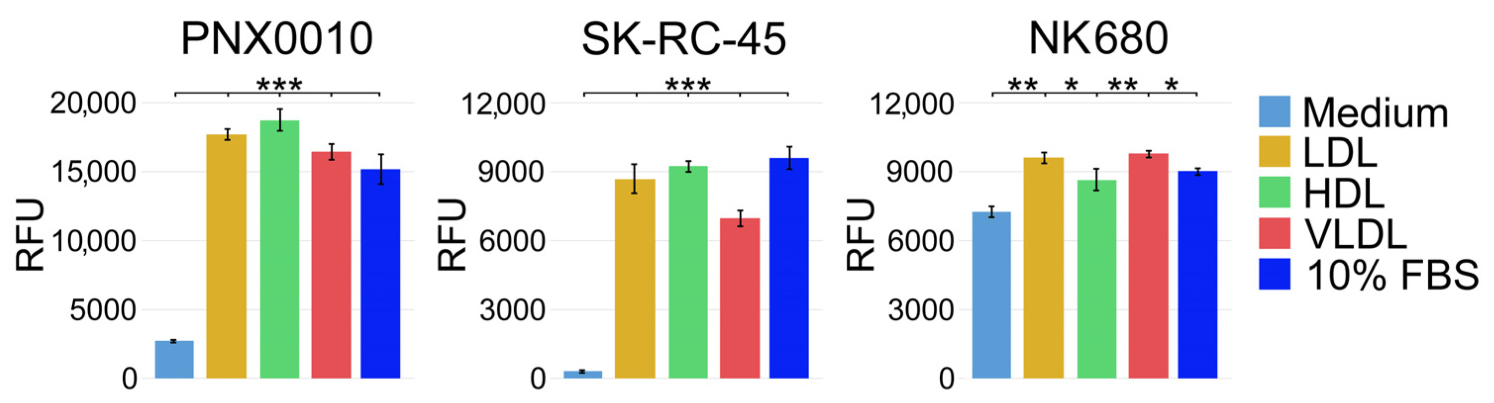
3.1. LDL, HDL, and VLDL Are Equally Effective in Supporting Viability of ccRCC Cells

Studies by Parinaud et al. demonstrate that all major lipoproteins, (i.e., LDL, HDL, and VLDL) have a similar ability to supply exogenous cholesterol [41]. Based on these findings, we examined the ability of different lipoproteins to support the viability of ccRCC cells. SK-RC-45 [8,42] and PNX0010 [43,44] ccRCC cell lines were cultured in RPMI-1640 medium supplemented with lipid-depleted fetal bovine serum (FBS) in the presence or absence of LDL, HDL, or VLDL. As demonstrated in Figure 1, LDL, HDL, and VLDL were equally effective in maintaining the viability of SK-RC-45 and PNX0010 ccRCC cells cultured under lipid-depleted conditions. The results of this experiment also show that ccRCC cells are largely dependent on exogenous cholesterol supply. In contrast, normal kidney epithelial NK680 cells were less dependent on exogenous cholesterol as indicated by their capacity to maintain viability under lipid-depleted conditions (Figure 1).

3.2. LDL, HDL, and VLDL Compromise the Antitumor Activity of TKIs

Our previous findings demonstrate that LDL cholesterol compromises the efficacy of TKIs against ccRCC and endothelial cells [37]. Therefore, we examined whether treatment with HDL and VLDL also affects the antitumor activity of TKIs. Our previous work has shown that TKIs may differ substantially with respect to the mechanism of their antitumor activity [42]. To demonstrate that our observations are not limited to a specific pharmacological agent, we treated SK-RC-45 and PNX0010 ccRCC cells with several clinically relevant TKIs, such as cabozantinib, sunitinib, axitinib, and pazopanib. As shown in Figure 2, LDL, HDL, and VLDL effectively rescued the viability of SK-RC-45 and PNX0010 cells treated with all tested TKIs.

3.3. Inhibition of Endosomal Cholesterol Trafficking Sensitizes ccRCC Cells to TKIs

Expression of NPC1 is increased in ccRCC at mRNA and protein levels (Figure 3A,B), and high expression of NPC1 is associated with poor prognosis based on TCGA data analysis (Figure 3C). Given that trafficking of de novo synthetized cholesterol in normal cells is not affected by pharmacological or genetic inhibition of NPC1 [22], we anticipated that ccRCC cells would be particularly sensitive to the inhibition of NPC-1-regulated endosomal cholesterol trafficking. Indeed, ccRCC cells manifested much higher sensitivity to the NPC1 inhibitor U18666A (90) compared with normal cells of various origins (Table 1). Of note, the effect of U1866A and posaconazole, an approved antifungal agent, which directly binds NPC1 and blocks endosomal cholesterol trafficking [45], on tumor cell viability was significantly increased under hypoxia, a central event in renal tumorigenesis (Table 2).
Unobstructed endolysosomal cholesterol trafficking is required to sustain the activity of several signaling pathways that confer drug resistance (i.e., Akt/mTOR, NF-κB, and Erk1/2) [37,46,47,48,49]. Given that, we tested whether NPC1 inhibition enhances the antitumor effect of TKIs. Our experiments reveal that administration of sunitinib, cabozantinib, or pazopanib in combination with U18666A showed a clear synergistic inhibitory effect on the viability of ccRCC cells at all effective dose levels (Table 3).
The results of these experiments were further validated using siRNA-mediated knockdown of NPC1. Genetic depletion of NPC1 reduced viability and sensitized ccRCC cells to TKI treatment (Figure 4 and Table 4).

4. Discussion

The lack of sensitivity of ccRCC to chemotherapy and radiation therapy prompted early research efforts into the development of new treatment options. The introduction of TKIs and immune checkpoint inhibitors (ICIs) notably changed the treatment paradigm of ccRCC. Recent clinical studies suggest that combining or sequencing TKIs with ICIs may provide effective treatments that reduce or delay disease progression [50,51,52,53,54,55,56,57]. However, despite the introduction of novel therapeutic approaches in the past decade, advanced ccRCC continues to be a treatment-resistant malignancy [3,4,5].
Multiple studies demonstrate that disruption of cholesterol homeostasis suppresses tumor growth and suggest that targeting cholesterol metabolism may be a promising strategy for antitumor therapy. Indeed, due to the absolute requirement of cholesterol for the synthesis of cell membranes, rapidly proliferating cancer cells have a greater requirement for cholesterol than normal cells (1,2). Our studies demonstrate that ccRCC cells can utilize lipoprotein-derived cholesterol irrespective of the particular class of cholesterol. donor and that all major classes of lipoproteins are capable of compromising the antitumor activity of TKIs. Thus, only simultaneous targeting of different lipoproteins is postulated to have therapeutic benefits. There are several potential approaches for targeting cholesterol homeostasis in cancer cells such as inhibition of cholesterol biosynthesis, blockade of cholesterol uptake, and modulation of intracellular cholesterol trafficking. Increased serum levels of cholesterol in humans can be effectively lowered by PCSK9 inhibitors and statins [58,59,60,61]. PCSK9 is a proprotein convertase that is involved in the degradation of LDLR in the liver [62]. PCSK9 inhibition increases LDLR expression by hepatocytes, which causes increased uptake of circulating LDL, thereby reducing plasma LDL-cholesterol. Statins are competitive inhibitors of HMG-CoA reductase, the rate-limiting enzyme of cholesterol biosynthesis [63,64]. However, these drugs up-regulate HDL levels [60,65,66,67,68], which can serve as a source of cholesterol for tumor cells. Also, statins demonstrate effects on a number of signaling pathways including protein isoprenylation [69]. Anticancer effects of statins have been largely attributed to the inhibition of this post-translational mechanism [69,70]. Moreover, cancer cells can bypass the effects of statins by unrestrained cholesterol importation [16,17]. Given that cholesterol uptake receptors have promiscuous ligand-binding properties, simultaneous targeting of different receptors may be necessary to reduce cellular cholesterol import, which may not be a clinically feasible approach.
As discussed above, tumor cells are largely dependent on exogenous lipids. Endolysosomal cholesterol transport is an attractive therapeutic target based on the fact that this is a point where LDL-, HDL, and VLDL-derived cholesterol trafficking routes converge and therefore may be simultaneously targeted. Furthermore, the trafficking of exogenous lipoprotein-derived cholesterol, in contrast to endogenously produced cholesterol, occurs through an NPC1-mediated endosomal trafficking system [23,24,25,26]. Therefore, the trafficking of de novo synthetized cholesterol in normal cells is not affected by NPC1 inhibition [22]. Indeed, our studies demonstrate that ccRCC cells are particularly sensitive to the inhibition of endosomal cholesterol trafficking by NPC1-specific inhibitor U18666A compared with normal cells of various origins. Importantly, the effect of U1866A and posaconazole on tumor cell viability was significantly increased under hypoxia, a central event in renal tumorigenesis. This observation could be potentially explained by the fact that hypoxia results in the depletion of cholesterol from the plasma membrane [71], increasing the demand for cholesterol transport from an endosomal compartment to the plasma membrane. These findings may widen the therapeutic window of NPC1 inhibitors for more selective targeting of malignant cells.

5. Conclusions

Our studies demonstrate that ccRCC cells are highly dependent on the uptake of exogenous cholesterol for their growth and survival and that all major lipoproteins have a comparable ability to supply cholesterol to tumor cells and compromise the antitumor activity of TKIs. Thus, concomitant targeting of all sources of cholesterol acquisition or common routes of cholesterol trafficking may be required to deprive tumor cells of cholesterol supply. Our findings indicate that ccRCC cells are particularly sensitive to the inhibition of NPC-1-dependent endosomal cholesterol trafficking. Importantly, NPC1 expression is increased in ccRCC, and high expression of NPC1 is associated with poor prognosis. Taken together, our work suggests that NPC1 may serve as a potential therapeutic target in ccRCC.

Supplementary Materials

The following supporting information can be downloaded at: https://www.mdpi.com/article/10.3390/cancers16030517/s1, Figure S1: full size blot of Figure 4A.

Author Contributions

Conceptualization, V.M.K.; methodology, V.M.K. and P.M.; investigation, P.M. and R.F.; writing—original draft preparation, V.M.K. and P.M.; writing—review and editing, V.M.K. and R.G.U.; supervision, V.M.K.; project administration, V.M.K.; funding acquisition, V.M.K. and P.M. All authors have read and agreed to the published version of the manuscript.

Funding

This work was supported in part by the National Institutes of Health Grants R21 CA263362 (to P.M.); the Department of Defense Idea Development Awards KC170127, KC220055, and PC210308 (to V.M.K.); MPI FCCC pilot application (to V.M.K.); and the NCI Core Grant NCI P30 CA006927 (to Fox Chase Cancer Center).

Institutional Review Board Statement

Not applicable.

Informed Consent Statement

Not applicable.

Data Availability Statement

The data presented in this study are available from the corresponding author (V.M.K.) upon request.

Acknowledgments

The authors acknowledge the support from the Cell Culture Facility at Fox Chase Cancer Center. PNX0010 ccRCC cell line was a kind gift from Vladimir Khazak.

Conflicts of Interest

The authors declare no conflicts of interest.

References

  1. Shuch, B.; Amin, A.; Armstrong, A.J.; Eble, J.N.; Ficarra, V.; Lopez-Beltran, A.; Martignoni, G.; Rini, B.I.; Kutikov, A. Understanding pathologic variants of renal cell carcinoma: Distilling therapeutic opportunities from biologic complexity. Eur. Urol. 2015, 67, 85–97. [Google Scholar] [CrossRef] [PubMed]
  2. Maines, F.; Caffo, O.; Veccia, A.; Trentin, C.; Tortora, G.; Galligioni, E.; Bria, E. Sequencing new agents after docetaxel in patients with metastatic castration-resistant prostate cancer. Crit. Rev. Oncol. Hematol. 2015, 96, 498–506. [Google Scholar] [CrossRef]
  3. Albiges, L.; Oudard, S.; Negrier, S.; Caty, A.; Gravis, G.; Joly, F.; Duclos, B.; Geoffrois, L.; Rolland, F.; Guillot, A.; et al. Complete remission with tyrosine kinase inhibitors in renal cell carcinoma. J. Clin. Oncol. 2012, 30, 482–487. [Google Scholar] [CrossRef] [PubMed]
  4. Park, K.; Lee, J.L.; Park, I.; Park, S.; Ahn, Y.; Ahn, J.H.; Ahn, S.; Song, C.; Hong, J.H.; Kim, C.S.; et al. Comparative efficacy of vascular endothelial growth factor (VEGF) tyrosine kinase inhibitor (TKI) and mammalian target of rapamycin (mTOR) inhibitor as second-line therapy in patients with metastatic renal cell carcinoma after the failure of first-line VEGF TKI. Med. Oncol. 2012, 29, 3291–3297. [Google Scholar] [CrossRef] [PubMed]
  5. Weinstock, M.; McDermott, D. Targeting PD-1/PD-L1 in the treatment of metastatic renal cell carcinoma. Ther. Adv. Urol. 2015, 7, 365–377. [Google Scholar] [CrossRef] [PubMed]
  6. Gotink, K.J.; Broxterman, H.J.; Labots, M.; de Haas, R.R.; Dekker, H.; Honeywell, R.J.; Rudek, M.A.; Beerepoot, L.V.; Musters, R.J.; Jansen, G.; et al. Lysosomal sequestration of sunitinib: A novel mechanism of drug resistance. Clin. Cancer Res. 2011, 17, 7337–7346. [Google Scholar] [CrossRef]
  7. Kutikov, A.; Makhov, P.; Golovine, K.; Canter, D.J.; Sirohi, M.; Street, R.; Simhan, J.; Uzzo, R.G.; Kolenko, V.M. Interleukin-6: A Potential Biomarker of Resistance to Multitargeted Receptor Tyrosine Kinase Inhibitors in Castration-Resistant Prostate Cancer. Urology 2011, 8, 968.e7–968.e11. [Google Scholar]
  8. Makhov, P.B.; Golovine, K.; Kutikov, A.; Teper, E.; Canter, D.J.; Simhan, J.; Uzzo, R.G.; Kolenko, V.M. Modulation of Akt/mTOR Signaling Overcomes Sunitinib Resistance in Renal and Prostate Cancer Cells. Mol. Cancer Ther. 2012, 11, 1510–1517. [Google Scholar] [CrossRef]
  9. Xin, H.; Zhang, C.; Herrmann, A.; Du, Y.; Figlin, R.; Yu, H. Sunitinib inhibition of Stat3 induces renal cell carcinoma tumor cell apoptosis and reduces immunosuppressive cells. Cancer Res. 2009, 69, 2506–2513. [Google Scholar] [CrossRef]
  10. Adelaiye, R.; Ciamporcero, E.; Miles, K.M.; Sotomayor, P.; Bard, J.; Tsompana, M.; Conroy, D.; Shen, L.; Ramakrishnan, S.; Ku, S.Y.; et al. Sunitinib dose escalation overcomes transient resistance in clear cell renal cell carcinoma and is associated with epigenetic modifications. Mol. Cancer Ther. 2015, 14, 513–522. [Google Scholar] [CrossRef]
  11. Gebhard, R.L.; Clayman, R.V.; Prigge, W.F.; Figenshau, R.; Staley, N.A.; Reesey, C.; Bear, A. Abnormal cholesterol metabolism in renal clear cell carcinoma. J. Lipid Res. 1987, 28, 1177–1184. [Google Scholar] [CrossRef] [PubMed]
  12. Llaverias, G.; Danilo, C.; Mercier, I.; Daumer, K.; Capozza, F.; Williams, T.M.; Sotgia, F.; Lisanti, M.P.; Frank, P.G. Role of cholesterol in the development and progression of breast cancer. Am. J. Pathol. 2011, 178, 402–412. [Google Scholar] [CrossRef] [PubMed]
  13. Freeman, M.R.; Di Vizio, D.; Solomon, K.R. The Rafts of the Medusa: Cholesterol targeting in cancer therapy. Oncogene 2010, 29, 3745–3747. [Google Scholar] [CrossRef] [PubMed]
  14. Riscal, R.; Skuli, N.; Simon, M.C. Even Cancer Cells Watch Their Cholesterol! Mol. Cell 2019, 76, 220–231. [Google Scholar] [CrossRef] [PubMed]
  15. Sundelin, J.P.; Stahlman, M.; Lundqvist, A.; Levin, M.; Parini, P.; Johansson, M.E.; Boren, J. Increased expression of the very low-density lipoprotein receptor mediates lipid accumulation in clear-cell renal cell carcinoma. PLoS ONE 2012, 7, e48694. [Google Scholar] [CrossRef] [PubMed]
  16. Gabitova, L.; Gorin, A.; Astsaturov, I. Molecular pathways: Sterols and receptor signaling in cancer. Clin. Cancer Res. 2014, 20, 28–34. [Google Scholar] [CrossRef] [PubMed]
  17. Guo, D.; Reinitz, F.; Youssef, M.; Hong, C.; Nathanson, D.; Akhavan, D.; Kuga, D.; Amzajerdi, A.N.; Soto, H.; Zhu, S.; et al. An LXR agonist promotes glioblastoma cell death through inhibition of an EGFR/AKT/SREBP-1/LDLR-dependent pathway. Cancer Discov. 2011, 1, 442–456. [Google Scholar] [CrossRef]
  18. Pompey, S.; Zhao, Z.; Luby-Phelps, K.; Michaely, P. Quantitative fluorescence imaging reveals point of release for lipoproteins during LDLR-dependent uptake. J. Lipid Res. 2013, 54, 744–753. [Google Scholar] [CrossRef]
  19. Trigatti, B.L.; Krieger, M.; Rigotti, A. Influence of the HDL receptor SR-BI on lipoprotein metabolism and atherosclerosis. Arterioscler. Thromb. Vasc. Biol. 2003, 23, 1732–1738. [Google Scholar] [CrossRef]
  20. Rigotti, A.; Miettinen, H.E.; Krieger, M. The role of the high-density lipoprotein receptor SR-BI in the lipid metabolism of endocrine and other tissues. Endocr. Rev. 2003, 24, 357–387. [Google Scholar] [CrossRef]
  21. Xu, G.H.; Lou, N.; Shi, H.C.; Xu, Y.C.; Ruan, H.L.; Xiao, W.; Liu, L.; Li, X.; Xiao, H.B.; Qiu, B.; et al. Up-regulation of SR-BI promotes progression and serves as a prognostic biomarker in clear cell renal cell carcinoma. BMC Cancer 2018, 18, 88. [Google Scholar] [CrossRef]
  22. Liscum, L.; Dahl, N.K. Intracellular cholesterol transport. J. Lipid Res. 1992, 33, 1239–1254. [Google Scholar] [CrossRef]
  23. Wojtanik, K.M.; Liscum, L. The transport of low density lipoprotein-derived cholesterol to the plasma membrane is defective in NPC1 cells. J. Biol. Chem. 2003, 278, 14850–14856. [Google Scholar] [CrossRef] [PubMed]
  24. Pfisterer, S.G.; Peranen, J.; Ikonen, E. LDL-cholesterol transport to the endoplasmic reticulum: Current concepts. Curr. Opin. Lipidol. 2016, 27, 282–287. [Google Scholar] [CrossRef] [PubMed]
  25. Kwon, H.J.; Abi-Mosleh, L.; Wang, M.L.; Deisenhofer, J.; Goldstein, J.L.; Brown, M.S.; Infante, R.E. Structure of N-terminal domain of NPC1 reveals distinct subdomains for binding and transfer of cholesterol. Cell 2009, 137, 1213–1224. [Google Scholar] [CrossRef] [PubMed]
  26. Peake, K.B.; Vance, J.E. Defective cholesterol trafficking in Niemann-Pick C-deficient cells. FEBS Lett. 2010, 584, 2731–2739. [Google Scholar] [CrossRef] [PubMed]
  27. Soffientini, U.; Graham, A. Intracellular cholesterol transport proteins: Roles in health and disease. Clin. Sci. 2016, 130, 1843–1859. [Google Scholar] [CrossRef] [PubMed]
  28. Lange, Y.; Ye, J.; Rigney, M.; Steck, T. Cholesterol movement in Niemann-Pick type C cells and in cells treated with amphiphiles. J. Biol. Chem. 2000, 275, 17468–17475. [Google Scholar] [CrossRef] [PubMed]
  29. O’Neill, K.I.; Kuo, L.W.; Williams, M.M.; Lind, H.; Crump, L.S.; Hammond, N.G.; Spoelstra, N.S.; Caino, M.C.; Richer, J.K. NPC1 Confers Metabolic Flexibility in Triple Negative Breast Cancer. Cancers 2022, 14, 3543. [Google Scholar] [CrossRef] [PubMed]
  30. Burns, V.E.; Kerppola, T.K. ATR-101 inhibits cholesterol efflux and cortisol secretion by ATP-binding cassette transporters, causing cytotoxic cholesterol accumulation in adrenocortical carcinoma cells. Br. J. Pharmacol. 2017, 174, 3315–3332. [Google Scholar] [CrossRef]
  31. Do, H.T.; Bruelle, C.; Tselykh, T.; Jalonen, P.; Korhonen, L.; Lindholm, D. Reciprocal regulation of very low density lipoprotein receptors (VLDLRs) in neurons by brain-derived neurotrophic factor (BDNF) and Reelin: Involvement of the E3 ligase Mylip/Idol. J. Biol. Chem. 2013, 288, 29613–29620. [Google Scholar] [CrossRef]
  32. Hong, C.; Duit, S.; Jalonen, P.; Out, R.; Scheer, L.; Sorrentino, V.; Boyadjian, R.; Rodenburg, K.W.; Foley, E.; Korhonen, L.; et al. The E3 ubiquitin ligase IDOL induces the degradation of the low density lipoprotein receptor family members VLDLR and ApoER2. J. Biol. Chem. 2010, 285, 19720–19726. [Google Scholar] [CrossRef] [PubMed]
  33. Ren, K.; Jiang, T.; Zhao, G.J. Quercetin induces the selective uptake of HDL-cholesterol via promoting SR-BI expression and the activation of the PPARgamma/LXRalpha pathway. Food Funct. 2018, 9, 624–635. [Google Scholar] [CrossRef] [PubMed]
  34. Venkateswaran, A.; Laffitte, B.A.; Joseph, S.B.; Mak, P.A.; Wilpitz, D.C.; Edwards, P.A.; Tontonoz, P. Control of cellular cholesterol efflux by the nuclear oxysterol receptor LXR alpha. Proc. Natl. Acad. Sci. USA 2000, 97, 12097–12102. [Google Scholar] [CrossRef] [PubMed]
  35. Hu, Y.W.; Wang, Q.; Ma, X.; Li, X.X.; Liu, X.H.; Xiao, J.; Liao, D.F.; Xiang, J.; Tang, C.K. TGF-beta1 up-regulates expression of ABCA1, ABCG1 and SR-BI through liver X receptor alpha signaling pathway in THP-1 macrophage-derived foam cells. J. Atheroscler. Thromb. 2010, 17, 493–502. [Google Scholar] [CrossRef] [PubMed]
  36. Golovine, K.; Makhov, P.; Naito, S.; Raiyani, H.; Tomaszewski, J.; Mehrazin, R.; Tulin, A.; Kutikov, A.; Uzzo, R.G.; Kolenko, V.M. Piperlongumine and its analogs down-regulate expression of c-Met in renal cell carcinoma. Cancer Biol. Ther. 2015, 16, 743–749. [Google Scholar] [CrossRef] [PubMed]
  37. Naito, S.; Makhov, P.; Astsaturov, I.; Golovine, K.; Tulin, A.; Kutikov, A.; Uzzo, R.G.; Kolenko, V.M. LDL cholesterol counteracts the antitumour effect of tyrosine kinase inhibitors against renal cell carcinoma. Br. J. Cancer 2017, 116, 1203–1207. [Google Scholar] [CrossRef] [PubMed]
  38. Golovine, K.; Makhov, P.; Uzzo, R.G.; Shaw, T.; Kunkle, D.; Kolenko, V.M. Overexpression of the zinc uptake transporter hZIP1 inhibits nuclear factor-kappaB and reduces the malignant potential of prostate cancer cells in vitro and in vivo. Clin. Cancer Res. 2008, 14, 5376–5384. [Google Scholar] [CrossRef]
  39. Chou, T.C. Theoretical basis, experimental design, and computerized simulation of synergism and antagonism in drug combination studies. Pharmacol. Rev. 2006, 58, 621–681. [Google Scholar] [CrossRef]
  40. Makhov, P.; Sohn, J.A.; Serebriiskii, I.G.; Fazliyeva, R.; Khazak, V.; Boumber, Y.; Uzzo, R.G.; Kolenko, V.M. CRISPR/Cas9 genome-wide loss-of-function screening identifies druggable cellular factors involved in sunitinib resistance in renal cell carcinoma. Br. J. Cancer 2020, 123, 1749–1756. [Google Scholar] [CrossRef]
  41. Parinaud, J.; Perret, B.; Ribbes, H.; Chap, H.; Pontonnier, G.; Douste-Blazy, L. High density lipoprotein and low density lipoprotein utilization by human granulosa cells for progesterone synthesis in serum-free culture: Respective contributions of free and esterified cholesterol. J. Clin. Endocrinol. Metab. 1987, 64, 409–417. [Google Scholar] [CrossRef]
  42. Canter, D.; Kutikov, A.; Golovine, K.; Makhov, P.; Simhan, J.; Uzzo, R.G.; Kolenko, V.M. Are all multi-targeted tyrosine kinase inhibitors created equal? An in vitro study of sunitinib and pazopanib in renal cell carcinoma cell lines. Can. J. Urol. 2011, 18, 5819–5825. [Google Scholar]
  43. Kirsanov, K.I.; Kotova, E.; Makhov, P.; Golovine, K.; Lesovaya, E.A.; Kolenko, V.M.; Yakubovskaya, M.G.; Tulin, A.V. Minor grove binding ligands disrupt PARP-1 activation pathways. Oncotarget 2014, 5, 428–437. [Google Scholar] [CrossRef] [PubMed]
  44. Thomas, C.; Ji, Y.; Lodhi, N.; Kotova, E.; Pinnola, A.D.; Golovine, K.; Makhov, P.; Pechenkina, K.; Kolenko, V.; Tulin, A.V. Non-NAD-Like poly(ADP-Ribose) Polymerase-1 Inhibitors effectively Eliminate Cancer in vivo. EBioMedicine 2016, 13, 90–98. [Google Scholar] [CrossRef] [PubMed]
  45. Trinh, M.N.; Lu, F.; Li, X.; Das, A.; Liang, Q.; De Brabander, J.K.; Brown, M.S.; Goldstein, J.L. Triazoles inhibit cholesterol export from lysosomes by binding to NPC1. Proc. Natl. Acad. Sci. USA 2017, 114, 89–94. [Google Scholar] [CrossRef]
  46. Li, Y.; Schwabe, R.F.; DeVries-Seimon, T.; Yao, P.M.; Gerbod-Giannone, M.C.; Tall, A.R.; Davis, R.J.; Flavell, R.; Brenner, D.A.; Tabas, I. Free cholesterol-loaded macrophages are an abundant source of tumor necrosis factor-alpha and interleukin-6: Model of NF-kappaB- and map kinase-dependent inflammation in advanced atherosclerosis. J. Biol. Chem. 2005, 280, 21763–21772. [Google Scholar] [CrossRef] [PubMed]
  47. Xu, J.; Dang, Y.J.; Ren, Y.R.Z.; Liu, J.O. Cholesterol trafficking is required for mTOR activation in endothelial cells. Proc. Natl. Acad. Sci. USA 2010, 107, 4764–4769. [Google Scholar] [CrossRef] [PubMed]
  48. Morales, M.L.; Arenas, A.; Ortiz-Ruiz, A.; Leivas, A.; Rapado, I.; Rodriguez-Garcia, A.; Castro, N.; Zagorac, I.; Quintela-Fandino, M.; Gomez-Lopez, G.; et al. MEK inhibition enhances the response to tyrosine kinase inhibitors in acute myeloid leukemia. Sci. Rep. 2019, 9, 18630. [Google Scholar] [CrossRef] [PubMed]
  49. Blakely, C.M.; Pazarentzos, E.; Olivas, V.; Asthana, S.; Yan, J.J.; Tan, I.; Hrustanovic, G.; Chan, E.; Lin, L.; Neel, D.S.; et al. NF-kappaB-activating complex engaged in response to EGFR oncogene inhibition drives tumor cell survival and residual disease in lung cancer. Cell Rep. 2015, 11, 98–110. [Google Scholar] [CrossRef]
  50. Bedke, J.; Stuhler, V.; Stenzl, A.; Brehmer, B. Immunotherapy for kidney cancer: Status quo and the future. Curr. Opin. Urol. 2018, 28, 8–14. [Google Scholar] [CrossRef]
  51. Mosillo, C.; Ciccarese, C.; Bimbatti, D.; Fantinel, E.; Volta, A.D.; Bisogno, I.; Zampiva, I.; Santoni, M.; Massar, F.; Brunelli, M.; et al. Renal cell carcinoma in one year: Going inside the news of 2017—A report of the main advances in RCC cancer research. Cancer Treat. Rev. 2018, 67, 29–33. [Google Scholar] [CrossRef] [PubMed]
  52. Makhov, P.; Joshi, S.; Ghatalia, P.; Kutikov, A.; Uzzo, R.G.; Kolenko, V.M. Resistance to Systemic Therapies in Clear Cell Renal Cell Carcinoma: Mechanisms and Management Strategies. Mol. Cancer Ther. 2018, 17, 1355–1364. [Google Scholar] [CrossRef] [PubMed]
  53. Atkins, M.B.; Plimack, E.R.; Puzanov, I.; Fishman, M.N.; McDermott, D.F.; Cho, D.C.; Vaishampayan, U.; George, S.; Olencki, T.E.; Tarazi, J.C.; et al. Axitinib in combination with pembrolizumab in patients with advanced renal cell cancer: A non-randomised, open-label, dose-finding, and dose-expansion phase 1b trial. Lancet Oncol. 2018, 19, 405–415. [Google Scholar] [CrossRef] [PubMed]
  54. Calvo, E.; Porta, C.; Grunwald, V.; Escudier, B. The Current and Evolving Landscape of First-Line Treatments for Advanced Renal Cell Carcinoma. Oncologist 2019, 24, 338–348. [Google Scholar] [CrossRef] [PubMed]
  55. Fuca, G.; de Braud, F.; Di Nicola, M. Immunotherapy-based combinations: An update. Curr. Opin. Oncol. 2018, 30, 345–351. [Google Scholar] [CrossRef] [PubMed]
  56. Vano, Y.A.; Ladoire, S.; Elaidi, R.; Dermeche, S.; Eymard, J.C.; Falkowski, S.; Gross-Goupil, M.; Malouf, G.; Narciso, B.; Sajous, C.; et al. First-Line Treatment of Metastatic Clear Cell Renal Cell Carcinoma: What Are the Most Appropriate Combination Therapies? Cancers 2021, 13, 5548. [Google Scholar] [CrossRef] [PubMed]
  57. Massari, F.; Rizzo, A.; Mollica, V.; Rosellini, M.; Marchetti, A.; Ardizzoni, A.; Santoni, M. Immune-based combinations for the treatment of metastatic renal cell carcinoma: A meta-analysis of randomised clinical trials. Eur. J. Cancer 2021, 154, 120–127. [Google Scholar] [CrossRef] [PubMed]
  58. Colhoun, H.M.; Leiter, L.A.; Muller-Wieland, D.; Cariou, B.; Ray, K.K.; Tinahones, F.J.; Domenger, C.; Letierce, A.; Israel, M.; Samuel, R.; et al. Effect of alirocumab on individuals with type 2 diabetes, high triglycerides, and low high-density lipoprotein cholesterol. Cardiovasc. Diabetol. 2020, 19, 14. [Google Scholar] [CrossRef]
  59. Mombelli, G.; Castelnuovo, S.; Pavanello, C. Potential of PCSK9 as a new target for the management of LDL cholesterol. Res. Rep. Clin. Cardiol. 2015, 6, 73–86. [Google Scholar] [CrossRef]
  60. Sabatine, M.S.; Giugliano, R.P.; Wiviott, S.D.; Raal, F.J.; Blom, D.J.; Robinson, J.; Ballantyne, C.M.; Somaratne, R.; Legg, J.; Wasserman, S.M.; et al. Efficacy and safety of evolocumab in reducing lipids and cardiovascular events. N. Engl. J. Med. 2015, 372, 1500–1509. [Google Scholar] [CrossRef]
  61. Moride, Y.; Hegele, R.A.; Langer, A.; McPherson, R.; Miller, D.B.; Rinfret, S. Clinical and public health assessment of benefits and risks of statins in primary prevention of coronary events: Resolved and unresolved issues. Can. J. Cardiol. 2008, 24, 293–300. [Google Scholar] [CrossRef] [PubMed]
  62. Hajar, R. PCSK 9 Inhibitors: A Short History and a New Era of Lipid-lowering Therapy. Heart Views 2019, 20, 74–75. [Google Scholar] [CrossRef] [PubMed]
  63. McFarlane, S.I.; Muniyappa, R.; Francisco, R.; Sowers, J.R. Clinical review 145: Pleiotropic effects of statins: Lipid reduction and beyond. J. Clin. Endocrinol. Metab. 2002, 87, 1451–1458. [Google Scholar] [CrossRef] [PubMed]
  64. Schaefer, E.J.; Asztalos, B.F. The effects of statins on high-density lipoproteins. Curr. Atheroscler. Rep. 2006, 8, 41–49. [Google Scholar] [CrossRef]
  65. Barter, P.J.; Brandrup-Wognsen, G.; Palmer, M.K.; Nicholls, S.J. Effect of statins on HDL-C: A complex process unrelated to changes in LDL-C: Analysis of the VOYAGER Database. J. Lipid Res. 2010, 51, 1546–1553. [Google Scholar] [CrossRef] [PubMed]
  66. McTaggart, F.; Jones, P. Effects of statins on high-density lipoproteins: A potential contribution to cardiovascular benefit. Cardiovasc. Drugs Ther. 2008, 22, 321–338. [Google Scholar] [CrossRef] [PubMed]
  67. Yamashita, S.; Tsubakio-Yamamoto, K.; Ohama, T.; Nakagawa-Toyama, Y.; Nishida, M. Molecular mechanisms of HDL-cholesterol elevation by statins and its effects on HDL functions. J. Atheroscler. Thromb. 2010, 17, 436–451. [Google Scholar] [CrossRef] [PubMed]
  68. Teramoto, T.; Kobayashi, M.; Tasaki, H.; Yagyu, H.; Higashikata, T.; Takagi, Y.; Uno, K.; Baccara-Dinet, M.T.; Nohara, A. Efficacy and Safety of Alirocumab in Japanese Patients With Heterozygous Familial Hypercholesterolemia or at High Cardiovascular Risk with Hypercholesterolemia Not Adequately Controlled With Statins- ODYSSEY JAPAN Randomized Controlled Trial. Circ. J. 2016, 80, 1980–1987. [Google Scholar] [CrossRef]
  69. Brusselmans, K.; Timmermans, L.; Van de Sande, T.; Van Veldhoven, P.P.; Guan, G.; Shechter, I.; Claessens, F.; Verhoeven, G.; Swinnen, J.V. Squalene synthase, a determinant of Raft-associated cholesterol and modulator of cancer cell proliferation. J. Biol. Chem. 2007, 282, 18777–18785. [Google Scholar] [CrossRef]
  70. Ribas, V.; Garcia-Ruiz, C.; Fernandez-Checa, J.C. Mitochondria, cholesterol and cancer cell metabolism. Clin. Transl. Med. 2016, 5, 22. [Google Scholar] [CrossRef]
  71. Huetsch, J.C.; Suresh, K.; Shimoda, L.A. When higher cholesterol is better: Membrane cholesterol loss and endothelial Ca2+ signaling. Am. J. Physiol. Heart Circ. Physiol. 2018, 314, H780–H783. [Google Scholar] [CrossRef]
Figure 1. LDL, HDL, and VLDL are equally effective in supporting viability of ccRCC cells. PNX0010 and SK-45 ccRCC cells and NK680 normal kidney epithelial cells were cultured in RPMI 1640 medium supplemented with lipid-depleted fetal bovine serum in the presence or absence of LDL, HDL, or VLDL (all at 100 μg/mL) for 96 h. Cell viability was analyzed by CellTiter Blue assay (Promega). Results are expressed as the mean (n = 3) ± s.e.m. * p < 0.01; ** p < 0.001; *** p < 0.0001. RFU-relative fluorescence units.
Cancers 16 00517 g001
Figure 2. LDL, HDL, and VLDL compromise the antitumor activity of TKIs. SK-45 and PNX0010 ccRCC cells were cultured with TKIs (all at 5 μM) with or without LDL, HDL, or VLDL (all at 100 μg/mL) in RPMI 1640 medium supplemented with regular 10% FBS for 96 h. Cell viability was analyzed by CellTiter Blue assay. Results are expressed as the mean (n = 3) ± s.e.m. * p < 0.01; ** p < 0.001; *** p < 0.0001; NS—Not significant.
Cancers 16 00517 g002
Figure 3. Relationship between NPC1 expression and clinical outcomes in ccRCC. (A) Expression of NPC1 is increased in ccRCC at (A) mRNA (TCGA) and (B) protein (CPTAC) levels according to the University of Alabama at Birmingham cancer data analysis portal (UALCAN). (C) NPC1 is an unfavorable prognostic marker in ccRCC based on TCGA analysis via cBioPortal.
Cancers 16 00517 g003
Figure 4. NPC1 depletion reduces viability of ccRCC cells. (A) Western blot analysis of NPC1 expression in 786-O and PNX00100 cells treated with control siRNA and NPC1 SmartPool siRNA. The uncropped blots are shown in the supplementary materials. (B) Aliquots of the cells used in panel A were cultured in RPMI1640 medium supplemented with 10% FBS for 72 h. Cell viability was analyzed as described in legend to Figure 2. *** p < 0.0001.
Cancers 16 00517 g004
Table 1. U18666A preferentially inhibits viability of ccRCC cells. ccRCC (786-O, SK-RC-45, and PNX0010), normal kidney epithelial (NKE and NK686), normal prostate epithelial (RWPE-1 and PZ-HPV-7), and human umbilical vein endothelial (HUVEC) cells were treated with escalating concentrations of U18666A for 72 h. Cell viability and effective doses (ED, μM) were evaluated as described in Materials and Methods.
Cell LineED25IC50 IC75 IC90
786-O2.12.83.74.9
SK-RC-450.090.41.88.2
PNX00105.210.415.931.3
NK68630.754.897.9174.7
NKE26.552.9105.9211
RWPE-121.830.342.158.5
PZ-HPV-713.728.057116.2
HUVEC19.734.159.4103.6
Table 2. The effectiveness of NPC1 inhibitors is enhanced under hypoxic conditions. 786-O and PNX0010 ccRCC cells were cultured under normoxic (N) (20% O2) or hypoxic (H) (2% O2) conditions in RPMI1640 medium supplemented with 10% FBS and treated with escalating concentrations of either U18666A or posaconazole for 72 h. Cell viability and effective doses (ED, μM) were evaluated as described in Materials and Methods.
DrugCell LineED25ED50ED75ED90
U18666A786-O (N)1.52.33.55.4
786-O (H)0.71.22.13.6
PNX0010 (N)2.23.34.97.2
PNX0010 (H)0.40.91.93.8
Posaconazole786-O (N)4.710.121.646.2
786-O (H)1.33.06.715.3
PNX0010 (N)4.68.014.224.9
PNX0010 (H)0.91.94.29.2
Table 3. The synergistic effect of combined treatment with U18666A and TKIs on the viability of ccRCC cells. The cells were treated with escalating concentrations of U18666A and TKIs for 72 h. Analysis of the synergistic interaction between U18666A and TKIs was performed as described in Materials and Methods. ED: effective doses. Combination index (CI) 0.85–0.9: slight synergism; CI 0.7–0.85: moderate synergism; CI 0.3–0.7: synergism; CI 0.1–0.3: strong synergism; CI < 0.1: very strong synergism.
DrugCell LineED25ED50ED75ED90
Sunitinib786-O0.560.520.470.41
SK-RC-450.450.410.370.32
PNX00100.500.460.420.39
Cabozantinib786-O0.440.400.350.30
SK-RC-450.390.330.270.22
PNX00100.520.450.410.38
Pazopanib786-O0.370.310.250.20
SK-RC-450.420.360.290.23
PNX00100.480.400.330.26
Table 4. Depletion of NPC1 sensitizes ccRCC cells to TKIs. Control and NPC1-depleted cells described in Figure 4 were treated with escalating concentrations of axitinib and cabozantinib for 72 h. Viability and EDs (μM) were calculated as described in the legend in Table 1.
DrugCell LineED25ED50ED75ED90
Axitinib786-O 5.47.410.113.9
786-O-NPC1KD1.32.55.09.9
PNX00104.16.911.519.3
PNX0010-NPC1KD1.52.64.78.5
Cabozantinib786-O5.68.512.819.3
786-O-NPC1KD2.54.06.410.4
PNX0010 4.66.810.014.9
PNX0010-NPC1KD2.73.95.78.2
Disclaimer/Publisher’s Note: The statements, opinions and data contained in all publications are solely those of the individual author(s) and contributor(s) and not of MDPI and/or the editor(s). MDPI and/or the editor(s) disclaim responsibility for any injury to people or property resulting from any ideas, methods, instructions or products referred to in the content.

