
Updated Epidemiology of Gastric Cancer in Asia: Decreased Incidence but Still a Big Challenge
 , ,
, ,
Abstract
:Simple Summary
Abstract
1. Introduction
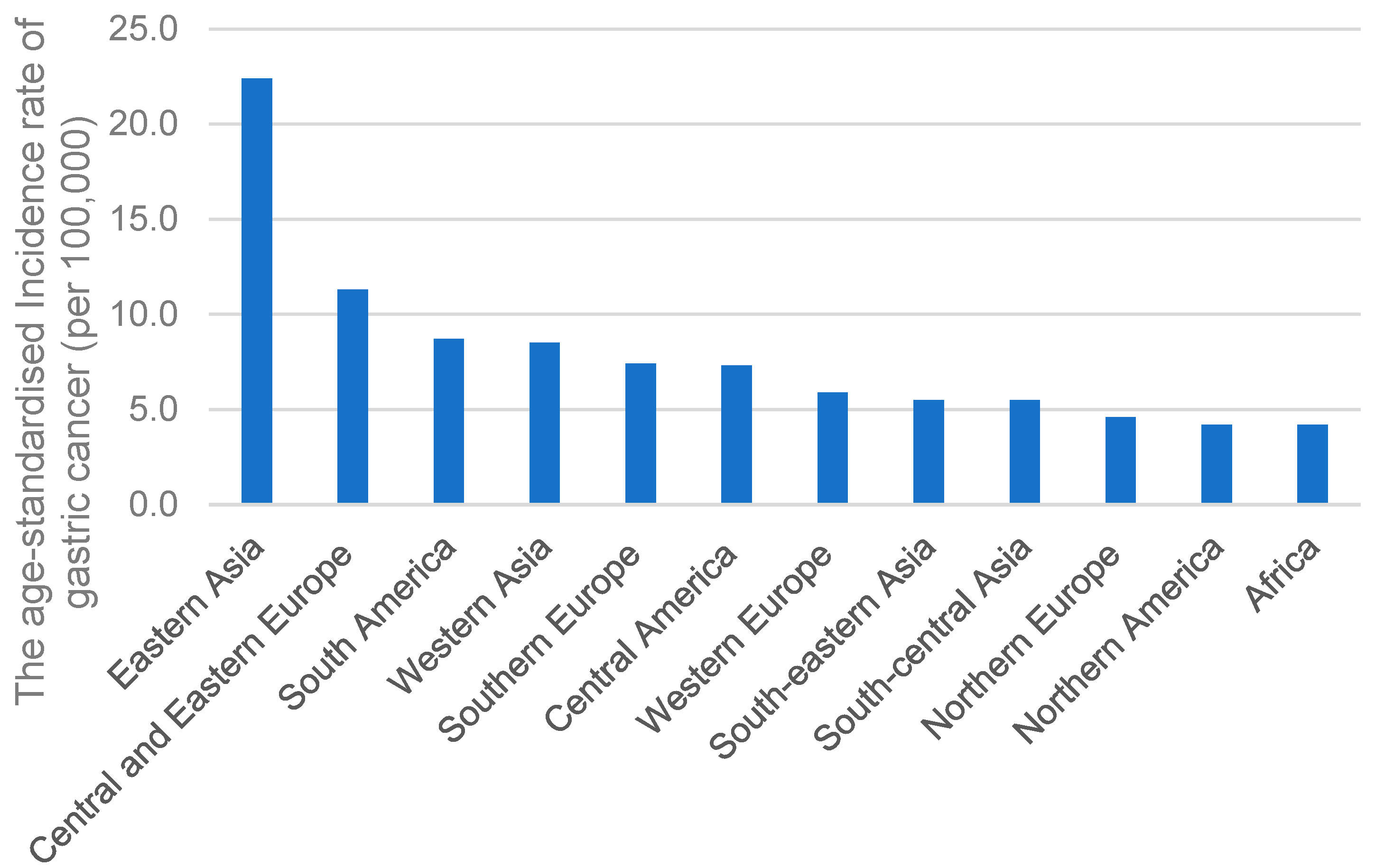
2. GC in Asia
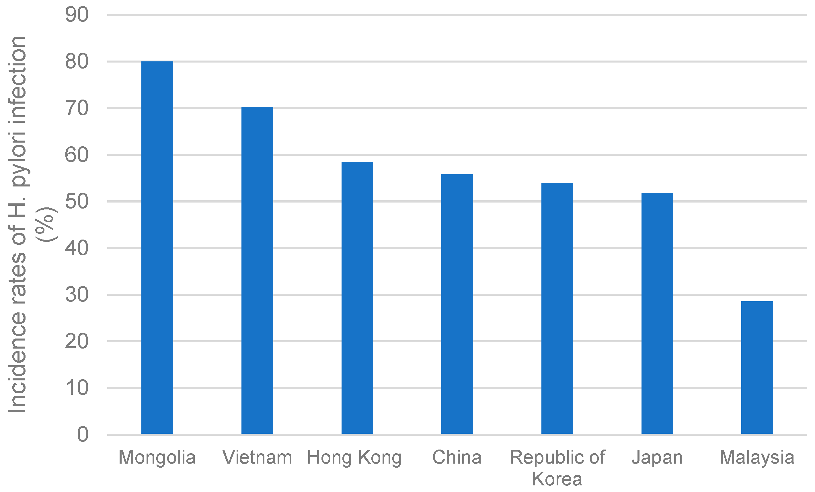
2.1. H. pylori Infection
2.2. Dietary Habits
2.3. Smoking Behaviors
2.4. Alcohol Consumption
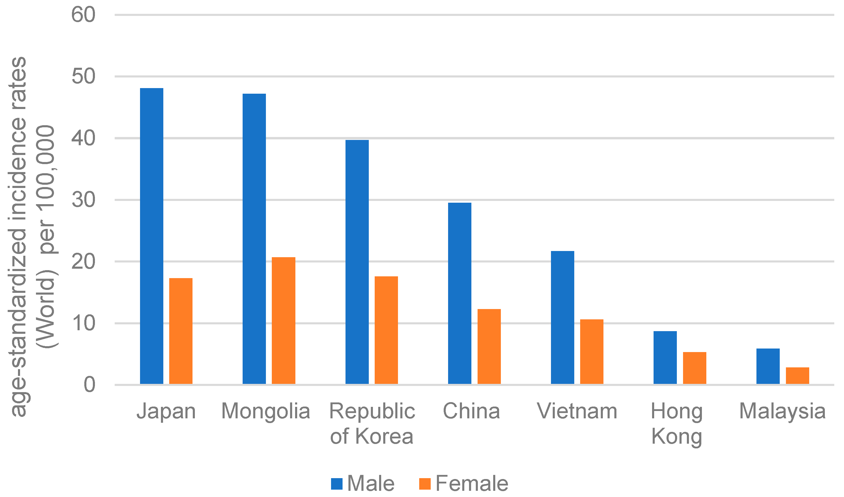
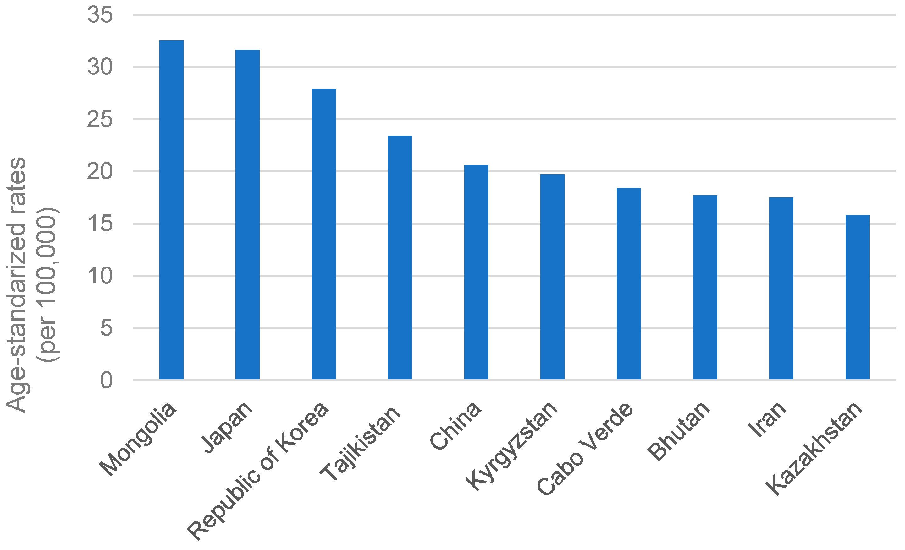
3. The Incidence of GC in Different Asia Regions
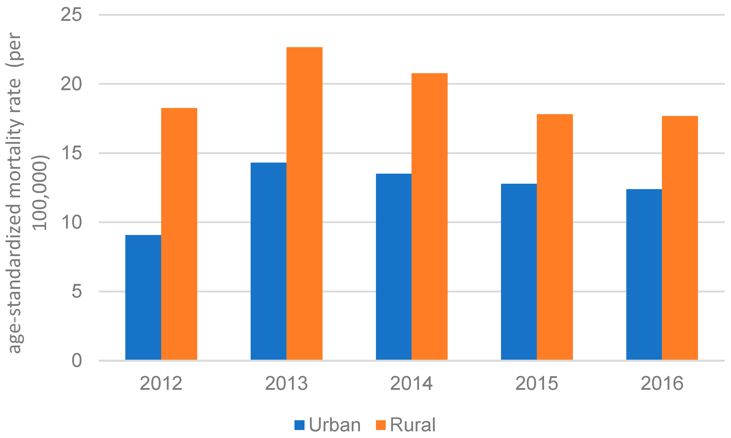
3.1. China
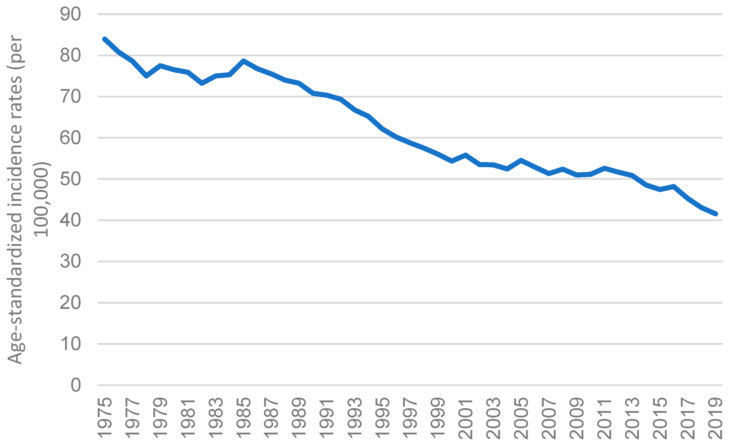
3.2. Japan
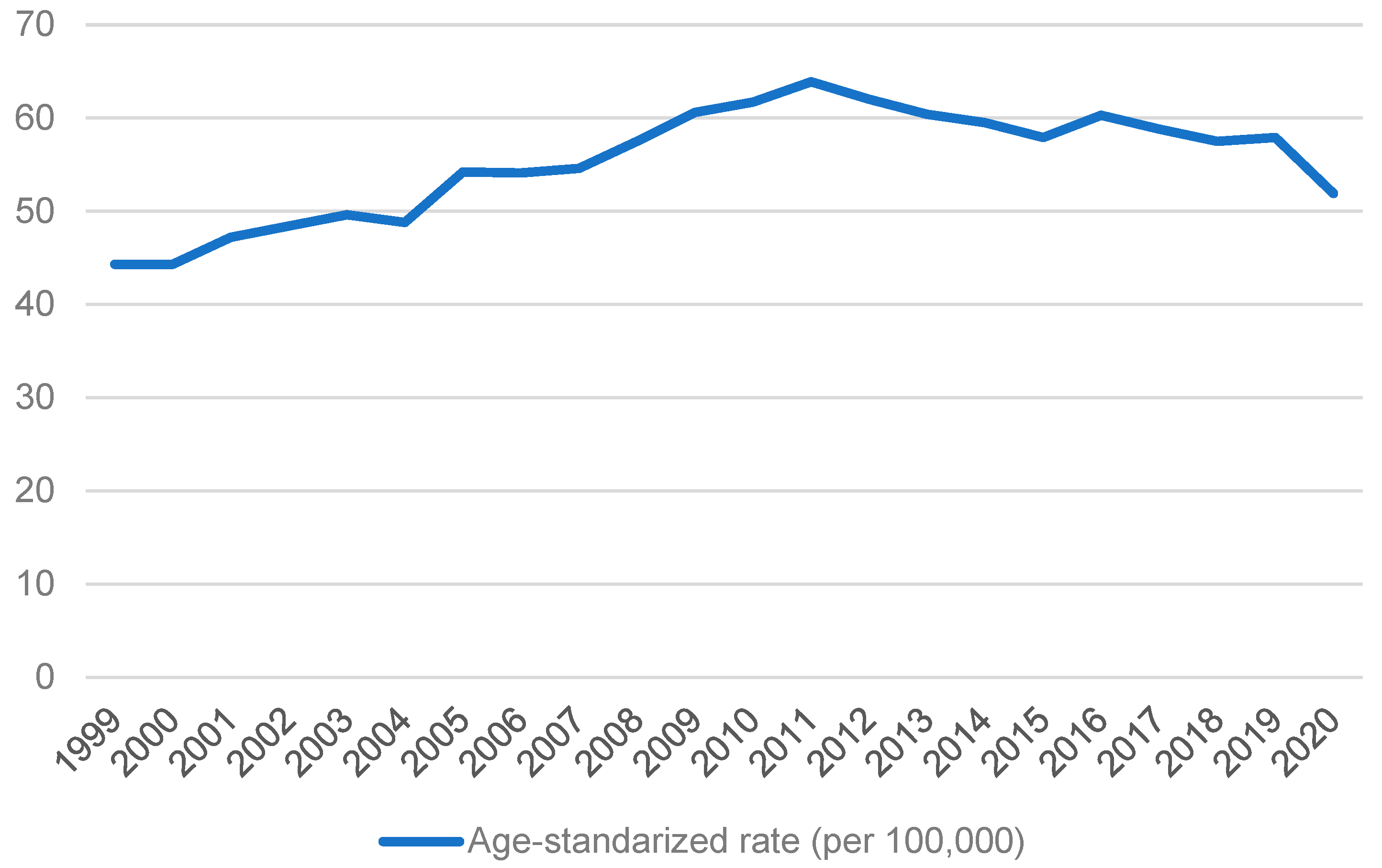
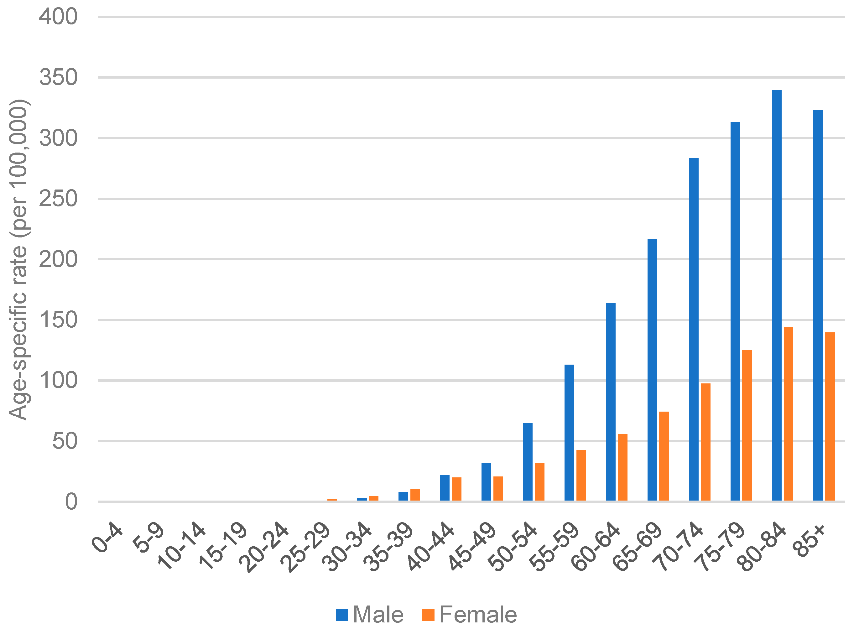
3.3. Republic of Korea
3.4. Hong Kong
3.5. Vietnam
3.6. Malaysia
3.7. Mongolia
4. Challenges for GC Treatment in Asian Countries
5. Forefronts in GC Treatment: Targeted Antibodies and Immunotherapy
6. Availability of GC Early Screening, Diagnosis, and Treatments Differs among Asian Countries in an Economy-Based Manner
7. Conclusions and Future Perspectives
Author Contributions
Funding
Acknowledgments
Conflicts of Interest
References
- Deo, S.; Sharma, J.; Kumar, S. GLOBOCAN 2020 Report on Global Cancer Burden: Challenges and Opportunities for Surgical Oncologists. Ann. Surg. Oncol. 2022, 29, 6497–6500. [Google Scholar] [CrossRef] [PubMed]
- Sung, H.; Ferlay, J.; Siegel, R.L.; Laversanne, M.; Soerjomataram, I.; Jemal, A.; Bray, F. Global cancer statistics 2020: GLOBOCAN estimates of incidence and mortality worldwide for 36 cancers in 185 countries. CA Cancer J. Clin. 2021, 71, 209–249. [Google Scholar] [CrossRef]
- Akshatha, C.; Bhat, S.; Sindhu, R.; Shashank, D.; Sommano, S.R.; Tapingkae, W.; Cheewangkoon, R.; Prasad, S.K. Current therapeutic options for gastric adenocarcinoma. Saudi J. Biol. Sci. 2021, 28, 5371–5378. [Google Scholar] [CrossRef]
- Sekiguchi, M.; Oda, I.; Matsuda, T.; Saito, Y. Epidemiological Trends and Future Perspectives of Gastric Cancer in Eastern Asia. Digestion 2022, 103, 22–28. [Google Scholar] [CrossRef] [PubMed]
- Puculek, M.; Machlowska, J.; Wierzbicki, R.; Baj, J.; Maciejewski, R.; Sitarz, R. Helicobacter pylori associated factors in the development of gastric cancer with special reference to the early-onset subtype. Oncotarget 2018, 9, 31146–31162. [Google Scholar] [CrossRef]
- Rawla, P.; Barsouk, A. Epidemiology of gastric cancer: Global trends, risk factors and prevention. Gastroenterol. Rev. Przegląd Gastroenterol. 2019, 14, 26–38. [Google Scholar] [CrossRef]
- Ilic, M.; Ilic, I. Epidemiology of stomach cancer. World J. Gastroenterol. 2022, 28, 1187. [Google Scholar] [CrossRef]
- Berlth, F.; Bollschweiler, E.; Drebber, U.; Hoelscher, A.H.; Moenig, S. Pathohistological classification systems in gastric cancer: Diagnostic relevance and prognostic value. World J. Gastroenterol. WJG 2014, 20, 5679. [Google Scholar] [CrossRef] [PubMed]
- Nagtegaal, I.D.; Odze, R.D.; Klimstra, D.; Paradis, V.; Rugge, M.; Schirmacher, P.; Washington, K.M.; Carneiro, F.; Cree, I.A. The 2019 WHO classification of tumours of the digestive system. Histopathology 2020, 76, 182. [Google Scholar] [CrossRef] [PubMed]
- Grabsch, H.I.; Tan, P. Gastric cancer pathology and underlying molecular mechanisms. Dig. Surg. 2013, 30, 150–158. [Google Scholar] [CrossRef]
- Wang, Q.; Xie, Q.; Liu, Y.; Guo, H.; Ren, Y.; Li, J.; Zhao, Q. Clinical characteristics and prognostic significance of TCGA and ACRG classification in gastric cancer among the Chinese population. Mol. Med. Rep. 2020, 22, 828–840. [Google Scholar] [CrossRef]
- Mabe, K.; Inoue, K.; Kamada, T.; Kato, K.; Kato, M.; Haruma, K. Endoscopic screening for gastric cancer in Japan: Current status and future perspectives. Dig Endosc 2022, 34, 412–419. [Google Scholar] [CrossRef]
- Abdelfatah, M.M.; Barakat, M.; Ahmad, D.; Ibrahim, M.; Ahmed, Y.; Kurdi, Y.; Grimm, I.S.; Othman, M.O. Long-term outcomes of endoscopic submucosal dissection versus surgery in early gastric cancer: A systematic review and meta-analysis. Eur. J. Gastroenterol. Hepatol. 2019, 31, 418–424. [Google Scholar] [CrossRef] [PubMed]
- Patel, T.H.; Cecchini, M. Targeted therapies in advanced gastric cancer. Curr. Treat. Options Oncol. 2020, 21, 1–14. [Google Scholar] [CrossRef] [PubMed]
- Kono, K.; Nakajima, S.; Mimura, K. Current status of immune checkpoint inhibitors for gastric cancer. Gastric Cancer 2020, 23, 565–578. [Google Scholar] [CrossRef] [PubMed]
- Sahin, U.; Türeci, Ö.; Manikhas, G.; Lordick, F.; Rusyn, A.; Vynnychenko, I.; Dudov, A.; Bazin, I.; Bondarenko, I.; Melichar, B.; et al. FAST: A randomised phase II study of zolbetuximab (IMAB362) plus EOX versus EOX alone for first-line treatment of advanced CLDN18.2-positive gastric and gastro-oesophageal adenocarcinoma. Ann. Oncol. Off. J. Eur. Soc. Med. Oncol. 2021, 32, 609–619. [Google Scholar] [CrossRef]
- Idowu, S.; Bertrand, P.P.; Walduck, A.K. Homeostasis and cancer initiation: Organoids as models to study the initiation of gastric cancer. Int. J. Mol. Sci. 2022, 23, 2790. [Google Scholar] [CrossRef] [PubMed]
- Jeon, J.; Cheong, J.-H. Clinical implementation of precision medicine in gastric cancer. J. Gastric Cancer 2019, 19, 235–253. [Google Scholar] [CrossRef]
- Cheung, K.S.; Leung, W.K. Risk of gastric cancer development after eradication of Helicobacter pylori. World J Gastrointest Oncol 2018, 10, 115–123. [Google Scholar] [CrossRef] [PubMed]
- Park, Y.; Ki, M. Population Attributable Fraction of Helicobacter pylori Infection–Related Gastric Cancer in Korea: A Meta-Analysis. Cancer Rese Treat 2021, 53, 744–753. [Google Scholar] [CrossRef]
- Khoder, G.; Muhammad, J.S.; Mahmoud, I.; Soliman, S.S.; Burucoa, C. Prevalence of Helicobacter pylori and its associated factors among healthy asymptomatic residents in the United Arab Emirates. Pathogens 2019, 8, 44. [Google Scholar] [CrossRef] [PubMed]
- Zamani, M.; Ebrahimtabar, F.; Zamani, V.; Miller, W.; Alizadeh-Navaei, R.; Shokri-Shirvani, J.; Derakhshan, M. Systematic review with meta-analysis: The worldwide prevalence of Helicobacter pylori infection. Aliment. Pharmacol. Ther. 2018, 47, 868–876. [Google Scholar] [CrossRef] [PubMed]
- Wong, M.C.S.; Huang, J.; Chan, P.S.F.; Choi, P.; Lao, X.Q.; Chan, S.M.; Teoh, A.; Liang, P. Global Incidence and Mortality of Gastric Cancer, 1980-2018. JAMA Netw. Open 2021, 4, e2118457. [Google Scholar] [CrossRef]
- Everhart, J.E.; Kruszon-Moran, D.; Perez-Perez, G.I.; Tralka, T.S.; McQuillan, G. Seroprevalence and Ethnic Differences in Helicobacter pylori Infection among Adults in the United States. J. Infect. Dis. 2000, 181, 1359–1363. [Google Scholar] [CrossRef] [PubMed]
- Pan, K.F.; Formichella, L.; Zhang, L.; Zhang, Y.; Ma, J.L.; Li, Z.X.; Liu, C.; Wang, Y.M.; Goettner, G.; Ulm, K. Helicobacter pylori antibody responses and evolution of precancerous gastric lesions in a Chinese population. Int. J. Cancer 2014, 134, 2118–2125. [Google Scholar]
- Ford, A.C.; Yuan, Y.; Moayyedi, P. Helicobacter pylori eradication therapy to prevent gastric cancer: Systematic review and meta-analysis. Gut 2020, 69, 2113–2121. [Google Scholar] [CrossRef]
- Chiang, T.-H.; Chang, W.-J.; Chen, S.L.-S.; Yen, A.M.-F.; Fann, J.C.-Y.; Chiu, S.Y.-H.; Chen, Y.-R.; Chuang, S.-L.; Shieh, C.-F.; Liu, C.-Y. Mass eradication of Helicobacter pylori to reduce gastric cancer incidence and mortality: A long-term cohort study on Matsu Islands. Gut 2021, 70, 243–250. [Google Scholar]
- Lucero, Y.; George, S.; O’Ryan, M. Indications for Helicobacter pylori Eradication: Do We Need to Consider to Screen and Treat Asymptomatic Children? J. Pediatr. Gastroenterol. Nutr. 2018, 67, e86–e87. [Google Scholar] [CrossRef]
- Wen, S.; Moss, S.F. Helicobacter pylori virulence factors in gastric carcinogenesis. Cancer Lett. 2009, 282, 1–8. [Google Scholar] [CrossRef]
- Mwangi, C.; Njoroge, S.; Tshibangu-Kabamba, E.; Moloo, Z.; Rajula, A.; Devani, S.; Matsumoto, T.; Nyerere, K.a.; Kariuki, S.; Revathi, G. Whole genome sequencing reveals virulence potentials of Helicobacter pylori strain KE21 isolated from a Kenyan patient with gastric signet ring cell carcinoma. Toxins 2020, 12, 556. [Google Scholar] [CrossRef]
- Keikha, M.; Karbalaei, M. Correlation between the geographical origin of Helicobacter pylori homB-positive strains and their clinical outcomes: A systematic review and meta-analysis. BMC Gastroenterol. 2021, 21, 1–8. [Google Scholar] [CrossRef] [PubMed]
- Enroth, H.; Kraaz, W.; Engstrand, L.; Nyrén, O.; Rohan, T. Helicobacter pylori strain types and risk of gastric cancer: A case-control study. Cancer Epidemiol. Biomark. Prev. 2000, 9, 981–985. [Google Scholar]
- Takahashi-Kanemitsu, A.; Knight, C.T.; Hatakeyama, M. Molecular anatomy and pathogenic actions of Helicobacter pylori CagA that underpin gastric carcinogenesis. Cell. Mol. Immunol. 2020, 17, 50–63. [Google Scholar] [CrossRef] [PubMed]
- Hatakeyama, M. Structure and function of Helicobacter pylori CagA, the first-identified bacterial protein involved in human cancer. Proc. Jpn. Acad. Ser. B 2017, 93, 196–219. [Google Scholar] [CrossRef] [PubMed]
- Suzuki, N.; Murata-Kamiya, N.; Yanagiya, K.; Suda, W.; Hattori, M.; Kanda, H.; Bingo, A.; Fujii, Y.; Maeda, S.; Koike, K. Mutual reinforcement of inflammation and carcinogenesis by the Helicobacter pylori CagA oncoprotein. Sci. Rep. 2015, 5, 1–14. [Google Scholar] [CrossRef]
- Chen, S.-Y.; Zhang, R.-G.; Duan, G.-C. Pathogenic mechanisms of the oncoprotein CagA in H. pylori-induced gastric cancer. Oncol. Rep. 2016, 36, 3087–3094. [Google Scholar] [CrossRef] [PubMed]
- Yong, X.; Tang, B.; Li, B.-S.; Xie, R.; Hu, C.-J.; Luo, G.; Qin, Y.; Dong, H.; Yang, S.-M. Helicobacter pylori virulence factor CagA promotes tumorigenesis of gastric cancer via multiple signaling pathways. Cell Commun. Signal. 2015, 13, 1–13. [Google Scholar] [CrossRef]
- Backert, S.; Tegtmeyer, N.; Selbach, M. The versatility of Helicobacter pylori CagA effector protein functions: The master key hypothesis. Helicobacter 2010, 15, 163–176. [Google Scholar] [CrossRef]
- Jones, K.R.; Whitmire, J.M.; Merrell, D.S. A tale of two toxins: Helicobacter pylori CagA and VacA modulate host pathways that impact disease. Front. Microbiol. 2010, 1, 115. [Google Scholar] [CrossRef]
- Li, N.; Tang, B.; Jia, Y.-p.; Zhu, P.; Zhuang, Y.; Fang, Y.; Li, Q.; Wang, K.; Zhang, W.-j.; Guo, G. Helicobacter pylori CagA protein negatively regulates autophagy and promotes inflammatory response via c-Met-PI3K/Akt-mTOR signaling pathway. Front. Cell. Infect. Microbiol. 2017, 7, 417. [Google Scholar] [CrossRef]
- Lamb, A.; Chen, L.F. Role of the Helicobacter pylori-Induced inflammatory response in the development of gastric cancer. J. Cell. Biochem. 2013, 114, 491–497. [Google Scholar] [CrossRef] [PubMed]
- Zhang, X.-Y.; Zhang, P.-Y.; Aboul-Soud, M.A. From inflammation to gastric cancer: Role of Helicobacter pylori. Oncol. Lett. 2017, 13, 543–548. [Google Scholar] [CrossRef] [PubMed]
- Wang, D.; Guo, Q.; Yuan, Y.; Gong, Y. The antibiotic resistance of Helicobacter pylori to five antibiotics and influencing factors in an area of China with a high risk of gastric cancer. BMC Microbiol. 2019, 19, 1–10. [Google Scholar] [CrossRef]
- Yang, L.; Ying, X.; Liu, S.; Lyu, G.; Xu, Z.; Zhang, X.; Li, H.; Li, Q.; Wang, N.; Ji, J. Gastric cancer: Epidemiology, risk factors and prevention strategies. Chin. J. Cancer Res. 2020, 32, 695. [Google Scholar] [CrossRef]
- Zeng, W.; Wen, W.; Deng, Y.; Tian, Y.; Sun, H.; Sun, Q. Chinese ethnic meat products: Continuity and development. Meat Sci. 2016, 120, 37–46. [Google Scholar] [CrossRef]
- Kim, S.Y.; Kwak, J.H.; Eun, C.S.; Han, D.S.; Kim, Y.S.; Song, K.S.; Choi, B.Y.; Kim, H.J. Gastric Cancer Risk Was Associated with Dietary Factors Irritating the Stomach Wall: A Case–Control Study in Korea. Nutrients 2022, 14, 2233. [Google Scholar] [CrossRef] [PubMed]
- Park, J.M.; Han, Y.M.; Park, Y.J.; Hahm, K.B. Dietary intake of walnut prevented Helicobacter pylori-associated gastric cancer through rejuvenation of chronic atrophic gastritis. J. Clin. Biochem. Nutr. 2021, 68, 37–50. [Google Scholar] [CrossRef]
- Lyons, K.; Le, L.C.; Pham, Y.T.-H.; Borron, C.; Park, J.Y.; Tran, C.T.; Tran, T.V.; Tran, H.T.-T.; Vu, K.T.; Do, C.D. Gastric cancer: Epidemiology, biology, and prevention: A mini review. Eur. J. Cancer Prev. 2019, 28, 397–412. [Google Scholar] [CrossRef]
- Karimi, P.; Islami, F.; Anandasabapathy, S.; Freedman, N.D.; Kamangar, F. Gastric Cancer: Descriptive Epidemiology, Risk Factors, Screening, and PreventionGastric Cancer. Cancer Epidemiol. Biomark. Prev. 2014, 23, 700–713. [Google Scholar] [CrossRef]
- Eusebi, L.H.; Telese, A.; Marasco, G.; Bazzoli, F.; Zagari, R.M. Gastric cancer prevention strategies: A global perspective. J. Gastroenterol. Hepatol. 2020, 35, 1495–1502. [Google Scholar] [CrossRef]
- Barati, N.; Momtazi-Borojeni, A.A.; Majeed, M.; Sahebkar, A. Potential therapeutic effects of curcumin in gastric cancer. J. Cell. Physiol. 2019, 234, 2317–2328. [Google Scholar] [CrossRef]
- Machlowska, J.; Baj, J.; Sitarz, M.; Maciejewski, R.; Sitarz, R. Gastric cancer: Epidemiology, risk factors, classification, genomic characteristics and treatment strategies. Int. J. Mol. Sci. 2020, 21, 4012. [Google Scholar] [CrossRef]
- Jensen, K.; Afroze, S.; Munshi, M.K.; Guerrier, M.; Glaser, S.S. Mechanisms for nicotine in the development and progression of gastrointestinal cancers. Transl. Gastrointest. Cancer 2012, 1, 81. [Google Scholar] [PubMed]
- Praud, D.; Rota, M.; Pelucchi, C.; Bertuccio, P.; Rosso, T.; Galeone, C.; Zhang, Z.-F.; Matsuo, K.; Ito, H.; Hu, J. Cigarette smoking and gastric cancer in the Stomach Cancer Pooling (StoP) Project. Eur. J. Cancer Prev. 2018, 27, 124–133. [Google Scholar] [CrossRef] [PubMed]
- Yoo, J.E.; Shin, D.W.; Han, K.; Kim, D.; Jeong, S.-M.; Koo, H.Y.; Yu, S.J.; Park, J.; Choi, K.S. Association of the frequency and quantity of alcohol consumption with gastrointestinal cancer. JAMA Netw. Open 2021, 4, e2120382. [Google Scholar] [CrossRef] [PubMed]
- Scherübl, H. Alcohol use and gastrointestinal cancer risk. Visc. Med. 2020, 36, 175–181. [Google Scholar] [CrossRef]
- Rota, M.; Pelucchi, C.; Bertuccio, P.; Matsuo, K.; Zhang, Z.F.; Ito, H.; Hu, J.; Johnson, K.C.; Palli, D.; Ferraroni, M. Alcohol consumption and gastric cancer risk—A pooled analysis within the StoP project consortium. Int. J. Cancer 2017, 141, 1950–1962. [Google Scholar] [CrossRef]
- He, Z.; Zhao, T.-T.; Xu, H.-M.; Wang, Z.-N.; Xu, Y.-Y.; Song, Y.-X.; Ni, Z.-R.; Xu, H.; Yin, S.-C.; Liu, X.-Y. Association between alcohol consumption and the risk of gastric cancer: A meta-analysis of prospective cohort studies. Oncotarget 2017, 8, 84459. [Google Scholar] [CrossRef]
- Choi, Y.J.; Lee, D.H.; Han, K.-D.; Kim, H.S.; Yoon, H.; Shin, C.M.; Park, Y.S.; Kim, N. The relationship between drinking alcohol and esophageal, gastric or colorectal cancer: A nationwide population-based cohort study of South Korea. PloS ONE 2017, 12, e0185778. [Google Scholar] [CrossRef]
- Wang, F.H.; Zhang, X.T.; Li, Y.F.; Tang, L.; Qu, X.J.; Ying, J.E.; Zhang, J.; Sun, L.Y.; Lin, R.B.; Qiu, H. The Chinese Society of Clinical Oncology (CSCO): Clinical guidelines for the diagnosis and treatment of gastric cancer, 2021. Cancer Commun. 2021, 41, 747–795. [Google Scholar] [CrossRef]
- He, Y.; Wang, Y.; Luan, F.; Yu, Z.; Feng, H.; Chen, B.; Chen, W. Chinese and global burdens of gastric cancer from 1990 to 2019. Cancer Med. 2021, 10, 3461–3473. [Google Scholar] [CrossRef]
- Lu, Y.; Xiao, F.; Wang, Y.; Wang, Z.; Liu, D.; Hong, F. Prevalence of Helicobacter pylori in Non-Cardia Gastric Cancer in China: A Systematic Review and Meta-Analysis. Front. Oncol. 2022, 12, 850389. [Google Scholar] [CrossRef] [PubMed]
- Fong, M.W. Digital divide between urban and rural regions in China. Electron. J. Inf. Syst. Dev. Ctries. 2009, 36, 1–12. [Google Scholar] [CrossRef]
- Zheng, W.; Wu, C.; Wu, X.; Cai, Y.; Liu, B.; Wang, C. Genetic variants of autophagy-related genes in the PI3K/Akt/mTOR pathway and risk of gastric cancer in the Chinese population. Gene 2021, 769, 145190. [Google Scholar] [CrossRef]
- Chen, P.; Lin, Y.; Zheng, K.; Liu, B.; Wu, C.; Yan, W.; Cai, Y. Risk factors of gastric cancer in high-risk region of China: A population-based case-control study. Asian Pac. J. Cancer Prev. 2019, 20, 775–781. [Google Scholar] [CrossRef] [PubMed]
- Lin, Y.; Guo, Z.; Huang, S.; Ma, J.; Xiang, Z.; Huang, Y.; Zhou, Y.; Chen, W. Time Trend of Upper Gastrointestinal Cancer Incidence in China from 1990 to 2019 and Analysis Using an Age–Period–Cohort Model. Curr. Oncol. 2022, 29, 7470–7481. [Google Scholar] [CrossRef] [PubMed]
- Hori, M.; Matsuda, T.; Shibata, A.; Katanoda, K.; Sobue, T.; Nishimoto, H. Cancer incidence and incidence rates in Japan in 2009: A study of 32 population-based cancer registries for the Monitoring of Cancer Incidence in Japan (MCIJ) project. Jpn. J. Clin. Oncol. 2015, 45, 884–891. [Google Scholar] [CrossRef]
- Hirabayashi, M.; Inoue, M.; Sawada, N.; Saito, E.; Abe, S.K.; Hidaka, A.; Iwasaki, M.; Yamaji, T.; Shimazu, T.; Shibuya, K. Effect of body-mass index on the risk of gastric cancer: A population-based cohort study in a Japanese population. Cancer Epidemiol. 2019, 63, 101622. [Google Scholar] [CrossRef]
- Hooi, J.K.; Lai, W.Y.; Ng, W.K.; Suen, M.M.; Underwood, F.E.; Tanyingoh, D.; Malfertheiner, P.; Graham, D.Y.; Wong, V.W.; Wu, J.C. Global prevalence of Helicobacter pylori infection: Systematic review and meta-analysis. Gastroenterology 2017, 153, 420–429. [Google Scholar] [CrossRef]
- Tumurbat, N.; Lonjid, T.; Dondov, G.; Badamjav, T.; Enkhbat, E.; Munkhjargal, C.; Davaa, B.; Tudev, B.-e. Current Status Gastric Cancer among Mongolian Population. Gut Liver 2019, 13, 14. [Google Scholar]
- Gantuya, B.; Oyuntsetseg, K.; Bolor, D.; Erdene-Ochir, Y.; Sanduijav, R.; Davaadorj, D.; Tserentogtokh, T.; Uchida, T.; Yamaoka, Y. Evaluation of serum markers for gastric cancer and its precursor diseases among high incidence and mortality rate of gastric cancer area. Gastric Cancer 2019, 22, 104–112. [Google Scholar] [CrossRef] [PubMed]
- Asaka, M.; Kobayashi, M.; Kudo, T.; Akino, K.; Asaka, Y.; Fujimori, K.; Kikuchi, S.; Kawai, S.; Kato, M. Gastric cancer deaths by age group in Japan: Outlook on preventive measures for elderly adults. Cancer Sci. 2020, 111, 3845–3853. [Google Scholar] [CrossRef] [PubMed]
- Tan, M.C.; Balakrishnan, M.; Graham, D.Y. Gastric cancer worldwide except Japan. In Gastric Cancer; Springer: Berlin/Heidelberg, Germany, 2019; pp. 17–28. [Google Scholar]
- Kim, D.; Lee, S.W.; Hwang, S.H.; Kim, S.Y.; Hyun, J.J.; Jung, S.W.; Koo, J.S. Characteristics and epidemiology of gastric cancer in Korea: Disparity in sex and age according to histologic classification. J. Clin. Oncol. 2019, 37, 21. [Google Scholar] [CrossRef]
- Lee, J.; Lee, M.A.; Kim, I.-H.; Roh, S.-Y. Clinical characteristics of young-age onset gastric cancer in Korea. BMC Gastroenterol. 2016, 16, 110. [Google Scholar] [CrossRef] [PubMed]
- Ryu, J.E.; Choi, E.; Lee, K.; Jun, J.K.; Suh, M.; Jung, K.W.; Choi, K.S. Trends in the performance of the Korean National Cancer Screening Program for gastric cancer from 2007 to 2016. Cancer Res. Treat. Off. J. Korean Cancer Assoc. 2022, 54, 842–849. [Google Scholar] [CrossRef]
- Arnold, M.; Park, J.Y.; Camargo, M.C.; Lunet, N.; Forman, D.; Soerjomataram, I. Is gastric cancer becoming a rare disease? A global assessment of predicted incidence trends to 2035. Gut 2020, 69, 823–829. [Google Scholar] [CrossRef]
- Suh, Y.S.; Lee, J.; Woo, H.; Shin, D.; Kong, S.H.; Lee, H.J.; Shin, A.; Yang, H.K. National cancer screening program for gastric cancer in Korea: Nationwide treatment benefit and cost. Cancer 2020, 126, 1929–1939. [Google Scholar] [CrossRef]
- Park, S.H.; Kang, M.J.; Yun, E.H.; Jung, K.-W. Epidemiology of gastric cancer in Korea: Trends in incidence and survival based on Korea Central Cancer Registry Data (1999–2019). J. Gastric Cancer 2022, 22, 160. [Google Scholar] [CrossRef]
- Hoang, T.; Woo, H.; Cho, S.; Lee, J.; Kazmi, S.Z.; Shin, A. Descriptive Analysis of Gastric Cancer Mortality in Korea, 2000–2020. Cancer Res. Treat. 2022, 55, 603–617. [Google Scholar] [CrossRef]
- Fock, K.M.; Ang, T.L. Epidemiology of Helicobacter pylori infection and gastric cancer in Asia. J. Gastroenterol. Hepatol. 2010, 25, 479–486. [Google Scholar] [CrossRef]
- Wong, B.; Lam, S.; Ching, C.; Hu, W.; Kwok, E.; Ho, J.; Yuen, S.; Gao, Z.; Chen, J.; Lai, K. Differential Helicobacter pylori infection rates in two contrasting gastric cancer risk regions of South China. J. Gastroenterol. Hepatol. 1999, 14, 120–125. [Google Scholar] [CrossRef] [PubMed]
- Leung, W.K.; Wong, I.O.; Cheung, K.S.; Yeung, K.F.; Chan, E.W.; Wong, A.Y.; Chen, L.; Wong, I.C.; Graham, D.Y. Effects of Helicobacter pylori treatment on incidence of gastric cancer in older individuals. Gastroenterology 2018, 155, 67–75. [Google Scholar] [CrossRef]
- Nguyen, N.-L.T.; Dang, N.-D.T.; Dang, Q.-H.; Tran, V.-C.; Vo, H.-L.; Yamaguchi, M.; Ta, T.-V. Polymorphism of MUC1 Gene in Vietnamese Gastric Cancer Patients: A Multicenter Case–Control Study. Front. Oncol. 2021, 11, 694977. [Google Scholar] [CrossRef] [PubMed]
- Nguyen, M.T.; Huynh, N.N.Y.; Nguyen, D.D.; Ta, N.H.; Van Nguyen, T.; Dang, H.T.; Le, N.T. Vitamin D intake and gastric cancer in Viet Nam: A case-control study. BMC cancer 2022, 22, 1–9. [Google Scholar]
- Le, H.X.; Truong, D.T.T.; Tran, L.B.; Le, P.H.; Pham, B.U.D.; Wada, K.; Ikeda, S.; Garidkhuu, A.; Van Phan, C.; Le, N.T. A prospective cohort study on the association between waterpipe tobacco smoking and gastric cancer mortality in Northern Vietnam. BMC Cancer 2022, 22, 1–11. [Google Scholar]
- Lim, K.G.; Palayan, K. A review of gastric cancer research in Malaysia. Asian Pac. J. Cancer Prev. APJCP 2019, 20, 5. [Google Scholar] [CrossRef]
- Miftahussurur, M.; Waskito, L.A.; Fauzia, K.A.; Mahmudah, I.; Doohan, D.; Adnyana, I.K.; Khomsan, A.; Ratnasari, N.; Rezkitha, Y.A.A. Overview of Helicobacter pylori Infection in Indonesia: What Distinguishes It from Countries with High Gastric Cancer Incidence? Gut Liver 2021, 15, 653. [Google Scholar] [CrossRef]
- Yang, X.; Zhang, T.; Zhang, H.; Sang, S.; Chen, H.; Zuo, X. Temporal trend of gastric cancer burden along with its risk factors in China from 1990 to 2019, and projections until 2030: Comparison with Japan, South Korea, and Mongolia. Biomark. Res. 2021, 9, 1–15. [Google Scholar] [CrossRef]
- Khasag, O.; Boldbaatar, G.; Tegshee, T.; Duger, D.; Dashdorj, A.; Uchida, T.; Matsuhisa, T.; Yamaoka, Y. The prevalence of Helicobacter pylori infection and other risk factors among Mongolian dyspeptic patients who have a high incidence and mortality rate of gastric cancer. Gut Pathog. 2018, 10, 14. [Google Scholar] [CrossRef]
- Tserentogtokh, T.; Gantuya, B.; Subsomwong, P.; Oyuntsetseg, K.; Bolor, D.; Erdene-Ochir, Y.; Azzaya, D.; Davaadorj, D.; Uchida, T.; Matsuhisa, T. Western-type Helicobacter pylori CagA are the most frequent type in Mongolian patients. Cancers 2019, 11, 725. [Google Scholar] [CrossRef]
- Necula, L.; Matei, L.; Dragu, D.; Neagu, A.I.; Mambet, C.; Nedeianu, S.; Bleotu, C.; Diaconu, C.C.; Chivu-Economescu, M. Recent advances in gastric cancer early diagnosis. World J. Gastroenterol. 2019, 25, 2029. [Google Scholar] [CrossRef] [PubMed]
- Johnston, F.M.; Beckman, M. Updates on management of gastric cancer. Curr. Oncol. Rep. 2019, 21, 67. [Google Scholar] [CrossRef] [PubMed]
- Terashima, M. The 140 years’ journey of gastric cancer surgery: From the two hands of Billroth to the multiple hands of the robot. Ann. Gastroenterol. Surg. 2021, 5, 270–277. [Google Scholar] [CrossRef] [PubMed]
- Chen, Z.-d.; Zhang, P.-F.; Xi, H.-Q.; Wei, B.; Chen, L.; Tang, Y. Recent advances in the diagnosis, staging, treatment, and prognosis of advanced gastric cancer: A literature review. Front. Med. 2021, 8, 1962. [Google Scholar] [CrossRef]
- Lei, Z.-N.; Teng, Q.-X.; Tian, Q.; Chen, W.; Xie, Y.; Wu, K.; Zeng, Q.; Zeng, L.; Pan, Y.; Chen, Z.-S. Signaling pathways and therapeutic interventions in gastric cancer. Signal Transduct. Target. Ther. 2022, 7, 358. [Google Scholar] [CrossRef]
- Pitt, J.M.; Marabelle, A.; Eggermont, A.; Soria, J.C.; Kroemer, G.; Zitvogel, L. Targeting the tumor microenvironment: Removing obstruction to anticancer immune responses and immunotherapy. Ann. Oncol. Off. J. Eur. Soc. Med. Oncol. 2016, 27, 1482–1492. [Google Scholar] [CrossRef]
- Xiao, Y.; Yu, D. Tumor microenvironment as a therapeutic target in cancer. Pharmacol. Ther. 2021, 221, 107753. [Google Scholar] [CrossRef]
- Pectasides, E.; Stachler, M.D.; Derks, S.; Liu, Y.; Maron, S.; Islam, M.; Alpert, L.; Kwak, H.; Kindler, H.; Polite, B.; et al. Genomic Heterogeneity as a Barrier to Precision Medicine in Gastroesophageal Adenocarcinoma. Cancer Discov. 2018, 8, 37–48. [Google Scholar] [CrossRef]
- Catenacci, D.V.T.; Moya, S.; Lomnicki, S.; Chase, L.M.; Peterson, B.F.; Reizine, N.; Alpert, L.; Setia, N.; Xiao, S.Y.; Hart, J.; et al. Personalized Antibodies for Gastroesophageal Adenocarcinoma (PANGEA): A Phase II Study Evaluating an Individualized Treatment Strategy for Metastatic Disease. Cancer Discov. 2021, 11, 308–325. [Google Scholar] [CrossRef]
- Sexton, R.E.; Al Hallak, M.N.; Diab, M.; Azmi, A.S. Gastric cancer: A comprehensive review of current and future treatment strategies. Cancer Metastasis Rev. 2020, 39, 1179–1203. [Google Scholar] [CrossRef]
- Schulz, C.; Schütte, K.; Mayerle, J.; Malfertheiner, P. The role of the gastric bacterial microbiome in gastric cancer: Helicobacter pylori and beyond. Ther. Adv. Gastroenterol. 2019, 12, 1756284819894062. [Google Scholar] [CrossRef]
- Coker, O.O.; Dai, Z.; Nie, Y.; Zhao, G.; Cao, L.; Nakatsu, G.; Wu, W.K.; Wong, S.H.; Chen, Z.; Sung, J.J.Y.; et al. Mucosal microbiome dysbiosis in gastric carcinogenesis. Gut 2018, 67, 1024–1032. [Google Scholar] [CrossRef]
- Joshi, S.S.; Badgwell, B.D. Current treatment and recent progress in gastric cancer. CA Cancer J. Clin. 2021, 71, 264–279. [Google Scholar] [CrossRef] [PubMed]
- Bang, Y.J.; Van Cutsem, E.; Feyereislova, A.; Chung, H.C.; Shen, L.; Sawaki, A.; Lordick, F.; Ohtsu, A.; Omuro, Y.; Satoh, T.; et al. Trastuzumab in combination with chemotherapy versus chemotherapy alone for treatment of HER2-positive advanced gastric or gastro-oesophageal junction cancer (ToGA): A phase 3, open-label, randomised controlled trial. Lancet 2010, 376, 687–697. [Google Scholar] [CrossRef] [PubMed]
- Shi, F.; Liu, Y.; Zhou, X.; Shen, P.; Xue, R.; Zhang, M. Disitamab vedotin: A novel antibody-drug conjugates for cancer therapy. Drug Deliv. 2022, 29, 1335–1344. [Google Scholar] [CrossRef]
- Peng, Z.; Liu, T.; Wei, J.; Wang, A.; He, Y.; Yang, L.; Zhang, X.; Fan, N.; Luo, S.; Li, Z.; et al. Efficacy and safety of a novel anti-HER2 therapeutic antibody RC48 in patients with HER2-overexpressing, locally advanced or metastatic gastric or gastroesophageal junction cancer: A single-arm phase II study. Cancer Commun. 2021, 41, 1173–1182. [Google Scholar] [CrossRef] [PubMed]
- Fuchs, C.S.; Tomasek, J.; Yong, C.J.; Dumitru, F.; Passalacqua, R.; Goswami, C.; Safran, H.; Dos Santos, L.V.; Aprile, G.; Ferry, D.R.; et al. Ramucirumab monotherapy for previously treated advanced gastric or gastro-oesophageal junction adenocarcinoma (REGARD): An international, randomised, multicentre, placebo-controlled, phase 3 trial. Lancet 2014, 383, 31–39. [Google Scholar] [CrossRef] [PubMed]
- Li, J.; Qin, S.; Xu, J.; Xiong, J.; Wu, C.; Bai, Y.; Liu, W.; Tong, J.; Liu, Y.; Xu, R.; et al. Randomized, Double-Blind, Placebo-Controlled Phase III Trial of Apatinib in Patients with Chemotherapy-Refractory Advanced or Metastatic Adenocarcinoma of the Stomach or Gastroesophageal Junction. J. Clin. Oncol. Off. J. Am. Soc. Clin. Oncol. 2016, 34, 1448–1454. [Google Scholar] [CrossRef]
- Fukuoka, S.; Hara, H.; Takahashi, N.; Kojima, T.; Kawazoe, A.; Asayama, M.; Yoshii, T.; Kotani, D.; Tamura, H.; Mikamoto, Y.; et al. Regorafenib Plus Nivolumab in Patients with Advanced Gastric or Colorectal Cancer: An Open-Label, Dose-Escalation, and Dose-Expansion Phase Ib Trial (REGONIVO, EPOC1603). J. Clin. Oncol. Off. J. Am. Soc. Clin. Oncol. 2020, 38, 2053–2061. [Google Scholar] [CrossRef]
- Kawazoe, A.; Fukuoka, S.; Nakamura, Y.; Kuboki, Y.; Wakabayashi, M.; Nomura, S.; Mikamoto, Y.; Shima, H.; Fujishiro, N.; Higuchi, T.; et al. Lenvatinib plus pembrolizumab in patients with advanced gastric cancer in the first-line or second-line setting (EPOC1706): An open-label, single-arm, phase 2 trial. Lancet Oncol. 2020, 21, 1057–1065. [Google Scholar] [CrossRef]
- Wainberg, Z.A.; Enzinger, P.C.; Kang, Y.K.; Qin, S.; Yamaguchi, K.; Kim, I.H.; Saeed, A.; Oh, S.C.; Li, J.; Turk, H.M.; et al. Bemarituzumab in patients with FGFR2b-selected gastric or gastro-oesophageal junction adenocarcinoma (FIGHT): A randomised, double-blind, placebo-controlled, phase 2 study. Lancet Oncol. 2022, 23, 1430–1440. [Google Scholar] [CrossRef] [PubMed]
- Meric-Bernstam, F.; Bahleda, R.; Hierro, C.; Sanson, M.; Bridgewater, J.; Arkenau, H.T.; Tran, B.; Kelley, R.K.; Park, J.O.; Javle, M.; et al. Futibatinib, an Irreversible FGFR1-4 Inhibitor, in Patients with Advanced Solid Tumors Harboring FGF/FGFR Aberrations: A Phase I Dose-Expansion Study. Cancer Discov. 2022, 12, 402–415. [Google Scholar] [CrossRef] [PubMed]
- Qi, C.; Gong, J.; Li, J.; Liu, D.; Qin, Y.; Ge, S.; Zhang, M.; Peng, Z.; Zhou, J.; Cao, Y.; et al. Claudin18.2-specific CAR T cells in gastrointestinal cancers: Phase 1 trial interim results. Nat. Med. 2022, 28, 1189–1198. [Google Scholar] [CrossRef]
- Maron, S.B.; Alpert, L.; Kwak, H.A.; Lomnicki, S.; Chase, L.; Xu, D.; O’Day, E.; Nagy, R.J.; Lanman, R.B.; Cecchi, F.; et al. Targeted Therapies for Targeted Populations: Anti-EGFR Treatment for EGFR-Amplified Gastroesophageal Adenocarcinoma. Cancer Discov. 2018, 8, 696–713. [Google Scholar] [CrossRef] [PubMed]
- Waddell, T.; Chau, I.; Cunningham, D.; Gonzalez, D.; Okines, A.F.; Okines, C.; Wotherspoon, A.; Saffery, C.; Middleton, G.; Wadsley, J.; et al. Epirubicin, oxaliplatin, and capecitabine with or without panitumumab for patients with previously untreated advanced oesophagogastric cancer (REAL3): A randomised, open-label phase 3 trial. Lancet Oncol. 2013, 14, 481–489. [Google Scholar] [CrossRef]
- Lee, J.; Kim, S.T.; Kim, K.; Lee, H.; Kozarewa, I.; Mortimer, P.G.S.; Odegaard, J.I.; Harrington, E.A.; Lee, J.; Lee, T.; et al. Tumor Genomic Profiling Guides Patients with Metastatic Gastric Cancer to Targeted Treatment: The VIKTORY Umbrella Trial. Cancer Discov. 2019, 9, 1388–1405. [Google Scholar] [CrossRef]
- Kang, Y.K.; Boku, N.; Satoh, T.; Ryu, M.H.; Chao, Y.; Kato, K.; Chung, H.C.; Chen, J.S.; Muro, K.; Kang, W.K.; et al. Nivolumab in patients with advanced gastric or gastro-oesophageal junction cancer refractory to, or intolerant of, at least two previous chemotherapy regimens (ONO-4538-12, ATTRACTION-2): A randomised, double-blind, placebo-controlled, phase 3 trial. Lancet 2017, 390, 2461–2471. [Google Scholar] [CrossRef]
- Bang, Y.J.; Ruiz, E.Y.; Van Cutsem, E.; Lee, K.W.; Wyrwicz, L.; Schenker, M.; Alsina, M.; Ryu, M.H.; Chung, H.C.; Evesque, L.; et al. Phase III, randomised trial of avelumab versus physician’s choice of chemotherapy as third-line treatment of patients with advanced gastric or gastro-oesophageal junction cancer: Primary analysis of JAVELIN Gastric 300. Ann. Oncol. Off. J. Eur. Soc. Med. Oncol. 2018, 29, 2052–2060. [Google Scholar] [CrossRef]
- Moehler, M.; Dvorkin, M.; Boku, N.; Özgüroğlu, M.; Ryu, M.H.; Muntean, A.S.; Lonardi, S.; Nechaeva, M.; Bragagnoli, A.C.; Coşkun, H.S.; et al. Phase III Trial of Avelumab Maintenance After First-Line Induction Chemotherapy Versus Continuation of Chemotherapy in Patients with Gastric Cancers: Results From JAVELIN Gastric 100. J. Clin. Oncol. Off. J. Am. Soc. Clin. Oncol. 2021, 39, 966–977. [Google Scholar] [CrossRef]
- Hecht, J.R.; Bang, Y.J.; Qin, S.K.; Chung, H.C.; Xu, J.M.; Park, J.O.; Jeziorski, K.; Shparyk, Y.; Hoff, P.M.; Sobrero, A.; et al. Lapatinib in Combination with Capecitabine Plus Oxaliplatin in Human Epidermal Growth Factor Receptor 2-Positive Advanced or Metastatic Gastric, Esophageal, or Gastroesophageal Adenocarcinoma: TRIO-013/LOGiC--A Randomized Phase III Trial. J. Clin. Oncol. Off. J. Am. Soc. Clin. Oncol. 2016, 34, 443–451. [Google Scholar] [CrossRef]
- Tabernero, J.; Hoff, P.M.; Shen, L.; Ohtsu, A.; Shah, M.A.; Cheng, K.; Song, C.; Wu, H.; Eng-Wong, J.; Kim, K.; et al. Pertuzumab plus trastuzumab and chemotherapy for HER2-positive metastatic gastric or gastro-oesophageal junction cancer (JACOB): Final analysis of a double-blind, randomised, placebo-controlled phase 3 study. Lancet Oncol. 2018, 19, 1372–1384. [Google Scholar] [CrossRef]
- Thuss-Patience, P.C.; Shah, M.A.; Ohtsu, A.; Van Cutsem, E.; Ajani, J.A.; Castro, H.; Mansoor, W.; Chung, H.C.; Bodoky, G.; Shitara, K.; et al. Trastuzumab emtansine versus taxane use for previously treated HER2-positive locally advanced or metastatic gastric or gastro-oesophageal junction adenocarcinoma (GATSBY): An international randomised, open-label, adaptive, phase 2/3 study. Lancet Oncol. 2017, 18, 640–653. [Google Scholar] [CrossRef] [PubMed]
- Shitara, K.; Bang, Y.J.; Iwasa, S.; Sugimoto, N.; Ryu, M.H.; Sakai, D.; Chung, H.C.; Kawakami, H.; Yabusaki, H.; Lee, J.; et al. Trastuzumab Deruxtecan in Previously Treated HER2-Positive Gastric Cancer. N. Engl. J. Med. 2020, 382, 2419–2430. [Google Scholar] [CrossRef] [PubMed]
- Ohtsu, A.; Shah, M.A.; Van Cutsem, E.; Rha, S.Y.; Sawaki, A.; Park, S.R.; Lim, H.Y.; Yamada, Y.; Wu, J.; Langer, B.; et al. Bevacizumab in combination with chemotherapy as first-line therapy in advanced gastric cancer: A randomized, double-blind, placebo-controlled phase III study. J. Clin. Oncol. Off. J. Am. Soc. Clin. Oncol. 2011, 29, 3968–3976. [Google Scholar] [CrossRef]
- Fuchs, C.S.; Shitara, K.; Di Bartolomeo, M.; Lonardi, S.; Al-Batran, S.E.; Van Cutsem, E.; Ilson, D.H.; Alsina, M.; Chau, I.; Lacy, J.; et al. Ramucirumab with cisplatin and fluoropyrimidine as first-line therapy in patients with metastatic gastric or junctional adenocarcinoma (RAINFALL): A double-blind, randomised, placebo-controlled, phase 3 trial. Lancet Oncol. 2019, 20, 420–435. [Google Scholar] [CrossRef] [PubMed]
- Wilke, H.; Muro, K.; Van Cutsem, E.; Oh, S.C.; Bodoky, G.; Shimada, Y.; Hironaka, S.; Sugimoto, N.; Lipatov, O.; Kim, T.Y.; et al. Ramucirumab plus paclitaxel versus placebo plus paclitaxel in patients with previously treated advanced gastric or gastro-oesophageal junction adenocarcinoma (RAINBOW): A double-blind, randomised phase 3 trial. Lancet Oncol. 2014, 15, 1224–1235. [Google Scholar] [CrossRef]
- Catenacci, D.V.T.; Tebbutt, N.C.; Davidenko, I.; Murad, A.M.; Al-Batran, S.E.; Ilson, D.H.; Tjulandin, S.; Gotovkin, E.; Karaszewska, B.; Bondarenko, I.; et al. Rilotumumab plus epirubicin, cisplatin, and capecitabine as first-line therapy in advanced MET-positive gastric or gastro-oesophageal junction cancer (RILOMET-1): A randomised, double-blind, placebo-controlled, phase 3 trial. Lancet Oncol. 2017, 18, 1467–1482. [Google Scholar] [CrossRef]
- Shah, M.A.; Bang, Y.J.; Lordick, F.; Alsina, M.; Chen, M.; Hack, S.P.; Bruey, J.M.; Smith, D.; McCaffery, I.; Shames, D.S.; et al. Effect of Fluorouracil, Leucovorin, and Oxaliplatin with or without Onartuzumab in HER2-Negative, MET-Positive Gastroesophageal Adenocarcinoma: The METGastric Randomized Clinical Trial. JAMA Oncol. 2017, 3, 620–627. [Google Scholar] [CrossRef]
- Lordick, F.; Kang, Y.K.; Chung, H.C.; Salman, P.; Oh, S.C.; Bodoky, G.; Kurteva, G.; Volovat, C.; Moiseyenko, V.M.; Gorbunova, V.; et al. Capecitabine and cisplatin with or without cetuximab for patients with previously untreated advanced gastric cancer (EXPAND): A randomised, open-label phase 3 trial. Lancet Oncol. 2013, 14, 490–499. [Google Scholar] [CrossRef]
- Dutton, S.J.; Ferry, D.R.; Blazeby, J.M.; Abbas, H.; Dahle-Smith, A.; Mansoor, W.; Thompson, J.; Harrison, M.; Chatterjee, A.; Falk, S.; et al. Gefitinib for oesophageal cancer progressing after chemotherapy (COG): A phase 3, multicentre, double-blind, placebo-controlled randomised trial. Lancet Oncol. 2014, 15, 894–904. [Google Scholar] [CrossRef]
- Van Cutsem, E.; Bang, Y.J.; Mansoor, W.; Petty, R.D.; Chao, Y.; Cunningham, D.; Ferry, D.R.; Smith, N.R.; Frewer, P.; Ratnayake, J.; et al. A randomized, open-label study of the efficacy and safety of AZD4547 monotherapy versus paclitaxel for the treatment of advanced gastric adenocarcinoma with FGFR2 polysomy or gene amplification. Ann. Oncol. Off. J. Eur. Soc. Med. Oncol. 2017, 28, 1316–1324. [Google Scholar] [CrossRef] [PubMed]
- Bang, Y.J.; Xu, R.H.; Chin, K.; Lee, K.W.; Park, S.H.; Rha, S.Y.; Shen, L.; Qin, S.; Xu, N.; Im, S.A.; et al. Olaparib in combination with paclitaxel in patients with advanced gastric cancer who have progressed following first-line therapy (GOLD): A double-blind, randomised, placebo-controlled, phase 3 trial. Lancet Oncol. 2017, 18, 1637–1651. [Google Scholar] [CrossRef]
- Ohtsu, A.; Ajani, J.A.; Bai, Y.X.; Bang, Y.J.; Chung, H.C.; Pan, H.M.; Sahmoud, T.; Shen, L.; Yeh, K.H.; Chin, K.; et al. Everolimus for previously treated advanced gastric cancer: Results of the randomized, double-blind, phase III GRANITE-1 study. J. Clin. Oncol. Off. J. Am. Soc. Clin. Oncol. 2013, 31, 3935–3943. [Google Scholar] [CrossRef]
- Shitara, K.; Özgüroğlu, M.; Bang, Y.J.; Di Bartolomeo, M.; Mandalà, M.; Ryu, M.H.; Fornaro, L.; Olesiński, T.; Caglevic, C.; Chung, H.C.; et al. Pembrolizumab versus paclitaxel for previously treated, advanced gastric or gastro-oesophageal junction cancer (KEYNOTE-061): A randomised, open-label, controlled, phase 3 trial. Lancet 2018, 392, 123–133. [Google Scholar] [CrossRef] [PubMed]
- Sun, J.M.; Shen, L.; Shah, M.A.; Enzinger, P.; Adenis, A.; Doi, T.; Kojima, T.; Metges, J.P.; Li, Z.; Kim, S.B.; et al. Pembrolizumab plus chemotherapy versus chemotherapy alone for first-line treatment of advanced oesophageal cancer (KEYNOTE-590): A randomised, placebo-controlled, phase 3 study. Lancet 2021, 398, 759–771. [Google Scholar] [CrossRef]
- Janjigian, Y.Y.; Shitara, K.; Moehler, M.; Garrido, M.; Salman, P.; Shen, L.; Wyrwicz, L.; Yamaguchi, K.; Skoczylas, T.; Campos Bragagnoli, A.; et al. First-line nivolumab plus chemotherapy versus chemotherapy alone for advanced gastric, gastro-oesophageal junction, and oesophageal adenocarcinoma (CheckMate 649): A randomised, open-label, phase 3 trial. Lancet 2021, 398, 27–40. [Google Scholar] [CrossRef] [PubMed]
- Smyth, E.C.; Nilsson, M.; Grabsch, H.I.; van Grieken, N.C.; Lordick, F. Gastric cancer. Lancet 2020, 396, 635–648. [Google Scholar] [CrossRef] [PubMed]
- Shen, L.; Shan, Y.S.; Hu, H.M.; Price, T.J.; Sirohi, B.; Yeh, K.H.; Yang, Y.H.; Sano, T.; Yang, H.K.; Zhang, X.; et al. Management of gastric cancer in Asia: Resource-stratified guidelines. Lancet Oncol. 2013, 14, e535–e547. [Google Scholar] [CrossRef] [PubMed]
- Cao, M.; Li, H.; Sun, D.; Chen, W. Cancer burden of major cancers in China: A need for sustainable actions. Cancer Commun. 2020, 40, 205–210. [Google Scholar] [CrossRef] [PubMed]
- Hamashima, C. Update version of the Japanese Guidelines for Gastric Cancer Screening. Jpn. J. Clin. Oncol. 2018, 48, 673–683. [Google Scholar] [CrossRef]
- Zhang, X.; Li, M.; Chen, S.; Hu, J.; Guo, Q.; Liu, R.; Zheng, H.; Jin, Z.; Yuan, Y.; Xi, Y.; et al. Endoscopic Screening in Asian Countries Is Associated with Reduced Gastric Cancer Mortality: A Meta-analysis and Systematic Review. Gastroenterology 2018, 155, 347–354.e349. [Google Scholar] [CrossRef] [PubMed]
- Swords, D.S.; Mulvihill, S.J.; Brooke, B.S.; Firpo, M.A.; Scaife, C.L. Size and Importance of Socioeconomic Status-Based Disparities in Use of Surgery in Nonadvanced Stage Gastrointestinal Cancers. Ann. Surg. Oncol. 2020, 27, 333–341. [Google Scholar] [CrossRef] [PubMed]
- Swaminathan, R.; Selvakumaran, R.; Vinodha, J.; Ferlay, J.; Sauvaget, C.; Esmy, P.O.; Shanta, V.; Sankaranarayanan, R. Education and cancer incidence in a rural population in south India. Cancer Epidemiol. 2009, 33, 89–93. [Google Scholar] [CrossRef] [PubMed]
- Kim, N.Y.; Oh, J.S.; Choi, Y.; Shin, J.; Park, E.C. Relationship between socioeconomic status and accessibility for endoscopic resection among gastric cancer patients: Using National Health Insurance Cohort in Korea: Poverty and endoscopic resection. Gastric Cancer Off. J. Int. Gastric Cancer Assoc. Jpn. Gastric Cancer Assoc. 2017, 20, 61–69. [Google Scholar] [CrossRef] [PubMed]


| Target | Drug | Phase | Study | Number of Patients | Outcome | Ref. |
|---|---|---|---|---|---|---|
| HER2 | Trastuzumab | III | TOGA | 594 | Trastuzumab-CP-FP combined group showed improved OS (HR 0.74, p < 0.01) and PFS (HR 0.71, p < 0.01), with an increased overall response rate (p < 0.01) when compared with the chemotherapy group. | [105] |
| Lapatinib | III | LOGIC | 545 | Lapatinib-OXC combined group showed improved PFS (HR 0.82, p = 0.038) and PFS (HR 0.82, p = 0.038), with increased overall response rate (p < 0.01) when compared with the chemotherapy group. | [121] | |
| Pertuzumab to trastuzumab | III | JACOB | 780 | No significant improvement in OS (HR 0.84, p = 0.057). | [122] | |
| Trastuzumab emtansine | II/III | GATSBY | 345 | Trastuzumab emtansine was inferior to taxane in patients with previously treated, HER2-positive advanced GC. | [123] | |
| Trastuzumab Deruxtecan | II | DESTINY-Gastric01 | 188 | Therapy with Trastuzumab deruxtecan led to significant improvements in response (p < 0.01) and OS (HR 0.59, p = 0.01) when compared with standard therapies. | [124] | |
| VEGF | Bevacizumab | II | AVAGAST | 774 | No significant improvement in OS, but increased PFS (HR 0.80, p = 0.0037) and overall response rate (p = 0.0315). | [125] |
| VEGFR2 | Ramucirumab | III | RAINFALL | 645 | No significant improvement in OS (HR 0.96, p = 0.68) and overall response rate (p = 0.17). | [126] |
| Ramucirumab | III | RAINBOW | 665 | The combination of ramucirumab with paclitaxel significantly increases overall survival (HR 0.80, p = 0.017) and PFS (HR 0.63. p < 0.01). | [127] | |
| Ramucirumab | III | REGARD | 355 | Single drug administration of Ramucirumab showed improved OS (HR 0.77, p = 0.047) and PFS (HR 0.48, p < 0.01). | [108] | |
| c-MET | Rilotumumab | III | RILOMET-1 | 609 | Not effective in improving clinical outcomes in MET-positive GC patients. | [128] |
| Onartuzumab | III | METGastric | 562 | No significant improvement in OS, PFS, and overall response rate. | [129] | |
| EGFR | Panitumumab | III | REAL3 | 553 | Adding panitumumab to EOC chemotherapy is ineffective in improving OS and PFS. | [116] |
| Cetuximab | III | EXPAND | 904 | No additional benefit to combining cetuximab with chemotherapy for advanced GC patients. | [130] | |
| Gefitinib | III | COG | 450 | No significant improvement in OS, but it has palliative benefits for patients with short life expectancy. | [131] | |
| FGFR2 | AZD4547 | II | SHINE | 71 | AZD4547 did not significantly improve PFS versus paclitaxel in FGFR2 amplification/polysomy GC patients. | [132] |
| PARP | Olaparib | III | GOLD | 643 | No significant improvement in OS with olaparib in the overall or ATM-negative population. | [133] |
| mTOR | Everolimus | III | GRANITE-1 | 656 | No significant improvement in OS (HR 0.90, p = 0.124) and overall response rate. | [134] |
| Claudin18.2 | Zolbetuximab | II | FAST | 252 | Adding zolbetuximab to first-line EOX provides longer PFS (HR 0.44, p < 0.001) and OS (HR 0.56, p < 0.001) versus EOX. | [16] |
| CT041 | I | NCT038-74897 | 123 | CT041 has promising efficacy with an acceptable safety profile in CLDN18.2-positive system cancers. | [114] | |
| Immuno-therapy | Pembro-lizumab | III | KEYNOTE-061 | 650 | Single drug administration of Pembrolizumab presented no significant improvement in OS compared to paclitaxel. | [135] |
| Pembro-lizumab | III | KEYNOTE-590 | 383 | The combined therapy presented improved OS (HR 0.62, p = 0.001) and PFS (HR 0.51, p < 0.001) when compared with chemotherapy alone. | [136] | |
| Nivolumab | III | ATTRAC-TION-02 | 493 | Improved OS (HR 0.63, p < 0.001) and PFS (HR 0.60, p < 0.001) of patients with advanced gastric or gastro-oesophageal junction cancer. | [118] | |
| Nivolumab | III | CheckMate-649 | 955 | Adding nivolumab to chemotherapy presented improved OS (HR 0.71, p < 0.001, PFS (HR 0.68, p < 0.001), and overall response rate (p < 0.01). | [137] |
Disclaimer/Publisher’s Note: The statements, opinions and data contained in all publications are solely those of the individual author(s) and contributor(s) and not of MDPI and/or the editor(s). MDPI and/or the editor(s) disclaim responsibility for any injury to people or property resulting from any ideas, methods, instructions or products referred to in the content. |
© 2023 by the authors. Licensee MDPI, Basel, Switzerland. This article is an open access article distributed under the terms and conditions of the Creative Commons Attribution (CC BY) license (https://creativecommons.org/licenses/by/4.0/).
Share and Cite
Shin, W.S.; Xie, F.; Chen, B.; Yu, P.; Yu, J.; To, K.F.; Kang, W. Updated Epidemiology of Gastric Cancer in Asia: Decreased Incidence but Still a Big Challenge. Cancers 2023, 15, 2639. https://doi.org/10.3390/cancers15092639
Shin WS, Xie F, Chen B, Yu P, Yu J, To KF, Kang W. Updated Epidemiology of Gastric Cancer in Asia: Decreased Incidence but Still a Big Challenge. Cancers. 2023; 15(9):2639. https://doi.org/10.3390/cancers15092639
Chicago/Turabian StyleShin, Wing Sum, Fuda Xie, Bonan Chen, Peiyao Yu, Jun Yu, Ka Fai To, and Wei Kang. 2023. "Updated Epidemiology of Gastric Cancer in Asia: Decreased Incidence but Still a Big Challenge" Cancers 15, no. 9: 2639. https://doi.org/10.3390/cancers15092639