Article

CD169+ Macrophages in Primary Breast Tumors Associate with Tertiary Lymphoid Structures, Tregs and a Worse Prognosis for Patients with Advanced Breast Cancer

1
Cancer Immunology, Department of Translational Medicine, Lund University, 214 28 Malmö, Sweden
2
Division of Oncology, Department of Clinical Sciences, Lund University, 223 81 Lund, Sweden
3
Division of Clinical Genetics, Department of Laboratory Medicine Lund, Lund University, 221 84 Lund, Sweden
4
Department of Oncology and Pathology, Karolinska Institutet, 171 77 Solna, Sweden
*
Author to whom correspondence should be addressed.
Cancers 2023, 15(4), 1262; https://doi.org/10.3390/cancers15041262
Submission received: 24 January 2023 / Revised: 13 February 2023 / Accepted: 13 February 2023 / Published: 16 February 2023

Abstract

:

Simple Summary

We here show that CD169+ TAMs in primary breast tumors are associated with tertiary lymphoid-like structures (TLLSs), Treg and Breg signatures, and a worse prognosis for the patient. In contrast, CD169+ TAMs and TLLSs present in lymph node metastases were associated with better prognosis. We propose that the negative prognostic value related to CD169+ TAMs and TLLSs in primary breast tumors is a unique consequence of an immunosuppressive tumor environment in advanced breast cancers. This knowledge is important for understanding the immune landscape in breast cancer and for future targeted therapies.

Abstract

The presence of CD169+ macrophages in the draining lymph nodes of cancer patients is, for unknown reasons, associated with a beneficial prognosis. We here investigated the prognostic impact of tumor-infiltrating CD169+ macrophages in primary tumors (PTs) and their spatial relation to tumor-infiltrating B and T cells. Using two breast cancer patient cohorts, we show that CD169+ macrophages were spatially associated with the presence of B and T cell tertiary lymphoid-like structures (TLLSs) in both PTs and lymph node metastases (LNMs). While co-infiltration of CD169+/TLLS in PTs correlated with a worse prognosis, the opposite was found when present in LNMs. RNA sequencing of breast tumors further confirmed that SIGLEC1 (CD169) expression was associated with mature tertiary lymphoid structure (TLS), and Treg and Breg signatures. We propose that the negative prognostic value related to CD169+ macrophages in PTs is a consequence of an immunosuppressive tumor environment rich in TLSs, Tregs and Bregs.

1. Introduction

Breast cancer is a high-impact disease in our society. With a high mortality rate, due to metastasis, breast cancer is the fifth deadliest cancer type worldwide and even passed lung cancer in incidence rate in 2020 [1]. The need for novel therapies and improvement in current treatment regimens is urgent.
In general, breast cancers are divided into various subtypes depending on hormone receptor expression status (estrogen receptor, ER, and progesterone receptor, PR) and human epidermal growth factor receptor 2 (HER2) status. Expression of these receptors has a large impact on choice of current treatment protocols and on breast cancer prognosis. While receptor positive breast cancers are more common (ER+/−PR+/−HER2+/−), triple negative breast cancers (ERPRHER2; TNBC) are less common and have the worst prognosis with few treatment options [2,3].
In breast cancer, the response rate to immune checkpoint blockade is still relatively low [4,5]. While immune checkpoint inhibitors focus on promoting cytotoxic T-cell activation, other immune cell populations infiltrating the tumor microenvironment (TME) are being further investigated in order to increase our understanding and the efficacy of current treatments [6,7]. Among the most important immune populations in the TME are macrophages and the myeloid immune cell compartment [8].
Macrophages are innate myeloid immune cells with a wide plasticity. They are broadly divided into either tissue-resident macrophages or recruited monocyte-derived macrophages [9]. Apart from this division, macrophage subsets are further characterized by their polarization state. There are two extreme macrophage polarization states, often being referred to as M1- and M2-like subsets, with a plethora of subpopulations ranging in between them, depending on localization, microenvironment and the type of disease in which they are active [10,11].
Lately, a tissue-resident macrophage subpopulation with expression of the surface marker CD169+ has been attracting attention, due to its highly prognostic impact in cancer and autoimmune disease [12]. CD169+ is expressed and upregulated predominantly on macrophages found in organs such as lungs, bone marrow and secondary lymphoid organs (SLOs) [13]. In the SLOs, the CD169+ macrophages are either subcapsular sinus (SCS) CD169+ macrophages or medullary CD169+ macrophages, with slightly different origin and function [14,15]. Their main function there is associated with lymphoid cell activation and regulation [16,17]. While the SCS CD169+ macrophages capture opsonized antigens or lymph-born antigens, allowing antigen encounters with underlying B-cell follicles and thus inducing a germinal center B-cell response, [18] the medullary sinus CD169+ macrophages are efficient at phagocytosis, pathogen clearance, sensing lipids and inducing tissue destruction [18,19]. In a tumor context, CD169+ macrophages can originate from activated monocytes [20] that infiltrate tumors, hence becoming tumor-associated macrophages (TAMs) [21].
In 2012, it was reported that CD169+ macrophages located in the paracortical region of lymph nodes were able to catch tumor antigens and use cross-presentation to activate CD8 T-cells [16]. It was also shown that SCS CD169+ macrophages could recognize sialic acid decorated apoptotic bodies from tumor cells, facilitating B cell anti-tumor immunity [22]. These initial findings were followed by several cohort studies presenting evidence that high presence of CD169+ macrophages in lymph nodes of cancer patients was associated with a beneficial prognosis [23,24,25,26]. We recently confirmed this phenomenon in breast cancer and showed that the presence of CD169+ macrophages in breast cancer lymph node metastasis (LNM) was associated with a better prognosis, while surprisingly the presence of CD169+ tumor-associated macrophages (CD169+ TAMs) in the primary tumor (PT) was not [27].
The functional localization of CD169+ macrophages surrounding lymphocyte follicles in SLOs led us to speculate whether infiltrating CD169+ macrophages in PTs (CD169+ TAMs) would localize with tumor-infiltrating lymphocytes (TILs). We specifically investigated whether the CD169+ TAMs in PTs would localize to tertiary lymphoid structures (TLS) or tertiary lymphoid-like structures (TLLSs), similar to the spatial positions they have in secondary lymphoid follicle structures, and the prognostic effect thereof. We here provide evidence that CD169+ TAMs associate with TLLS, Treg and Breg signatures in breast cancers, leading to an adverse clinical outcome when present in PTs, while the opposite effect was observed in LNMs.

2. Materials and Methods

2.1. Ethical Declarations

Written informed consent was received from the patients included in the clinical trials presented in this study, and ethical approvals for the clinical trials were obtained from the regional ethics committees in Sweden: Stockholm (Dnr KI 02-206 and KI 02-205) and Lund (Dnr 2009/658) [28,29].

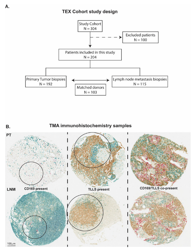
2.2. Patient Cohorts and Study Design

Two patient cohorts were used in this study; the first cohort was a retrospective cohort study based on primary tumors and lymph node metastases from patients with locally advanced and metastatic breast cancer from the randomized phase III TEX trial performed between 2002–2007 [28]. Detailed information about the clinical trial is found at clinicaltrials.gov with identification number NCT01433614. Briefly, the clinical trial comprised 304 women with advanced or inoperable metastatic breast cancer. Participants received two types of combination chemotherapy as the first line of treatment: Epirubicin and Paclitaxel alone or combined with Capecitabine. Among several criteria that have been described in detail previously [28], the enrolled participants had to have a life expectancy of at least 3 months, no brain metastases and they were not permitted to join the study if they had performed previous chemotherapy treatment cycles. From the 304 participants, formalin-fixed, paraffin-embedded blocks from primary tumors and synchronous lymph node metastases were collected wherever possible for tissue microarray (TMA) construction, as described previously [29], enabling further analysis of the tissue with immunohistochemistry (IHC). A simplified study design of the cohort is illustrated in Figure 1A.
The second cohort was a broad prospective, population-based cohort used in order to validate our findings from the smaller cohort using RNA sequencing data. This cohort was compromised of 8164 patients enrolled in the Sweden Cancerome Analysis Network—Breast (SCAN-B) initiative [30], and was approved by the regional ethical review board in Lund, Sweden. Detailed information from the cohort is found at ClinicalTrials.gov with identification number NCT02306096. Fresh biopsy samples were taken from each patient during the primary surgery by pathologists performing their routine clinical diagnostics. All analyses were performed in accordance with patient consent and ethical regulations, and the biopsies were used to gather RNA sequencing data.

2.3. Immunohistochemistry and Scoring

IHC was performed on the TMA cohort where all primary tumor and lymph node metastases were scored for the different immune cell surface markers, CD169, CD20 and CD3, using the protocol previously described [29,31]. FoxP3 had been annotated previously [31]. In brief, TMA blocks were sectioned to a thickness of 4 mm prior to mounting. The sections contained cores with diameters of 800 μm and were pre-treated with the PT-link system before staining with an Autostainer Plus (DAKO, Santa Clara, CA, USA) at pH6 with an overnight staining protocol. The following antibodies and dilutions were used for staining: anti-CD169+ macrophages (1:100, Invitrogen, Clone SP216, Waltham, MA, USA), anti-CD20+ B-cells (1:100, Abcam, Clone L-26, Cambridge, UK), anti-CD3+ T-cells (1:100, Abcam, Clone 11084, Cambridge, UK) and developed with a triple staining IHC kit from Abcam. A previous staining performed by authors (J.S. and C.H.) used mouse monoclonal anti-FoxP3 (ab20034, clone 236A/E7, Abcam, 1:400, Cambridge, UK) to annotate Tregs as previously published [31].
CD3+ T-cells, CD20+ B-cells and CD169+ macrophages were annotated individually by authors O.B., E.K. and K.L. The following scores were used: for CD169, CD169+ expression present = 1, and CD169+ expression absent = 0; for CD20, CD20+ clusters in spatial contact with CD3+ T-cells present = 1, and CD20+ absent or present as dispersed single cells (not in clusters) or without spatial contact with CD3+ T-cells = 0. Since we did not include a follicular dendritic cell marker, the CD20+/CD3+ B/T cell clusters will be referred to as TLLS, and not TLS. The purpose of this scoring was to classify immune cell infiltration into three different categories: (1) CD169+ macrophages positive tumors/metastases; (2) TLLS positive tumors/metastases (CD20+/CD3+); (3) tumors/metastases with presence of CD169+ and TLLS (CD169+/CD20+/CD3+), as represented in Figure 1B.
Treg (FoxP3+) annotations were published previously and performed by authors J.S. and C.H. [31]. The Tregs scoring strategy ranged from 0–3 and furthermore also categorized absence–presence (0–1). In the present study, the Tregs (FoxP3+) (0–1) score was used, solely exploring correlation between presence or absence of Tregs and its effect within the three different immune cell infiltration categories (CD169+, TLLS, CD169+/TLLS).

2.4. Statistical Analyses

Statistical analyses were performed with IBM SPSS statistics (version 27), with all statistical tests being two-sided with p ≤ 0.05 considered as significant results. In the TMA cohort, age at diagnosis ranged from 27 to 71 years old with an overall median age at diagnosis of 51 years. A total of 21% of included patients were alive at the time of data collection (July 2013) and the median follow-up time for patients alive was 10.5 years. Age, tumor size, lymph node status, metastatic stage, PT receptor status, lymph node receptor status and adjuvant therapy given are presented in Table 1.
Correlations between clinicopathological factors and immune cell infiltration in PTs and LNMs were assessed using cross tabulation tables. Odds ratios with a 95% confidence interval were correlated to 5-year recurrence-free interval (RFI), 5-year breast-cancer-specific survival (BCSS), tumor sizes above 20 mm, expression of the receptors (ER, PR, HER2), high Ki67 levels (>15%) and presence of Tregs, TLLSs or CD169+ macrophages. All clinicopathological factors were set as binary values; thus, significant correlations with immune cell infiltration were analyzed with the chi-square test or with Fisher’s exact test when fewer observations than 20 were seen.
The prognostic outcome of immune cell infiltration was analyzed with Kaplan–Meier plots and log-rank tests to exclude the null hypothesis of equal prognostic effect for BCSS or RFI based on specific immune cell infiltration in tumor tissues. Effects on BCSS and RFI were calculated based on infiltration of CD169+ macrophages alone, TLLSs alone, or dual infiltration of CD169+ macrophages and TLLSs (CD169+/TLLS). The follow-up data from the TMA cohort enabled a long time to event scale for BCSS and RFI, since all patients had presented with locally advanced or metastatic disease at the time of inclusion into the clinical trial. The time to event scale for BCSS and RFI in the current analysis was censored at 25 years after primary tumor diagnosis. For LNM biopsies, the time to event scale for BCSS and RFI was set to 10 and 25 years after diagnosis in order to focus on both early and long-term prognostic effects, since lymph node metastases were present at primary diagnosis in the majority of patients in the TEX cohort.
Univariable followed by multivariable Cox regression analyses were also performed to estimate hazard ratios (HRs) for recurrence or death from breast cancer according to CD169+, TLLS and CD169+/TLLS infiltration in PTs and LNMs. The same time to event scale as for the Kaplan–Meier analyses was maintained and the multivariable models accounted for hormone receptor/growth factor expression status (ER, PR, Her2), Tregs presence, Ki67 levels, tumor size and age at primary diagnosis. Results were illustrated using forest plots showing HRs with a 95% confidence interval.

2.5. Gene Expression Analyses

Gene expression analyses of TLS gene signature [32], Breg signature [33], Treg (FoxP3) signature [34] and CD169+ TAMs (SIGLEC1) were performed using RNA sequencing data from the SCAN-B cohort, following the same procedure as previously described [30,35]. Expression data were extracted as fragments per kilobase per million reads for each case and transformed into a logarithmic scale. Five gene classifiers representing different subtype predictors were used to classify samples into the intrinsic breast cancer subtypes according to the PAM50 gene signature [36]. Prior to analysis, a batch correction was performed via ComBat in order to remove potential bias associated with technical variations. After correction, the data were uploaded unto The Institute for Genomic Research MultiExperiment Viewer (TIGR MeV) version 3.1, and differences in gene expression were determined through hierarchical clustering using median-centered gene correlations where status 1 or above represented upregulated expression and -1 or below represented downregulated expression. RNA sequencing results are presented with heat maps, showing Pearson correlation distance and complete hierarchal clustering linkages.

3. Results

3.1. CD169+ TAMs Associate with TLLSs in PTs

Since SCS CD169+ macrophages in lymph nodes have a functional localization surrounding B cell follicles and are associated with a beneficial prognosis in cancer patients, we first set out to investigate the localization pattern of PT-infiltrating CD169+ macrophages (CD169+ TAMs) in relation to CD20+/CD3+ B/T cell clusters (referred to as TLLSs) in primary breast cancer tumors (Figure 1B).
Localization patterns were investigated with odds ratios (ORs), as shown in Table 2 and Table S1. Firstly, CD169+ TAMs in PTs were indeed correlated with the presence of TLLSs (OR = 3.77, p = 0.004) and furthermore showed a trend for Treg infiltration (OR = 2.06, p = 0.057). CD169+ TAMs also correlated with B cells as only marker, unrelated to TLLSs (OR = 5.26, p = 0.017). In LNMs, CD169+ presence (CD169+ LNM) was found to also correlate with TLLS presence (OR = 4.76, p = 0.0001) and Treg infiltration (OR = 2.87, p = 0.046).
CD169+ LNMs further correlated with decreased odds of tumor size above 20 mm (OR = 0.42, p = 0.041) and increased odds of surviving beyond 5 years (OR = 2.20, p = 0.045), while CD169 in PTs (CD169+ TAMs) correlated with high Ki67 levels (OR = 2.33, p = 0.021) (Table 2). This was in line with our previously published data using another breast cancer cohort regarding CD169+ infiltration in PTs (CD169+ TAM) and LNMs (CD169+ LNM) [27]. CD169+ TAMs also correlated with decreased odds of tumor size above 20 mm (OR = 0.47, p = 0.019) and decreased odds for expression of ER (OR = 0.28, p = 0.0001).
Additionally, in PTs, the presence of CD169+/TLLS structures showed a trend towards association with decreased odds of surviving the first 5 years (OR = 0.29, p = 0.053), decreased expression of ER in both PTs and LNMs (ORPT-ER = 0.26, p = 0.042, ORLNM-ER = 0.10, p = 0.051), and high Ki67 levels in PTs (OR = 5.43, p = 0.018). In sharp contrast, CD169+ /TLLS in LNMs was significantly correlated with increased odds of surviving breast cancer the first 5 years (OR = 3.51, p = 0.005; Table S1).
To explore the potential univariable role of TLLS infiltration (CD20+/CD3+; “TLLS”) without CD169+ co-localization, further OR analysis was performed between TLLSs and other patient and tumor characteristics (Table S1). Results indicated that TLLSs in PTs per se (TLLS+ PT) was significantly correlated with reduced odds of breast-cancer-related death (OR = 0.41, p = 0.045) and lower odds for recurrence (OR = 0.31, p = 0.018) within 5 years after diagnosis. Furthermore, TLLS was significantly correlated with greater odds for infiltration of Tregs (FoxP3) (OR = 8.54, p = 0.001) into PTs. Again, and in contrast, TLLSs in the LMNs per se (TLLS+ LNM) was significantly correlated with increased odds of surviving beyond 5 years (OR = 2.63, p = 0.018), and decreased odds of PR expression in LNM (OR = 0.43, p = 0.044).
In summary, CD169+ macrophages were associated with TLLSs both in PTs and in LNMs, but when present in PTs this was associated to a worse prognosis for the patients, the opposite of what was seen in LNMs.

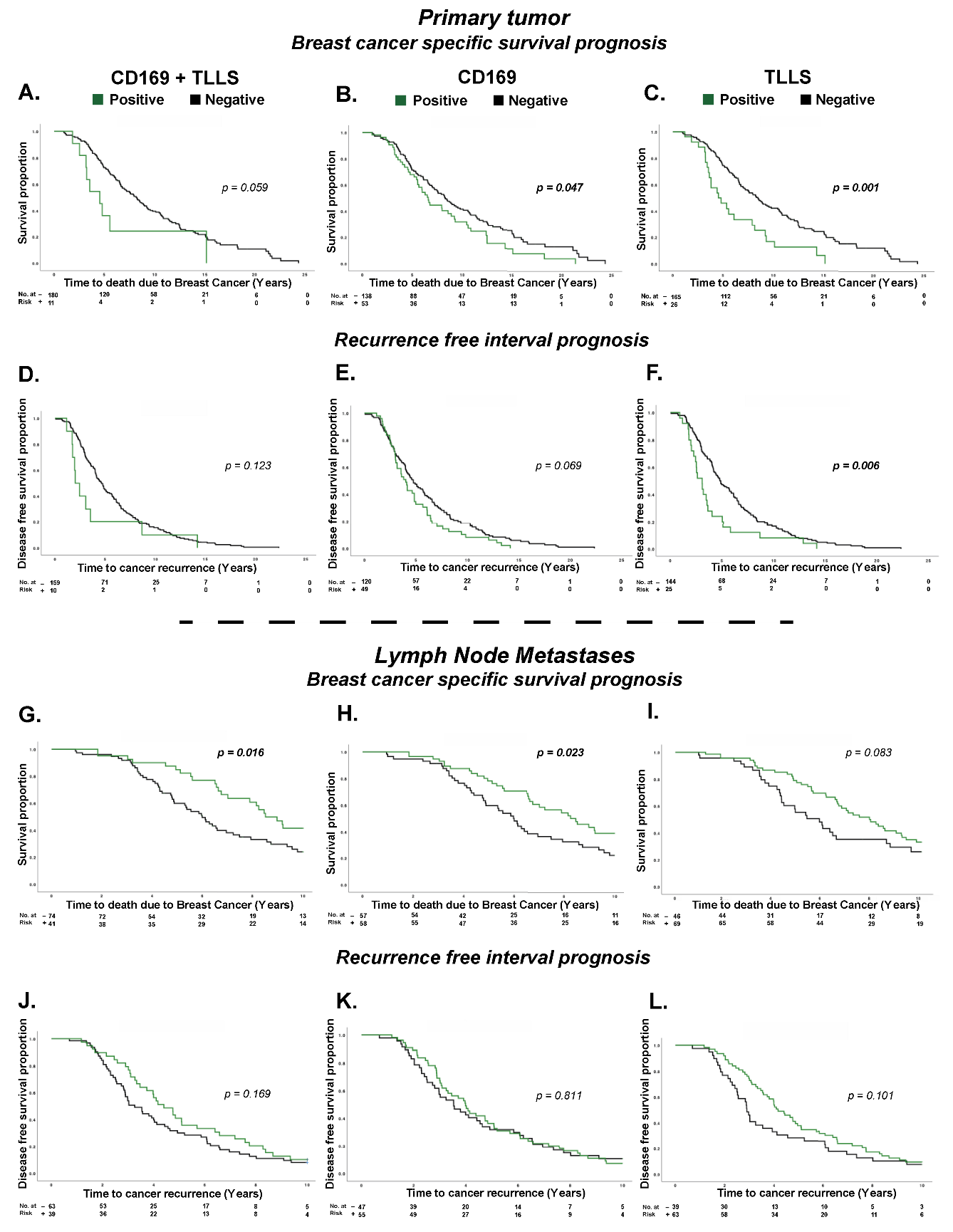
3.2. CD169+ TAMs and TLLSs as Prognostic Markers for Breast Cancer Patients

Investigating prognostic impact, Kaplan–Meier plots for each variable (CD169+/TLLS; CD169+; TLLS) showed unique survival patterns. In general, a worse prognosis was seen with CD169+ and TLLS infiltration in PTs, while infiltration in LNMs showed a better prognosis. In PTs, CD169+/TLLS co-infiltration was a borderline prognostic marker associated with worse BCSS (p = 0.059) (Figure 2A). To estimate if the observed effect was caused by the dual infiltration pattern (CD169+/TLLS), or was solely from one type of cell infiltration (CD169+ or TLLS), individual Kaplan–Meier plots with corresponding log-rank tests were performed. Both CD169+ infiltration (p = 0.047) (Figure 2B) and TLLS infiltration (p = 0.001) (Figure 2C) in PTs showed evidence for an adverse BCSS. A similar significant inferior outcome regarding RFI was seen only for TLLS infiltration (p = 0.006) (Figure 2D–F). Infiltrating B cells as only variable (CD20) however, did not have an impact on survival (BCSS p = 0.38; RFI p = 0.82).
Conversely, CD169+/TLLS presence in LNMs showed significant evidence of improved BCSS in the first 10 years (p = 0.016) (Figure 2G). Individually, CD169+ LNM presence remained significant (p = 0.023), while TLLSs showed weaker evidence (p = 0.083) for a beneficial prognostic effect on BCSS (Figure 2H,I). For RFI, no statistically significant correlations were observed for any type of immune cell infiltration; the survival curves, however, did trend towards longer RFI upon TLLS infiltration alone or CD169+/TLLS co-presence, suggesting a potential beneficial prognostic effect (Figure 2J–L). The beneficial prognostic effects for CD169+ and TLLS in LNMs, however, were lost in the long-term 25-year follow-up (Figure S1).
Because the prognostic effect was opposite that based on tumor localization (PT vs. LNM), we further investigated the prognostic impact for patient matched biopsies. Interestingly, the results suggested that CD169+ TAM infiltration in PTs was relevant as a prognostic factor only if CD169+ macrophages in LNMs were absent, and vice versa. The same finding was true for TLLSs (Figure S2).
Hence, the observed opposite prognostic effects in PTs and LNMs were seen both for individual (CD169+ or TLLS) and dual infiltration patterns (CD169+/TLLS).

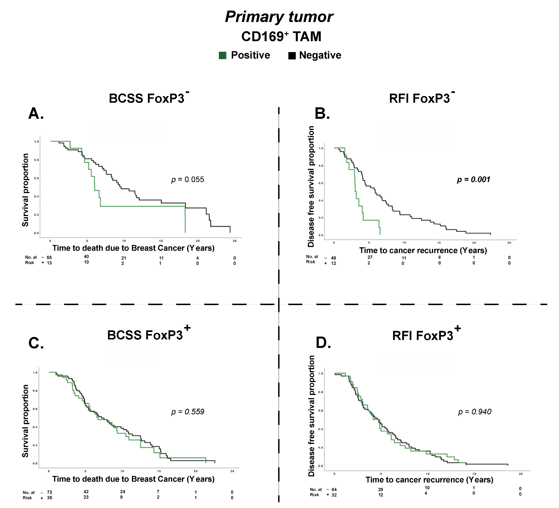
3.3. Treg Infiltration Impacts the Prognostic Effect of CD169+ TAMs

In previous research using the TMA cohort, Treg infiltration in PTs was found to be an independent prognostic factor for decreased BCSS, but the prognostic effect was lost in LNMs [31]. In line with this, in the present study we show that CD169+ TAMs trended towards an association with infiltration of Tregs (OR = 2.06, p = 0.057), and that TLLSs correlated with Tregs (OR = 8.54, p = 0.001) (Table 2 and Table S1). Interestingly, CD169+/TLLS dual infiltration in PTs (CD169+/TLLS PT) was significantly associated with the opposite, meaning a decreased presence of Tregs (FoxP3+) in PTs (OR = 0.59, p = 0.007) (Table S1). To interpret this, we further investigated the impact of CD169+ TAMs and TLLS infiltration based on a FoxP3+ (Treg) strata in PT biopsies only. To our surprise, we found that PTs lacking Treg infiltration also always lacked CD169+/TLLS dual infiltration, implying that co-infiltration of CD169+ TAMs and TLLSs is necessary for the presence of Tregs, and vice versa. Therefore, Kaplan–Meier plots with corresponding log-rank tests could not be performed with a FoxP3+ strata for CD169+/TLLS and TLLs. Importantly, however, individual analysis of CD169+ infiltration alone (CD169+ TAM) associated with shortened RFI (PRFI = 0.001) and a trend towards association with shortened BCSS (PBCSS = 0.055) only in the absence of FoxP3+ Tregs in PTs (Figure 3A,B). Hence, the prognostic effects of CD169+ TAMs alone for BCSS and RFI were completely lost in the FoxP3+ Tregs positive patient strata (Figure 3C,D).
Altogether this suggests that co-infiltration of CD169+/TLLS may be necessary for the presence of Tregs, and that the presence of CD169+ TAMs alone may only have a prognostic impact in breast tumors lacking Tregs.

3.4. CD169+ TAMs and TLLSs Show Unique Independent Prognostic Effects

Multivariable Cox regression analyses were done to compare the effects from each type of cell infiltration biomarker adjusted for several potential confounders. Included confounders taken into account were: age, nodal status, tumor size, Ki67, receptor status (ER, PR, HER2), Tregs presence, TLLS (CD20+/CD3+) presence and CD169+ macrophage presence (Figures S3 and S4). The prognostic impact was calculated with HR with a 25-year timeline in PT samples and a 10-year timeline in LNM samples for BCSS and RFI. For BCSS, after multivariable adjustments, dual infiltration of CD169+/TLLS in PTs showed an independent HR value correlating to a worse prognosis (HR = 2.88, 95%CI: (1.33–6.2), p = 0.007), hence even stronger than the univariable effect (HR = 1.90, 95%CI: (0.97–3.75), p = 0.063) (Figure S3A). In contrast, in LNMs, CD169+/TLLS dual infiltration was correlated with improved survival in univariable analysis only (HR = 0.54, 95%CI: (0.33–0.90), p = 0.017). In the multivariable analysis, CD169+/TLLS dual infiltration in LNMs showed a similar trend but with weaker statistical evidence (HR = 0.45, 95%CI: (0.20–1.02), p = 0.057) (Figure S3B). CD169+ and TLLS infiltration were next analyzed separately. Multivariable adjustments further strengthened TLLSs as an independent prognostic marker in PTs (HR TLLS = 1.73, 95%CI (1.03–2.93), p = 0.040), while evidence for CD169+ TAMs as a prognostic factor in PTs decreased (HR CD169 = 1.07, 95%CI: (0.67–1.71), p = 0.77) compared to their univariable effects (HR TLLS = 2.14, 95%CI: (1.38–3.35), p = 0.001; HR CD169 = 1.43, 95%CI (1.002–2.04), p = 0.049) (Figure S3C,E). In contrast to PTs, multivariable analyses of CD169+ LNMs showed both stronger HRs and stronger correlation to decreased risk of death from breast cancer, while evidence for TLLS infiltration being an independent prognostic factor decreased (HR CD169 = 0.48, 95%CI: (0.23–0.99), p = 0.046; HR TLLS = 0.72, 95%CI: (0.40–1.31), p = 0.28) compared to their univariable effects (HR CD169 = 0.59, 95%CI: (0.37–0.93), p = 0.025; HR TLLS = 0.66, 95%CI: (0.41–1.06), p = 0.085) (Figure S3D,F).
Regarding the independent prognostic impact on RFI, it was clear that the prognostic effect was not as important as for BCSS. Multivariable analysis for CD169+ TAMs and TLLS infiltration, respectively, showed non-significant HR correlations in both PTs and LNMs. However, CD169+/TLLS dual infiltration in PTs was significantly correlated with higher HRs of recurrence (HR = 2.15, 95%CI: (1.06–4.38), p = 0.035) (Figure S4).
Our data thus showed that CD169+/TLLS dual infiltration in PTs of advanced breast cancer patients was an independent prognostic marker with regards to both BCSS and RFI.

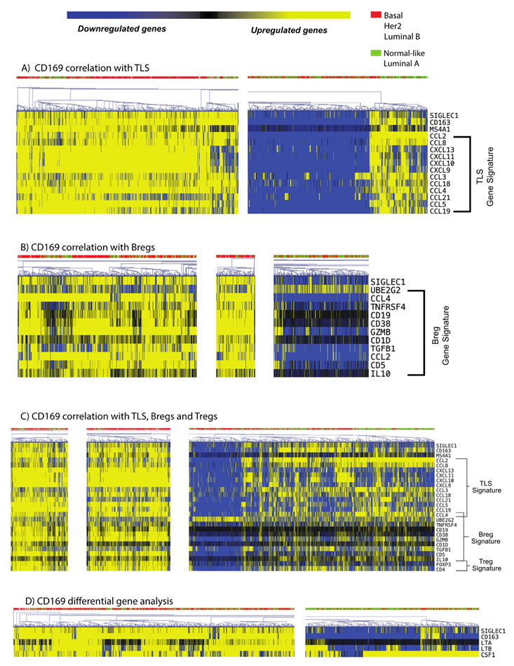
3.5. CD169+ TAMs Associate with Both Mature TLS and Breg Gene Signatures

To investigate whether the TLLSs associating with CD169+ TAMs in PTs were functional tertiary lymphoid follicles, and to confirm the association between CD169+ TAMs and TLLSs in breast cancer, gene signatures of mature tertiary lymphoid structure (TLS) [32] from bulk RNAseq from 8164 patients of the SCAN-B cohort were investigated, allowing analysis in a larger, contemporary and representative cohort. Initial results (Figure 4A), showed that CD169+ expression (gene name, SIGLEC1) indeed correlated with a mature TLS gene signature for a specific cluster of patients, indicating functional TLS formations. This specific cluster also had upregulated levels of the gene MS4A1, encoding CD20. For all clusters, the molecular subtype, although being dominant for more aggressive subtypes (luminal B, HER2-enriched, basal), was not restricted to one subtype cluster. This implies that TLS formation can occur in all subtypes of breast cancer.
To further investigate the possible immunological role of the CD169+ TAMs in the PT, we analyzed other immunoregulatory gene signatures [33,34]. CD169+ TAM upregulated clusters correlated with the Bregs gene signature (Figure 4B). Notably, a cluster of patients with CD169+ TAMs further associated with TLS, Breg and Treg gene signatures (Figure 4C), indicating an immunosuppressive function for these TLSs. More importantly, gene signatures for TLS formation were not seen in patient clusters that lacked CD169+ TAMs, and the same observations were true for Treg and Breg gene signatures. Lastly, CD169+ TAM positive clusters also associated with genes important for CD169+ macrophage biology, such as CD163, CSF-1, LTA and LTB (Figure 4D). Complete heatmaps with the highlighted clusters are presented in Figure S5.
In summary, this implies that CD169+ TAMs are closely connected to mature TLS formation, and Treg and Breg infiltration, in PTs of breast cancer patients.

4. Discussion

In secondary lymphoid organs (SLOs), the CD169+ SCS lymph node macrophages surround B cell follicles to aid in antigen delivery and to regulate immune responses [19]. Our initial hypothesis was therefore that CD169+ TAMs would associate with TLLSs in PTs, just like CD169+ SCS macrophages do in SLOs, hence aiding or regulating immune activity. CD169 is primarily expressed on activated monocytes and macrophages, with occasional expression on T cells and mature dendritic cells (DCs) [37,38]. Here, co-stainings with CD3 ruled out T cells expressing CD169; however, we cannot exclude that follicular DCs could potentially express microvesicles containing CD169 derived from macrophages [39]. Our original published data, which were supported by this present study, showed that CD169+ TAMs infiltrating PTs were indeed associated with a worse prognosis for breast cancer patients [27]. We here show that CD169+ TAMs infiltrating PTs actually do associate with TLLSs in PTs, similar to the CD169+ SCS resident lymph node macrophages and B-cell follicles in SLOs. We show that CD169+ macrophages are in close spatial association with TLLSs in PTs, and surprisingly also with presence of Tregs. In spleen, CD169+ macrophages are dependent on lymphotoxin α1ß1 generated from B-cell follicles [40,41]. An explanation for the co-localization of CD169+ TAMs and TLLSs in breast tumors could therefore be a local secretion of lymphotoxin α1ß1 in tumors with TLSs. Indeed, our bulk RNA-sequencing data from the SCAN-B cohort showed that upregulation of CD169/SIGLEC1 also correlates with LTA1 and LTB1 upregulation.
Both CD169+ TAMs and TLLSs in PTs were clearly associated with a worse prognosis, in contrast to CD169+ macrophages present in LNMs, which had a beneficial effect on prognosis. This finding is in disagreement with previous literature where TLS presence in primary breast tumors was a positive prognostic factor [42,43], as a meta-analysis has shown TLS-presence to generally be associated with a beneficial prognosis in breast cancer [43]. However, in previous studies, the beneficial effect of TLSs as a prognostic marker in PTs was highly dependent on breast cancer molecular subtypes (HER2 amplified [44] or TNBC [45]). These data should be put into relation with the TMA cohort used in the present study, which was comprised mostly of luminal tumors (n = 143) and very few HER2+ (n = 9) and TNBC (n = 24) tumors. All the patients used in the TMA cohort, furthermore, had developed metastatic disease and therefore had a poor prognosis. This could indicate differential impact of TLS depending on the molecular subtype or due to the advanced stage. One observation supporting this was made by Figenschau et al. [46], who verified that tumors with a higher level of tumor infiltrating immune cells correlated with intra-tumoral TLS formation, higher tumor grade and a higher degree of inflammation, thus leading to worse prognosis. We also found that presence of CD169+ LNM and CD169+ PT showed lower odds of having a large tumor size. This is surprising given the worse prognosis seen for CD169+ TAMs in PTs, although when adjusting for multiple variables the prognostic effect of CD169+ PT was lost. Nonetheless, these findings could indicate a more aggressive behavior and hence microenvironment of primary tumors with CD169+ TAMs already at a low tumor size in this cohort with advanced breast cancer patients. Lastly, a general difficulty when investigating TLSs in lymph node metastases is the lobular structure of the SLO, since secondary lymphoid follicles may be difficult to separate from TLSs in the sectioned lymph node metastases. How to discriminate TLS from secondary lymphoid follicles will be an important issue to solve for the TLS immune oncology field in the future.
Using RNA sequencing data from the large SCAN-B breast cancer cohort, we showed that SIGLEC1 (CD169) expression in primary breast tumors clustered with the expression of functional TLS signatures, indicating that CD169+ TAMs actually do associate with mature tertiary lymphoid follicles also in primary tumors. A fraction of these were enriched for Breg and Treg signatures, thus possibly inducing immunosuppression and adverse prognostic effects in breast cancer patients. This would be supported by a recent study showing that the presence of TLSs with Breg and Treg infiltration was associated with a worse prognosis in primary breast tumors from invasive ductal carcinoma (IDC) and ductal carcinoma in situ (DCIS) [47]. In the present study, we evaluated this hypothesis using RNA sequencing data. In fact, there was a clear subcluster of patients with high transcript levels for CD169+, TLS gene signatures, and Breg and Treg signatures, possibly leading to immunosuppression and hence a worse prognosis. Furthermore, TLS and Breg gene signature transcripts were only present for subsets of patients with a higher infiltration of CD169+ TAMs, implying that CD169+ TAMs, TLS and Bregs may interact. On the other hand, patients with both CD169 and TLS signatures, presenting with lower expression of the Bregs signature, were numerous in comparison. The significance of these findings in relation to outcome will be interesting to evaluate and will be a future goal.
Another important result from our study was the correlation between TLS, CD169+ TAMs and FoxP3+ Tregs. We found a strong correlation between FoxP3+/CD4+ Treg signatures and CD169+ expression (SIGLEC1) in the SCAN-B cohort, and the Treg gene signature also correlated with TLS and Breg gene signatures. Indeed, based on our IHC results using the TMA cohort, TLS infiltration was almost exclusively present when Tregs were also present in PTs. In fact, several papers have shown that the presence of Tregs is associated with a worse prognosis in breast cancer [31,48,49]. As the main purpose of Tregs is to suppress lymphocytes, it is likely that FoxP3+ Treg infiltration is a natural feedback response following high levels of TLLS formation, suppressing these lymphocytes. A previous in vivo study investigating the correlation between Tregs and TLS in lung cancer showed that Tregs actively suppress the anti-tumor response from TLSs. In the same model, Tregs depletion reversed this effect and led to T-cell expansion starting at the TLS sites and promoting tumor destruction [50]. It was also recently shown that tumors affect local lymph node immune tolerance epigenetically via type I IFNs, eventually promoting distant metastasis facilitated by tumor-antigen specific Tregs in a malignant melanoma model [51]. As lymph node CD169+ macrophages are known type I IFN producers [12], the correlation seen here between CD169+ macrophages and Tregs could possibly be involved in this epigenetic reprograming [51]. Even more interesting is that Tregs infiltration is associated with a higher risk of death and relapse, especially for ER positive breast cancer patients [49], which is in line with our TMA cohort that was predominantly of luminal subtype. The presence of Tregs likely inhibits reactivation of T-cell responses; thus, despite the presence of TLLSs, the immune system is unable to counteract tumor growth. A final interesting finding regarding Tregs from the present study was that co-infiltration of CD169+ TAMs/TLLS is necessary for the presence of Tregs, and that CD169+ TAMs alone only had a prognostic impact in tumors lacking Tregs. This most probably means that Tregs have a dominant functional role over CD169+ TAMs. CD169+ TAMs may also play another important role here in relation to Tregs infiltration, as it has been shown that CD169+ macrophages can upregulate CCL22 upon interaction with apoptotic cells, leading to FoxP3+ Treg recruitment via a CCL22/CCR4-mediated chemotaxis gradient [52]. The same mechanism might apply within breast cancer patients with high tumor grade, with an abundance of apoptotic cells or necrotic cells, thus recruiting CD169+ TAMs which release CCL22 and recruit Tregs.
Lastly, the results we obtained, where the presence of CD169+ TAMs and TLLSs were correlated with prognosis only for patients who had immune cell infiltration in either PT or in LNM, but not for both, are interesting. Subsequently, patients who had CD169+ TAMs or TLLS infiltration in both PT and LNM had similar survival compared to patients that lacked the same in both PT and LNM. This result either implies that the harmful prognostic effect of immune cell infiltration seen in PTs cancels out the beneficial prognostic effect seen in LNMs, or that infiltration of CD169+ TAMs into PTs is concurrent with the depletion of CD169+ macrophages from the LNMs in breast cancer patients with a higher tumor grade, therefore giving rise to worse prognosis within our cohort that only includes patients with advanced breast cancer.

5. Conclusions

In conclusion, CD169+ macrophages present in breast cancer PTs and LNMs correlate to the presence of TLS, Treg and Breg signatures for a subset of patients. This was associated to worse survival when present in PTs, while conferring a better prognosis when present in LNMs (Figure 5). We propose that attraction and polarization of CD169+ TAMs occur in tumors where formation of TLSs occurs, and that these inflamed environments cause enrichment of immunosuppressive regulatory lymphocytes, Tregs and Bregs, thus fueling even more immunosuppressive environments and breast tumor progression.

Supplementary Materials

The following supporting information can be downloaded at: https://www.mdpi.com/article/10.3390/cancers15041262/s1, Figure S1: Kaplan–Meier survival showing the differences in 25-year breast cancer specific survival (BCSS) and recurrence free interval (RFI) for specific immune cell populations infiltrating lymph node metastases (LNMs); Figure S2: Kaplan–Meier analyses performed on matched donors; Figure S3: Forest plots with Cox regression analyses (BCSS); Figure S4: Forest plots with Cox regression analyses (RFI); Figure S5: Complete heat maps containing sample hierarchical clusters with Pearson correlations between SIGLEC1 (CD169) and gene signatures for tertiary lymphoid structures (TLSs), Bregs or Tregs; Table S1: Odds ratio table comparing CD169+/TLLS+ co-infiltration and TLLS+ infiltration with tumor and metastasis clinicopathological features and other immune cell infiltration.

Author Contributions

The work reported in the paper was performed by the authors. O.B., E.K. and K.L. performed the annotation of CD169, CD20 and CD3 IHC in the TEX cohort. O.B. was responsible for managing the data analyses in SPSS and TIGR MeV, for interpretation of results and drafting of the manuscript. S.K., I.H. and T.H. collected clinicopathological traits and outcome data for all patients and constructed TMAs for the TEX cohort. S.V. supported the RNA sequencing analysis and batch correction in the SCAN-B cohort. J.S. and C.H. were responsible for the annotation and analysis of FoxP3 in the TEX cohort. T.H. was responsible for the design of the TEX cohort and follow-up. K.L. was responsible for designing this study, interpretation of the results (together with OB) and drafting of the manuscript. All authors have read and agreed to the published version of the manuscript.

Funding

This work was generously supported by grants from the Swedish Research Council (2020-01842), the Swedish Cancer Foundation (21 1392 Pj 01 H), Malmö Allmänna Sjukhuset (MAS) för bekämpande av Cancer Foundation (2022-11-07), regional grants from the Swedish state under the agreement between the Swedish government and the county councils, (the ALF agreement) (13734 ALF Proj K Leandersson), the Gyllenstiernska Krapperups Foundation (KR2019-0078), and the B Kamprad Foundation (FBKS-2018-14, FBKS-2020-9, and FBKS-2022-35).

Institutional Review Board Statement

This study was conducted in accordance with the Declaration of Helsinki, and ethical approvals for the clinical trials were obtained from the regional ethics committees in Sweden: Stockholm, Dnr KI 02-206 and KI 02-205 (date of approval 14 November 2002), and Lund Dnr 2009/658 (date of approval 17 December 2009) [28,29].

Informed Consent Statement

A written informed consent was received from the included patients in the clinical trials presented in this study.

Data Availability Statement

Gene expression data are available at Mendeley Data as a publicly accessible dataset (https://data.mendeley.com/datasets/yzxtxn4nmd, accessed on 17 May 2022) (https://doi.org/10.17632/yzxtxn4nmd.1) [49].

Acknowledgments

For the TMA cohort used in this study, the authors thank the patients and the TEX Trialists Group‘s Coordinating Investigator, Thomas Hatschek. The authors also thank the patients, clinicians and hospital staff participating in the SCAN-B study; the staff at the central SCAN-B laboratory at the Division of Oncology, Lund University; the Swedish National Quality Register for Breast Cancer (NKBC); the Regional Cancer Center South; and the South Swedish Breast Cancer Group (SSBCG).

Conflicts of Interest

K.L. is a board member of Cantargia A.B., a company developing IL1RAP inhibitors. Cantargia A.B. had no role in the funding of the study or in the design of the study; in the collection, analyses or interpretation of data; in the writing of the manuscript; or in the decision to publish the results. The authors otherwise declare no competing interest. The funders had no role in the design of the study; in the collection, analyses or interpretation of data; in the writing of the manuscript; or in the decision to publish the results.

References

  1. World Health Organization. Cancer. Available online: https://www.who.int/news-room/fact-sheets/detail/cancer (accessed on 4 March 2022).
  2. Aysola, K.; Desai, A.; Welch, C.; Xu, J.; Qin, Y.; Reddy, V.; Matthews, R.; Owens, C.; Okoli, J.; Beech, D.J.; et al. Triple Negative Breast Cancer—An Overview. Hered. Genet. Curr. Res. 2013, 2013 (Suppl. S2), 1. [Google Scholar]
  3. Andrahennadi, S.; Sami, A.; Manna, M.; Pauls, M.; Ahmed, S. Current Landscape of Targeted Therapy in Hormone Receptor-Positive and HER2-Negative Breast Cancer. Curr. Oncol. 2021, 28, 1803–1822. [Google Scholar] [CrossRef] [PubMed]
  4. Darvin, P.; Toor, S.M.; Sasidharan Nair, V.; Elkord, E. Immune checkpoint inhibitors: Recent progress and potential biomarkers. Exp. Mol. Med. 2018, 50, 1–11. [Google Scholar] [CrossRef] [PubMed] [Green Version]
  5. Michel, L.L.; von Au, A.; Mavratzas, A.; Smetanay, K.; Schütz, F.; Schneeweiss, A. Immune Checkpoint Blockade in Patients with Triple-Negative Breast Cancer. Target. Oncol. 2020, 15, 415–428. [Google Scholar] [CrossRef]
  6. Tang, H.; Qiao, J.; Fu, Y.-X. Immunotherapy and tumor microenvironment. Cancer Lett. 2016, 370, 85–90. [Google Scholar] [CrossRef] [Green Version]
  7. Tang, T.; Huang, X.; Zhang, G.; Hong, Z.; Bai, X.; Liang, T. Advantages of targeting the tumor immune microenvironment over blocking immune checkpoint in cancer immunotherapy. Signal Transduct. Target. Ther. 2021, 6, 72. [Google Scholar] [CrossRef]
  8. Panni, R.Z.; Linehan, D.C.; DeNardo, D.G. Targeting tumor-infiltrating macrophages to combat cancer. Immunotherapy 2013, 5, 1075–1087. [Google Scholar] [CrossRef] [Green Version]
  9. Cortez-Retamozo, V.; Etzrodt, M.; Newton, A.; Rauch, P.J.; Chudnovskiy, A.; Berger, C.; Ryan, R.J.; Iwamoto, Y.; Marinelli, B.; Gorbatov, R.; et al. Origins of tumor-associated macrophages and neutrophils. Proc. Natl. Acad. Sci. USA 2012, 109, 2491–2496. [Google Scholar] [CrossRef] [Green Version]
  10. Zhang, C.; Yang, M.; Ericsson, A.C. Function of Macrophages in Disease: Current Understanding on Molecular Mechanisms. Front. Immunol. 2021, 12, 620510. [Google Scholar] [CrossRef]
  11. Wu, K.; Lin, K.; Li, X.; Yuan, X.; Xu, P.; Ni, P.; Xu, D. Redefining Tumor-Associated Macrophage Subpopulations and Functions in the Tumor Microenvironment. Front. Immunol. 2020, 11, 1731. [Google Scholar] [CrossRef]
  12. Liu, Y.; Xia, Y.; Qiu, C.-H. Functions of CD169 positive macrophages in human diseases (Review). Biomed. Rep. 2021, 14, 26. [Google Scholar] [CrossRef]
  13. Uhlén, M.; Fagerberg, L.; Hallström, B.M.; Lindskog, C.; Oksvold, P.; Mardinoglu, A.; Sivertsson, Å.; Kampf, C.; Sjöstedt, E.; Asplund, A.; et al. Tissue-based map of the human proteome. Science 2015, 347, 1260419. [Google Scholar] [CrossRef]
  14. Mondor, I.; Baratin, M.; Lagueyrie, M.; Saro, L.; Henri, S.; Gentek, R.; Suerinck, D.; Kastenmuller, W.; Jiang, J.X.; Bajenoff, M. Lymphatic Endothelial Cells Are Essential Components of the Subcapsular Sinus Macrophage Niche. Immunity 2019, 50, 1453–1466.e54. [Google Scholar] [CrossRef]
  15. Camara, A.; Cordeiro, O.G.; Alloush, F.; Sponsel, J.; Chypre, M.; Onder, L.; Asano, K.; Tanaka, M.; Yagita, H.; Ludewig, B.; et al. Lymph Node Mesenchymal and Endothelial Stromal Cells Cooperate via the RANK-RANKL Cytokine Axis to Shape the Sinusoidal Macrophage Niche. Immunity 2019, 50, 1467–1481.e66. [Google Scholar] [CrossRef]
  16. Martinez-Pomares, L.; Gordon, S. CD169+ macrophages at the crossroads of antigen presentation. Trends Immunol. 2012, 33, 66–70. [Google Scholar] [CrossRef]
  17. Shaabani, N.; Duhan, V.; Khairnar, V.; Gassa, A.; Ferrer-Tur, R.; Häussinger, D.; Recher, M.; Zelinskyy, G.; Liu, J.; Dittmer, U.; et al. CD169+ macrophages regulate PD-L1 expression via type I interferon and thereby prevent severe immunopathology after LCMV infection. Cell Death Dis. 2016, 7, e2446. [Google Scholar] [CrossRef] [Green Version]
  18. Gray, E.E.; Cyster, J.G. Lymph node macrophages. J. Innate Immun. 2012, 4, 424–436. [Google Scholar] [CrossRef]
  19. Louie, D.A.P.; Liao, S. Lymph Node Subcapsular Sinus Macrophages as the Frontline of Lymphatic Immune Defense. Front. Immunol. 2019, 10, 347. [Google Scholar] [CrossRef] [Green Version]
  20. Affandi, A.J.; Olesek, K.; Grabowska, J.; Nijen Twilhaar, M.K.; Rodríguez, E.; Saris, A.; Zwart, E.S.; Nossent, E.J.; Kalay, H.; de Kok, M.; et al. CD169 Defines Activated CD14(+) Monocytes With Enhanced CD8(+) T Cell Activation Capacity. Front. Immunol. 2021, 12, 697840. [Google Scholar] [CrossRef]
  21. Zhang, Y.; Li, J.Q.; Jiang, Z.Z.; Li, L.; Wu, Y.; Zheng, L. CD169 identifies an anti-tumour macrophage subpopulation in human hepatocellular carcinoma. J. Pathol. 2016, 239, 231–241. [Google Scholar] [CrossRef]
  22. Fraschilla, I.; Pillai, S. Viewing Siglecs through the lens of tumor immunology. Immunol. Rev. 2017, 276, 178–191. [Google Scholar] [CrossRef] [PubMed] [Green Version]
  23. Asano, T.; Ohnishi, K.; Shiota, T.; Motoshima, T.; Sugiyama, Y.; Yatsuda, J.; Kamba, T.; Ishizaka, K.; Komohara, Y. CD169-positive sinus macrophages in the lymph nodes determine bladder cancer prognosis. Cancer Sci. 2018, 109, 1723–1730. [Google Scholar] [CrossRef] [PubMed] [Green Version]
  24. Takeya, H.; Shiota, T.; Yagi, T.; Ohnishi, K.; Baba, Y.; Miyasato, Y.; Kiyozumi, Y.; Yoshida, N.; Takeya, M.; Baba, H.; et al. High CD169 expression in lymph node macrophages predicts a favorable clinical course in patients with esophageal cancer. Pathol. Int. 2018, 68, 685–693. [Google Scholar] [CrossRef] [PubMed] [Green Version]
  25. Ohnishi, K.; Yamaguchi, M.; Erdenebaatar, C.; Saito, F.; Tashiro, H.; Katabuchi, H.; Takeya, M.; Komohara, Y. Prognostic significance of CD169-positive lymph node sinus macrophages in patients with endometrial carcinoma. Cancer Sci. 2016, 107, 846–852. [Google Scholar] [CrossRef] [PubMed] [Green Version]
  26. Saito, Y.; Ohnishi, K.; Miyashita, A.; Nakahara, S.; Fujiwara, Y.; Horlad, H.; Motoshima, T.; Fukushima, S.; Jinnin, M.; Ihn, H.; et al. Prognostic Significance of CD169+ Lymph Node Sinus Macrophages in Patients with Malignant Melanoma. Cancer Immunol. Res. 2015, 3, 1356–1363. [Google Scholar] [CrossRef] [Green Version]
  27. Björk Gunnarsdottir, F.; Auoja, N.; Bendahl, P.-O.; Rydén, L.; Fernö, M.; Leandersson, K. Co-localization of CD169(+) macrophages and cancer cells in lymph node metastases of breast cancer patients is linked to improved prognosis and PDL1 expression. Oncoimmunology 2020, 9, 1848067. [Google Scholar] [CrossRef]
  28. Hatschek, T.; Carlsson, L.; Einbeigi, Z.; Lidbrink, E.; Linderholm, B.; Lindh, B.; Loman, N.; Malmberg, M.; Rotstein, S.; Söderberg, M.; et al. Individually tailored treatment with epirubicin and paclitaxel with or without capecitabine as first-line chemotherapy in metastatic breast cancer: A randomized multicenter trial. Breast Cancer Res. Treat. 2012, 131, 939–947. [Google Scholar] [CrossRef] [Green Version]
  29. Kimbung, S.; Kovács, A.; Bendahl, P.-O.; Malmström, P.; Fernö, M.; Hatschek, T.; Hedenfalk, I. Claudin-2 is an independent negative prognostic factor in breast cancer and specifically predicts early liver recurrences. Mol. Oncol. 2014, 8, 119–128. [Google Scholar] [CrossRef]
  30. Saal, L.H.; Vallon-Christersson, J.; Häkkinen, J.; Hegardt, C.; Grabau, D.; Winter, C.; Brueffer, C.; Tang, M.-H.E.; Reuterswärd, C.; Schulz, R.; et al. The Sweden Cancerome Analysis Network—Breast (SCAN-B) Initiative: A large-scale multicenter infrastructure towards implementation of breast cancer genomic analyses in the clinical routine. Genome Med. 2015, 7, 20. [Google Scholar] [CrossRef] [Green Version]
  31. Stenström, J.; Hedenfalk, I.; Hagerling, C. Regulatory T lymphocyte infiltration in metastatic breast cancer-an independent prognostic factor that changes with tumor progression. Breast Cancer Res. 2021, 23, 27. [Google Scholar] [CrossRef]
  32. Zhu, G.; Falahat, R.; Wang, K.; Mailloux, A.; Artzi, N.; Mulé, J.J. Tumor-Associated Tertiary Lymphoid Structures: Gene-Expression Profiling and Their Bioengineering. Front. Immunol. 2017, 8, 767. [Google Scholar] [CrossRef] [Green Version]
  33. Dubois, F.; Limou, S.; Chesneau, M.; Degauque, N.; Brouard, S.; Danger, R. Transcriptional meta-analysis of regulatory B cells. Eur. J. Immunol. 2020, 50, 1757–1769. [Google Scholar] [CrossRef]
  34. Bhairavabhotla, R.; Kim, Y.C.; Glass, D.D.; Escobar, T.M.; Patel, M.C.; Zahr, R.; Nguyen, C.K.; Kilaru, G.K.; Muljo, S.A.; Shevach, E.M. Transcriptome profiling of human FoxP3+ regulatory T cells. Hum. Immunol. 2016, 77, 201–213. [Google Scholar] [CrossRef] [Green Version]
  35. Brueffer, C.; Vallon-Christersson, J.; Grabau†, D.; Ehinger, A.; Häkkinen, J.; Hegardt, C.; Malina, J.; Chen, Y.; Bendahl, P.-O.; Manjer, J.; et al. Clinical Value of RNA Sequencing–Based Classifiers for Prediction of the Five Conventional Breast Cancer Biomarkers: A Report From the Population-Based Multicenter Sweden Cancerome Analysis Network—Breast Initiative. JCO Precis. Oncol. 2018, in press. [Google Scholar] [CrossRef]
  36. Parker, J.S.; Mullins, M.; Cheang, M.C.; Leung, S.; Voduc, D.; Vickery, T.; Davies, S.; Fauron, C.; He, X.; Hu, Z.; et al. Supervised risk predictor of breast cancer based on intrinsic subtypes. J. Clin. Oncol. 2009, 27, 1160–1167. [Google Scholar] [CrossRef]
  37. Puryear, W.B.; Akiyama, H.; Geer, S.D.; Ramirez, N.P.; Yu, X.; Reinhard, B.M.; Gummuluru, S. Interferon-inducible mechanism of dendritic cell-mediated HIV-1 dissemination is dependent on Siglec-1/CD169. PLoS Pathog. 2013, 9, e1003291. [Google Scholar] [CrossRef] [Green Version]
  38. Zhang, J.; Xu, J.; Zhang, R.X.; Zhang, Y.; Ou, Q.J.; Li, J.Q.; Jiang, Z.Z.; Wu, X.J.; Fang, Y.J.; Zheng, L. CD169 identifies an activated CD8(+) T cell subset in regional lymph nodes that predicts favorable prognosis in colorectal cancer patients. Oncoimmunology 2016, 5, e1177690. [Google Scholar] [CrossRef] [Green Version]
  39. Chen, X.; Zheng, Y.; Liu, S.; Yu, W.; Liu, Z. CD169(+) subcapsular sinus macrophage-derived microvesicles are associated with light zone follicular dendritic cells. Eur. J. Immunol. 2022, 52, 1581–1594. [Google Scholar] [CrossRef]
  40. Tumanov, A.V.; Kuprash, D.V.; Lagarkova, M.A.; Grivennikov, S.I.; Abe, K.; Shakhov, A.N.; Drutskaya, L.N.; Stewart, C.L.; Chervonsky, A.V.; Nedospasov, S.A. Distinct Role of Surface Lymphotoxin Expressed by B Cells in the Organization of Secondary Lymphoid Tissues. Immunity 2002, 17, 239–250. [Google Scholar] [CrossRef] [Green Version]
  41. Xu, H.C.; Huang, J.; Khairnar, V.; Duhan, V.; Pandyra, A.A.; Grusdat, M.; Shinde, P.; McIlwain, D.R.; Maney, S.K.; Gommerman, J.; et al. Deficiency of the B cell-activating factor receptor results in limited CD169+ macrophage function during viral infection. J. Virol. 2015, 89, 4748–4759. [Google Scholar] [CrossRef] [Green Version]
  42. Sautès-Fridman, C.; Lawand, M.; Giraldo, N.A.; Kaplon, H.; Germain, C.; Fridman, W.H.; Dieu-Nosjean, M.-C. Tertiary Lymphoid Structures in Cancers: Prognostic Value, Regulation, and Manipulation for Therapeutic Intervention. Front. Immunol. 2016, 7, 407. [Google Scholar] [CrossRef] [PubMed] [Green Version]
  43. Zhang, N.-N.; Qu, F.-J.; Liu, H.; Li, Z.-J.; Zhang, Y.-C.; Han, X.; Zhu, Z.-Y.; Lv, Y. Prognostic impact of tertiary lymphoid structures in breast cancer prognosis: A systematic review and meta-analysis. Cancer Cell Int. 2021, 21, 536. [Google Scholar] [CrossRef] [PubMed]
  44. Liu, X.; Tsang, J.Y.S.; Hlaing, T.; Hu, J.; Ni, Y.-B.; Chan, S.K.; Cheung, S.Y.; Tse, G.M. Distinct Tertiary Lymphoid Structure Associations and Their Prognostic Relevance in HER2 Positive and Negative Breast Cancers. Oncologist 2017, 22, 1316–1324. [Google Scholar] [CrossRef] [PubMed] [Green Version]
  45. Lee, H.J.; Park, I.A.; Song, I.H.; Shin, S.-J.; Kim, J.Y.; Yu, J.H.; Gong, G. Tertiary lymphoid structures: Prognostic significance and relationship with tumour-infiltrating lymphocytes in triple-negative breast cancer. J. Clin. Pathol. 2016, 69, 422. [Google Scholar] [CrossRef] [PubMed]
  46. Figenschau, S.L.; Fismen, S.; Fenton, K.A.; Fenton, C.; Mortensen, E.S. Tertiary lymphoid structures are associated with higher tumor grade in primary operable breast cancer patients. BMC Cancer 2015, 15, 101. [Google Scholar] [CrossRef] [Green Version]
  47. Ishigami, E.; Sakakibara, M.; Sakakibara, J.; Masuda, T.; Fujimoto, H.; Hayama, S.; Nagashima, T.; Sangai, T.; Nakagawa, A.; Nakatani, Y.; et al. Coexistence of regulatory B cells and regulatory T cells in tumor-infiltrating lymphocyte aggregates is a prognostic factor in patients with breast cancer. Breast Cancer 2019, 26, 180–189. [Google Scholar] [CrossRef]
  48. Shang, B.; Liu, Y.; Jiang, S.-j.; Liu, Y. Prognostic value of tumor-infiltrating FoxP3+ regulatory T cells in cancers: A systematic review and meta-analysis. Sci. Rep. 2015, 5, 15179. [Google Scholar] [CrossRef] [Green Version]
  49. Gobert, M.; Treilleux, I.; Bendriss-Vermare, N.; Bachelot, T.; Goddard-Leon, S.; Arfi, V.; Biota, C.; Doffin, A.C.; Durand, I.; Olive, D.; et al. Regulatory T cells recruited through CCL22/CCR4 are selectively activated in lymphoid infiltrates surrounding primary breast tumors and lead to an adverse clinical outcome. Cancer Res. 2009, 69, 2000–2009. [Google Scholar] [CrossRef] [Green Version]
  50. Joshi, N.S.; Akama-Garren, E.H.; Lu, Y.; Lee, D.Y.; Chang, G.P.; Li, A.; DuPage, M.; Tammela, T.; Kerper, N.R.; Farago, A.F.; et al. Regulatory T Cells in Tumor-Associated Tertiary Lymphoid Structures Suppress Anti-tumor T Cell Responses. Immunity 2015, 43, 579–590. [Google Scholar] [CrossRef] [Green Version]
  51. Reticker-Flynn, N.E.; Zhang, W.; Belk, J.A.; Basto, P.A.; Escalante, N.K.; Pilarowski, G.O.W.; Bejnood, A.; Martins, M.M.; Kenkel, J.A.; Linde, I.L.; et al. Lymph node colonization induces tumor-immune tolerance to promote distant metastasis. Cell 2022, 185, 1942.e23. [Google Scholar] [CrossRef]
  52. Ravishankar, B.; Shinde, R.; Liu, H.; Chaudhary, K.; Bradley, J.; Lemos, H.P.; Chandler, P.; Tanaka, M.; Munn, D.H.; Mellor, A.L.; et al. Marginal zone CD169+ macrophages coordinate apoptotic cell-driven cellular recruitment and tolerance. Proc. Natl. Acad. Sci. USA 2014, 111, 4215–4220. [Google Scholar] [CrossRef] [Green Version]
Figure 1. The TEX cohort study design flow chart and immunohistochemical staining examples from the cohort. (A) 204 patients were included from the retrospective TEX cohort, giving altogether 192 primary tumor and 115 lymph node metastasis biopsy samples; excluded patients lacked or had missing biopsy cores. (B) Tissue microarray (TMA) sections and immunohistochemistry using the three markers CD169 (red), CD20 (brown) and CD3 (blue). The staining panel allowed for identification of three types of cell infiltration/presence patterns, which are highlighted in circles, in primary tumors (PTs) and lymph node metastases (LNMs); CD169+ macrophages only (CD169+), tertiary lymphoid-like structures only (TLLS), and CD169+ macrophages together with TLLS (CD169+/TLLS).
Cancers 15 01262 g001
Figure 2. Kaplan–Meier survival plots investigating differences in 25-year breast cancer specific survival (BCSS) and recurrence free interval (RFI) for specific immune cell populations infiltrating tumors. P values by the log-rank test are highlighted in bold when significant. In panels (AF), the impact of immune cell infiltration for CD169+ TAMs, TLLSs and CD169+ TAMS/TLLS was investigated as prognostic markers for BCSS and RFI in primary tumors (PTs). In panels (GL), the impact of CD169+ TAMs, TLLSs and CD169+ TAMS/TLLS on BCSS and RFI was investigated in lymph node metastases (LNMs). Green lines indicate PTs and LNMs with CD169+ TAMs, TLLSs or CD169+ TAMS/TLLS infiltration, and black lines indicate the absence of CD169+ TAMs, TLLSs or CD169+ TAMS/TLLS.
Cancers 15 01262 g002
Figure 3. Kaplan–Meier survival plots with a FoxP3 strata on BCSS and RFI in PTs only. p values by the log-rank test and highlighted in bold when significant. Panels (A,B) show correlations for BCCS and RFI with CD169+ TAMs in FoxP3 negative cases, while panels (C,D) show correlations for BCCS and RFI with CD169+ TAMs in FoxP3 positive tumors. For all panels, green lines indicate PTs with CD169+ TAMs and black lines indicate patients with the absence of CD169+ TAMs.
Cancers 15 01262 g003
Figure 4. Heat map associations between SIGLEC1 (CD169) and gene signatures for (A) tertiary lymphoid structures (TLSs), (B) Bregs and (C) Tregs. Patients are characterized based on their molecular subtype of breast cancer, aggressive subtypes in red (basal-like, HER2+ and luminal B), and luminal A or normal-like subtypes in green. Upregulated genes are shown in yellow while downregulated genes are shown in blue. The threshold for upregulation/downregulation was set at 1/-1 based on median-centered genes relations. The highlighted areas represent clusters with positive cell infiltration, taken from full-scale analysis shown in Supplementary Figure S4. (D) SIGLEC1 (CD169) was correlated with the CD169+ macrophage differentiation markers CD163, LTA, LTB and CSF1.
Cancers 15 01262 g004
Figure 5. Schematic summary of results. Made in biorender.com. https://biorender.com (accessed on 9 February 2023).
Cancers 15 01262 g005
Table 1. Patient characteristics and clinicopathological features of patients included in the TEX study [28].
Patient Characteristics No. of PatientsPercent (%)
Age>509245.1
<5011254.9
Tumor size (T)T1 (0–20 mm)8340.7
T2 (20–50 mm)9546.6
T3 (>50 mm)167.8
T4 (Invasion)94.4
Missing10.5
Regional lymph nodes (N)N06531.9
N112460.8
N294.4
N321.0
Missing42.0
Metastasis (M)M018590.7
M1188.8
Missing10.5
PT receptor status
ERNeg3617.6
Pos15292.2
Missing167.8
PRNeg8039.2
Pos10752.5
Missing178.3
HER2Neg17284.3
Pos178.3
Missing157.4
LNM receptor status
ERNeg2813.7
Pos7436.3
Missing10250.0
PRNeg6431.4
Pos3818.6
Missing10250.0
HER2Neg7737.7
Pos136.4
Missing11455.9
Adjuvant therapy given
ChemotherapyNo10652.0
Yes9848.0
EndocrineNo9245.1
Yes11254.9
RadiotherapyNo5527.0
Yes14973.0
Abbreviations: ER = estrogen receptor; PR = progesterone receptor; HER2 = human epidermal growth factor receptor 2.
Table 2. Odds ratio table comparing CD169+ macrophage infiltration in PTs and LNMs with tumor and metastasis clinicopathological features as well as other immune cell infiltration.
Clinicopathological FeaturesCD169+ PTCD169+ LNM
OR95% CIp-Value anOR95% CIp-Value an
Age>50 851 48
<500.960.51–1.810.901060.890.43–1.880.7767
Overall Survival>5 years1 671 42
<5 years1.210.61–2.360.591242.201.01–4.790.04573
Relapse free interval>5 years1 1081 77
<5 years0.610.31–1.220.16731.370.61–3.060.4435
Tumor sizeT11 801 34
>T10.470.24–0.890.0191100.420.19–0.970.04180
Ki67+ PTNeg1 241 37
Pos2.331.183–4.6000.021241.260.535–2.9890.67 b16
Ki67+ LNMNeg1 141 31
Pos1.470.550–3.9230.45b90.690.285–1.6620.51b12
ER PTNeg1 361 21
Pos0.280.13–0.600.0011471.430.55–3.750.6385
ER LNMNeg1 241 28
Pos0.760.26–2.280.77690.530.22–1.290.1870
PR PTNeg1 791 42
Pos0.590.31–1.120.111031.180.54–2.590.6861
PR LNMNeg1 571 60
Pos0.340.11–1.010.053370.680.30–1.560.3637
HER2 PTNeg1 1661 95
Pos1.770.61–5.170.37b160.650.17–2.460.74 b10
HER2 LNMNeg1 691 77
Pos0.990.24–4.061b131.410.41–4.760.76 b12
Cell infiltration association
FoxP3 PTNeg1 681 40
Pos2.060.99–4.260.0571070.760.34–1.720.5156
FoxP3 LNMNeg1 241 26
Pos0.700.23–2.130.57492.870.99–8.270.04654
TLLS PTNeg1 1651 91
Pos3.771.61–8.820.004262.040.58–7.270.36 b12
TLLS LNMNeg1 391 46
Pos1.020.40–2.620.97644.762.12–10.710.000169
Abbreviations: PT = primary tumor; LNM = lymph node metastases; OR = odds ratio; 95% CI = 95% confidence interval; n = number of patients; ER = estrogen receptor; PR = progesterone receptor; HER2 = human epidermal growth factor receptor 2; a = Fisher exact test unless otherwise stated, two-tailed; b = Pearson’s chi square test, two-tailed.
Disclaimer/Publisher’s Note: The statements, opinions and data contained in all publications are solely those of the individual author(s) and contributor(s) and not of MDPI and/or the editor(s). MDPI and/or the editor(s) disclaim responsibility for any injury to people or property resulting from any ideas, methods, instructions or products referred to in the content.

