Review

The Multifaceted Functions of Prion Protein (PrPC) in Cancer

by
Roland Abi Nahed
1,2,3,
Hasan Safwan-Zaiter
1,2,3,
Kevin Gemy
1,2,3,
Camille Lyko
1,2,3,
Mélanie Boudaud
1,2,3,
Morgane Desseux
1,2,3,
Christel Marquette
1,2,3,
Tiphaine Barjat
1,2,3,
Nadia Alfaidy
1,2,3,† and
Mohamed Benharouga
1,2,3,*,†
1
U1292, Laboratoire de BioSanté, Institut National de la Santé et de la Recherche Médicale (INSERM), F-38058 Grenoble, France
2
Commissariat à l’Energie Atomique (CEA), DSV-IRIG, F-38054 Grenoble, France
3
University of Grenoble Alpes (UGA), F-38058 Grenoble, France
*
Author to whom correspondence should be addressed.
These authors contributed equally to this work.
Cancers 2023, 15(20), 4982; https://doi.org/10.3390/cancers15204982
Submission received: 4 August 2023 / Revised: 23 September 2023 / Accepted: 11 October 2023 / Published: 13 October 2023
(This article belongs to the Topic Advance in Tumorigenesis Research and Cancer Cell Therapy)

Abstract

:

Simple Summary

Despite its involvement in several human pathophysiological processes, the cellular prion protein (PrPC) remains enigmatic. During the last ten years, PrPC has also been reported to be implicated in several human cancers, the molecular mechanisms of which are under investigation. In some tumors, elevated expression of PrPC protein is associated with poor patient prognosis. At the cellular level, high PrPC expression in tumor cells is associated with the acquisition of stemness1-like characteristics, metastatic and invasive process, and resistance to chemotherapy. This review explores PrPC’s expression in different types of cancer and addresses its potential as a target for their treatment.

Abstract

The cellular prion protein (PrPC) is a glycoprotein anchored to the cell surface by glycosylphosphatidylinositol (GPI). PrPC is expressed both in the brain and in peripheral tissues. Investigations on PrPC’s functions revealed its direct involvement in neurodegenerative and prion diseases, as well as in various physiological processes such as anti-oxidative functions, copper homeostasis, trans-membrane signaling, and cell adhesion. Recent findings have revealed the ectopic expression of PrPC in various cancers including gastric, melanoma, breast, colorectal, pancreatic, as well as rare cancers, where PrPC promotes cellular migration and invasion, tumor growth, and metastasis. Through its downstream signaling, PrPC has also been reported to be involved in resistance to chemotherapy and tumor cell apoptosis. This review summarizes the variance of expression of PrPC in different types of cancers and discusses its roles in their development and progression, as well as its use as a potential target to treat such cancers.

1. Introduction

1.1. Background

Cellular prion protein (PrPC) is a glycosylphosphatidylinositol (GPI)-anchored glycoprotein expressed on the cell surface in various organs and tissues [1]. The PrPC protein is encoded by the PRNP gene that is localized on Chromosome 20 and 2 in humans and in mice, respectively [1]. PrPC is first synthesized as a pre-pro-protein with a leader peptide at the N-terminal tail, and a GPI anchor signaling peptide (GPI-PSS) at the C-terminal tail. The leader peptide guides the pre-pro-PrPC into the endoplasmic reticulum (ER) where it is cleaved to generate the pro-PrPC (Figure 1). Like other GPI-anchored proteins, pro-PrPC is then translocated from the ER to the Golgi with the help of post-GPI attachment proteins 1 and 5 (PGAP1 and PGAP5) [2,3]. In this compartment, PrPC becomes a mature protein by undergoing further processes such as N-linked glycosylation, GPI-PSS removal, and addition of the pre-assembled GPI anchor [4,5] (Figure 1). PrPC is translocated from the Golgi to the outer leaflet of the plasma membrane where it is inserted via its GPI domain [4,5] (Figure 1).
PrPC contains a flexible N-terminal domain (Nt) located between residues 23 and 124. It comprises five repetitive motifs of eight amino acids (PHGGGWGQ) that exhibit a high affinity for copper ions (Cu2+). This binding takes place within the HGGGW residues that showed, in vitro, more affinity for Cu2+ than for Cu+ or to any other metal ion [6] (Figure 1 and Figure 2). PrPC also exhibits a globular C-terminal domain (Ct), anchored to the plasma membrane, of about 100 amino acids, from residues 125 to 228. This domain is composed of three α-helices, corresponding to amino acids 144–154, 173–194, and 200–228, interspersed by two antiparallel β sheets of residues 128–131 and 161–164 (Figure 2).
The third α helix and the second β sheet are connected by a flexible loop. There are two N-glycosylation sites (residues 183 and 199), which might not be partially or fully glycosylated, resulting in three distinct forms of the PrPC: the non-glycosylated (~25 kDa), the mono-glycosylated (~25 to 30 kDa) and the bi-glycosylated forms (~35 kDa), respectively. PrPC is also characterized by the presence of a single disulfide bridge between the two cysteine residues 179 and 214, which allows the link between helix 2 and 3 and serves to stabilize the tertiary structure of the PrPC (Figure 2). Between the Nt and Ct domains (residues 110 to 135), there is a strongly conserved hydrophobic region called the TMD (transmembrane domain). The function of this region is not yet known, but seems to be involved in the conversion of PrPC to a pathogenic form [7]. PrPC exhibits a highly conserved structure in mammals regardless of the degree of the sequence’s homology.

1.2. PrPC Expression and Functions

The expression of PrPC begins at embryogenesis [1]. The highest level of PrPC expression was found in the central and peripheral nervous systems [1]. In adults, strong expressions were detected in the brain, spinal cord, neurons, and glial cells [8,9]. PrPC expression was also ubiquitously detected in various cells of the peripheral tissues [10,11,12,13].
The interest in the study of PrPC was mainly related to its incrimination in the pathogenesis of the neurodegenerative disorders known as spongiform encephalopathies (SE) or prion diseases [14,15].
The SE was mainly associated with bovines (BES), and commonly referred to as mad cow disease, which refers to an untreatable and inevitably fatal neurodegenerative illness that affects cattle [16]. BES is characterized by the aggregation of an abnormal beta-sheet rich isoform of the PrPC protein called scrapie (PrPSc) [14,15]. In humans, the corresponding form of BES is Creutzfeldt-Jakob’s Disease (CJD), which is also characterized as a brain degenerative disorder [17]. Fatal familial insomnia (FFI) stands as an exceptionally rare prion disease that induces neurodegeneration and primarily manifests through insomnia, making it incredibly challenging to sleep. The predominant instances of this condition are hereditary in nature, resulting from a mutation in the PRNP gene, while sporadic cases make up the remaining occurrences [18]. Scrapie gives rise to a lethal degenerative ailment that targets the nervous systems of goats and sheep. Classified as one of several transmissible spongiform encephalopathies (TSEs), it is believed to stem from prions [19]. Numerous other prion diseases affect both animals and humans, including kuru, Gerstmann–Sträussler–Scheinker syndrome, and chronic wasting disease. These disorders are characterized by prolonged incubation periods, abnormal behavior, and rapid deterioration of brain function. Unfortunately, they always culminate in fatality and currently lack any known cure.
In addition to its involvement in prion diseases, several studies have attributed plenty of physiological roles to PrPC including anti/pro-apoptosis, metal homeostasis, anti-oxidative damage, cell adhesion and migration, signaling, immune modulation, cell differentiation, and epithelial junctions [20,21,22,23,24,25,26,27]. Yet, the PrPC physiological function is still enigmatic, since no obvious phenotype was observed in PrPC knockout mice [28,29].
In the last decade, PrPC has also been shown to play a significant role in cancer biology. PrPC has been found to be upregulated or ectopically expressed in different types of cancer tissues, such as hepatocellular carcinoma, gastric cancer, melanoma, breast cancer, colorectal cancer, pancreatic ductal adenocarcinoma, prostate cancer, osteosarcoma, and glioblastoma [30,31,32,33,34,35,36,37,38,39]. The increased expression of PrPC appears to play a crucial role in cancer growth, development, differentiation, invasion, migration, metastasis, chemotherapy resistance, and resistance to apoptosis [30,31,32,33,34,35,36,37]. The growing body of evidence linking PrPC to cancer has opened up new avenues for cancer research [40,41].
The interaction of PrPC with various proteins and receptors leads to the activation of intracellular signaling pathways that promote tumorigenesis [42,43]. The differential expression of PrPC in various types of cancer, its involvement in protein–protein interactions and its activation of downstream pathways confers to this protein a likely role in cancer (Table 1).
While much progress has been made in the last few years, there is still much to do to fully understand the role of PrPC in cancer and to develop effective therapies targeting this protein. One promising area of research is the development of new compounds that can directly reduce the levels of expression of PrPC in cancer cells [74].
These compounds have shown great potential in preclinical studies, and could represent a new class of anti-cancer agents. Additionally, the development of monoclonal antibodies against PrPC and PrPC-specific T cells represents exciting new approaches for cancer immunotherapy [75].

2. Aims

While much remains to be done, the growing body of evidence suggests that PrPC is a promising target for cancer treatment and that sustained research in this area is warranted. Understanding the mechanisms by which PrPC contributes to cancer progression is of major interest that may help to develop new and more effective therapies targeting this protein. With continued effort and innovation, PrPC could become an important target for cancer treatment in the years to come.
Our review aims to create a summary of the specific roles of PrPC in each type of cancer and discuss the underlying mechanisms that may shed light on potential cancer-targeted therapy involving this protein.

3. PrPC in Human Cancers

3.1. PrPC and Gastric Cancer

The expression of PrPC has been reported to be highly elevated in gastric cancer tissue, indicating its potential involvement in the pathogenesis of this disease [31]. Moreover, PrPC has been shown to promote multidrug resistance in gastric cancer cells by inhibiting apoptosis [31]. Due to its ability to bind to certain extracellular matrix and adhesive proteins, PrPC exhibits an adhesive feature, indicating its involvement in cell adhesion [33,37,76]. A comparison of the PrPC expression in primary and metastatic sites was conducted in patients with metastatic and non-metastatic gastric cancer [34]. Although no significant difference in the PrPC expression was observed between the primary and metastatic sites, a higher staining score for PrPC was observed in the metastatic compared to the non-metastatic cancers, indicating a potential correlation between the PrPC expression and gastric cancer aggressiveness. Moreover, the expression of the PrPC protein has been shown to enhance the adhesive, invasive, and metastatic abilities of cancer cells through the activation of the ERK1/2 signaling pathway and transactivation of MMP11, a metalloproteinase responsible for extracellular matrix (ECM) degradation in cancer [34,36]. In vitro invasion assays showed a strong invasiveness profile of two gastric cancer cell lines that constitutively express PrPC (SGC7901 and MKN45), confirming the role of PrPC in the invasion process. Additionally, using a tail vein metastasis model, these cell lines exhibited metastatic capacity to the liver and other organs [34]. To validate the role of PrPC in the development of gastric cancer, its expression was downregulated using siRNA vectors named PrPsi1. The knockdown of PrPC resulted in a decrease in the adhesive and invasive abilities of both SGC7901 and MKN45 cells, and to a reduction in the metastasis process in vivo [34]. Mechanistically, the metastatic potential of PrPC-expressing gastric cancer cells is mediated by MMP11 [36,44]. Inhibition of MMP11 using an anti-MMP11 antibody decreased the number of invasive cells in a concentration-dependent manner [34]. Further studies revealed that the NH2-terminal region of PrPC was critical for conferring invasive properties to gastric cancer cells, by using the ERK1/2 signaling pathway [34].
Molecular studies using three different PrPC constructs, NH2-terminal deleted (PrPΔN), Octarepeat-copper binding region (PrPΔOR), and C-terminal deleted (PrPΔC), confirmed the critical role of the N-terminal region of the PrPC protein in promoting the invasive properties of gastric cancer cells [34,77]. Therefore, these findings suggest that PrPC plays a significant role in promoting the adhesive, invasive, and metastatic abilities of gastric cancer cells and that targeting PrPC or its downstream effectors may represent a potential therapeutic strategy for gastric cancer.

3.2. PrPC and Melanoma

Previous studies have shown that PrPC interacts with Filamin A (FLNA) to promote cancer progression [47]. PrPC-silenced FLNA-deficient M2 melanoma cells exhibited decreased M2 cell migration in wound healing assays [21]. This was further reversed by reintroducing PrPC in PRNP-null M2 cells [46]. Despite the fact that PrPC enhances cell migration and alters the cell cytoskeleton organization through FLNA disruption, M2 cells do not express FLNA. Indeed, the effect of PRNP deletion on cell migration was shown to be associated with F-actin protein. The latter, in wild-type M2 cells that are characterized with a higher mobility, shows an expression level which varies according to that of PrPC [46]. These findings demonstrate that PrPC negatively regulates F-actin without binding to FLNA. To determine the pathway through which PrPC affects F-actin, Hsp27 was assessed based on its importance for cell motility and its ability to reduce actin aggregation [78,79]. The levels and phosphorylation of Hsp27 were evaluated in the presence or absence of PrPC. There was a significant decrease in phosphorylated Hsp27 at Ser82 when PRNP was deleted, and P-Hsp27 levels were rescued when PRNP was re-expressed in PRNP-null M2 cells [46]. To identify the kinase responsible for this observation, the inhibition of P38MAPK, Akt, PKD, PKA, and PKC was assessed, as these kinases have been reported to act on Hsp27 [80,81,82,83,84]. Only Akt inhibition decreased the P-Hsp27 levels that were also decreased when PRNP was silenced. The Akt expression was rescued when PrPC was re-expressed. The binding between Akt and Hsp27 was confirmed by co-immunoprecipitation and co-purification in the presence of PrPC and was higher in comparison to their binding in PrPC-null M2 cells [46]. These findings support the correlation between PrPC and Akt levels, which will disturb the downstream Akt/Hsp27 interaction, inducing the regulation of actin polymerization and cell migration. PrPC interaction with FLNA also promotes FLNA interaction with β1 integrin, contributing to melanomagenesis [45,48]. A7 cells, which express FLNA, exhibited higher spreading and migration ability compared to M2 cells that do not express FLNA [85]. PrPC exists as Pro- PrPC in both A7 and M2 cells, retaining its glycosylphosphatidylinositol anchor peptide signal sequence (GPI-PSS) with an FLNA binding motif. Reducing the PrPC expression in A7 cells altered the distribution of FLNA and the organization of actin, diminishing cell migration. Integrin β1 also binds FLNA as an independent complex from PrPC-FLNA, but reducing the PrPC expression caused a decrease in FLNA-Integrin β1 binding. Therefore, in A7 cells, FLNA interacts with Integrin β1, which is enhanced by Pro-PrPC, leading to spreading and migration.
The in situ detection of Pro-PrPC in melanoma and its increased expression in invasive melanoma indicates that PrPC is directly involved in the development of this cancer [45,48].

3.3. PrPC and Breast Cancer

The resistance of cancer cells to apoptosis or drug treatment is one of the main features of tumorigenesis. Epigenetic modifications [86], ectopic gene expression [50,87,88], and oncogene overexpression can lead to aberrant expression of anti- or pro-apoptotic proteins. In breast cancer, PrPC has been reported to contribute to cancer resistance to apoptosis and drug treatment. Chemotherapy of TNF-resistant breast carcinoma cells was effective in patients who were PrPC-negative. However, PrPC overexpression in estrogen receptor (ER)-negative breast cancer patients was linked to decreased sensitivity to chemotherapy, indicating that PrPC could potentially be used as a predictor of adjuvant chemotherapy benefit in ER-negative patients [50].
Overexpression of PrPC has also been shown to cause resistance to TRAIL (Tumor necrosis factor-Related Apoptosis Inducing Ligand)-induced apoptosis in Adriamycin (MCF7/ADR) [49,51,52,53,54,55]. The elevated expression of PrPC in MCF7/ADR and 2101 cell lines compared to MCF7 cells correlates with the breast carcinoma cells’ resistance to Adriamycin and TRAIL-induced cell death [89].
Nevertheless, the knockdown of PrPC using the siRNA-PrPC strategy in resistant cell lines only restores sensitivity to TRAIL-mediated apoptosis by up to 25% in MCF7/ADR and 60% in 2101 cells. This is achieved through the enhancement of Bid cleavage and caspase-3 processing, concomitantly with Mcl-1 downregulation and activation of pro-apoptotic Bax through the downregulation of Bcl-2 [89]. In addition to its role in acquiring resistance, PrPC has been shown to be a crucial factor for invasion and migration of MCF7 breast cancer cells. PrPC overexpression increases matrix metalloprotease-9 (MMP-9) expression by enhancing the association of NF-κB with the promoter of the MMP-9 gene and ERK signaling, similar to that observed in gastric cancer [57] (Figure 3). Furthermore, PrPC physically associates with P-glycoprotein (P-gp), an ATP-binding cassette (ABC) drug efflux pump, leading to higher invasive capacity and advanced malignancies in MCF7/ADR cells treated with paclitaxel [58] (Figure 3). Indeed, paclitaxel had no effect on the invasion of P-gp (+)/PrPC (−) and P-gp (−)/PrPC (+) cells, confirming that this drug promotes the invasion in multidrug-resistant (MDR) breast cancer cells through a mechanism that involves the interaction of P-gp with PrPC [58] (Figure 3).

3.4. PrPC and Colorectal Cancer

Colorectal adenocarcinoma (CRC) cells exhibit high levels of expression of PrPC compared to normal colorectal cells. PrPC plays a crucial role in tumor growth and survival by promoting the Warburg effect, which involves increased reliance on glucose metabolism, in the presence of oxygen. This process ensures rapid proliferation and survival of cancer cells [59,61]. Through the Fyn-HIF-2α pathway, PrPC increases the expression of GLUT-1, the main glucose transporter, thereby enhancing the dependency of CRC cells on the glycolytic pathway for tumor growth (Figure 3). In contrast, the depletion of PrPC suppresses glucose utilization by suppressing GLUT-1 expression, leading to the inhibition of tumor growth both in vitro and in vivo [60]. Cell surface proteomics studies have identified the differential expression of GLUT-1 and PrPC as potential biomarkers of colorectal adenoma to carcinoma progression. Hence, these proteins can serve as potential targets for the emerging molecular imaging modalities [90]. Functional assays have revealed a molecular mechanism that links the levels of PrPC expression to the regulation of CRC metastasis. Ectopic PrPC expression was found to promote the in vitro metastatic potential of CRC cells, while inhibition of PrPC significantly reduced cancer cell motility [63]. The pathway involving PrPC-mediated upregulation of SATB1 is a matrix attachment region-binding protein that regulates higher-order chromatin organization and tissue-specific gene expression. This pathway uses a novel PrPC-dependent pathway that involves the activation of Fyn-SP1-SATB1 complex protein. The depletion of PrPC abolished the activity of Fyn and SP1, resulting in reduced SATB1 expression [63]. PrPC has also been found to increase the growth of LS-174T colon cancer cells and promote their invasion and migration abilities [56]. Additionally, cancer stem cells expressing CD44+/PrPC+ exhibited a higher liver metastatic capacity compared to CD44+/PrPC- stem cells from CRC, emphasizing the contribution of PrPC to cancer metastasis [62]. Recently, PrPC was shown to interact with c-Met in colorectal cancer cells to regulate cancer stem cell properties [91].
In colorectal and pancreatic ductal adenocarcinoma (PDAC), the overexpression of PrPC has been shown to confer resistance to anti-cancer drugs, including doxorubicin, etoposide, and vincristine sulfate [64,65,66,67]. In LS-174T cells overexpressing PrPC, a higher cell viability and less apoptosis were observed compared to non-transfected cells. The PrPC anti-apoptotic effect is thought to be mediated through the upregulation of the three proteins that are involved in the inhibition of apoptotic pathway. These include the inhibitor of apoptosis proteins (IAPs)-survivin, the X-linked inhibitor of apoptosis protein (XIAP), and the cellular inhibitor of apoptosis protein-1 (cIAP-1) [56] (Figure 3). On the other hand, the silencing of PrPC has been shown to enhance the anti-cancer effect of fucoidan in HT29 colon cancer cells [67].
Fucoidan (a sulfated polysaccharide with anti-inflammatory and anti-cancer properties) treatment led to reduced PrPC expression, which results in an anti-proliferative and pro-apoptotic effect. When PrPC expression was further downregulated using siRNA, in addition to fucoidan treatment, a further increase in apoptotic cells and a significant reduction in cell migration were observed [67].
At the molecular level, it was proposed that the PrPC involvement in PDAC is mediated upon its interaction with filamin A (FLNA). This interaction affects the cytoskeleton organization and the expression of different signaling proteins, triggering the cellular proliferation and invasiveness, leading to overall tumor growth [64,66] (Figure 3). In addition, the expression of PrPC in PDAC has been associated with a poor prognosis and reduced patient survival. One study found that the risk of death was four times higher (HR = 3.8; 95% CI: 2.2, 6.5) in 108 PDAC cases with PrPC+ tumors (median survival 5 months) compared to the 34 cases with PrPC- tumors (median survival 20 months), indicating that PrPC may serve as a potential prognostic biomarker of PDAC [67]. Hence, targeting PrPC could be a potential therapeutic approach to overcoming drug resistance and improve the efficacy of anti-cancer treatment.

4. The Potential Diagnostic and Therapeutic Value of PrPC in Different Types of Cancer

PrPC expression has been investigated in various types of cancer, including bladder and prostate cancer, osteosarcoma, and glioblastoma [41,68,69,70,92]. In prostate spheroids, PrPC expression was inversely correlated with the spheroid diameter and related to the intracellular redox state, potentially by contributing to anti-oxidative defense. Moreover, PrPC was found to be overexpressed in 90% of prostate cancer biopsies, although its diagnostic or prognostic value remains unknown [68]. In osteosarcoma, the most common bone malignancy, PrPC was differentially overexpressed and appeared to be associated with tumor development and aggressiveness, as well as a negative regulator of apoptosis [69]. In glioblastoma, a CNS solid tumor, PrPC was highly expressed and found to contribute to tumorigenesis through its interaction with the stress-inducible protein-1 STI1 [70].
PrPC expression was directly correlated with the proliferation of glioma stem cells (GSC), and its downregulation reduced GSC stemness, cell growth, clonogenicity, and spherogenicity, as well as the ability to develop tumors in animal models. The results imply that PrPC plays a crucial role in preserving GSC stemness [93,94].
Hence, blocking its activity could enhance the sensitivity of cancer cells to chemotherapy [70,95]. Interestingly, PrPC expression was found to increase the sensitivity to doxorubicin in MDA-MB-435 breast cancer cells, unlike colorectal cancer, suggesting a tumor type-specific mechanism [71].
Recent studies have also shown that PrPC is expressed in human lung epithelial cells and is involved in anti-oxidative defense and the maintenance of tight junctions in the epithelial barrier [72]. Furthermore, PrPC has been reported to be implicated in the invasiveness and metastasis of lung cancer [73], highlighting its crucial role in both lung physiology and lung tumorigenesis, as observed in other types of cancer.
Given the well-known association between smoking and lung cancer, it would be interesting to investigate the effect of nicotine on the levels of expression of PrPC in lung epithelial cells, and to determine how smoking may affect PrPC expression. Hence, further studies are required to decipher the contribution of PrPC to lung tumorigenesis.

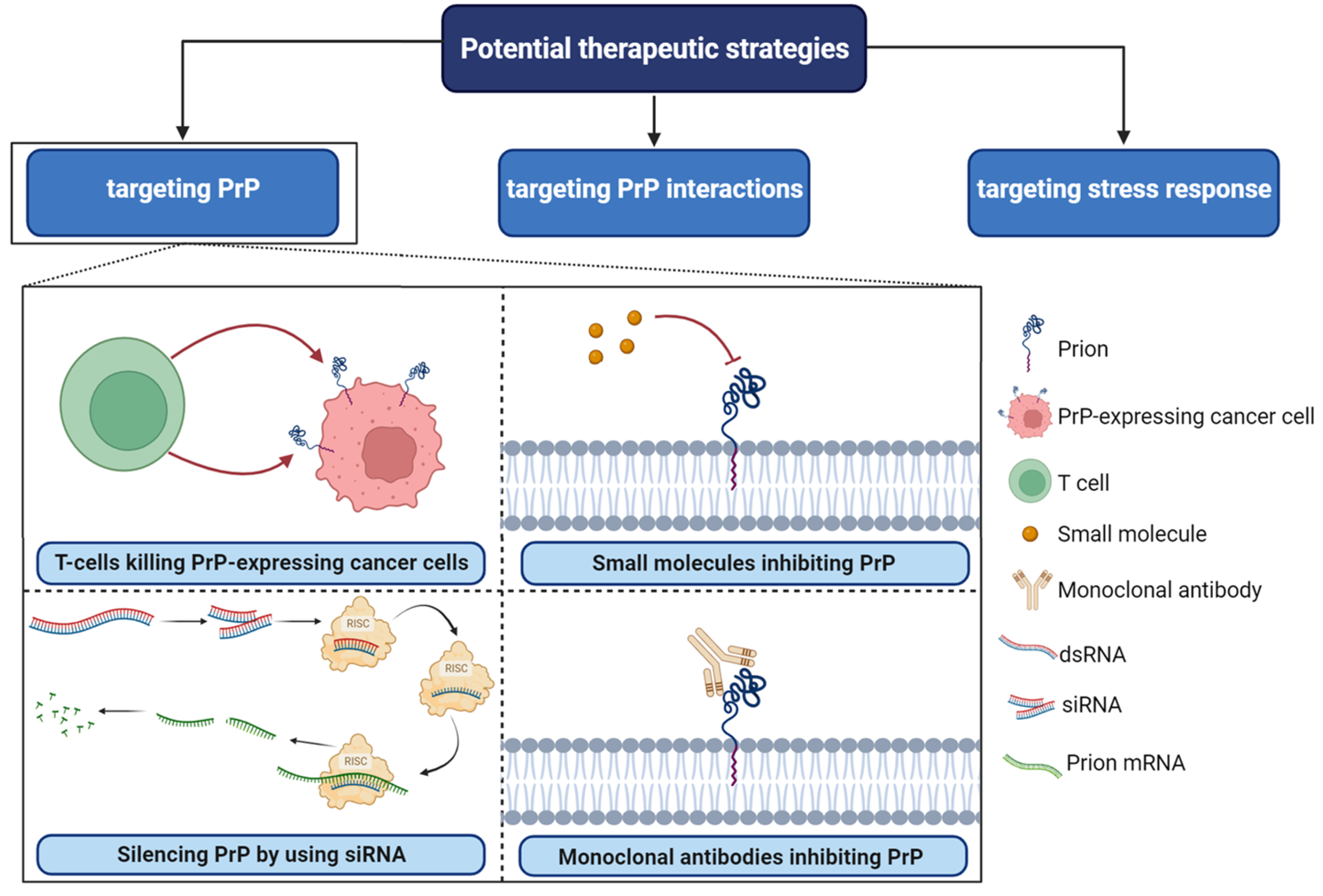
5. Targeting PrPC Interactions in Cancer: New Insights and Potential Strategies (Figure 4)

PrPC plays a central role as a scaffold protein by forming multiprotein complexes with receptors or extracellular molecules. These interactions may contribute to the activation of downstream signal pathways that control numerous biological functions, including cancer stem cell self-renewal, the central entity of tumor maintenance and dissemination [41,96]. One potential strategy for targeting PrPC in cancer is to disrupt its interactions with other molecules known to be involved in cancer progression [95]. For example, PrPC has been shown to interact with several cell surface receptors, including integrins and laminin receptors. Importantly, these proteins have been reported to play important roles in cancer cell adhesion, migration, and invasion [68]. Inhibiting these interactions could potentially prevent cancer cells from spreading and invading surrounding tissues. In breast cancer, PrPC interaction with P-gp was associated with drug resistance, higher aggressiveness, invasion, and migration. PrPC also interfered in neo-adjuvant chemotherapy response in this cancer [96]. In addition, the PrPC-STI1 interaction has been shown to be involved in many tumors, including glioblastoma [96]. In PDAC and melanomas, pro-PrPC has been shown to interact with FLNA to promote tumorigenesis and was associated with worse prognoses. These results strongly suggested that inhibition of these interactions by specific compounds could constitute a promising therapy to treat cancer [97]. Additionally, in some categories of breast cancer that are resistant to conventional treatment, ER stress increased the PrPC expression, contributing to their survival. Therefore, targeting ER stress response and PrPC may provide synergistic effects [98]. PrPC silencing has been shown to sensitize breast cancer cell lines to TRAIL-, Bax, TNF-α, and adjuvant chemotherapy-mediated cell death, which can also be considered as alternative treatments in breast cancer [96] (Figure 3). The direct targeting of PrPC protein, may also be considered as an interesting alternative. Several compounds such as small molecules, peptides, and siRNA, have been identified as reducers of PrPC expression in cancer cells [74]. These compounds have shown promising results in preclinical studies and may represent a new class of anti-cancer agents (Figure 4).
Figure 4. Potential therapeutic strategies in PrPC-associated cancer. RISC; RNA-induced silencing complex, SiRNA; small interfering RNA, dsRNA; double-stranded RNA. Several small molecules, including Quinacrine, Chlorpromazine, Amphotericin B, Pentosan polysulfate, and Suramin, have been identified as potential inhibitors of PrPC.
Cancers 15 04982 g004
Finally, immunotherapy approaches targeting PrPC have also been explored [99]. For example, monoclonal antibodies against PrPC have been shown to inhibit cancer cell proliferation and migration in vitro and in vivo [99]. Additionally, PrPC-specific T cells have been generated and have been shown to recognize and kill PrPC-expressing cancer cells. Overall, these studies strongly suggest that the targeting of PrPC interactions with tumor-associated proteins may represent a promising new avenue for cancer therapy.
While much more research is needed to fully understand the role of PrPC in cancer development and progression and to develop effective therapies targeting this protein, the growing body of evidence suggests that PrPC is a promising target for cancer treatment [100] (Figure 4).

6. Conclusions

Despite the lack of solid evidence for the precise physiological role of PrPC, its involvement in human diseases, especially cancer, is now well-established. A growing body of evidence linking PrPC to cancer has opened up new avenues for cancer research and treatment.
Through this review, we shed light on the role of PrPC in numerous types of cancers where it is highly expressed. In addition, we believe that PrPC may contribute to tumorigenic processes by regulating tumor cell invasion, migration, and metastasis. PrPC also appears to exhibit anti-apoptotic and drug resistance effects.
At the therapeutic level, one promising area of research is the development of new drugs that can directly reduce the PrPC expression in cancer cells or target its interactions with other molecules involved in cancer progression. Some of these drugs have shown great potential in preclinical studies, and could represent a new class of anti-cancer agents. Moreover, the development of monoclonal antibodies against PrPC and PrPC-specific T cells represents exciting new approaches for cancer immunotherapy.
Future research should focus on a better understanding of the mechanisms by which PrPC contributes to cancer progression, as well as on developing new and more effective therapies targeting this protein. With continued effort and innovation, PrPC could become an important target for cancer treatment in the years to come.
In conclusion, the discovery of the link between PrPC and the etiology of cancer will open up exciting new avenues for cancer research and therapy. While much remains to be done, the growing body of evidence suggests that PrPC is a promising target for cancer treatment. Hence, sustained research in this area is warranted.

Author Contributions

R.A.N., H.S.-Z. and K.G., preparation of the original draft; C.L., M.B. (Mélanie Boudaud) and M.D., illustration and visualization; C.M. and T.B., resources and proof reading; N.A. and M.B. (Mohamed Benharouga), supervision, writing, review and editing. All authors have read and agreed to the published version of the manuscript.

Funding

The authors acknowledge the following sources of funding: Institut National de la Santé et de la Recherche Médicale (INSERM), University Grenoble-Alpes, VALO-GRAL CBH-EUR-GS (ANR-17-EURE-0003), Région Auvergne-Rhône-Alpes/Cancéropôle Lyon Au-vergne Rhône-Alpes, Ligues Départementales (Isère & Savoie) contre le Cancer, ARC (association de recherche contre le cancer) (ARCPJ2021050003618) and Fondation pour la Recherche Médicale (FRM) SPF20150934074. Part of this work has been performed at the CMBA platform—IRIG-DS-BGE-Gen&Chem-CMBA, CEA-Grenoble, F-38054 Grenoble, (a member of GIS-IBISA and ChemBioFrance infrastructure) supported by GRAL, a program of the Chemistry Biology Health Graduate School of Université Grenoble Alpes (ANR-17-EURE-0003).

Acknowledgments

We acknowledge the following sources of funding: University Grenoble-Alpes, Institut National de la Santé et de la Recherche Médicale (INSERM), and Ligue Départementale (Savoie) contre le Cancer.

Conflicts of Interest

The authors declare no conflict of interest.

Abbreviations

ABCATP-Binding Cassette
AktProtein Kinase B
ATPAdenosine Triphosphate
BESBovine encephalopathies spongiform
CD44Cluster of Differentiation 44
cIAP-1Cellular Inhibitor of Apoptosis Protein-1
CJDCreutzfeldt-Jakob’s Disease
CNSCentral Nervous System
CRCColorectal Cancer
CSCCancer Stem Cells
CtC-terminal domain
Cu2+Copper ions
dsRNADouble-Stranded RNA
ECMExtracellular Matrix
EREndoplasmic Reticulum
ERK1/2Extracellular Signal-Regulated Kinases 1 and 2
FFI Fatal Familial Insomnia
FLNA Filamin A
FynProto-oncogene tyrosine-protein kinase Fyn
GAGolgi Apparatus
GLUT-1Glucose Transporter 1
GPIGlycosylphosphatidylinositol
GSCGlioma Stem Cells
HIF-2αHypoxia-Inducible Factor-2 Alpha
HSP27Heat Shock Protein 27
HT29A colorectal carcinoma cell line
IAPsInhibitor of Apoptosis Proteins
LS-174TA colorectal adenocarcinoma cell line
MCF7/ADRMCF7 Adriamycin-Resistant Cells
MDA-MB-435A breast cancer cell line
MDRMultidrug Resistance
MKN45A gastric cancer cell line
MMPMatrix Metalloprotease
NtN-terminal domain
P38MAPKp38 Mitogen-Activated Protein Kinase
PDACPancreatic Ductal Adenocarcinoma
PGAP1/PGAP5Post-GPI Attachment Proteins 1 and 5
P-gpP-glycoprotein
PKAProtein Kinase A
PKDProtein Kinase D
PRNPPrion Protein Gene
PrPCCellular Prion Protein
RISCRNA-Induced Silencing Complex
SATB1Special AT-Rich Sequence-Binding Protein 1
SEspongiform encephalopathies
Ser82Serine 82
SGC7901A gastric cancer cell line
siRNASmall Interfering RNA
Sp1Specificity Protein 1
TMDTrans Membrane Domain
TNF-αTumor Necrosis Factor Alpha
TRAILTumor Necrosis Factor-Related Apoptosis Inducing Ligand
TSETransmissible spongiform encephalopathies
XIAPX-Linked Inhibitor of Apoptosis

References

  1. Bendheim, P.E.; Brown, H.R.; Rudelli, R.D.; Scala, L.J.; Goller, N.L.; Wen, G.Y.; Kascsak, R.J.; Cashman, N.R.; Bolton, D.C. Nearly ubiquitous tissue distribution of the scrapie agent precursor protein. Neurology 1992, 42, 149–156. [Google Scholar] [CrossRef]
  2. Bonnon, C.; Wendeler, M.W.; Paccaud, J.-P.; Hauri, H.-P. Selective export of human GPI-anchored proteins from the endoplasmic reticulum. J. Cell Sci. 2010, 123 Pt 10, 1705–1715. [Google Scholar] [CrossRef]
  3. Tanaka, S.; Maeda, Y.; Tashima, Y.; Kinoshita, T. Inositol deacylation of glycosylphosphatidylinositol-anchored proteins is mediated by mammalian PGAP1 and yeast Bst1p. J. Biol. Chem. 2004, 279, 14256–14263. [Google Scholar] [CrossRef]
  4. Campana, V.; Sarnataro, D.; Zurzolo, C. The highways and byways of prion protein trafficking. Trends Cell Biol. 2005, 15, 102–111. [Google Scholar] [CrossRef]
  5. Sarnataro, D.; Campana, V.; Paladino, S.; Stornaiuolo, M.; Nitsch, L.; Zurzolo, C. PrP(C) association with lipid rafts in the early secretory pathway stabilizes its cellular conformation. Mol. Biol. Cell 2004, 15, 4031–4042. [Google Scholar] [CrossRef]
  6. Burns, C.S.; Aronoff-Spencer, E.; Dunham, C.M.; Lario, P.; Avdievich, N.I.; Antholine, W.E.; Olmstead, M.M.; Vrielink, A.; Gerfen, G.J.; Peisach, J.; et al. Molecular features of the copper binding sites in the octarepeat domain of the prion protein. Biochemistry 2002, 41, 3991–4001. [Google Scholar] [CrossRef]
  7. Harrison, C.F.; Lawson, V.A.; Coleman, B.M.; Kim, Y.-S.; Masters, C.L.; Cappai, R.; Barnham, K.J.; Hill, A.F. Conservation of a glycine-rich region in the prion protein is required for uptake of prion infectivity. J. Biol. Chem. 2010, 285, 20213–20223. [Google Scholar] [CrossRef]
  8. Harris, D.A.; Lele, P.; Snider, W.D. Localization of the mRNA for a chicken prion protein by in situ hybridization. Proc. Natl. Acad. Sci. USA 1993, 90, 4309–4313. [Google Scholar] [CrossRef]
  9. Manson, J.; West, J.D.; Thomson, V.; McBride, P.; Kaufman, M.H.; Hope, J. The prion protein gene: A role in mouse embryogenesis? Development 1992, 115, 117–122. [Google Scholar] [CrossRef]
  10. Brown, D.R.; Schmidt, B.; Groschup, M.H.; Kretzschmar, H.A. Prion protein expression in muscle cells and toxicity of a prion protein fragment. Eur. J. Cell Biol. 1998, 75, 29–37. [Google Scholar] [CrossRef]
  11. Li, R.; Liu, D.; Zanusso, G.; Liu, T.; Fayen, J.D.; Huang, J.-H.; Petersen, R.B.; Gambetti, P.; Sy, M.-S. The expression and potential function of cellular prion protein in human lymphocytes. Cell. Immunol. 2001, 207, 49–58. [Google Scholar] [CrossRef]
  12. Pammer, J.; Cross, H.S.; Frobert, Y.; Tschachler, E.; Oberhuber, G. The pattern of prion-related protein expression in the gastrointestinal tract. Virchows Arch. 2000, 436, 466–472. [Google Scholar] [CrossRef]
  13. Pammer, J.; Weninger, W.; Tschachler, E. Human keratinocytes express cellular prion-related protein in vitro and during inflammatory skin diseases. Am. J. Pathol. 1998, 153, 1353–1358. [Google Scholar] [CrossRef]
  14. Liemann, S.; Glockshuber, R. Transmissible spongiform encephalopathies. Biochem. Biophys. Res. Commun. 1998, 250, 187–193. [Google Scholar] [CrossRef]
  15. Prusiner, S.B. Novel proteinaceous infectious particles cause scrapie. Science 1982, 216, 136–144. [Google Scholar] [CrossRef]
  16. Jeffrey, M.; Gonzalez, L. Pathology and pathogenesis of bovine spongiform encephalopathy and scrapie. Curr. Top Microbiol. Immunol. 2004, 284, 65–97. [Google Scholar]
  17. Bregman, N.; Shiner, T.; Kavé, G.; Alcalay, R.; Gana-Weisz, M.; Goldstein, O.; Glinka, T.; Aizenstein, O.; Ben Bashat, D.; Alcalay, Y.; et al. Correction: The natural history study of preclinical genetic Creutzfeldt-Jakob Disease (CJD): A prospective longitudinal study protocol. BMC Neurol. 2023, 23, 229. [Google Scholar] [CrossRef]
  18. Li, B.; Wang, S.; Han, S.; Hu, N.; Shang, X. Case report: Creutzfeldt-Jakob disease: A case that initiated with the onset of obsessive-compulsive state. Front. Neurol. 2023, 14, 1227566. [Google Scholar] [CrossRef]
  19. Khan, Z.; Bollu, P.C. Fatal Familial Insomnia. In StatPearls; StatPearls: Treasure Island, FL, USA, 2023. [Google Scholar]
  20. Bounhar, Y.; Zhang, Y.; Goodyer, C.G.; LeBlanc, A.C. Prion protein protects human neurons against Bax-mediated apoptosis. J. Biol. Chem. 2001, 276, 39145–39149. [Google Scholar] [CrossRef]
  21. Brown, D.R.; Qin, K.; Herms, J.W.; Madlung, A.; Manson, J.; Strome, R.; Fraser, P.E.; Kruck, T.; von Bohlen, A.; Schulz-Schaeffer, W.; et al. The cellular prion protein binds copper in vivo. Nature 1997, 390, 684–687. [Google Scholar] [CrossRef]
  22. Kuwahara, C.; Takeuchi, A.M.; Nishimura, T.; Haraguchi, K.; Kubosaki, A.; Matsumoto, Y.; Saeki, K.; Matsumoto, Y.; Yokoyama, T.; Itohara, S.; et al. Prions prevent neuronal cell-line death. Nature 1999, 400, 225–226. [Google Scholar] [CrossRef]
  23. Mouillet-Richard, S.; Ermonval, M.; Chebassier, C.; Laplanche, J.L.; Lehmann, S.; Launay, J.M.; Kellermann, O. Signal transduction through prion protein. Science 2000, 289, 1925–1928. [Google Scholar] [CrossRef]
  24. Paitel, E.; Fahraeus, R.; Checler, F. Cellular prion protein sensitizes neurons to apoptotic stimuli through Mdm2-regulated and p53-dependent caspase 3-like activation. J. Biol. Chem. 2003, 278, 10061–10066. [Google Scholar] [CrossRef]
  25. Stella, R.; Massimino, M.L.; Sandri, M.; Sorgato, M.C.; Bertoli, A. Cellular prion protein promotes regeneration of adult muscle tissue. Mol. Cell. Biol. 2010, 30, 4864–4876. [Google Scholar] [CrossRef]
  26. Stürmer, C.; Langhorst, M.F.; Wiechers, M.F.; Legler, D.F.; von Hanwehr, S.H.; Guse, A.H.; Plattner, H. PrPc capping in T cells promotes its association with the lipid raft proteins reggie-1 and reggie-2 and leads to signal transduction. FASEB J. 2004, 18, 1731–1733. [Google Scholar] [CrossRef]
  27. Grimaldi, I.; Leser, F.S.; Janeiro, J.M.; da Rosa, B.G.; Campanelli, A.C.; Romão, L.; Lima, F.R.S. The multiple functions of PrP(C) in physiological, cancer, and neurodegenerative contexts. J. Mol. Med. 2022, 100, 1405–1425. [Google Scholar] [CrossRef]
  28. Büeler, H.; Fischer, M.; Lang, Y.; Bluethmann, H.; Lipp, H.-P.; DeArmond, S.J.; Prusiner, S.B.; Aguet, M.; Weissmann, C. Normal development and behaviour of mice lacking the neuronal cell-surface PrP protein. Nature 1992, 356, 577–582. [Google Scholar] [CrossRef]
  29. Makarava, N.; Katorcha, E.; Chang, J.C.-Y.; Lau, J.T.Y.; Baskakov, I.V. Deficiency in ST6GAL1, one of the two alpha2,6-sialyltransferases, has only a minor effect on the pathogenesis of prion disease. Front. Mol. Biosci. 2022, 9, 1058602. [Google Scholar] [CrossRef]
  30. Cavallaro, U.; Christofori, G. Cell adhesion and signalling by cadherins and Ig-CAMs in cancer. Nat. Rev. Cancer 2004, 4, 118–132. [Google Scholar] [CrossRef]
  31. Du, J.; Pan, Y.; Shi, Y.; Guo, C.; Jin, X.; Sun, L.; Liu, N.; Qiao, T.; Fan, D. Overexpression and significance of prion protein in gastric cancer and multidrug-resistant gastric carcinoma cell line SGC7901/ADR. Int. J. Cancer 2005, 113, 213–220. [Google Scholar] [CrossRef]
  32. Graner, E.; Mercadante, A.F.; Zanata, S.M.; Forlenza, O.V.; Cabral, A.L.; Veiga, S.S.; Juliano, M.A.; Roesler, R.; Walz, R.; Minetti, A.; et al. Cellular prion protein binds laminin and mediates neuritogenesis. Brain Res. Mol. Brain Res. 2000, 76, 85–92. [Google Scholar] [CrossRef]
  33. Pan, T.; Wong, B.-S.; Liu, T.; Li, R.; Petersen, R.B.; Sy, M.-S. Cell-surface prion protein interacts with glycosaminoglycans. Biochem. J. 2002, 368 Pt 1, 81–90. [Google Scholar] [CrossRef]
  34. Pan, Y.; Zhao, L.; Liang, J.; Liu, J.; Shi, Y.; Liu, N.; Zhang, G.; Jin, H.; Gao, J.; Xie, H.; et al. Cellular prion protein promotes invasion and metastasis of gastric cancer. FASEB J. 2006, 20, 1886–1888. [Google Scholar] [CrossRef]
  35. Rieger, R.; Edenhofer, F.; Lasmézas, C.I.; Weiss, S. The human 37-kDa laminin receptor precursor interacts with the prion protein in eukaryotic cells. Nat. Med. 1997, 3, 1383–1388. [Google Scholar] [CrossRef]
  36. Satoh, J.-I.; Kuroda, Y.; Katamine, S. Gene expression profile in prion protein-deficient fibroblasts in culture. Am. J. Pathol. 2000, 157, 59–68. [Google Scholar] [CrossRef]
  37. Schmitt-Ulms, G.; Legname, G.; Baldwin, M.A.; Ball, H.L.; Bradon, N.; Bosque, P.J.; Crossin, K.L.; Edelman, G.M.; DeArmond, S.J.; Cohen, F.E.; et al. Binding of neural cell adhesion molecules (N-CAMs) to the cellular prion protein. J. Mol. Biol. 2001, 314, 1209–1225. [Google Scholar] [CrossRef]
  38. Cheng, Q.; Zheng, H.; Li, M.; Wang, H.; Guo, X.; Zheng, Z.; Chen, C.; Liu, J.; Zhan, T.; Li, Z.; et al. LGR4 cooperates with PrPc to endow the stemness of colorectal cancer stem cells contributing to tumorigenesis and liver metastasis. Cancer Lett. 2022, 540, 215725. [Google Scholar] [CrossRef]
  39. Kim, M.-J.; Cho, Y.-A.; Kim, E.; Choe, J.-Y.; Park, J.-W.; Lee, J.; Lee, J.-W.; Moon, S.-H.; Kim, Y.-S.; Kim, S.-E.; et al. Cellular Prion Protein Is Closely Associated with Early Recurrence and Poor Survival in Patients with Hepatocellular Carcinoma. Diagnostics 2022, 12, 1635. [Google Scholar] [CrossRef]
  40. Ding, M.; Chen, Y.; Lang, Y.; Cui, L. The Role of Cellular Prion Protein in Cancer Biology: A Potential Therapeutic Target. Front. Oncol. 2021, 11, 742949. [Google Scholar] [CrossRef]
  41. Yousaf, S.; Ahmad, M.; Wu, S.; Zia, M.A.; Ahmed, I.; Iqbal, H.M.; Liu, Q.; Rehman, S.U. Cellular Prion Protein Role in Cancer Biology: Is It A Potential Therapeutic Target? Biomedicines 2022, 10, 2833. [Google Scholar] [CrossRef]
  42. Blasi, F.; Carmeliet, P. uPAR: A versatile signalling orchestrator. Nat. Rev. Mol. Cell Biol. 2002, 3, 932–943. [Google Scholar] [CrossRef]
  43. Lee, K.S.; Linden, R.; Brentani, R.R.; Martins, V.R.; Prado, M.A.M. Towards cellular receptors for prions. Rev. Med. Virol. 2003, 13, 399–408. [Google Scholar] [CrossRef]
  44. Khasigov, P.Z.; Podobed, O.V.; Gracheva, T.S.; Salbiev, K.D.; Grachev, S.V.; Berezov, T.T. Role of matrix metalloproteinases and their inhibitors in tumor invasion and metastasis. Biochemistry 2003, 68, 711–717. [Google Scholar]
  45. Feng, Y.; Walsh, C.A. The many faces of filamin: A versatile molecular scaffold for cell motility and signalling. Nat. Cell Biol. 2004, 6, 1034–1038. [Google Scholar] [CrossRef]
  46. Ke, J.; Wu, G.; Zhang, J.; Li, H.; Gao, S.; Shao, M.; Gao, Z.; Sy, M.-S.; Cao, Y.; Yang, X.; et al. Melanoma migration is promoted by prion protein via Akt-hsp27 signaling axis. Biochem. Biophys. Res. Commun. 2020, 523, 375–381. [Google Scholar] [CrossRef]
  47. Li, C.; Yu, S.; Nakamura, F.; Pentikäinen, O.T.; Singh, N.; Yin, S.; Xin, W.; Sy, M.S. Pro-prion binds filamin A, facilitating its interaction with integrin beta1, and contributes to melanomagenesis. J. Biol. Chem. 2010, 285, 30328–30339. [Google Scholar] [CrossRef]
  48. Stossel, T.P.; Condeelis, J.; Cooley, L.; Hartwig, J.H.; Noegel, A.; Schleicher, M.; Shapiro, S.S. Filamins as integrators of cell mechanics and signalling. Nat. Rev. Mol. Cell Biol. 2001, 2, 138–145. [Google Scholar] [CrossRef]
  49. Kandasamy, K.; Srinivasula, S.M.; Alnemri, E.S.; Thompson, C.B.; Korsmeyer, S.J.; Bryant, J.L.; Srivastava, R.K. Involvement of proapoptotic molecules Bax and Bak in tumor necrosis factor-related apoptosis-inducing ligand (TRAIL)-induced mitochondrial disruption and apoptosis: Differential regulation of cytochrome c and Smac/DIABLO release. Cancer Res. 2003, 63, 1712–1721. [Google Scholar]
  50. Kang, T.H.; Noh, K.H.; Kim, J.H.; Bae, H.C.; Lin, K.Y.; Monie, A.; Pai, S.I.; Hung, C.-F.; Wu, T.-C.; Kim, T.W. Ectopic expression of X-linked lymphocyte-regulated protein pM1 renders tumor cells resistant to antitumor immunity. Cancer Res. 2010, 70, 3062–3070. [Google Scholar] [CrossRef]
  51. Keane, M.M.; Ettenberg, S.A.; Nau, M.M.; Russell, E.K.; Lipkowitz, S. Chemotherapy augments TRAIL-induced apoptosis in breast cell lines. Cancer Res. 1999, 59, 734–741. [Google Scholar]
  52. Kim, E.J.; Suliman, A.; Lam, A.; Srivastava, R.K. Failure of Bcl-2 to block mitochondrial dysfunction during TRAIL-induced apoptosis. Tumor necrosis-related apoptosis-inducing ligand. Int. J. Oncol. 2001, 18, 187–194. [Google Scholar] [CrossRef]
  53. Simstein, R.; Burow, M.; Parker, A.; Weldon, C.; Beckman, B. Apoptosis, chemoresistance, and breast cancer: Insights from the MCF-7 cell model system. Exp. Biol. Med. 2003, 228, 995–1003. [Google Scholar] [CrossRef]
  54. Sprick, M.R.; Rieser, E.; Stahl, H.; Grosse-Wilde, A.; Weigand, M.A.; Walczak, H. Caspase-10 is recruited to and activated at the native TRAIL and CD95 death-inducing signalling complexes in a FADD-dependent manner but can not functionally substitute caspase-8. EMBO J. 2002, 21, 4520–4530. [Google Scholar] [CrossRef]
  55. Srivastava, R.K. Intracellular mechanisms of TRAIL and its role in cancer therapy. Mol. Cell Biol. Res. Commun. 2000, 4, 67–75. [Google Scholar] [CrossRef]
  56. Chieng, C.K.-L.; Say, Y.-H. Cellular prion protein contributes to LS 174T colon cancer cell carcinogenesis by increasing invasiveness and resistance against doxorubicin-induced apoptosis. Tumour. Biol. 2015, 36, 8107–8120. [Google Scholar] [CrossRef]
  57. Gil, M.; Kim, Y.K.; Kim, K.-E.; Kim, W.; Park, C.-S.; Lee, K.J. Cellular prion protein regulates invasion and migration of breast cancer cells through MMP-9 activity. Biochem. Biophys. Res. Commun. 2016, 470, 213–219. [Google Scholar] [CrossRef]
  58. Li, Q.-Q.; Cao, X.-X.; Xu, J.-D.; Chen, Q.; Wang, W.-J.; Tang, F.; Chen, Z.-Q.; Liu, X.-P.; Xu, Z.-D. The role of P-glycoprotein/cellular prion protein interaction in multidrug-resistant breast cancer cells treated with paclitaxel. Cell. Mol. Life Sci. 2009, 66, 504–515. [Google Scholar] [CrossRef]
  59. Fantin, V.R.; St-Pierre, J.; Leder, P. Attenuation of LDH-A expression uncovers a link between glycolysis, mitochondrial physiology, and tumor maintenance. Cancer Cell 2006, 9, 425–434. [Google Scholar] [CrossRef]
  60. Li, Q.-Q.; Sun, Y.-P.; Ruan, C.-P.; Xu, X.-Y.; Ge, J.-H.; He, J.; Xu, Z.-D.; Wang, Q.; Gao, W.-C. Cellular prion protein promotes glucose uptake through the Fyn-HIF-2α-Glut1 pathway to support colorectal cancer cell survival. Cancer Sci. 2011, 102, 400–406. [Google Scholar] [CrossRef]
  61. Warburg, O. On the origin of cancer cells. Science 1956, 123, 309–314. [Google Scholar] [CrossRef]
  62. Du, L.; Rao, G.; Wang, H.; Li, B.; Tian, W.; Cui, J.; He, L.; Laffin, B.; Tian, X.; Hao, C.; et al. CD44-positive cancer stem cells expressing cellular prion protein contribute to metastatic capacity in colorectal cancer. Cancer Res. 2013, 73, 2682–2694. [Google Scholar] [CrossRef]
  63. Wang, Q.; Qian, J.; Wang, F.; Ma, Z. Cellular prion protein accelerates colorectal cancer metastasis via the Fyn-SP1-SATB1 axis. Oncol. Rep. 2012, 28, 2029–2034. [Google Scholar] [CrossRef]
  64. Han, H.; Bearss, D.J.; Browne, L.W.; Calaluce, R.; Nagle, R.B.; Von Hoff, D.D. Identification of differentially expressed genes in pancreatic cancer cells using cDNA microarray. Cancer Res. 2002, 62, 2890–2896. [Google Scholar]
  65. Lee, J.H.; Yun, C.W.; Lee, S.H. Cellular Prion Protein Enhances Drug Resistance of Colorectal Cancer Cells via Regulation of a Survival Signal Pathway. Biomol. Ther. 2018, 26, 313–321. [Google Scholar] [CrossRef]
  66. Li, C.; Yu, S.; Nakamura, F.; Yin, S.; Xu, J.; Petrolla, A.A.; Singh, N.; Tartakoff, A.; Abbott, D.W.; Xin, W.; et al. Binding of pro-prion to filamin A disrupts cytoskeleton and correlates with poor prognosis in pancreatic cancer. J. Clin. Investig. 2009, 119, 2725–2736. [Google Scholar] [CrossRef]
  67. Sy, M.-S.; Altekruse, S.F.; Li, C.; Lynch, C.F.; Goodman, M.T.; Hernandez, B.Y.; Zhou, L.; Saber, M.S.; Hewitt, S.M.; Xin, W. Association of prion protein expression with pancreatic adenocarcinoma survival in the SEER residual tissue repository. Cancer Biomark 2011, 10, 251–258. [Google Scholar] [CrossRef]
  68. Sauer, H.; Dagdanova, A.; Hescheler, J.; Wartenberg, M. Redox-regulation of intrinsic prion expression in multicellular prostate tumor spheroids. Free. Radic. Biol. Med. 1999, 27, 1276–1283. [Google Scholar] [CrossRef]
  69. Sollazzo, V.; Galasso, M.; Volinia, S.; Carinci, F. Prion proteins (PRNP and PRND) are over-expressed in osteosarcoma. J. Orthop. Res. 2012, 30, 1004–1012. [Google Scholar] [CrossRef]
  70. Thellung, S.; Corsaro, A.; Bosio, A.; Zambito, M.; Barbieri, F.; Mazzanti, M.; Florio, T. Emerging Role of Cellular Prion Protein in the Maintenance and Expansion of Glioma Stem Cells. Cells 2019, 8, 1458. [Google Scholar] [CrossRef]
  71. Yu, G.; Jiang, L.; Xu, Y.; Guo, H.; Liu, H.; Zhang, Y.; Yang, H.; Yuan, C.; Ma, J. Silencing prion protein in MDA-MB-435 breast cancer cells leads to pleiotropic cellular responses to cytotoxic stimuli. PLoS ONE 2012, 7, e48146. [Google Scholar] [CrossRef]
  72. Kouadri, A.; El Khatib, M.; Cormenier, J.; Chauvet, S.; Zeinyeh, W.; El Khoury, M.; Macari, L.; Richaud, P.R.; Coraux, C.; Michaud-Soret, I.; et al. Involvement of the Prion Protein in the Protection of the Human Bronchial Epithelial Barrier Against Oxidative Stress. Antioxid Redox Signal. 2019, 31, 59–74. [Google Scholar] [CrossRef]
  73. Lin, S.-C.; Lin, C.-H.; Shih, N.-C.; Liu, H.-L.; Wang, W.-C.; Lin, K.-Y.; Liu, Z.-Y.; Tseng, Y.-J.; Chang, H.-K.; Lin, Y.-C.; et al. Cellular prion protein transcriptionally regulated by NFIL3 enhances lung cancer cell lamellipodium formation and migration through JNK signaling. Oncogene 2020, 39, 385–398. [Google Scholar] [CrossRef]
  74. Ryskalin, L.; Biagioni, F.; Busceti, C.L.; Giambelluca, M.A.; Morelli, L.; Frati, A.; Fornai, F. The Role of Cellular Prion Protein in Promoting Stemness and Differentiation in Cancer. Cancers 2021, 13, 170. [Google Scholar] [CrossRef]
  75. Perrier, V.; Solassol, J.; Crozet, C.; Frobert, Y.; Mourton-Gilles, C.; Grassi, J.; Lehmann, S. Anti-PrP antibodies block PrPSc replication in prion-infected cell cultures by accelerating PrPC degradation. J. Neurochem. 2004, 89, 454–463. [Google Scholar] [CrossRef]
  76. Limone, A.; Maggisano, V.; Sarnataro, D.; Bulotta, S. Emerging roles of the cellular prion protein (PrP(C)) and 37/67 kDa laminin receptor (RPSA) interaction in cancer biology. Cell. Mol. Life Sci. 2023, 80, 207. [Google Scholar] [CrossRef]
  77. Vassallo, N.; Herms, J. Cellular prion protein function in copper homeostasis and redox signalling at the synapse. J. Neurochem. 2003, 86, 538–544. [Google Scholar] [CrossRef]
  78. Lavoie, J.; Gingras-Breton, G.; Tanguay, R.; Landry, J. Induction of Chinese hamster HSP27 gene expression in mouse cells confers resistance to heat shock. HSP27 stabilization of the microfilament organization. J. Biol. Chem. 1993, 268, 3420–3429. [Google Scholar] [CrossRef]
  79. Nomura, N.; Nomura, M.; Sugiyama, K.; Hamada, J.-I. Phorbol 12-myristate 13-acetate (PMA)-induced migration of glioblastoma cells is mediated via p38MAPK/Hsp27 pathway. Biochem. Pharmacol. 2007, 74, 690–701. [Google Scholar] [CrossRef]
  80. Clerk, A.; Michael, A.; Sugden, P.H. Stimulation of multiple mitogen-activated protein kinase sub-families by oxidative stress and phosphorylation of the small heat shock protein, HSP25/27, in neonatal ventricular myocytes. Biochem. J. 1998, 333 Pt 3, 581–589. [Google Scholar] [CrossRef]
  81. McLaughlin, M.M.; Kumar, S.; McDonnell, P.C.; Van Horn, S.; Lee, J.C.; Livi, G.P.; Young, P.R. Identification of mitogen-activated protein (MAP) kinase-activated protein kinase-3, a novel substrate of CSBP p38 MAP kinase. J. Biol. Chem. 1996, 271, 8488–8492. [Google Scholar] [CrossRef]
  82. New, L.; Jiang, Y.; Zhao, M.; Liu, K.; Zhu, W.; Flood, L.J.; Kato, Y.; Parry, G.C.; Han, J. PRAK, a novel protein kinase regulated by the p38 MAP kinase. EMBO J. 1998, 17, 3372–3384. [Google Scholar] [CrossRef]
  83. Rouse, J.; Cohen, P.; Trigon, S.; Morange, M.; Alonso-Llamazares, A.; Zamanillo, D.; Hunt, T.; Nebreda, A.R. A novel kinase cascade triggered by stress and heat shock that stimulates MAPKAP kinase-2 and phosphorylation of the small heat shock proteins. Cell 1994, 78, 1027–1037. [Google Scholar] [CrossRef]
  84. Santell, L.; Bartfeld, N.S.; Levin, E.G. Identification of a protein transiently phosphorylated by activators of endothelial cell function as the heat-shock protein HSP27. A possible role for protein kinase C. Biochem. J. 1992, 284 Pt 3, 705–710. [Google Scholar] [CrossRef]
  85. Morelli, J.G.; Yohn, J.J.; Zekman, T.; Norris, D.A. Melanocyte movement in vitro: Role of matrix proteins and integrin receptors. J. Investig. Dermatol. 1993, 101, 605–608. [Google Scholar] [CrossRef]
  86. Baxter, E.; Windloch, K.; Gannon, F.; Lee, J.S. Epigenetic regulation in cancer progression. Cell Biosci. 2014, 4, 45. [Google Scholar] [CrossRef]
  87. Gonzalez-Malerva, L.; Park, J.; Zou, L.; Hu, Y.; Moradpour, Z.; Pearlberg, J.; Sawyer, J.; Stevens, H.; Harlow, E.; LaBaer, J. High-throughput ectopic expression screen for tamoxifen resistance identifies an atypical kinase that blocks autophagy. Proc. Natl. Acad. Sci. USA 2011, 108, 2058–2063. [Google Scholar] [CrossRef]
  88. Meijer, D.; van Agthoven, T.; Bosma, P.T.; Nooter, K.; Dorssers, L.C. Functional screen for genes responsible for tamoxifen resistance in human breast cancer cells. Mol. Cancer Res. 2006, 4, 379–386. [Google Scholar] [CrossRef]
  89. Meslin, F.; Hamai, A.; Gao, P.; Jalil, A.; Cahuzac, N.; Chouaib, S.; Mehrpour, M. Silencing of prion protein sensitizes breast adriamycin-resistant carcinoma cells to TRAIL-mediated cell death. Cancer Res. 2007, 67, 10910–10919. [Google Scholar] [CrossRef]
  90. De Wit, M.; Jimenez, C.R.; Carvalho, B.; Belien, J.A.; Delis-van Diemen, P.M.; Mongera, S.; Piersma, S.R.; Vikas, M.; Navani, S.; Pontén, F.; et al. Cell surface proteomics identifies glucose transporter type 1 and prion protein as candidate biomarkers for colorectal adenoma-to-carcinoma progression. Gut 2012, 61, 855–864. [Google Scholar] [CrossRef]
  91. Lim, J.H.; Go, G.; Lee, S.H. PrPC Regulates the Cancer Stem Cell Properties via Interaction With c-Met in Colorectal Cancer Cells. Anticancer Res. 2021, 41, 3459–3470. [Google Scholar] [CrossRef]
  92. Yang, C.C.; Chuang, F.C.; Chang, C.L.; Huang, C.R.; Chen, H.H.; Yip, H.K.; Chen, Y.T. Melatonin-Assisted Cisplatin Suppresses Urinary Bladder Cancer Cell Proliferation and Growth through Inhibiting PrP(C)-Regulated Cell Stress and Cell Proliferation Signaling. Int. J. Mol. Sci. 2023, 24, 3353. [Google Scholar] [CrossRef]
  93. Ameyar-Zazoua, M.; Larochette, N.; Dorothée, G.; Daugas, E.; Haddada, H.; Gouloumet, V.; Métivier, D.; Stancou, R.; Mami-Chouaib, F.; Kroemer, G.; et al. Wild-type p53 induced sensitization of mutant p53 TNF-resistant cells: Role of caspase-8 and mitochondria. Cancer Gene Ther. 2002, 9, 219–227. [Google Scholar] [CrossRef]
  94. Lenzi, P.; Busceti, C.L.; Lazzeri, G.; Ferese, R.; Biagioni, F.; Salvetti, A.; Pompili, E.; De Franchis, V.; Puglisi-Allegra, S.; Frati, A.; et al. Autophagy Activation Associates with Suppression of Prion Protein and Improved Mitochondrial Status in Glioblastoma Cells. Cells 2023, 12, 221. [Google Scholar] [CrossRef]
  95. Mouillet-Richard, S.; Martin-Lannerée, S.; Le Corre, D.; Hirsch, T.Z.; Ghazi, A.; Sroussi, M.; Pilati, C.; de Reyniès, A.; Djouadi, F.; Vodovar, N.; et al. A proof of concept for targeting the PrP(C)—Amyloid beta peptide interaction in basal prostate cancer and mesenchymal colon cancer. Oncogene 2022, 41, 4397–4404. [Google Scholar] [CrossRef]
  96. Linden, R.; Martins, V.R.; Prado, M.A.M.; Cammarota, M.; Izquierdo, I.; Brentani, R.R. Physiology of the prion protein. Physiol. Rev. 2008, 88, 673–728. [Google Scholar] [CrossRef]
  97. Santos, T.G.; Lopes, M.H.; Martins, V.R. Targeting prion protein interactions in cancer. Prion 2015, 9, 165–173. [Google Scholar] [CrossRef]
  98. Déry, M.-A.; Jodoin, J.; Ursini-Siegel, J.; Aleynikova, O.; Ferrario, C.; Hassan, S.; Basik, M.; LeBlanc, A.C. Endoplasmic reticulum stress induces PRNP prion protein gene expression in breast cancer. Breast Cancer Res. 2013, 15, R22. [Google Scholar] [CrossRef]
  99. Ma, Y.; Ma, J. Immunotherapy against Prion Disease. Pathogens 2020, 9, 216. [Google Scholar] [CrossRef]
  100. Rovis, T.L.; Legname, G. Prion protein-specific antibodies-development, modes of action and therapeutics application. Viruses 2014, 6, 3719–3737. [Google Scholar] [CrossRef]
Figure 1. Cellular biosynthetic pathway of PrPC protein. PrPC is synthetized as a pro-pre-protein in the endoplasmic reticulum compartment before trafficking to the Golgi apparatus and plasma membrane where it is anchored as a glycosylphosphatidylinositol (GPI) protein.
Cancers 15 04982 g001
Figure 2. Schematic representation of the PrPC protein. Linear representation of the primary sequence of human PrPC showing important protein domains.
Cancers 15 04982 g002
Figure 3. Cellular regulatory pathways involving PrPC protein in cancer. The PrPC protein is involved in different cellular tumorigenesis process, where its activities are regulated directly through protein–protein interactions or/and indirectly using different cellular regulatory pathways. P-gp; P-glycoprotein, GLUT-1; Glucose transporter-1; FLNA; Filamin A.
Cancers 15 04982 g003
Table 1. Identified roles of PrPC protein in different types of human cancers.
Cancer TypeRole of PrPCReferences
Gastric CancerPromotion of multidrug resistance [31]
Enhancement of adhesive and invasive abilities [33,34,36,44]
MelanomaPromotion of cancer migration [21,45,46,47,48]
Disruption of Filamin A [21,45,46,47,48]
Breast CancerResistance to apoptosis and drug treatment [30,49,50,51,52,53,54,55]
Promotion of invasion and migration [56,57,58]
Colorectal CancerPromotion of tumor growth via Warburg effect [59,60,61]
Enhancement of metastasis [62,63]
Confer resistance to anti-cancer drugs [64,65,66,67]
Prostate CancerPotential involvement in tumor development [68]
OsteosarcomaAssociation with tumor development [69]
GlioblastomaEnhancement of glioma stem cell proliferation [70,71]
Lung CancerPossible involvement in tumorigenesis [72,73]
Disclaimer/Publisher’s Note: The statements, opinions and data contained in all publications are solely those of the individual author(s) and contributor(s) and not of MDPI and/or the editor(s). MDPI and/or the editor(s) disclaim responsibility for any injury to people or property resulting from any ideas, methods, instructions or products referred to in the content.

