
Immunotherapy-Related Oral Adverse Effects: Immediate Sequelae, Chronicity and Secondary Cancer

Abstract
:Simple Summary
Abstract
1. Introduction
2. Methods
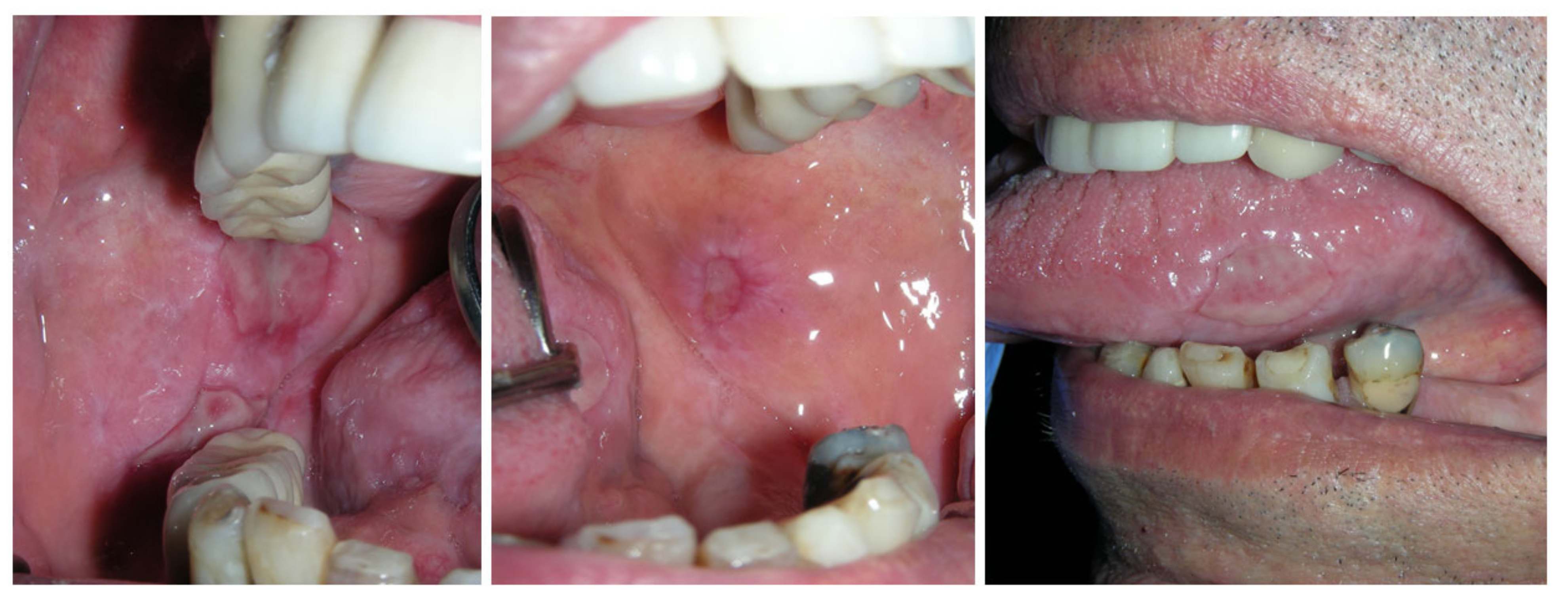
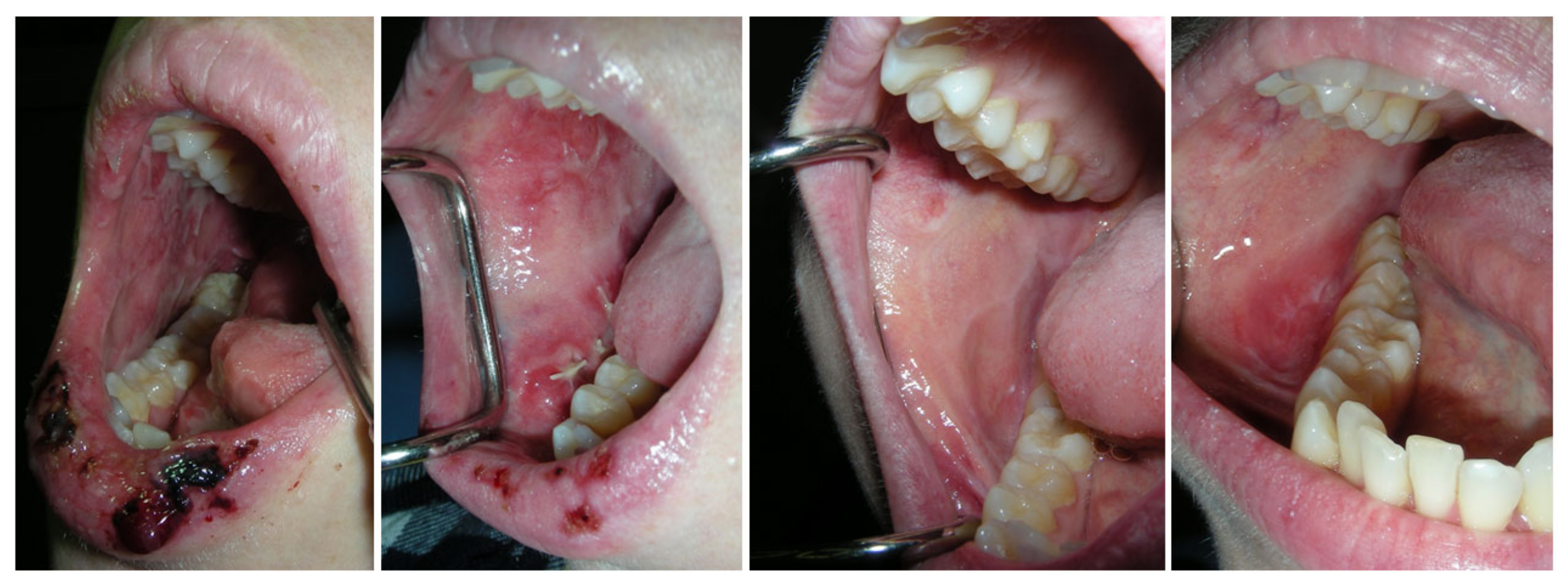
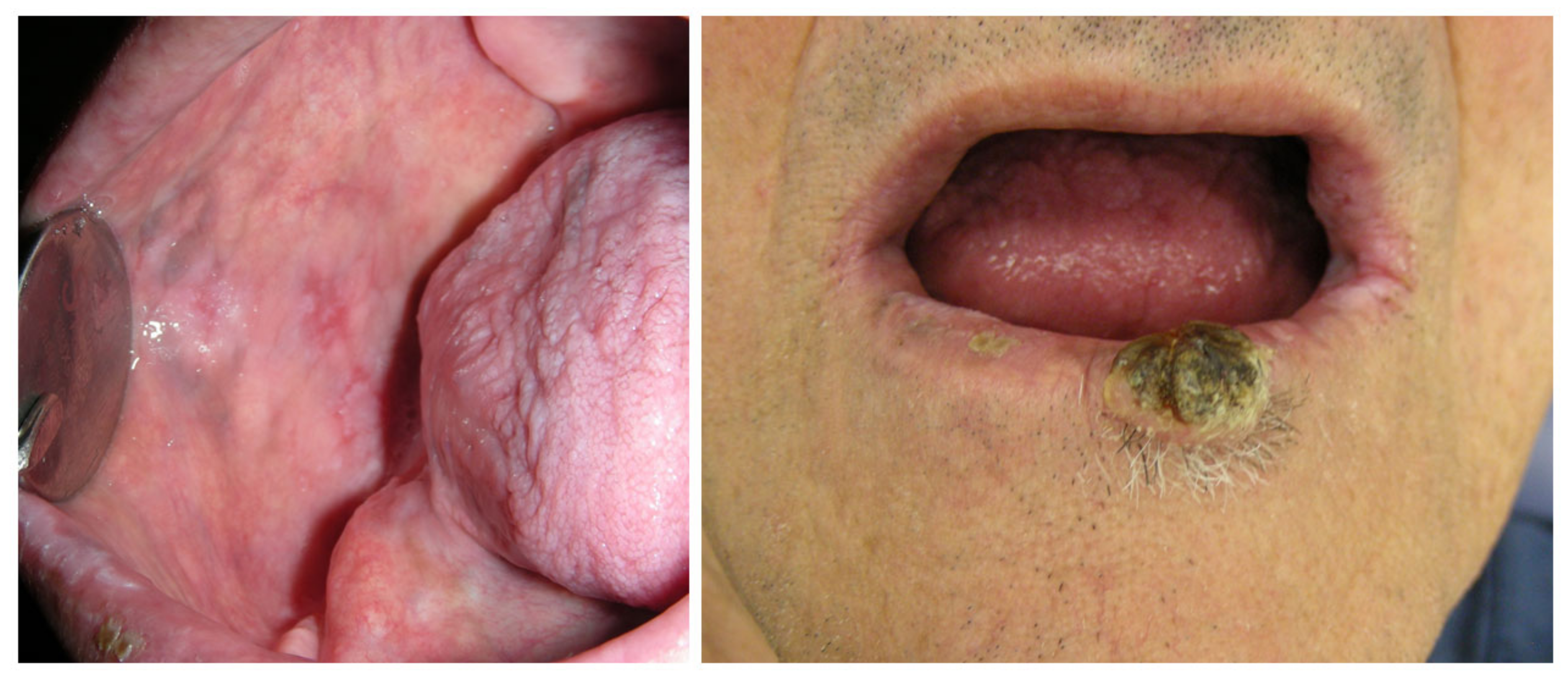
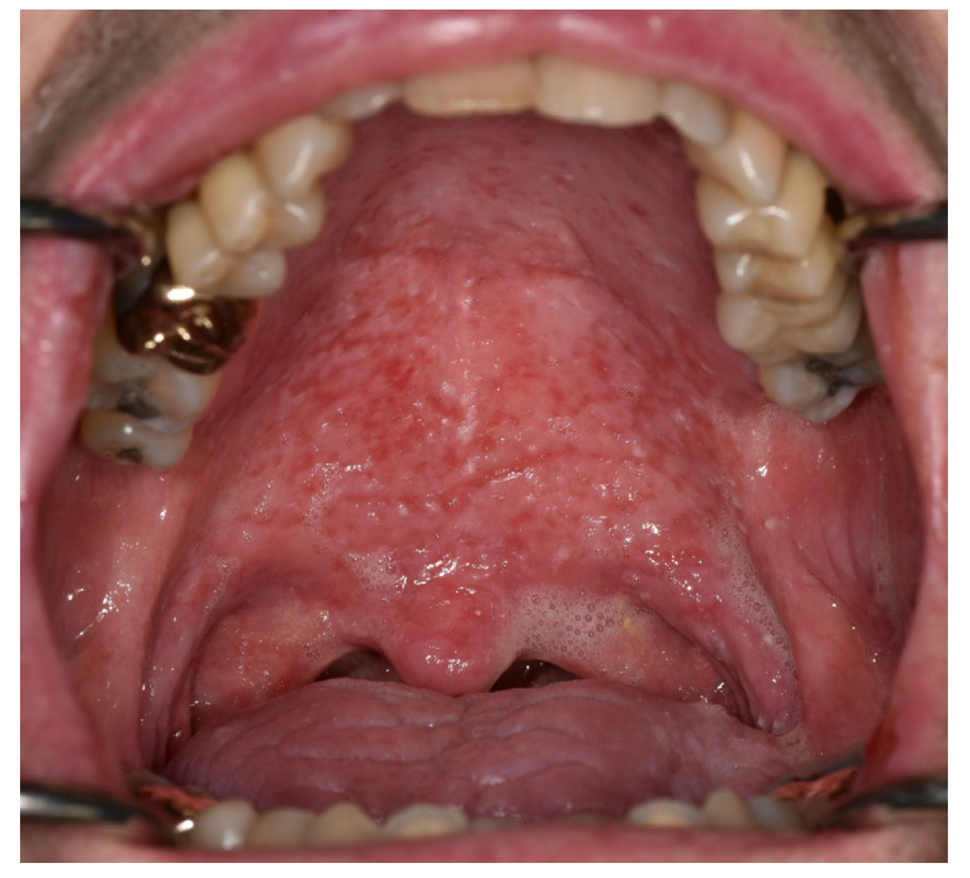
3. Results
4. Discussion
5. Conclusions
Author Contributions
Funding
Institutional Review Board Statement
Informed Consent Statement
Data Availability Statement
Acknowledgments
Conflicts of Interest
References
- Brahmer, J.R.; Lacchetti, C.; Thompson, J.A. Management of Immune-Related Adverse Events in Patients Treated with Immune Checkpoint Inhibitor Therapy: American Society of Clinical Oncology Clinical Practice Guideline Summary. J. Oncol. Pract. 2018, 14, 247–249. [Google Scholar] [CrossRef]
- Kennedy, L.B.; Salama, A.K.S. A review of cancer immunotherapy toxicity. CA Cancer J. Clin. 2020, 70, 86–104. [Google Scholar] [CrossRef]
- Lee, D.J.; Lee, H.J., Jr.; Farmer, J.R.; Reynolds, K.L. Mechanisms Driving Immune-Related Adverse Events in Cancer Patients Treated with Immune Checkpoint Inhibitors. Curr. Cardiol. Rep. 2021, 23, 98. [Google Scholar] [CrossRef]
- Chen, T.W.; Razak, A.R.; Bedard, P.L.; Siu, L.L.; Hansen, A.R. A systematic review of immune-related adverse event reporting in clinical trials of immune checkpoint inhibitors. Ann. Oncol. 2015, 26, 1824–1829. [Google Scholar] [CrossRef]
- Martins, F.; Sofiya, L.; Sykiotis, G.P.; Lamine, F.; Maillard, M.; Fraga, M.; Shabafrouz, K.; Ribi, C.; Cairoli, A.; Guex-Crosier, Y.; et al. Adverse effects of immune-checkpoint inhibitors: Epidemiology, management and surveillance. Nat. Rev. Clin. Oncol. 2019, 16, 563–580. [Google Scholar] [CrossRef] [PubMed]
- Ramos-Casals, M.; Brahmer, J.R.; Callahan, M.K.; Flores-Chavez, A.; Keegan, N.; Khamashta, M.A.; Lambotte, O.; Mariette, X.; Prat, A.; Suarez-Almazor, M.E. Immune-related adverse events of checkpoint inhibitors. Nat. Rev. Dis. Prim. 2020, 6, 38. [Google Scholar] [CrossRef] [PubMed]
- Klein, B.A.; Alves, F.A.; de Santana Rodrigues Velho, J.; Vacharotayangul, P.; Hanna, G.J.; LeBoeuf, N.R.; Shazib, M.A.; Villa, A.; Woo, S.B.; Sroussi, H.; et al. Oral manifestations of immune-related adverse events in cancer patients treated with immune checkpoint inhibitors. Oral Dis. 2022, 28, 9–22. [Google Scholar] [CrossRef]
- Bustillos, H.; Indorf, A.; Alwan, L.; Thompson, J.; Jung, L. Xerostomia: An immunotherapy-related adverse effect in cancer patients. Support. Care Cancer 2022, 30, 1681–1687. [Google Scholar] [CrossRef] [PubMed]
- Amy, D.P.B.; Shalabi, A.; Finfter, O.; Birenzweig, Y.; Zadik, Y. Severe chronic nonlichenoid oral mucositis in pembrolizumab-treated patients: New cases and a review of the literature. Immunotherapy 2020, 12, 777–784. [Google Scholar] [CrossRef] [PubMed]
- Elad, S.; Yarom, N.; Zadik, Y.; Kuten-Shorrer, M.; Sonis, S.T. The broadening scope of oral mucositis and oral ulcerative mucosal toxicities of anticancer therapies. CA Cancer J. Clin. 2022, 72, 57–77. [Google Scholar] [CrossRef] [PubMed]
- Jacob, J.S.; Dutra, B.E.; Garcia-Rodriguez, V.; Panneerselvam, K.; Abraham, F.O.; Zou, F.; Ma, W.; Grivas, P.; Thompson, J.A.; Altan, M.; et al. Clinical Characteristics and Outcomes of Oral Mucositis Associated with Immune Checkpoint Inhibitors in Patients with Cancer. J. Natl. Compr. Cancer Netw. 2021, 19, 1415–1424. [Google Scholar] [CrossRef] [PubMed]
- Decaux, J.; Magremanne, M. Medication-related osteonecrosis of the jaw related to epacadostat and pembrolizumab. J. Stomatol. Oral Maxillofac. Surg. 2020, 121, 740–742. [Google Scholar] [CrossRef] [PubMed]
- Warner, B.M.; Baer, A.N.; Lipson, E.J.; Allen, C.; Hinrichs, C.; Rajan, A.; Pelayo, E.; Beach, M.; Gulley, J.L.; Madan, R.A.; et al. Sicca Syndrome Associated with Immune Checkpoint Inhibitor Therapy. Oncologist 2019, 24, 1259–1269. [Google Scholar] [CrossRef]
- Xu, Y.; Wen, N.; Sonis, S.T.; Villa, A. Oral side effects of immune checkpoint inhibitor therapy (ICIT): An analysis of 4683 patients receiving ICIT for malignancies at Massachusetts General Hospital, Brigham & Women’s Hospital, and the Dana-Farber Cancer Institute, 2011 to 2019. Cancer 2021, 127, 1796–1804. [Google Scholar] [CrossRef]
- Yura, Y.; Hamada, M. Oral Immune-Related Adverse Events Caused by Immune Checkpoint Inhibitors: Salivary Gland Dysfunction and Mucosal Diseases. Cancers 2022, 14, 792. [Google Scholar] [CrossRef]
- Ioannides, D.; Vakirlis, E.; Kemeny, L.; Marinovic, B.; Massone, C.; Murphy, R.; Nast, A.; Ronnevig, J.; Ruzicka, T.; Cooper, S.M.; et al. European S1 guidelines on the management of lichen planus: A cooperation of the European Dermatology Forum with the European Academy of Dermatology and Venereology. J. Eur. Acad. Dermatol. Venereol. 2020, 34, 1403–1414. [Google Scholar] [CrossRef]
- Cheng, Y.S.; Gould, A.; Kurago, Z.; Fantasia, J.; Muller, S. Diagnosis of oral lichen planus: A position paper of the American Academy of Oral and Maxillofacial Pathology. Oral Surg. Oral Med. Oral Pathol. Oral Radiol. 2016, 122, 332–354. [Google Scholar] [CrossRef]
- Duan, S.; Zhang, X.; Wang, F.; Shi, Y.; Wang, J.; Zeng, X. Coexistence of oral mucous membrane pemphigoid and lichenoid drug reaction: A case of toripalimab-triggered and pembrolizumab-aggravated oral adverse events. Oral Surg. Oral Med. Oral Pathol. Oral Radiol. 2021, 132, e86–e91. [Google Scholar] [CrossRef]
- Abdalla-Aslan, R.; Keshet, N.; Zadik, Y.; Aframian, D.J.; Nadler, C. Standardization of terminology, imaging features, and interpretation of CBCT sialography of major salivary glands: A clinical review. Quintessence Int. 2021, 52, 728–740. [Google Scholar] [CrossRef]
- US Department of Health and Human Services, National Institutes of Health, National Cancer Institute. Common Terminology Criteria for Adverse Events (CTCAE) Version 5.0. 2017. Available online: https://ctep.cancer.gov/protocoldevelopment/electronic_applications/docs/CTCAE_v5_Quick_Reference_8.5x11.pdf (accessed on 16 September 2023).
- Harris, J.A.; Huang, K.; Miloslavsky, E.; Hanna, G.J. Sicca syndrome associated with immune checkpoint inhibitor therapy. Oral Dis. 2022, 28, 2083–2092. [Google Scholar] [CrossRef] [PubMed]
- Hakeem, A.; Bhattacharyya, I.; Aljabri, M.; Bindakhil, M.; Pachigar, K.; Islam, M.N.; Cohen, D.M.; Fitzpatrick, S.G. Lichenoid reaction with granulomatous stomatitis: A retrospective histologic study of 47 patients. J. Oral Pathol. Med. 2019, 48, 846–854. [Google Scholar] [CrossRef]
- James, D.G. A clinicopathological classification of granulomatous disorders. Postgrad. Med. J. 2000, 76, 457–465. [Google Scholar] [CrossRef]
- Atsuta, Y.; Suzuki, R.; Yamashita, T.; Fukuda, T.; Miyamura, K.; Taniguchi, S.; Iida, H.; Uchida, T.; Ikegame, K.; Takahashi, S.; et al. Continuing increased risk of oral/esophageal cancer after allogeneic hematopoietic stem cell transplantation in adults in association with chronic graft-versus-host disease. Ann. Oncol. 2014, 25, 435–441. [Google Scholar] [CrossRef]
- Warnakulasuriya, S.; Kujan, O.; Aguirre-Urizar, J.M.; Bagan, J.V.; Gonzalez-Moles, M.A.; Kerr, A.R.; Lodi, G.; Mello, F.W.; Monteiro, L.; Ogden, G.R.; et al. Oral potentially malignant disorders: A consensus report from an international seminar on nomenclature and classification, convened by the WHO Collaborating Centre for Oral Cancer. Oral Dis. 2021, 27, 1862–1880. [Google Scholar] [CrossRef] [PubMed]
- Iocca, O.; Sollecito, T.P.; Alawi, F.; Weinstein, G.S.; Newman, J.G.; De Virgilio, A.; Di Maio, P.; Spriano, G.; Pardinas Lopez, S.; Shanti, R.M. Potentially malignant disorders of the oral cavity and oral dysplasia: A systematic review and meta-analysis of malignant transformation rate by subtype. Head Neck 2020, 42, 539–555. [Google Scholar] [CrossRef] [PubMed]
- Buchbinder, E.I.; Desai, A. CTLA-4 and PD-1 Pathways: Similarities, Differences, and Implications of Their Inhibition. Am. J. Clin. Oncol. 2016, 39, 98–106. [Google Scholar] [CrossRef] [PubMed]
- Villa, A.; Kuten-Shorrer, M. Pathogenesis of Oral Toxicities Associated with Targeted Therapy and Immunotherapy. Int. J. Mol. Sci. 2023, 24, 8188. [Google Scholar] [CrossRef]
- El-Howati, A.; Thornhill, M.H.; Colley, H.E.; Murdoch, C. Immune mechanisms in oral lichen planus. Oral Dis. 2023, 29, 1400–1415. [Google Scholar] [CrossRef]
- Finfter, O.; Avni, B.; Grisariu, S.; Haviv, Y.; Nadler, C.; Rimon, O.; Zadik, Y. Photobiomodulation (low-level laser) therapy for immediate pain relief of persistent oral ulcers in chronic graft-versus-host disease. Support. Care Cancer 2021, 29, 4529–4534. [Google Scholar] [CrossRef]
- Finfter, O.; Cohen, R.; Hanut, A.; Gavish, L.; Zadik, Y. High-power laser photobiomodulation therapy for immediate pain relief of refractory oral mucositis. Oral Dis. 2023. [Google Scholar] [CrossRef]
- Aframian, D.J.; Baaton, S.; Mazor, S.; Nadler, C.; Keshet, N.; Haviv, Y.; Zadik, Y.; Schwimmer-Noy, R.; Shay, B.; Almoznino, G. Improvement of dry mouth following intraductal irrigation of salivary glands. Oral Dis. 2019, 25, 1735–1743. [Google Scholar] [CrossRef] [PubMed]
- Haanen, J.; Obeid, M.; Spain, L.; Carbonnel, F.; Wang, Y.; Robert, C.; Lyon, A.R.; Wick, W.; Kostine, M.; Peters, S.; et al. Management of toxicities from immunotherapy: ESMO Clinical Practice Guideline for diagnosis, treatment and follow-up. Ann. Oncol. 2022, 33, 1217–1238. [Google Scholar] [CrossRef] [PubMed]


| (a) | |||||||
| Patient # | A1 | A2 | A3 | A4 | A5 | A6 | A7 |
| Age | 72 | 62 | 33 | 60 | 52 | 56 | 63 |
| Gender | M | M | F | M | M | F | M |
| Cancer Diagnosis | Melanoma | Adenocarcinoma of lung | Melanoma | Nasopharyngeal carcinoma | Melanoma | Malignant peripheral nerve sheet tumor of lung | Adenocarcinoma of lung |
| Metastasis | Y | Y | Y | N | Y | Y | Y |
| Metastasis to | Liver, mediastinum | Brain, spinal cord | Lung | - | Lymph nodes | Adrenal, subcutaneous pancreas | Lymph nodes |
| Smoking | N | Y-current | N | N | N | N | N-former |
| Alcohol | N | N | N | N | Occasionally | N | N |
| Smoking History | - | 50 years (=100 year-pack) | - | - | - | - | 20 years (=20 year-packs) |
| Co-morbidities | None prior to cancer | DM, GERD | None | None | HTN, Hypothyroidism, BPH | GERD | PE |
| Medications | Zoledronic acid, Calcium, Prednisone *, Lenvatinib | Zoledronic acid, Lansoprazole, Amlodipine + Valsartan (Duplex), Simvastatin, Aspirin, Thiazide, Metformin | None | None | Ramipril | Omeprazole | Erdafitinib, Enoxaparin |
| Cancer Treatment | Surgery | RT (not to H&N) | Surgery, RT (not to H&N) | Surgery, RT to H&N, CT | Surgery | CT | RT (not to H&N), CT, bevacizumab |
| Immunotherapy Agent | Nivolumab + Ipilimumab | Nivolumab + Ipilimumab | Nivolumab + Ipilimumab, Nivolumab | Pembrolizumab | Nivolumab | Pembrolizumab | Nivolumab |
| Dose | 1 mg/kg × 1/3 wk. + 3 mg/kg × 1/3 wk. | 1 mg/kg × 1/3 wk. + 3 mg/kg × 1/3 wk. | 1 mg/kg × 1/3 wk. + 3 mg/kg × 1/3 wk; 240 mg × 1/2 wk. | 200 mg × 1/3 wk. | 240 mg × 1/2 wk. | 200 mg × 1/3 wk. | 240 mg × 1/2 wk. |
| # Doses by the first visit in Oral Medicine | 2 | 5 | 1, 4 | 9 | 9 | 3 | 15 |
|
(b) | |||||||
| Patient # | A8 | B1 | B2 | B3 | C1 | C2 | C3 |
| Age | 81 | 52 | 83 | 81 | 79 | 68 | 61 |
| Gender | M | F | F | M | M | M | M |
| Cancer Diagnosis | Renal cell carcinoma | Melanoma | Cervical | Melanoma | Melanoma, Eye | Esophageal Adenocarcinoma | Tongue SCC |
| Metastasis | Y | Y | Y | Y | Y | N | Y |
| Metastasis to | Skeletal, lungs, prostate | Lung, mediastinal lymph nodes, bone | Lung, peritoneal | Lymph node | Parotid gland | - | Lymph node, scalp |
| Smoking | N | N | N | N | N | N-former | - |
| Alcohol | N | N | N | N | 1 glass/day | N | - |
| Smoking History | - | - | - | - | - | 2 ppd, 80 pack-years | - |
| Co-morbidities | Osteoarthritis, Renal failure, Hypothyroidism, GERD | DM | DM, HTN, Osteoporosis, Hyperthyroidism, Dyslipidemia, AF | BPH, HTN | Prostate cancer, Sarcoidosis, Chronic kidney disease, Aortic valve disorder, Mitral valve disorder | BPH, HTN, AF, CAP, Urothelial cancer, GERD | Factor VII deficiency, hyperlipidemia, HTN, Hypothyroidism, PE |
| Medications | Zoledronic acid, Prednisone, * Levothyroxine, Atorvastatin, Tamsulosin, Famotidine | Metformin/Sitagliptin, Amitriptyline, Oxycodone + Acetaminophen, Oxycodone + Naloxone, Acetazolamide | Candesartan, Mirabegron, Levothyroxine, Metformin, Atenolol, Amiodarone, Simvastatin, Rivaroxaban | Doxazosin Escitalopram Spironolactone, Zolpidem, Fesoterodine, Valsartan | Prednisone, Nifedipine, Atorvastatin, Vitamin B-12 | Omeprazole, Magnesium hydroxide, Tamsulosin | Tramadol, Lorazepam, Aspirin, Metronidazole, Famotidine, Duloxetine |
| Cancer Treatment | Surgery, RT (not to H&N) | CT, RT (not to H&N), Lenvatinib | CT, RT (not to H&N), encorafenib + binimetinib, Lenvatinib | None | Surgery | Surgery, CT, RT to the N | Surgery, CT, RT to H&N, Cetuximab |
| Immunotherapy Agent | Nivolumab + Ipilimumab, Nivolumab | Pembrolizumab | Pembrolizumab | Pembrolizumab | Pembrolizumab | Nivolumab | Pembrolizumab |
| Dose | 1 mg/kg × 1/3 wk. + 3 mg/kg × 1/3 wk. for 8 mo; 240 mg × 1/2 wk. for 26 mo. | 200 mg × 1/3 wk. | 200 mg × 1/3 wk. | 200 mg × 1/3 wk. | 400 mg × 1/6 wk. | 240 mg × 1/2 wk. | 200 mg × 1/3 wk. |
| # Doses by the first visit in Oral Medicine | 11, 52 | 22 | 16 | 10 | 5 | 2 | 14 |
| Patient # | A1 | A2 | A3 | A4 | A6 | A7 | A8 | B1 | B2 | B3 | C1 | C2 | |
|---|---|---|---|---|---|---|---|---|---|---|---|---|---|
| Pain Level | 0 | 0 | 10 | 5 | 7 | 10 | 0 | 9 | 10 | 10 | N/A | 7 | |
| Sensitivity Level | 10 | 0 | 7 | 0 | 0 | 0 | 0 | 9 | 10 | 10 | N/A | N/A | |
| Type of symptom | Burning, difficulty eating, 26 lbs. weight loss | Burning, dysphagia, cannot eat or drink for a week | Pain | Pain, dysphagia | Pain, difficulty eating | Pain, difficulty swallowing, bleeding | Pain, sensitivity to spicy/acidic foods, difficulty swallowing | Pain, burning sensation, difficulty eating and swallowing | Sensitivity to touch/foods, cracked and swollen lips, mouth bleeding | Pain, swelling, taste loss, bleeding, unable to eat, 30 lbs. weight loss | |||
| Onset Time since immunotherapy | 11 mo. | 12 mo. | 5 mo. | 2 mo. | 12 mo. | 2 mo. | 10 mo. | 8 mo. | 2 mo. | 2–3 wks. | |||
| Aggravating Factors | Eating | Salt and sour intake | Salt and sour intake | Eating | RT to the left parotid | RT to esophagus 8 mo. prior | |||||||
| Type of lesions | |||||||||||||
| Lichenoid | x | x | x | x | x | x | x | x | x | x | |||
| Erythema | x | x | x | x | x | x | x | x | x | ||||
| Ulcer | x | x | x | x | x | x | x | x | |||||
| Other | Lip crusts | Bleeding, Pigmentation | Thinning of the mucosa | Thinning of the mucosa | Thinning of the mucosa | Mucoid saliva, thin white plaque | Mucoid saliva | ||||||
| Surface involved | |||||||||||||
| Lips | x | x | x | x | x | ||||||||
| Buccal mucosa | x | x | x | x | x | x | x | x | x | ||||
| Labial mucosa | x | x | x | x | x | x | x | ||||||
| Tongue Dorsum | x | x | x | x | |||||||||
| Tongue Sides | x | x | x | x | |||||||||
| Palate–Hard | x | x | x | x | x | ||||||||
| Palate–Soft | x | ||||||||||||
| Floor of Mouth | x | ||||||||||||
| Gingival tissue | x | x | x | x | x | ||||||||
| Microscopic assessment | |||||||||||||
| H&E | N/A | Mucosal acanthosis, hyperkeratosis, and mild neutrophilic infiltration. | Lichenoid reaction with ulceration (no dysplasia) | Lichen planus pemphigoides | N/A | Lichenoid reaction (no dysplasia) | N/A | Subepithelial split | N/A | Peripheral giant cell granuloma | Marked mixed inflammation involving squamous mucosa | Ulcerated and markedly inflamed squamous mucosa | |
| -squamous mucosa with lichenoid pattern esophagitis | |||||||||||||
| DIF | N/A | IgA, IgG, IgM, C3, Fibrinogen—negative | N/A | IgA, IgG, Fibrinogen—intercellular surface staining IgM, C3—weak intercellular surface staining | N/A | IgA, IgG, IgM, C3—no staining Fibrinogen—linear basement membrane zone | N/A | IgA—+/- junction IgG—positive +3 junction C3—positive +2 junction IgM, Fibrinogen—negative | N/A | N/A | N/A | IgG, IgA, IgM, C3, Fibrinogen–negative | |
| Patient # | A1 | A3 | A4 | A6 | A7 | A8 | B1 | B2 | B3 | C1 | C2 |
|---|---|---|---|---|---|---|---|---|---|---|---|
| Immunotherapy reduced/held temporarily/stopped | No * | Stopped | No | No | No | No | Stopped | Stopped | Stopped | Stopped | Stopped |
| Treatment (systemic) | None | Prednisone 50 mg | None | None | None | None | Prednisone 40 mg | None | None | Prednisone 60 mg | Prednisone 40 mg; Clotrimazole; Fluconazole; Nystatin |
| Treatment (topical) | Nystatin | Dexa. 0.05% mouthwash | None | Bud. 0.025% mouthwash; PBM | Dexa. 0.04% mouthwash; Benzydamine mouthwash 0.15% | None | Dexa. 0.05%. mouthwash; later, clobetasol in individual tray | Dexa. 0.05% mouthwash, reduced to 0.02% due to adverse effects | Dexa. 0.05% mouthwash | Nystatin, CHX, moistening agents | Diphenhydramine–Lidocaine–Maalox compound, Benzocaine 20% gel, Lidocaine 2% rinse, salty water rinse, Hydrogen peroxide rinse, Dexa.-Nystatin-Lidocaine compound, Vaseline |
| Response | Fair | Excellent | Lost to F/U | No response to topical Bud; good response to PBM | No response with Dexa. ** Relief for 30 min with Benzydamine | N/A | Initial good response to systemic prednisone and topical Dexa. Exacerbation following tapering down. Very good response to clobetasol in individual tray | Good response | Good response | Good response-rapid, improvement on systemic prednisone, but slow healing over months | Fair |
| Patient # | A1 | A5 | A6 | B1 | B2 | B3 | C1 | C3 |
| Dry mouth (0–10) | 7.5 | 9 | 10 | 9 | 8 | 4 | ||
| Type of symptoms | Xerostomia, loss of appetite, waking from sleep | Dryness, cannot tolerate dry intake, waking from sleep because of xerostomia | Severe dryness, dysphagia, difficulty speaking | Severe dryness | Dryness, dysphagia | Dry lips | Dryness, sticky mucous | |
| Onset time since immunotherapy | 11 mo. | 4 mo. | 8 mo. | 22 mo. | 2 mo. | 8 mo. | 1 mo. | 20 mo. |
| Aggravating factors | Mouth breathing | Talking, eating | RT *, medication, mouth breathing | |||||
| Signs | Thick, sticky, frothy saliva | Thick, sticky, frothy saliva, glossy appearance, depapillated tongue, oral candidiasis | Glossy appearance | Glossy appearance; mirror sticks to surface | Glossy appearance, sticky saliva | Dry and cracked lips | Thick, sticky, frothy saliva; dry and cracked lips; mucoid strings; glossy tongue appearance, oral surface is wet | Oral surface is very dry |
| Patient # | A1 | A5 | A6 | B1 | B2 | B3 | C1 |
| Immunotherapy reduced/held temporarily/stopped | No * | No | No | Stopped long before dry mouth | Stopped | Stopped | Stopped |
| Treatment (systemic) | None | Pilocarpine 5 mg ×3/d | None | Prednisone 15 mg | None | None | Prednisone 60 mg |
| Treatment (topical) | Sipping water, gustatory stimulants, liquid moistening agents | Sipping water with lemon, gustatory stimulants, liquid moistening agents, nystatin | Sipping water, gustatory stimulants, liquid moistening agents | Liquid moistening agent | Dissolving moistening agents | None | Acupuncture, nystatin, CHX, gustatory stimulants, dissolving moistening agents, Salivary gland intraductal irrigations |
| Response | Fair | Fair | Lost to F/U | Partial improvement | No improvement | Gradual spontaneous improvement | Rapid improvement since on prednisone; additional immediate improvement after salivary gland intraductal irrigations. |
Disclaimer/Publisher’s Note: The statements, opinions and data contained in all publications are solely those of the individual author(s) and contributor(s) and not of MDPI and/or the editor(s). MDPI and/or the editor(s) disclaim responsibility for any injury to people or property resulting from any ideas, methods, instructions or products referred to in the content. |
© 2023 by the authors. Licensee MDPI, Basel, Switzerland. This article is an open access article distributed under the terms and conditions of the Creative Commons Attribution (CC BY) license (https://creativecommons.org/licenses/by/4.0/).
Share and Cite
Elad, S.; Yarom, N.; Zadik, Y. Immunotherapy-Related Oral Adverse Effects: Immediate Sequelae, Chronicity and Secondary Cancer. Cancers 2023, 15, 4781. https://doi.org/10.3390/cancers15194781
Elad S, Yarom N, Zadik Y. Immunotherapy-Related Oral Adverse Effects: Immediate Sequelae, Chronicity and Secondary Cancer. Cancers. 2023; 15(19):4781. https://doi.org/10.3390/cancers15194781
Chicago/Turabian StyleElad, Sharon, Noam Yarom, and Yehuda Zadik. 2023. "Immunotherapy-Related Oral Adverse Effects: Immediate Sequelae, Chronicity and Secondary Cancer" Cancers 15, no. 19: 4781. https://doi.org/10.3390/cancers15194781