Review

PIWI-RNAs Small Noncoding RNAs with Smart Functions: Potential Theranostic Applications in Cancer

1
Institute of Translational Pharmacology (IFT), National Research Council (CNR), 90146 Palermo, Italy
2
Department of Biomedicine, Neurosciences and Advanced Diagnostics, Institute of Clinical Biochemistry, Clinical Molecular Medicine, Laboratory Medicine, University of Palermo, 90127 Palermo, Italy
*
Authors to whom correspondence should be addressed.
Cancers 2023, 15(15), 3912; https://doi.org/10.3390/cancers15153912
Submission received: 23 June 2023 / Revised: 26 July 2023 / Accepted: 27 July 2023 / Published: 1 August 2023

Abstract

:

Simple Summary

P-element-induced wimpy testis-interacting RNAs (piRNAs) are a novel class of small regulatory RNAs that often bind to PIWI proteins. First identified in animal germ line cells, piRNAs have key roles in germ line development. New insights into the functions of PIWI-piRNA complexes demonstrate that they regulate protein-coding genes. Aberrant piRNA expression has been also associated with different diseases, including cancer. Recently, piRNAs have been described in extracellular vesicles. EVs are one of the components of liquid biopsy, a revolutionary technique for detecting specific molecular biomarkers. This review focuses on piRNAs as potential biomarkers in different cancer types. Furthermore, piRNAs contained in extracellular vesicles could represent a new route for early diagnosis and therapies in a personalized medicine approach.

Abstract

P-element-induced wimpy testis (PIWI)-interacting RNAs (piRNAs) are a new class of small noncoding RNAs (ncRNAs) that bind components of the PIWI protein family. piRNAs are specifically expressed in different human tissues and regulate important signaling pathways. Aberrant expressions of piRNAs and PIWI proteins have been associated with tumorigenesis and cancer progression. Recent studies reported that piRNAs are contained in extracellular vesicles (EVs), nanosized lipid particles, with key roles in cell–cell communication. EVs contain several bioactive molecules, such as proteins, lipids, and nucleic acids, including emerging ncRNAs. EVs are one of the components of liquid biopsy (LB) a non-invasive method for detecting specific molecular biomarkers in liquid samples. LB could become a crucial tool for cancer diagnosis with piRNAs as biomarkers in a precision oncology approach. This review summarizes the current findings on the roles of piRNAs in different cancer types, focusing on potential theranostic applications of piRNAs contained in EVs (EV-piRNAs). Their roles as non-invasive diagnostic and prognostic biomarkers and as new therapeutic options have been also discussed.

1. Introduction

Only 1–2% of transcriptomes are protein encoding. The latest evidence proved that a large scale of mammal genomes is transcribed to noncoding RNAs (ncRNAs). ncRNAs have emerged as an important class of genetic regulators, and their value in human diseases is becoming progressively more evident [1]. These molecules are often dysregulated in human cancers and can affect cancer progression via different mechanisms such as transcriptional and post-transcriptional modifications, epigenetics, and signal transduction [2]. The pivotal role of many ncRNAs in cancer is widely demonstrated, and they can be functionally classified into oncogenes or tumor suppressors [3,4,5]. ncRNAs are a heterogeneous family characterized by different lengths, biogenesis, and biological function, including (i) short ncRNAs as microRNAs (miRNAs), piwi-interacting RNAs (piRNAs), small nuclear-RNAs (snRNAs), and small nucleolar-RNAs (snoRNAs), and (ii) long ncRNAs, including circular RNAs (circRNAs) and long noncoding RNAs (lncRNAs) [4,6,7]. Among the short ncRNAs, piRNAs have emerged as the newest members of this family that are being recognized as important mediators of cell biology [8]. Several studies report that extracellular vesicles (EVs) deliver several biologically active molecules with a key role in cell–cell communication. EVs carry a wide range of cargo, such as ncRNAs, including piRNAs, which are selectively loaded into the vesicles [9]. Thus, strategies to specifically target ncRNAs contained in EVs (EV-ncRNAs) are an attractive therapeutic option [10]. This review focuses on piRNA functions in cancers and their potential clinical implications. Moreover, we discuss the potential of EV-ncRNAs as non-invasive diagnostic and prognostic biomarkers and as new therapeutic options.

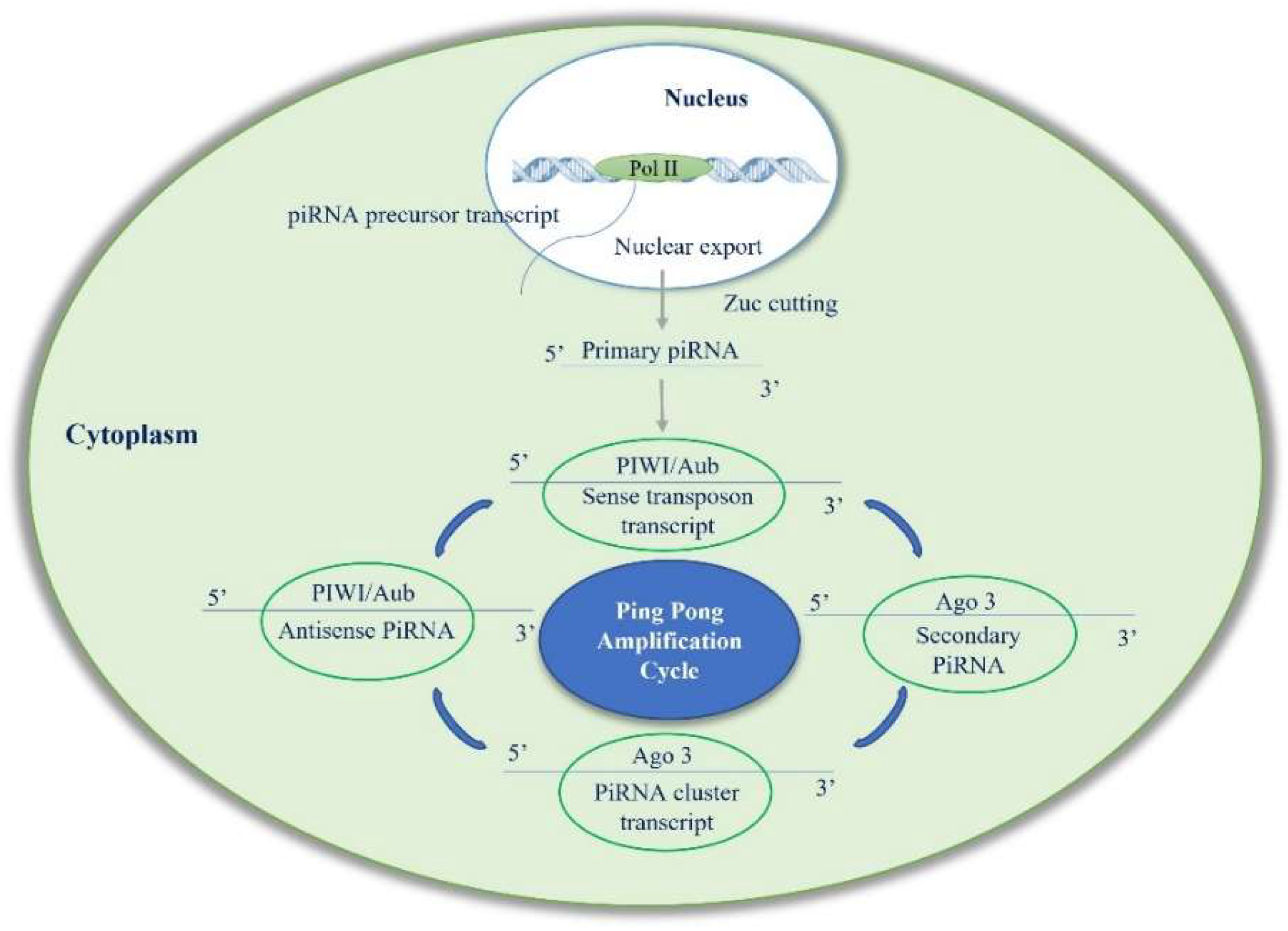
2. Biogenesis of piRNAs

piRNAs were first found in germline cells and are considered critical regulators of germline maintenance. These animal-specific short-chain RNAs have a size of 24–32 nucleotides and a 2′-O-methylation at the 3′ end, a distinctive and exclusive feature of all piRNAs, and are associated with PIWI proteins. These proteins belong to the Argonaute protein family [9], which were discovered, for the first time, in Drosophila melanogaster ovarian germ cells and follicular cells [10]. piRNAs can bind DNA sequences of specific genes via complementary base pairing to silence transposons and regulate gene expression. The human genome contains over 30,000 piRNA genes that are mainly derived from intergenic regions. According to multiple origins, piRNAs are divided into three subclasses: mRNA-derived, lncRNA-derived, and transposon-derived piRNAs [11]. piRNAs transcribed from transposons are known as “piRNA clusters”. These clusters are mainly located in the pericentromeric and sub-telomeric parts of the chromosomes. piRNA clusters are transcribed to form piRNA precursors via bidirectional or unidirectional transcription. Moreover, piRNAs can be generated from mRNA 3′ untranslated region (3′ UTR) and some long non-coding regions in the genome [12]. The mechanism of piRNA production includes two steps: a primary and a secondary amplification cycle described as the “ping-pong cycle”, in which piRNAs are bound to PIWI proteins [13]. In primary amplification, newly transcribed piRNAs are exported through the nuclear envelope, processed, and matured. In the cytoplasm, the secondary structures are resolved via RNA helicase Armitage (Armi). After, piRNA precursors are cleaved by mitochondria-associated endonuclease Zucchini (Zuc) and transformed into pre-piRNAs with a 5′ monophosphate. Then, pre-piRNAs are loaded onto PIWI proteins and cut at the 3′ ends by a 3′ to 5′ exonuclease, Nibbler (Nbr) [12]. After, 3′ terminal ends are methylated at 2′ oxygen by RNA2′-O-methyltransferase Hen1 [13]. piRNAs produced in this way are named primary piRNAs. In secondary amplification, piRNAs’ generation is increased with the involvement of Argonauta 3 (Ago 3) and Aubergine (Aub) proteins. Aub binds to antisense strand piRNAs and cleaves sense piRNA precursors, giving rise to sense piRNAs bound by Ago3. In contrast, Ago3 binds to sense-strand piRNAs and cleaves antisense piRNA precursors, producing antisense piRNAs that load onto Aub [14]. The round of cleavage repeats and produces several piRNA molecules. These piRNAs are then bound by PIWI proteins and transported back to the nucleus to silence target genes. Two Tudor-domain containing piRNA factors, Krimper (Krimp) and Qin/Kumo, play crucial roles in making Aub-AGO3 heterotypic ping-pong robust. This maintains the levels of piRNAs loaded onto Piwi and Aub to efficiently repress transposons at transcriptional and post-transcriptional levels, respectively [15].
To summarize, in the primary pathway, piRNA forms a complex with PIWI proteins to mediate transposon silencing. In the secondary pathway, piRNA binds to Aub protein to produce secondary piRNA, while secondary piRNA binds to Ago3 to produce primary piRNA, and the cycle continues [9] (Figure 1). Although with some differences, the piRNA ping-pong mechanism exists not only in germ cells but also in somatic cells [14]. Despite increasing interest in the role of piRNAs in human diseases, their homeostasis in cells is still a poorly understood process. RNAs’ cellular concentration is maintained by a balance of biogenesis and degradation. PIWI proteins protect piRNAs from this degradation. However, when piRNAs are released from the PIWI complex, their 5′ end and 3′ end become unprotected and can easily be accessed by exoribonucleases. The degradation of human piRNAs is mainly dependent on the 5′-3′ exoribonuclease pathway mediated by XRN1 and XRN2, the two major 5′-3′ exoribonucleases involved in piRNA degradation in human somatic cells (Figure 2). It was also reported that the presence of 3′-end 2′-O-methylation in piRNAs reduced their degradation through an exosome-mediated decay pathway [16].

3. Function of piRNAs and PIWI Proteins

Depending on the context, piRNAs can act in the following different ways: transposon silencing, epigenetic regulation, germ stem cell maintenance, and genome rearrangement. Recently, piRNAs have been described in tumorigenesis and in various steps of cancer progression, such as proliferation, invasion, metastasis, apoptosis, and drug resistance [17] (Figure 3). piRNAs act in reproduction, in a PIWI-dependent manner, and in fertility regulation by attaching to PIWI proteins and forming a silencing ribonucleoprotein complex [9]. It seems likely their ancestral function was an adaptive mechanism to silence active transposable elements (TEs). Through complementary sequences, piRNA cluster transcripts recognize TEs, avoiding their expression, which might lead to a loss of genome integrity. piRNAs can regulate cellular processes with different mechanisms, also independently of PIWI proteins, such as increasing translation or stabilizing mRNA [18]. Recent findings on piRNAs’ biological significance suggest that they can somatically regulate gene expression also via epigenetic alterations [19]. Epigenetic global changes in cancer include DNA methylation, DNA hypomethylation, CpG island methylation, and gene-specific DNA hypermethylation, leading to oncogene activation (Ras, cyclin D2) [20] and tumor suppressor silencing (RB1, p16) [21]. It was demonstrated that aberrant DNA methylation in tumor cells is linked to PIWI/piRNA disorders. Therefore, piRNA deregulation may influence the expression and stability of the genome, causing cell signaling alteration, which, in turn, may induce disease onset and progression [22,23,24,25,26,27,28].
PIWI family proteins consist of four members crucial for the biogenesis and function of small ncRNAs: PIWIL1, PIWIL2, PIWIL3, and PIWIL4. PIWIL proteins bind piRNAs, as a unique type of small ncRNA, forming a PIWI/piRNA complex. This complex exerts the gene regulation function, playing an important role in the stability and integrity maintenance of the germ cell genome [8]. PIWI/piRNA acts as an epigenetic modulator recruiting other epigenetic regulatory factors, such as DNA methylase, beyond its function of directly cutting and degrading target RNA, acting like an Ago protein/miRNA complex. PIWI family proteins have been considered prognostic markers for various malignancies [29]. Although specific mechanisms need further investigation, various studies have demonstrated that PIWI proteins, expressed in many cancers, affect multiple biological processes, also without interacting with piRNAs, including different steps of cancer progression such as cell proliferation, apoptosis, migration, invasion, cell cycle regulation, and self-renewal [30] (Figure 4). Specific examples of PIWIL proteins involved in cancer are described below. PIWIL1 is the most studied PIWI protein that regulates gene expression, apoptosis, cell cycle, and proliferation. PIWIL1 is a coactivator of adenomatous polyposis coli C-terminal domain C complex that targets cell adhesion protein, Pinin, for proteolytic ubiquitination, thus promoting metastasis in pancreatic cancer (PC) [31]. PIWIL2 overexpression functions as an oncogene; its deregulation plays an important role in cancer progression and is associated with poor survival and aggressive clinicopathological properties of patients [32]. The deregulation of PIWIL3 is reported in many cancer types; it is highly expressed in both primary ovarian cancer (OC) and metastatic tissues [33]. PIWIL3 plays an important role in melanoma, and its expression correlates with the tumor stage [34]. In gastric cancer (GC), PIWIL3 upregulation increases cell proliferation, migration, and invasion [35]. Conversely, PIWIL3 overexpression seems to have a protective effect in glioma cell lines and decreased tumor size in vivo [36]. Moreover, PIWIL3 is considered a prognostic biomarker of breast cancer (BC) since its upregulation is significantly associated with poor overall survival [37].
PIWIL4 is involved in chromatin modifications in human somatic cells [38], and it can process precursor hairpins generating miRNAs in DICER independent manner [39]. PIWIL4 role in tumorigenesis is controversial; high expressions of PIWIL4 are found in colorectal, cervical, gastric, and ovarian cancer [40,41]. However, other studies reported that low PIWIL4 expression is associated with a poor prognosis in different cancer types [37]. Furthermore, the lack of PIWIL4 expression triggered by CpG island hypermethylation has been found in testicular tumors [42] (Table 1).

4. piRNAs in Cancer

Cancer and germ cells share important biological characteristics such as rapid proliferation and the ability for self-renewal. Recently, a growing number of studies have revealed the role of piRNAs in cancers, launching a new biological concept in which piRNAs mediate a gene regulation mechanism typical of germline cells in somatic cells [43]. It is plausible that cancer cells with a high proliferation rate can adopt and utilize self-renewal machinery like germ cells. In malignant cells, piRNAs are involved in epigenetic regulation and may be crucial to sustaining cancer stemness [30].

4.1. Role of piRNAs in Cancer Initiation and Progression

Several piRNAs affect cancer stem cells (CSCs) and somatic cells by regulating gene expression via epigenetic processes. CSCs are a small population of cancer cells with high heterogeneity and a great capacity to renew tumors. Aberrant expressions of piRNAs and PIWI proteins also in CSCs can regulate tumor initiation and progression. Examples of specific piRNAs involved in cancer are described below. In non-small cell lung cancer (NSCLC), piR-651 upregulation correlates with a significant increase in tumor growth and metastasis, affecting cell cycle arrest and inducing cyclin D1 and CDK4 and suggesting piR-651 as a potential oncogene. piR-651 overexpression promotes proliferation and invasion and reduces cell apoptosis by inducing different oncogene (CDK4, Cyclin D1, and MDM2) expressions [44]. In addition, piR-651 can increase phosphatase and tensin homolog (PTEN) methylation via DNA (cytosine-5)-methyltransferase 1 (DNMT1) [45]. It was also demonstrated that piR-651 is down-regulated in patients with Hodgkin lymphoma (HL) with respect to healthy controls; low levels of piR-651 correlate with poor prognosis in HL patients [46]. High expressions of piR-30473 support the aggressive phenotype of diffuse large B-cell lymphoma, exerting its oncogenic role through a mechanism involving the upregulation of Wilms Tumor-1 Associated Protein (WTAP), an m6A mRNA methylase, that enhances the global m6A level. WTAP induces the expression of its critical target gene, hexokinase 2 (HK2), by enhancing HK2 m6A level, thereby promoting lymphoma progression [47]. An imbalance in piRNA regulatory processes modifies several levels of gene regulation that control DNA damage repair, chromatin organization, and mitogenic signals, inducing uncontrolled cell proliferation [48]. piR-55490 expression is downregulated in lung cancer (LC), restoring piR-55490 can reduce LC cell proliferation rates, whereas suppressing piR-55490 increases cell proliferation rates. piR-55490 acts by inhibiting the serine/threonine kinase 1 (AKT)/mTOR pathway, thereby suppressing cell growth [49]. piR-211106 binding to pyruvate carboxylase can inhibit the progression of LC-enhancing chemotherapy sensitivity, suggesting that it is a potential therapeutic target [50]. In BC has been described an aberrant expression of various piRNAs. piR-36712 is lowly expressed in this cancer type compared with non-tumor tissues and acts as a possible tumor suppressor. It was also reported that piR-36712 upregulation has a synergistic anticancer effect with chemotherapy on BC cells via the Interaction with SEPW1 pseudogene SEPW1P RNA [51]. piR-36712 can be considered a novel tumor suppressor and a prognostic predictor of BC. Moreover, piR-36712 modulates the expression levels of tumor suppressor genes p53 and P21; its increase leads to cell cycle arrest in the G0/G1 phase of cancer cells [52]. piR-021285 induces methylation at cancer-relevant genes, and it is considered a potential modulator of BC invasiveness by remodeling the cancer epigenome. The exogenous expression of piR-021285 induces significant methylation differences at BC-related genes, including the attenuated methylation of 5′ UTR first exon at the pro-invasive ARHGAP11A gene. There is an increased ARHGAP11A mRNA expression and enhanced invasiveness in variant versus WT piR-021285 mimic-transfected BC cell lines, supporting the role of this piRNA in tumorigenesis via a piRNA-mediated epigenetic mechanism [53]. In clear cells renal carcinoma (ccRC), by using piRNA microarray in a large cohort study, three piRNAs (piR-30924, piR-57125, and piR-38756) have been identified as piRNAs significantly associated with tumor recurrence and overall survival [54]. In cells and plasma of various cancer patients, an altered expression of piR-823 has been observed, with a role in regulating tumor cell growth. In GC, piR-823 acts as a tumor suppressor, and its expression is dramatically decreased in GC tissues. The restoration of piR-823 in GC cells inhibits cancer cell growth both in vitro and in vivo [55]. Also, in colorectal cancer (CRC), piR-823 downregulation inhibits cell proliferation and increases cell apoptosis by inducing an apoptosis activator gene, the transcription factor HSF1 [56]. Moreover, in CRC tissue and serum, piR-54265 is upregulated and induces cancer progression activating STAT3 signaling [57]. Furthermore, it was reported that piR-18 is involved and contributed to the tumorigenesis and progression of CRC. The overexpression of piR-18 inhibits the cell proliferation, migration, and invasion of CRC; thus, it could potentially be used as a new biomarker for diagnosis and therapy [52]. In hepatocellular carcinoma (HCC), a new piRNA, piR-Hep1 has been identified; it is upregulated in HCC with respect to non-tumoral liver cells. The silencing of piR-Hep1 inhibits cell viability, migration, and invasion, with a concomitant decrease in AKT phosphorylation [58,59] (Figure 5).
In BC, piR-2158 is downregulated in CSCs; it was demonstrated that the overexpression of piR-2158 prevents mammary gland tumorigenesis via regulating CSCs and tumor angiogenesis. piR-2158 acts as a transcriptional repressor of Interleukin 11 (IL11) by competing with AP-1 transcription factor subunit FOSL1 to bind the promoter of IL11. piR-2158 can also provide a potential therapeutic strategy in BC treatment [60]. Moreover, the piRNA/PIWI complex can selectively control the phosphorylation of target proteins. It was reported that piR-54265-binding PIWIL2 promotes the formation of PIWIL2/STAT3/phosphorylated SRC complex, inducing phosphorylated SRC-mediated STAT3 phosphorylation that, in turn, causes the proliferation, metastasis, and chemotherapy resistance of CRC cells [19]. The piR-823/PIWIL2 complex mediates STAT3 phosphorylation and the activation of the STAT3/BCL-xl/cyclin D1 pathway, inducing the expression of cyclin-dependent kinase inhibitors and controlling G1 phase regulators Cyclin D1 and CDK4, thus promoting CRC progression [39]. piRNA/PIWI complex can interact with other ncRNAs, including miRNAs and lncRNAs, to regulate cancer progression. piR-30188/PIWIL3 binds to OIP5-AS1, a cancer-associated lncRNA, a target of miR-367-3p in gliomas. miR-367-3p negatively regulates CEBPA mRNA expression and increases TRAF4 expression [36]. The combination of OIP5-AS1 knockdown with the over-expression of PIWIL3 and miR-367-3p leads to tumor regression, identifying a novel molecular pathway in glioma cells that may provide a potential innovative approach for cancer therapy [23]. Overall, these findings indicate that piRNAs can have several potential clinical applications in diagnosis, prognosis, and therapy (Table 2).

4.2. piRNAs as Cancer Biomarkers

ncRNAs have received a lot of attention as one factor contributing to genetic and epigenetic instability. Clinically, ncRNA alterations have a relevant diagnostic and prognostic significance [26]. Epigenetic biomarkers can be useful in predicting therapeutic drug responses, and piRNAs are considered emerging biomarkers for therapy monitoring. A biomarker is a significant indicator that can be used to assess a target’s diagnostic potential, risk of recurrence, and clinical prognosis [63]. Aberrant expressions of piRNAs and their correlation with cancer patient features suggest that these may have an important clinical impact, not only as diagnostic biomarkers but also as druggable targets [67]. Alterated piRNA levels may be considered good cancer biomarkers, with higher sensitivity and specificity than miRNAs [69]. Nowadays, few studies investigate the role of piRNAs as cancer biomarkers, but the field is in progress and updated [70]. Some studies indicate that piRNAs have a better diagnostic ability than traditional biomarkers. It was reported that piR-13643 and piR-21238 performed better than conventional biomarkers, such as hector battifora mesothelial antigen-1 (HBME1), conventionally used to discriminate malignant nodules from benign ones in papillary thyroid carcinoma [65]. Moreover, serum piR-5937 and piR-28876 were able to distinguish CRC from healthy controls with higher sensitivity and specificity than traditional markers such as Carcinoembryonic Antigen (CEA) and Carbohydrate antigen 19-9 (CA199) [61]. It was also suggested that piR-54265 can be used as a biomarker for the early detection and clinical monitoring of CRC [77].

5. piRNAs and Liquid Biopsy

Liquid biopsy is considered an ideal tool for discovering new cancer biomarkers, and in the current era of personalized medicine, LB has acquired high relevance in cancer patient management [66]. LB serves as a safe alternative to solid biopsies; its main components are circulating tumor cells (CTCs), circulating tumor DNA (ctDNA), circulating tumor RNA (ctRNA), tumor-associated platelets, and EVs [78]. Nowadays, clinical oncology adopts next-generation sequencing (NGS)-based diagnostics; these high throughput technologies allow for the identification of ncRNA profiles and significant genetic mutations across the human genome. LB is increasingly being used for early diagnosis and to determine the best therapeutic option for cancer patients [79].

5.1. piRNAs in Extracellular Vesicles

Extracellular vesicles (EVs) are nanoscale membrane particles released by all cytotypes in physiological and pathological conditions. EVs are internalized by target cells through a different mechanism and transport a plethora of bioactive molecules, including proteins, lipids, and nucleic acids [68,75,80,81,82]. EVs have a key role in several steps of cancer progression; they are involved in proliferation, migration, premetastatic niche formation, and immune response [62,76]. EVs have been reported to contain an abundance of RNAs, such as ncRNAs, including miRNAs, lncRNAs, circRNAs, and piRNAs [74]. The packaging of different ncRNAs into EVs is selective and mirrors the status of parental cells. EV-RNAs, with regulatory effects, have been implicated in many EV-mediated biological functions [64,75]. EVs are widely present in biofluids, such as blood, urine, saliva, and malignant effusions. Moreover, piRNAs can cross the plasma membrane, and EVs released by cancer cells contained piRNAs that remain stable in body fluids. This feature suggests that piRNAs can easily be detected in body fluids as accurate biomarkers [44]. Several studies have focused on plasma EV’s role as an early and non-invasive diagnostic biomarker for cancers. EV-cargo represents a valuable source of genetic materials, mainly ncRNA biomarkers [83]. These findings indicate that LB can allow for the detection of piRNAs that may become therapeutic and diagnostic tools for various cancer types. Differentially expressed EV-piRNAs have been identified in patients with specific disease conditions compared to healthy controls, suggesting an association between piRNA and progression in various diseases [72]. Although we are currently lacking guidelines on piRNA bioinformatic analysis, EV-piRNA can be considered a new research niche in human cancer pathology. piRNAs encapsulated in EVs are stable in body fluids and have the potential to be promising markers for cancer diagnosis and prognosis and new tools for innovative therapies [73]. Examples of specific piRNAs shuttled by EVs are described below and summarized in Table 3.
It was reported that piR-26925 and piR-5444 are upregulated In EVs isolated from the serum of LC patients [84]. A high-throughput sequencing of small ncRNAs from EVs, cancerous and adjacent noncancerous tissues in patients with NSCLC, was applied to recognize candidate piRNAs as diagnostic biomarkers. This study reveals that piR-164586 was significantly upregulated in paracancerous tissues and EVs from serum samples of healthy individuals [85].
Recently, it was demonstrated that EVs isolated from the plasma of neuroblastoma (NB) patients contain high levels of piR-1089. In vitro, studies indicated that EV-piR-1089 promoted NB cell proliferation and migration by inhibiting Kelch-like ECH-associated protein 1 (KEAP1) expression. Low KEAP1 expressions were associated with NB progression in clinical samples [86]. piRNAs are also involved in the crosstalk between multiple myeloma (MM) cells and bone marrow microenvironment. In MM, piR-823 silencing causes cell cycle dysregulation, reduction of apoptosis-related proteins, and de novo DNA methyltransferases, thus inhibiting the tumorigenicity of MM in vitro and in vivo [87]. piR-823 contained in EVs from peripheral blood of MM patients and cell lines plays an important role in cell–cell communication between endothelial and myeloma cells in the tumor microenvironment. EV-piR-823 from MM cells can be effectively transferred to endothelial cells and alter their biological characteristics, promoting anti-apoptotic and pro-angiogenic activity and creating a favorable microenvironment for MM cell survival. The upregulation of piR-823 can increase the expressions of IL-6, VEGF, and ICAM-1, promoting endothelial cell proliferation, invasion, and tube formation [88]. Granulocytic myeloid-derived suppressor cells (MDSCs) can trigger piR-823 expression, and silencing piR-823 can decrease the stemness of MM stem cells maintained by granulocytic MDSCs [89]. Moreover, piR-823 expression has been correlated with the stage and prognosis of MM, indicating its potential as a therapeutic target and prognostic stratification biomarker [88,90]. Gu et al. reported that the EV-piRNA population is altered in the plasma of cholangiocarcinoma (CCA) and gallbladder carcinoma (GBC) patients compared to healthy individuals. EV-piRNA profiling revealed unique piRNA signatures of CCA and GBC. Small RNA sequencing data, obtained by the NGS system, showed piR-2660989, piR-10506469, piR-20548188, piR-10822895, piR-23209, and piR-18044111 in EVs isolated from CCA and GBC plasma. Interestingly, expression level analyses of EV-piRNAs in plasma of CCA and GBC patients, before and after surgeries, have demonstrated that piR-10506469 and piR-20548188 were significantly decreased in patients who underwent surgeries [91]. In OC, EVs drive the crosstalk between cancer cells and omental fibroblasts. EV-piR-25783 activates TGF-β/SMAD2/SMAD3 pathway in fibroblasts and promotes the fibroblast-to-myofibroblast transition in the omentum, secretion of various cytokines, proliferation, migration, and invasion, contributing to the premetastatic niche formation. piR-25783-induced myofibroblasts improve tumor implantation and growth in the omentum. EV-piR-25783 upregulation is thus associated with adverse clinicopathological characteristics and shorter survival [92]. Moreover, EV-piRNAs can be considered promising non-invasive diagnostic biomarkers for GC, piR-019308, piR-004918, and piR-018569 contained in serum EVs were significantly increased in GC patients compared to healthy controls [93].
In prostate cancer (PCa), piRNAs were involved in the proliferation, migration, and invasion of PCa cells by activating different signal pathways, which may represent a new marker of PCa diagnosis. It was reported that piRNAs contained in urinary EVs, isolated from patients with PCa and detected using NGS technology, are useful for diagnosis. The fraction of PCa-derived EVs in urine is larger than in plasma and allows for a better detection and tracking of PCa-derived RNAs [94]. The expressions of piR-349843, piR-382289, piR-158533, and piR-002468 in urinary EVs were significantly increased in PCa patients compared with healthy controls [95]. Interestingly, it was also indicated that in plasma EVs of patients with bladder cancer (BCa), two small ncRNAs, miR-4508 and piR-5936, were associated with the risk class: miR-4508 with a descendant trend going from controls to high-risk BCa; piR-5936 with an ascending trend [96]. Taken together, these reports indicate that EV-piRNAs have an important role in tumor–host crosstalk, contributing to cancer progression and showing a great potential as theranostic biomarkers.

5.2. piRNA-Based Therapeutic Approaches in Cancer

On-target specificity is a significant difficulty to overcome for an RNA-based therapeutic strategy that involves gene silencing. Moreover, RNAs are unstable molecules, and the development of ways to enhance RNA stability and its uptake are crucial points. Although siRNA molecules are structurally susceptible to RNases and degradation, some advances to overcome these limits are ongoing, employing various chemical and structural modifications that make RNAi delivery more physiologically amenable. Synthetic lipid nanoparticles have been developed and have enabled successful systemic delivery of RNAi. Each of these RNA components can be modified to overcome their susceptibility to degradation [94]. Stability and barriers to delivery were overcome thanks to the success of the mRNA vaccine for COVID-19 in early 2021, opening new routes for targeted RNA therapies [95]. siRNAs are double-stranded ncRNAs with similar biological functions to miRNAs but are synthetically derived (exogenously) via the process of RNA interference (RNAi). siRNAs, like miRNAs, degrade target mRNAs in a sequence-specific manner, leading to gene silencing. RNAi machinery targets and can silence pathologic mRNA sequences [94].
Some typical limitations of siRNAs and miRNAs can be overcome using piRNAs and EVs as their shuttles. Nowadays, the potential role of piRNAs as therapeutic tools has been reported; piRNA treatment is performed via the correction of pathological alteration of piRNA expression, including the reactivation of endogenous piRNA as a pathological inhibitor and functional blocking or reduction in piRNA expression as a pathological driver [30]. Synthetic piRNAs are very intriguing as they could block the synthesis of cancer-related proteins by binding to mRNAs. Different from miRNAs, which need to be processed by enzymes and regulate several mRNAs, piRNAs have the advantage of not requiring enzyme processing and better specificity than other ncRNAs to targets [97]. Since epigenetic regulatory mechanisms are closely related to cancer progression, the epigenetic reprogramming of cancer cells can be a potentially powerful therapeutic approach [43]. Epigenetic drugs (epidrugs) consist of small molecules targeting epigenetic key signaling pathways, promoting transcriptional and post-transcriptional modifications and acting mainly on tumor suppressor and DNA repair gene activation [44]. Since piRNAs regulate protein-coding genes involved in the development of various diseases, they can be considered molecules useful as innovative epidrugs [45]. Although the studies on piRNAs as new therapeutic methods are still in the early stage, understanding the mechanisms and functions of piRNAs could enhance therapeutic alternatives in cancer. Recently, EV-mediated ncRNA delivery systems have attracted tremendous attention in the research of next-generation therapeutics for the treatment of several pathologies, including cancer [98]. EVs, thanks to their small size, low immunogenicity, and high biocompatibility due to their natural origin [99,100], are more attractive than synthetic vehicles. Moreover, EV cargos are protected from degradation in the bloodstream by a lipid bilayer membrane; they have the potential to escape from immune system clearance and cross the physiological barriers [101]. Despite these advantages, natural EVs show some pitfalls. The procedures for EV isolation are time consuming, monitoring methods for cargo dosage are insufficient, and EV stability after storage is not easy to monitor [102]. Currently, the yield of EVs isolated from body fluids or culture media is insufficient to supply clinical demands, and EV-quality control is a challenge that limits their clinical application [103]. To overcome this limit, EVs collected from non-human sources such as plants, bacteria, and milk are considered a potential alternative for cancer therapy. Since EV-mediated communication is involved in all the domains of life and in several cellular physiological and pathological processes, EVs are involved in a universal, evolutionarily conserved mechanism for inter-kingdom and intra-kingdom communication [104]. It was reported that plant EVs can be used as nanoplatforms to transfer ncRNAs in inter-kingdom communication [28,71]. ncRNAs contained in plant EVs are considered a new class of cross-kingdom modulators, capable of mediating animal-plant interactions at the molecular level [105]. Applications of plant EVs in nanomedicine and nutraceutics are based on their intrinsic biological properties, such as anti-cancer, anti-inflammatory, and anti-aging, and on their use as shuttles of therapeutic biomolecules [106]. It was also reported that bacterial EVs isolated from the host gut microbiome can enter the circulatory system to disseminate to distant organs and tissues and be detected in human biofluids. Bacterial EVs in microbiome-based liquid biopsies might be useful for cancer diagnostics and bioengineering strategies for cancer therapy [107]. Moreover, bovine milk-derived EVs have been shown to increase the oral bioavailability of drugs and are optimal vehicles to transport bioactive compounds for nutritional and therapeutic purposes [108]. In cancer therapy, milk EVs can be functionalized with ligands such as folic acid to achieve cancer targeting [109]. In addition, milk-derived EVs have shown several therapeutic effects, such as a selective interaction with macrophages and an increase in intestinal stem cell proliferation as potential adjuvant therapy to standard clinical management of malnourished children [110].

6. Conclusions and Outlook

Recently, significant advances in the piRNA field have been made, providing mechanistic insights into the transcription of piRNA clusters, piRNA biogenesis, and piRNA function. Although the knowledge of piRNAs in cancers remains in its infancy, a growing number of studies have shown that piRNAs can be considered good diagnostic and prognostic markers and new therapeutic tools. However, it needs to report a few studies indicating that piRNAs could be fragments of other ncRNAs and could correspond to false positives due to the use of noncurated piRNA databases [111,112]. Many studies have found aberrant piRNAs in biofluids of cancer patients; thus, LB is considered an ideal tool for developing cancer biomarkers. Even if a single piRNA can be sufficient to distinguish cancer patients from healthy controls, combined biomarkers may be more diagnostically accurate than one. EVs can have a dual role in cancer management as a source of diagnostic biomarkers with the identification of specific piRNAs and as a therapeutic tool consisting of engineered EVs with piRNAs useful as potential and breakthrough epidrugs (Figure 6). Future research efforts should concentrate on wide studies on piRNA profiles in different cancer types and stages. This effort can allow for the identification of comprehensive diagnostic panels able to compliant the heterogeneous landscape of cancer. Preclinical studies show how smart EVs have the potential to expand into next-generation drugs to address the current demand for precision cancer therapies. However, numerous milestones on piRNAs contained in EVs are yet to be achieved. To date, the clinical trials with piRNAs and EV-piRNAs are still a hard challenge, and only a few clinical studies on these emerging ncRNAs are ongoing (www.clinical.trials.org, accessed on 20 April 2023). Further clinical studies are needed to demonstrate that EV-piRNAs are smart tools useful for theranostic applications in precision oncology.

Author Contributions

Conceptualization: S.T. and G.C.; methodology, data curation; writing—original draft preparation: S.T., A.M. and G.C.; writing—review and editing: S.T. and G.C.; visualization: S.T., A.M. and G.C.; supervision: S.T. and G.C. All authors have read and agreed to the published version of the manuscript.

Funding

This research received no external funding.

Institutional Review Board Statement

Not applicable.

Informed Consent Statement

Not applicable.

Data Availability Statement

Not applicable.

Conflicts of Interest

The authors declare no conflict of interest.

References

  1. Dhamija, S.; Menon, M.B. Non-Coding Transcript Variants of Protein-Coding Genes-What Are They Good For? RNA Biol. 2018, 15, 1025–1031. [Google Scholar] [CrossRef] [PubMed] [Green Version]
  2. Kumar, S.; Gonzalez, E.A.; Rameshwar, P.; Etchegaray, J.-P. Non-Coding RNAs as Mediators of Epigenetic Changes in Malignancies. Cancers 2020, 12, 3657. [Google Scholar] [CrossRef] [PubMed]
  3. Ye, J.; Li, J.; Zhao, P. Roles of NcRNAs as CeRNAs in Gastric Cancer. Genes 2021, 12, 1036. [Google Scholar] [CrossRef]
  4. Cammarata, G.; Barraco, N.; Giusti, I.; Gristina, V.; Dolo, V.; Taverna, S. Extracellular Vesicles-CeRNAs as Ovarian Cancer Biomarkers: Looking into CircRNA-MiRNA-MRNA Code. Cancers 2022, 14, 3404. [Google Scholar] [CrossRef] [PubMed]
  5. Pucci, M.; Reclusa Asiain, P.; Durendez Saez, E.; Jantus-Lewintre, E.; Malarani, M.; Khan, S.; Fontana, S.; Naing, A.; Passiglia, F.; Raez, L.E.; et al. Extracellular Vesicles As MiRNA Nano-Shuttles: Dual Role in Tumor Progression. Target. Oncol. 2018, 13, 175–187. [Google Scholar] [CrossRef] [PubMed]
  6. Ameli Mojarad, M.; Ameli Mojarad, M.; Shojaee, B.; Nazemalhosseini-Mojarad, E. PiRNA: A Promising Biomarker in Early Detection of Gastrointestinal Cancer. Pathol. Res. Pract. 2022, 230, 153757. [Google Scholar] [CrossRef] [PubMed]
  7. Cammarata, G.; de Miguel-Perez, D.; Russo, A.; Peleg, A.; Dolo, V.; Rolfo, C.; Taverna, S. Emerging Noncoding RNAs Contained in Extracellular Vesicles: Rising Stars as Biomarkers in Lung Cancer Liquid Biopsy. Ther. Adv. Med. Oncol. 2022, 14, 17588359221131228. [Google Scholar] [CrossRef]
  8. Weng, W.; Li, H.; Goel, A. Piwi-Interacting RNAs (PiRNAs) and Cancer: Emerging Biological Concepts and Potential Clinical Implications. Biochim. Biophys. Acta Rev. Cancer 2019, 1871, 160–169. [Google Scholar] [CrossRef]
  9. Koch, B.; Geßner, A.; Farmand, S.; Fuhrmann, D.C.; Chiocchetti, A.G.; Schubert, R.; Baer, P.C. Effects of Hypoxia on RNA Cargo in Extracellular Vesicles from Human Adipose-Derived Stromal/Stem Cells. Int. J. Mol. Sci. 2022, 23, 7384. [Google Scholar] [CrossRef]
  10. Huang, M.; Peng, X.; Yang, L.; Yang, S.; Li, X.; Tang, S.; Li, B.; Jin, H.; Wu, B.; Liu, J.; et al. Non-Coding RNA Derived from Extracellular Vesicles in Cancer Immune Escape: Biological Functions and Potential Clinical Applications. Cancer Lett. 2021, 501, 234–246. [Google Scholar] [CrossRef]
  11. Sadoughi, F.; Mirhashemi, S.M.; Asemi, Z. Epigenetic Roles of PIWI Proteins and PiRNAs in Colorectal Cancer. Cancer Cell Int. 2021, 21, 328. [Google Scholar] [CrossRef] [PubMed]
  12. Houwing, S.; Kamminga, L.M.; Berezikov, E.; Cronembold, D.; Girard, A.; van den Elst, H.; Filippov, D.V.; Blaser, H.; Raz, E.; Moens, C.B.; et al. A Role for Piwi and PiRNAs in Germ Cell Maintenance and Transposon Silencing in Zebrafish. Cell 2007, 129, 69–82. [Google Scholar] [CrossRef] [PubMed] [Green Version]
  13. Billi, A.C.; Alessi, A.F.; Khivansara, V.; Han, T.; Freeberg, M.; Mitani, S.; Kim, J.K. The Caenorhabditis Elegans HEN1 Ortholog, HENN-1, Methylates and Stabilizes Select Subclasses of Germline Small RNAs. PLoS Genet. 2012, 8, e1002617. [Google Scholar] [CrossRef] [PubMed] [Green Version]
  14. Zhang, Z.; Xu, J.; Koppetsch, B.S.; Wang, J.; Tipping, C.; Ma, S.; Weng, Z.; Theurkauf, W.E.; Zamore, P.D. Heterotypic PiRNA Ping-Pong Requires Qin, a Protein with Both E3 Ligase and Tudor Domains. Mol. Cell 2011, 44, 572–584. [Google Scholar] [CrossRef] [Green Version]
  15. Sato, K.; Iwasaki, Y.W.; Siomi, H.; Siomi, M.C. Tudor-Domain Containing Proteins Act to Make the PiRNA Pathways More Robust in Drosophila. Fly 2015, 9, 86–90. [Google Scholar] [CrossRef] [Green Version]
  16. Balaratnam, S.; Hoque, M.E.; West, N.; Basu, S. Decay of Piwi-Interacting RNAs in Human Cells Is Primarily Mediated by 5’ to 3’ Exoribonucleases. ACS Chem. Biol. 2022, 17, 1723–1732. [Google Scholar] [CrossRef]
  17. Fonseca Cabral, G.; Azevedo Dos Santos Pinheiro, J.; Vidal, A.F.; Santos, S.; Ribeiro-Dos-Santos, Â. PiRNAs in Gastric Cancer: A New Approach Towards Translational Research. Int. J. Mol. Sci. 2020, 21, 2126. [Google Scholar] [CrossRef] [Green Version]
  18. Tóth, K.F.; Pezic, D.; Stuwe, E.; Webster, A. The PiRNA Pathway Guards the Germline Genome Against Transposable Elements. Adv. Exp. Med. Biol. 2016, 886, 51–77. [Google Scholar] [CrossRef] [Green Version]
  19. Li, Z.; Tang, X.; Shen, E.-Z. How Mammalian PiRNAs Instruct de Novo DNA Methylation of Transposons. Signal Transduct. Target. Ther. 2020, 5, 190. [Google Scholar] [CrossRef]
  20. Wilson, A.S.; Power, B.E.; Molloy, P.L. DNA Hypomethylation and Human Diseases. Biochim. Biophys. Acta-Rev. Cancer 2007, 1775, 138–162. [Google Scholar] [CrossRef]
  21. Baylin, S.B. DNA Methylation and Gene Silencing in Cancer. Nat. Clin. Pract. Oncol. 2005, 2, S4–S11. [Google Scholar] [CrossRef] [PubMed]
  22. Yao, J.; Xie, M.; Ma, X.; Song, J.; Wang, Y.; Xue, X. PIWI-Interacting RNAs in Cancer: Biogenesis, Function, and Clinical Significance. Front. Oncol. 2022, 12, 965684. [Google Scholar] [CrossRef] [PubMed]
  23. Chénais, B. Transposable Elements and Human Cancer: A Causal Relationship? Biochim. Biophys. Acta-Rev. Cancer 2013, 1835, 28–35. [Google Scholar] [CrossRef] [PubMed]
  24. Fabry, M.H.; Ciabrelli, F.; Munafò, M.; Eastwood, E.L.; Kneuss, E.; Falciatori, I.; Falconio, F.A.; Hannon, G.J.; Czech, B. PiRNA-Guided Co-Transcriptional Silencing Coopts Nuclear Export Factors. Elife 2019, 8, e47999. [Google Scholar] [CrossRef]
  25. Jia, D.-D.; Jiang, H.; Zhang, Y.-F.; Zhang, Y.; Qian, L.-L.; Zhang, Y.-F. The Regulatory Function of PiRNA/PIWI Complex in Cancer and Other Human Diseases: The Role of DNA Methylation. Int. J. Biol. Sci. 2022, 18, 3358–3373. [Google Scholar] [CrossRef]
  26. Ali, R.; Laskar, S.A.; Khan, N.J.; Wahab, S.; Khalid, M. Non-Coding RNA’s Prevalence as Biomarkers for Prognostic, Diagnostic, and Clinical Utility in Breast Cancer. Funct. Integr. Genomics 2023, 23, 195. [Google Scholar] [CrossRef]
  27. Baylin, S.B.; Jones, P.A. A Decade of Exploring the Cancer Epigenome—Biological and Translational Implications. Nat. Rev. Cancer 2011, 11, 726–734. [Google Scholar] [CrossRef]
  28. Siddiqi, S.; Matushansky, I. Piwis and Piwi-Interacting RNAs in the Epigenetics of Cancer. J. Cell. Biochem. 2012, 113, 373–380. [Google Scholar] [CrossRef]
  29. Mentis, A.-F.A.; Dardiotis, E.; Romas, N.A.; Papavassiliou, A.G. PIWI Family Proteins as Prognostic Markers in Cancer: A Systematic Review and Meta-Analysis. Cell. Mol. Life Sci. 2020, 77, 2289–2314. [Google Scholar] [CrossRef]
  30. Zhou, J.; Xie, H.; Liu, J.; Huang, R.; Xiang, Y.; Tian, D.; Bian, E. PIWI-Interacting RNAs: Critical Roles and Therapeutic Targets in Cancer. Cancer Lett. 2023, 562, 216189. [Google Scholar] [CrossRef]
  31. Li, F.; Yuan, P.; Rao, M.; Jin, C.-H.; Tang, W.; Rong, Y.-F.; Hu, Y.-P.; Zhang, F.; Wei, T.; Yin, Q.; et al. PiRNA-Independent Function of PIWIL1 as a Co-Activator for Anaphase Promoting Complex/Cyclosome to Drive Pancreatic Cancer Metastasis. Nat. Cell Biol. 2020, 22, 425–438. [Google Scholar] [CrossRef] [PubMed]
  32. Zhao, X.; Huang, L.; Lu, Y.; Jiang, W.; Song, Y.; Qiu, B.; Tao, D.; Liu, Y.; Ma, Y. PIWIL2 Interacting with IKK to Regulate Autophagy and Apoptosis in Esophageal Squamous Cell Carcinoma. Cell Death Differ. 2021, 28, 1941–1954. [Google Scholar] [CrossRef]
  33. Chen, C.; Liu, J.; Xu, G. Overexpression of PIWI Proteins in Human Stage III Epithelial Ovarian Cancer with Lymph Node Metastasis. Cancer Biomark. 2013, 13, 315–321. [Google Scholar] [CrossRef] [PubMed]
  34. Gambichler, T.; Kohsik, C.; Höh, A.-K.; Lang, K.; Käfferlein, H.U.; Brüning, T.; Stockfleth, E.; Stücker, M.; Dreißigacker, M.; Sand, M. Expression of PIWIL3 in Primary and Metastatic Melanoma. J. Cancer Res. Clin. Oncol. 2017, 143, 433–437. [Google Scholar] [CrossRef]
  35. Jiang, L.; Wang, W.-J.; Li, Z.-W.; Wang, X.-Z. Downregulation of Piwil3 Suppresses Cell Proliferation, Migration and Invasion in Gastric Cancer. Cancer Biomark. 2017, 20, 499–509. [Google Scholar] [CrossRef]
  36. Liu, X.; Zheng, J.; Xue, Y.; Yu, H.; Gong, W.; Wang, P.; Li, Z.; Liu, Y. PIWIL3/OIP5-AS1/MiR-367-3p/CEBPA Feedback Loop Regulates the Biological Behavior of Glioma Cells. Theranostics 2018, 8, 1084–1105. [Google Scholar] [CrossRef]
  37. Li, W.; Martinez-Useros, J.; Garcia-Carbonero, N.; Fernandez-Aceñero, M.J.; Orta, A.; Ortega-Medina, L.; Garcia-Botella, S.; Perez-Aguirre, E.; Diez-Valladares, L.; Celdran, A.; et al. The Clinical Significance of PIWIL3 and PIWIL4 Expression in Pancreatic Cancer. J. Clin. Med. 2020, 9, 1252. [Google Scholar] [CrossRef] [PubMed]
  38. Sugimoto, K.; Kage, H.; Aki, N.; Sano, A.; Kitagawa, H.; Nagase, T.; Yatomi, Y.; Ohishi, N.; Takai, D. The Induction of H3K9 Methylation by PIWIL4 at the P16Ink4a Locus. Biochem. Biophys. Res. Commun. 2007, 359, 497–502. [Google Scholar] [CrossRef] [PubMed]
  39. Coley, W.; Van Duyne, R.; Carpio, L.; Guendel, I.; Kehn-Hall, K.; Chevalier, S.; Narayanan, A.; Luu, T.; Lee, N.; Klase, Z.; et al. Absence of DICER in Monocytes and Its Regulation by HIV-1. J. Biol. Chem. 2010, 285, 31930–31943. [Google Scholar] [CrossRef] [PubMed] [Green Version]
  40. Zoch, A.; Auchynnikava, T.; Berrens, R.V.; Kabayama, Y.; Schöpp, T.; Heep, M.; Vasiliauskaitė, L.; Pérez-Rico, Y.A.; Cook, A.G.; Shkumatava, A.; et al. SPOCD1 Is an Essential Executor of PiRNA-Directed de Novo DNA Methylation. Nature 2020, 584, 635–639. [Google Scholar] [CrossRef]
  41. Sohn, E.J.; Oh, S.-O. P-Element-Induced Wimpy Testis Proteins and P-Element-Induced Wimpy Testis-Interacting RNAs Expression in Ovarian Cancer Stem Cells. Genet. Test. Mol. Biomark. 2023, 27, 56–64. [Google Scholar] [CrossRef] [PubMed]
  42. Ferreira, H.J.; Heyn, H.; Garcia del Muro, X.; Vidal, A.; Larriba, S.; Muñoz, C.; Villanueva, A.; Esteller, M. Epigenetic Loss of the PIWI/PiRNA Machinery in Human Testicular Tumorigenesis. Epigenetics 2014, 9, 113–118. [Google Scholar] [CrossRef] [PubMed]
  43. Cheng, Y.; Wang, Q.; Jiang, W.; Bian, Y.; Zhou, Y.; Gou, A.; Zhang, W.; Fu, K.; Shi, W. Emerging Roles of PiRNAs in Cancer: Challenges and Prospects. Aging 2019, 11, 9932–9946. [Google Scholar] [CrossRef]
  44. Li, D.; Luo, Y.; Gao, Y.; Yang, Y.; Wang, Y.; Xu, Y.; Tan, S.; Zhang, Y.; Duan, J.; Yang, Y. PiR-651 Promotes Tumor Formation in Non-Small Cell Lung Carcinoma through the Upregulation of Cyclin D1 and CDK4. Int. J. Mol. Med. 2016, 38, 927–936. [Google Scholar] [CrossRef] [PubMed] [Green Version]
  45. Liu, T.; Wang, J.; Sun, L.; Li, M.; He, X.; Jiang, J.; Zhou, Q. Piwi-Interacting RNA-651 Promotes Cell Proliferation and Migration and Inhibits Apoptosis in Breast Cancer by Facilitating DNMT1-Mediated PTEN Promoter Methylation. Cell Cycle 2021, 20, 1603–1616. [Google Scholar] [CrossRef]
  46. Cordeiro, A.; Navarro, A.; Gaya, A.; Díaz-Beyá, M.; Gonzalez-Farré, B.; Castellano, J.J.; Fuster, D.; Martínez, C.; Martínez, A.; Monzó, M. PiwiRNA-651 as Marker of Treatment Response and Survival in Classical Hodgkin Lymphoma. Oncotarget 2016, 7, 46002–46013. [Google Scholar] [CrossRef] [Green Version]
  47. Han, H.; Fan, G.; Song, S.; Jiang, Y.; Qian, C.; Zhang, W.; Su, Q.; Xue, X.; Zhuang, W.; Li, B. PiRNA-30473 Contributes to Tumorigenesis and Poor Prognosis by Regulating M6A RNA Methylation in DLBCL. Blood 2021, 137, 1603–1614. [Google Scholar] [CrossRef]
  48. Wang, X.; Ramat, A.; Simonelig, M.; Liu, M.-F. Emerging Roles and Functional Mechanisms of PIWI-Interacting RNAs. Nat. Rev. Mol. Cell Biol. 2022, 24, 123–141. [Google Scholar] [CrossRef]
  49. Peng, L.; Song, L.; Liu, C.; Lv, X.; Li, X.; Jie, J.; Zhao, D.; Li, D. PiR-55490 Inhibits the Growth of Lung Carcinoma by Suppressing MTOR Signaling. Tumour Biol. 2016, 37, 2749–2756. [Google Scholar] [CrossRef]
  50. Liu, Y.; Dong, Y.; He, X.; Gong, A.; Gao, J.; Hao, X.; Wang, S.; Fan, Y.; Wang, Z.; Li, M.; et al. PiR-Hsa-211106 Inhibits the Progression of Lung Adenocarcinoma Through Pyruvate Carboxylase and Enhances Chemotherapy Sensitivity. Front. Oncol. 2021, 11, 651915. [Google Scholar] [CrossRef]
  51. Tan, L.; Mai, D.; Zhang, B.; Jiang, X.; Zhang, J.; Bai, R.; Ye, Y.; Li, M.; Pan, L.; Su, J.; et al. PIWI-Interacting RNA-36712 Restrains Breast Cancer Progression and Chemoresistance by Interaction with SEPW1 Pseudogene SEPW1P RNA. Mol. Cancer 2019, 18, 9. [Google Scholar] [CrossRef] [PubMed] [Green Version]
  52. Liu, Q.; Chen, Q.; Zhou, Z.; Tian, Z.; Zheng, X.; Wang, K. PiRNA-18 Inhibition Cell Proliferation, Migration and Invasion in Colorectal Cancer. Biochem. Genet. 2023. [Google Scholar] [CrossRef] [PubMed]
  53. Fu, A.; Jacobs, D.I.; Hoffman, A.E.; Zheng, T.; Zhu, Y. PIWI-Interacting RNA 021285 Is Involved in Breast Tumorigenesis Possibly by Remodeling the Cancer Epigenome. Carcinogenesis 2015, 36, 1094–1102. [Google Scholar] [CrossRef] [PubMed] [Green Version]
  54. Busch, J.; Ralla, B.; Jung, M.; Wotschofsky, Z.; Trujillo-Arribas, E.; Schwabe, P.; Kilic, E.; Fendler, A.; Jung, K. Piwi-Interacting RNAs as Novel Prognostic Markers in Clear Cell Renal Cell Carcinomas. J. Exp. Clin. Cancer Res. 2015, 34, 61. [Google Scholar] [CrossRef] [PubMed] [Green Version]
  55. Cheng, J.; Deng, H.; Xiao, B.; Zhou, H.; Zhou, F.; Shen, Z.; Guo, J. PiR-823, a Novel Non-Coding Small RNA, Demonstrates in Vitro and in Vivo Tumor Suppressive Activity in Human Gastric Cancer Cells. Cancer Lett. 2012, 315, 12–17. [Google Scholar] [CrossRef] [PubMed]
  56. Yin, J.; Jiang, X.-Y.; Qi, W.; Ji, C.-G.; Xie, X.-L.; Zhang, D.-X.; Cui, Z.-J.; Wang, C.-K.; Bai, Y.; Wang, J.; et al. PiR-823 Contributes to Colorectal Tumorigenesis by Enhancing the Transcriptional Activity of HSF1. Cancer Sci. 2017, 108, 1746–1756. [Google Scholar] [CrossRef] [Green Version]
  57. Mai, D.; Ding, P.; Tan, L.; Zhang, J.; Pan, Z.; Bai, R.; Li, C.; Li, M.; Zhou, Y.; Tan, W.; et al. PIWI-Interacting RNA-54265 Is Oncogenic and a Potential Therapeutic Target in Colorectal Adenocarcinoma. Theranostics 2018, 8, 5213–5230. [Google Scholar] [CrossRef]
  58. Law, P.T.-Y.; Qin, H.; Ching, A.K.-K.; Lai, K.P.; Co, N.N.; He, M.; Lung, R.W.-M.; Chan, A.W.-H.; Chan, T.-F.; Wong, N. Deep Sequencing of Small RNA Transcriptome Reveals Novel Non-Coding RNAs in Hepatocellular Carcinoma. J. Hepatol. 2013, 58, 1165–1173. [Google Scholar] [CrossRef]
  59. Yuan, C.; Qin, H.; Ponnusamy, M.; Chen, Y.; Lin, Z. PIWI-interacting RNA in Cancer: Molecular Mechanisms and Possible Clinical Implications (Review). Oncol. Rep. 2021, 46, 209. [Google Scholar] [CrossRef]
  60. Zhao, Q.; Qian, L.; Guo, Y.; Lü, J.; Li, D.; Xie, H.; Wang, Q.; Ma, W.; Liu, P.; Liu, Y.; et al. IL11 Signaling Mediates PiR-2158 Suppression of Cell Stemness and Angiogenesis in Breast Cancer. Theranostics 2023, 13, 2337–2349. [Google Scholar] [CrossRef]
  61. Vychytilova-Faltejskova, P.; Stitkovcova, K.; Radova, L.; Sachlova, M.; Kosarova, Z.; Slaba, K.; Kala, Z.; Svoboda, M.; Kiss, I.; Vyzula, R.; et al. Circulating PIWI-Interacting RNAs PiR-5937 and PiR-28876 Are Promising Diagnostic Biomarkers of Colon Cancer. Cancer Epidemiol. Biomark. Prev. 2018, 27, 1019–1028. [Google Scholar] [CrossRef] [PubMed] [Green Version]
  62. de Miguel-Perez, D.; Russo, A.; Arrieta, O.; Ak, M.; Barron, F.; Gunasekaran, M.; Mamindla, P.; Lara-Mejia, L.; Peterson, C.B.; Er, M.E.; et al. Extracellular Vesicle PD-L1 Dynamics Predict Durable Response to Immune-Checkpoint Inhibitors and Survival in Patients with Non-Small Cell Lung Cancer. J. Exp. Clin. Cancer Res. 2022, 41, 186. [Google Scholar] [CrossRef] [PubMed]
  63. Califf, R.M. Biomarker Definitions and Their Applications. Exp. Biol. Med. 2018, 243, 213–221. [Google Scholar] [CrossRef] [PubMed]
  64. Rigogliuso, S.; Donati, C.; Cassarà, D.; Taverna, S.; Salamone, M.; Bruni, P.; Vittorelli, M.L. An Active Form of Sphingosine Kinase-1 Is Released in the Extracellular Medium as Component of Membrane Vesicles Shed by Two Human Tumor Cell Lines. J. Oncol. 2010, 2010, 509329. [Google Scholar] [CrossRef] [PubMed] [Green Version]
  65. Chen, Y.-J.; Zhao, R.-M.; Zhao, Q.; Li, B.-Y.; Ma, Q.-Y.; Li, X.; Chen, X. Diagnostic Significance of Elevated Expression of HBME-1 in Papillary Thyroid Carcinoma. Tumour Biol. 2016, 37, 8715–8720. [Google Scholar] [CrossRef] [PubMed]
  66. Serrano, M.J.; Garrido-Navas, M.C.; Diaz Mochon, J.J.; Cristofanilli, M.; Gil-Bazo, I.; Pauwels, P.; Malapelle, U.; Russo, A.; Lorente, J.A.; Ruiz-Rodriguez, A.J.; et al. Precision Prevention and Cancer Interception: The New Challenges of Liquid Biopsy. Cancer Discov. 2020, 10, 1635–1644. [Google Scholar] [CrossRef]
  67. Perera, B.P.U.; Morgan, R.K.; Polemi, K.M.; Sala-Hamrick, K.E.; Svoboda, L.K.; Dolinoy, D.C. PIWI-Interacting RNA (PiRNA) and Epigenetic Editing in Environmental Health Sciences. Curr. Environ. Health Rep. 2022, 9, 650–660. [Google Scholar] [CrossRef]
  68. Galvano, A.; Taverna, S.; Badalamenti, G.; Incorvaia, L.; Castiglia, M.; Barraco, N.; Passiglia, F.; Fulfaro, F.; Beretta, G.; Duro, G.; et al. Detection of RAS Mutations in Circulating Tumor DNA: A New Weapon in an Old War against Colorectal Cancer. A Systematic Review of Literature and Meta-Analysis. Ther. Adv. Med. Oncol. 2019, 11, 1758835919874653. [Google Scholar] [CrossRef]
  69. Ray, S.K.; Mukherjee, S. Piwi-Interacting RNAs (PiRNAs) and Colorectal Carcinoma: Emerging Non-Invasive Diagnostic Biomarkers with Potential Therapeutic Target Based Clinical Implications. Curr. Mol. Med. 2023, 23, 300–311. [Google Scholar] [CrossRef]
  70. Pardini, B.; Sabo, A.A.; Birolo, G.; Calin, G.A. Noncoding RNAs in Extracellular Fluids as Cancer Biomarkers: The New Frontier of Liquid Biopsies. Cancers 2019, 11, 1170. [Google Scholar] [CrossRef] [Green Version]
  71. He, B.; Cai, Q.; Qiao, L.; Huang, C.-Y.; Wang, S.; Miao, W.; Ha, T.; Wang, Y.; Jin, H. RNA-Binding Proteins Contribute to Small RNA Loading in Plant Extracellular Vesicles. Nat. Plants 2021, 7, 342–352. [Google Scholar] [CrossRef]
  72. Goh, T.-X.; Tan, S.-L.; Roebuck, M.M.; Teo, S.-H.; Kamarul, T. A Systematic Review of Extracellular Vesicle-Derived Piwi-Interacting RNA in Human Body Fluid and Its Role in Disease Progression. Tissue Eng. Part C. Methods 2022, 28, 511–528. [Google Scholar] [CrossRef]
  73. Mokarram, P.; Niknam, M.; Sadeghdoust, M.; Aligolighasemabadi, F.; Siri, M.; Dastghaib, S.; Brim, H.; Ashktorab, H. PIWI Interacting RNAs Perspectives: A New Avenues in Future Cancer Investigations. Bioengineered 2021, 12, 10401–10419. [Google Scholar] [CrossRef] [PubMed]
  74. Zimta, A.-A.; Sigurjonsson, O.E.; Gulei, D.; Tomuleasa, C. The Malignant Role of Exosomes as Nanocarriers of Rare RNA Species. Int. J. Mol. Sci. 2020, 21, 5866. [Google Scholar] [CrossRef] [PubMed]
  75. Monteleone, F.; Taverna, S.; Alessandro, R.; Fontana, S. SWATH-MS Based Quantitative Proteomics Analysis Reveals That Curcumin Alters the Metabolic Enzyme Profile of CML Cells by Affecting the Activity of MiR-22/IPO7/HIF-1alpha Axis. J. Exp. Clin. Cancer Res. 2018, 37, 170. [Google Scholar] [CrossRef] [Green Version]
  76. Oliveres, H.; Caglevic, C.; Passiglia, F.; Taverna, S.; Smits, E.; Rolfo, C. Vaccine and Immune Cell Therapy in Non-Small Cell Lung Cancer. J. Thorac. Dis. 2018, 10, S1602–S1614. [Google Scholar] [CrossRef]
  77. Mai, D.; Zheng, Y.; Guo, H.; Ding, P.; Bai, R.; Li, M.; Ye, Y.; Zhang, J.; Huang, X.; Liu, D.; et al. Serum PiRNA-54265 Is a New Biomarker for Early Detection and Clinical Surveillance of Human Colorectal Cancer. Theranostics 2020, 10, 8468–8478. [Google Scholar] [CrossRef] [PubMed]
  78. Al-Obeidi, E.; Riess, J.W.; Malapelle, U.; Rolfo, C.; Gandara, D.R. Convergence of Precision Oncology and Liquid Biopsy in Non-Small Cell Lung Cancer. Hematol. Oncol. Clin. N. Am. 2023, 37, 475–487. [Google Scholar] [CrossRef]
  79. Connors, D.; Allen, J.; Alvarez, J.D.; Boyle, J.; Cristofanilli, M.; Hiller, C.; Keating, S.; Kelloff, G.; Leiman, L.; McCormack, R.; et al. International Liquid Biopsy Standardization Alliance White Paper. Crit. Rev. Oncol. Hematol. 2020, 156, 103112. [Google Scholar] [CrossRef]
  80. Candela, M.E.; Geraci, F.; Turturici, G.; Taverna, S.; Albanese, I.; Sconzo, G. Membrane Vesicles Containing Matrix Metalloproteinase-9 and Fibroblast Growth Factor-2 Are Released into the Extracellular Space from Mouse Mesoangioblast Stem Cells. J. Cell. Physiol. 2010, 224, 144–151. [Google Scholar] [CrossRef]
  81. Taverna, S.; Giusti, I.; D’Ascenzo, S.; Pizzorno, L.; Dolo, V. Breast Cancer Derived Extracellular Vesicles in Bone Metastasis Induction and Their Clinical Implications as Biomarkers. Int. J. Mol. Sci. 2020, 21, 3573. [Google Scholar] [CrossRef] [PubMed]
  82. Reclusa, P.; Verstraelen, P.; Taverna, S.; Gunasekaran, M.; Pucci, M.; Pintelon, I.; Claes, N.; de Miguel-Pérez, D.; Alessandro, R.; Bals, S.; et al. Improving Extracellular Vesicles Visualization: From Static to Motion. Sci. Rep. 2020, 10, 6494. [Google Scholar] [CrossRef] [PubMed] [Green Version]
  83. Kalluri, R.; LeBleu, V.S. The Biology, Function, and Biomedical Applications of Exosomes. Science 2020, 367, eaau6977. [Google Scholar] [CrossRef]
  84. Li, J.; Wang, N.; Zhang, F.; Jin, S.; Dong, Y.; Dong, X.; Chen, Y.; Kong, X.; Tong, Y.; Mi, Q.; et al. PIWI-Interacting RNAs Are Aberrantly Expressed and May Serve as Novel Biomarkers for Diagnosis of Lung Adenocarcinoma. Thorac. Cancer 2021, 12, 2468–2477. [Google Scholar] [CrossRef] [PubMed]
  85. Li, Y.; Dong, Y.; Zhao, S.; Gao, J.; Hao, X.; Wang, Z.; Li, M.; Wang, M.; Liu, Y.; Yu, X.; et al. Serum-Derived PiR-Hsa-164586 of Extracellular Vesicles as a Novel Biomarker for Early Diagnosis of Non-Small Cell Lung Cancer. Front. Oncol. 2022, 12, 850363. [Google Scholar] [CrossRef] [PubMed]
  86. Wang, H.; Shi, B.; Zhang, X.; Shen, P.; He, Q.; Yin, M.; Pan, Y.; Ma, J. Exosomal Hsa-PiR1089 Promotes Proliferation and Migration in Neuroblastoma via Targeting KEAP1. Pathol. Res. Pract. 2023, 241, 154240. [Google Scholar] [CrossRef]
  87. Yan, H.; Wu, Q.-L.; Sun, C.-Y.; Ai, L.-S.; Deng, J.; Zhang, L.; Chen, L.; Chu, Z.-B.; Tang, B.; Wang, K.; et al. PiRNA-823 Contributes to Tumorigenesis by Regulating de Novo DNA Methylation and Angiogenesis in Multiple Myeloma. Leukemia 2015, 29, 196–206. [Google Scholar] [CrossRef]
  88. Li, B.; Hong, J.; Hong, M.; Wang, Y.; Yu, T.; Zang, S.; Wu, Q. PiRNA-823 Delivered by Multiple Myeloma-Derived Extracellular Vesicles Promoted Tumorigenesis through Re-Educating Endothelial Cells in the Tumor Environment. Oncogene 2019, 38, 5227–5238. [Google Scholar] [CrossRef]
  89. Ai, L.; Mu, S.; Sun, C.; Fan, F.; Yan, H.; Qin, Y.; Cui, G.; Wang, Y.; Guo, T.; Mei, H.; et al. Myeloid-Derived Suppressor Cells Endow Stem-like Qualities to Multiple Myeloma Cells by Inducing PiRNA-823 Expression and DNMT3B Activation. Mol. Cancer 2019, 18, 88. [Google Scholar] [CrossRef]
  90. Feng, J.; Yang, M.; Wei, Q.; Song, F.; Zhang, Y.; Wang, X.; Liu, B.; Li, J. Novel Evidence for Oncogenic PiRNA-823 as a Promising Prognostic Biomarker and a Potential Therapeutic Target in Colorectal Cancer. J. Cell. Mol. Med. 2020, 24, 9028–9040. [Google Scholar] [CrossRef]
  91. Gu, X.; Wang, C.; Deng, H.; Qing, C.; Liu, R.; Liu, S.; Xue, X. Exosomal PiRNA Profiling Revealed Unique Circulating PiRNA Signatures of Cholangiocarcinoma and Gallbladder Carcinoma. Acta Biochim. Biophys. Sin. 2020, 52, 475–484. [Google Scholar] [CrossRef]
  92. Li, G.; Yi, X.; Du, S.; Gong, L.; Wu, Q.; Cai, J.; Sun, S.; Cao, Y.; Chen, L.; Xu, L.; et al. Tumour-Derived Exosomal PiR-25783 Promotes Omental Metastasis of Ovarian Carcinoma by Inducing the Fibroblast to Myofibroblast Transition. Oncogene 2023, 42, 421–433. [Google Scholar] [CrossRef] [PubMed]
  93. Ge, L.; Zhang, N.; Li, D.; Wu, Y.; Wang, H.; Wang, J. Circulating Exosomal Small RNAs Are Promising Non-Invasive Diagnostic Biomarkers for Gastric Cancer. J. Cell. Mol. Med. 2020, 24, 14502–14513. [Google Scholar] [CrossRef] [PubMed]
  94. Bajo-Santos, C.; Brokāne, A.; Zayakin, P.; Endzeliņš, E.; Soboļevska, K.; Belovs, A.; Jansons, J.; Sperga, M.; Llorente, A.; Radoviča-Spalviņa, I.; et al. Plasma and Urinary Extracellular Vesicles as a Source of RNA Biomarkers for Prostate Cancer in Liquid Biopsies. Front. Mol. Biosci. 2023, 10, 980433. [Google Scholar] [CrossRef] [PubMed]
  95. Peng, Q.; Chiu, P.K.-F.; Wong, C.Y.-P.; Cheng, C.K.-L.; Teoh, J.Y.-C.; Ng, C.-F. Identification of PiRNA Targets in Urinary Extracellular Vesicles for the Diagnosis of Prostate Cancer. Diagnostics 2021, 11, 1828. [Google Scholar] [CrossRef] [PubMed]
  96. Sabo, A.A.; Birolo, G.; Naccarati, A.; Dragomir, M.P.; Aneli, S.; Allione, A.; Oderda, M.; Allasia, M.; Gontero, P.; Sacerdote, C.; et al. Small Non-Coding RNA Profiling in Plasma Extracellular Vesicles of Bladder Cancer Patients by Next-Generation Sequencing: Expression Levels of MiR-126-3p and PiR-5936 Increase with Higher Histologic Grades. Cancers 2020, 12, 1507. [Google Scholar] [CrossRef]
  97. Rayford, K.J.; Cooley, A.; Rumph, J.T.; Arun, A.; Rachakonda, G.; Villalta, F.; Lima, M.F.; Pratap, S.; Misra, S.; Nde, P.N. PiRNAs as Modulators of Disease Pathogenesis. Int. J. Mol. Sci. 2021, 22, 2373. [Google Scholar] [CrossRef]
  98. Bobis-Wozowicz, S.; Marbán, E. Editorial: Extracellular Vesicles as Next Generation Therapeutics. Front. Cell Dev. Biol. 2022, 10, 919426. [Google Scholar] [CrossRef]
  99. Armstrong, J.P.K.; Stevens, M.M. Strategic Design of Extracellular Vesicle Drug Delivery Systems. Adv. Drug Deliv. Rev. 2018, 130, 12–16. [Google Scholar] [CrossRef]
  100. Huang, J.; Xiao, K. Nanoparticles-Based Strategies to Improve the Delivery of Therapeutic Small Interfering RNA in Precision Oncology. Pharmaceutics 2022, 14, 1586. [Google Scholar] [CrossRef]
  101. Liu, J.; Peng, X.; Yang, S.; Li, X.; Huang, M.; Wei, S.; Zhang, S.; He, G.; Zheng, H.; Fan, Q.; et al. Extracellular Vesicle PD-L1 in Reshaping Tumor Immune Microenvironment: Biological Function and Potential Therapy Strategies. Cell Commun. Signal. 2022, 20, 14. [Google Scholar] [CrossRef] [PubMed]
  102. Taverna, S.; Pucci, M.; Alessandro, R. Extracellular Vesicles: Small Bricks for Tissue Repair/Regeneration. Ann. Transl. Med. 2017, 5, 83. [Google Scholar] [CrossRef] [Green Version]
  103. Kadota, T.; Yoshioka, Y.; Fujita, Y.; Kuwano, K.; Ochiya, T. Extracellular Vesicles in Lung Cancer-From Bench to Bedside. Semin. Cell Dev. Biol. 2017, 67, 39–47. [Google Scholar] [CrossRef] [PubMed]
  104. Giancaterino, S.; Boi, C. Alternative Biological Sources for Extracellular Vesicles Production and Purification Strategies for Process Scale-Up. Biotechnol. Adv. 2023, 63, 108092. [Google Scholar] [CrossRef]
  105. Rome, S. Biological Properties of Plant-Derived Extracellular Vesicles. Food Funct. 2019, 10, 529–538. [Google Scholar] [CrossRef] [PubMed]
  106. Fang, Z.; Liu, K. Plant-Derived Extracellular Vesicles as Oral Drug Delivery Carriers. J. Control. Release 2022, 350, 389–400. [Google Scholar] [CrossRef]
  107. Chronopoulos, A.; Kalluri, R. Emerging Role of Bacterial Extracellular Vesicles in Cancer. Oncogene 2020, 39, 6951–6960. [Google Scholar] [CrossRef] [PubMed]
  108. Carobolante, G.; Mantaj, J.; Ferrari, E.; Vllasaliu, D. Cow Milk and Intestinal Epithelial Cell-Derived Extracellular Vesicles as Systems for Enhancing Oral Drug Delivery. Pharmaceutics 2020, 12, 226. [Google Scholar] [CrossRef] [Green Version]
  109. Munagala, R.; Aqil, F.; Jeyabalan, J.; Gupta, R.C. Bovine Milk-Derived Exosomes for Drug Delivery. Cancer Lett. 2016, 371, 48–61. [Google Scholar] [CrossRef] [Green Version]
  110. Maghraby, M.K.; Li, B.; Chi, L.; Ling, C.; Benmoussa, A.; Provost, P.; Postmus, A.C.; Abdi, A.; Pierro, A.; Bourdon, C.; et al. Extracellular Vesicles Isolated from Milk Can Improve Gut Barrier Dysfunction Induced by Malnutrition. Sci. Rep. 2021, 11, 7635. [Google Scholar] [CrossRef]
  111. Tosar, J.P.; García-Silva, M.R.; Cayota, A. Circulating SNORD57 Rather than PiR-54265 Is a Promising Biomarker for Colorectal Cancer: Common Pitfalls in the Study of Somatic PiRNAs in Cancer. RNA 2021, 27, 403–410. [Google Scholar] [CrossRef] [PubMed]
  112. Tosar, J.P.; Rovira, C.; Cayota, A. Non-Coding RNA Fragments Account for the Majority of Annotated PiRNAs Expressed in Somatic Non-Gonadal Tissues. Commun. Biol. 2018, 1, 2. [Google Scholar] [CrossRef] [PubMed] [Green Version]
Figure 1. Simplified representation of the currently proposed model of piRNA biogenesis. Two steps of piRNA biogenesis: (1) primary amplification cycle in nucleus and (2) secondary amplification cycle described as “ping-pong cycle” in cytoplasm. Abbreviations: Pol II: polymerase II; Zuc: endonuclease Zucchini; Aub: Aubergine protein; Ago 3: Argonauta 3 protein.
Cancers 15 03912 g001
Figure 2. Homeostasis of piRNA in human cells. The degradation of human piRNAs depends on 5′-3′ exoribonuclease.
Cancers 15 03912 g002
Figure 3. piRNA involvement in different biological processes, hallmarks of cancer, such as tumor progression, epigenetic regulation, transposon deregulation, DNA methylation, metastasis, genomic integrity, drug resistance, and stem cell maintenance.
Cancers 15 03912 g003
Figure 4. Roles of the four members of PIWI protein family. PIWI1, PIWI2, PIWI3, and PIWI4 have several roles in the steps of cancer progression (such as proliferation, migration, invasion, cell survival, tumor escape, chemoresistance, self-renewal properties, metastasis, apoptosis, EMT: epithelial–mesenchymal transition) and epigenetic regulation (DNA methylation).
Cancers 15 03912 g004
Figure 5. Schematic representation of the piRNAs localization in various cancer types. 1: Breast and ovarian cancer; 2: lung cancer; 3: prostate cancer; 4: renal cancer; 5: colon rectal cancer; 6: gastric cancer; 7: pancreatic and biliary cancer; 8: other cancer type.
Cancers 15 03912 g005
Figure 6. Working hypothesis of piRNA use in diagnosis and therapies. The identification of EV-piRNAs collected by the blood of cancer patients can be useful as diagnostic biomarkers. The engineering of EVs, loading ectopic piRNA, may allow shuttling piRNA useful as new epidrugs.
Cancers 15 03912 g006
Table 1. PIWI protein expression and function in different cancers.
Piwi ProteinCancer typeExpressionFunctionReferences
PIWIL1GasricUpregulatedMigration proliferation invasion[32]
Mieloma MultipleUpregulatedChemoresistance[30]
LungUpregulatedproliferation migration[31]
BreastUpregulatedDNA methylation[33]
PIWIL2Colon RectalUperegulationChemoresistance Proliferation[19,22]
GastricDownregulatedSelf renewal properties[19]
OvarianUpregulatedApoptosis[20]
BreastUpregulatedChemoresistance[19]
PIWIL3BreastUpregulatedProliferation Cell survival[24]
OvarianMetastasisMetastasis[20]
Mieloma MultipleUpregulatedApoptosis[21]
GastricUpregulatedproliferation migration invasion[22]
GliomaDownregulatedTumor immune escape[23]
PIWIL4LungUpregulatedProliferation[39]
LiverUpregulatedMetastasis[27]
GastricUpregulatedApoptosis[28]
TesticularDownpregulatedProliferation[29]
OvarianUpregulatedMetastasis[20]
Table 2. piRNA localization and their potential clinical applications in different cancers.
Cancer TypepiRNAExpressionSample TypeTargetPotential Clinical ApplicationReferences
LungpiR-651UpregulationCellCDK4
Cyclin D1
MDM2
PTEN
DNMT1
Diagnosis[34,51]
piR-211106 UpregulationCellPyruvate carboxylaseTherapy[56]
piR-55490 DownregulationCellAKT
mTOR
Therapy[55]
piR-5444UpregulationEV Diagnosis Prognosis Therapy[61]
piR-26925 UpregulationEV Diagnosis Prognosis Therapy[61]
piR-164586 UpregulationEV Diagnosis[62]
GastricpiR-823DownregulationCell
Plasma
Diagnosis[63]
piR-019308UpregulationEV Diagnosis[64]
piR-004918 UpregulationEV Diagnosis[64]
piR-018569 UpregulationEV Diagnosis[64]
BreastpiR-2158 DownregulationStem cellIL11Therapy[65]
piR-823UpregulationStem cellDNMTTherapy[66]
piR-36712 UpregulationCellSEPW1
p53
p21
Prognosis[57,58]
piR-021285 UpregulationCellARHGAP11A Therapy[53,61]
Colon RectalpiR-823DownregulationCellHSF1 [67]
piR-54265 UpregulationTissue SerumSTAT3Prognosis[35]
piR-5937 UpregulationSerum Diagnosis[68]
piR-28876 UpregulationSerum Diagnosis[68]
piR-18UpregulationTissue
Cell
Diagnosis Therapy[58]
LiverpiR-Hep1UpregulationTissue
Cell
AKTDiagnosis[69,70]
NeuroblastomapiR-1089UpregulationEVKEAP1Prognosis[71]
Lymphoma piR-651DownregulationSerum Prognosis[52]
RenalpiR-38756UpregulationTissue Prognosis[60]
piR-57125UpregulationTissue Prognosis[60]
piR-30924UpregulationTissue Prognosis[60]
B-cell lymphomapiR-30473 UpregulationSerumWTAP
HK2
Prognosis[53]
BladderpiR-5936UpregulationPlasma
EV
Diagnosis[72,73]
Ovarian piR-25783 UpregulationPlasma
EV
Diagnosis Prognosis [74]
ThyroidpiR-13643 UpregulationTissue Diagnosis[75]
piR-21238 UpregulationTissue Diagnosis[75]
Cholangio GallbladderpiR-2660989UpregulationEV Diagnosis Prognosis[76]
piR-10506469UpregulationEV Diagnosis Prognosis[76]
piR-20548188UpregulationEV Diagnosis Prognosis[76]
piR-10822895UpregulationEV Diagnosis Prognosis[76]
piR-23209 UpregulationEV Diagnosis Prognosis[76]
piR-18044111UpregulationEV Diagnosis Prognosis[76]
Table 3. piRNAs content in EVs collected from different cancer types.
Cancer TypepiRNAEV OriginTarget/RoleReferences
LungpiR-26925
piR-5444
piR-164586
SerumBiomarker[61,62]
OvarianpiR-25783CMTGF-β/SMAD2
/SMAD3
pathway
[74]
GastricpiR-019308
piR-004918
piR-018569
SerumBiomarker[64]
ProstatepiR-349843
piR-382289
piR-158533
piR-002468
UrineBiomarker[72]
NeuroblastomapiR-1089PlasmaKEAP1[71]
Multiple MielomapiR-823CM, PlasmaIL-6, VEGF
ICAM-1
[105]
Cholangio Gallbladder carcinomapiR-2660989
piR-10506469
piR-20548188
piR-10822895
piR23209
piR-18044111
PlasmaBiomarker[76]
BladderpiR-5936PlasmaBiomarker[73]
Disclaimer/Publisher’s Note: The statements, opinions and data contained in all publications are solely those of the individual author(s) and contributor(s) and not of MDPI and/or the editor(s). MDPI and/or the editor(s) disclaim responsibility for any injury to people or property resulting from any ideas, methods, instructions or products referred to in the content.

