
deepPERFECT: Novel Deep Learning CT Synthesis Method for Expeditious Pancreatic Cancer Radiotherapy

Abstract
:Simple Summary
Abstract
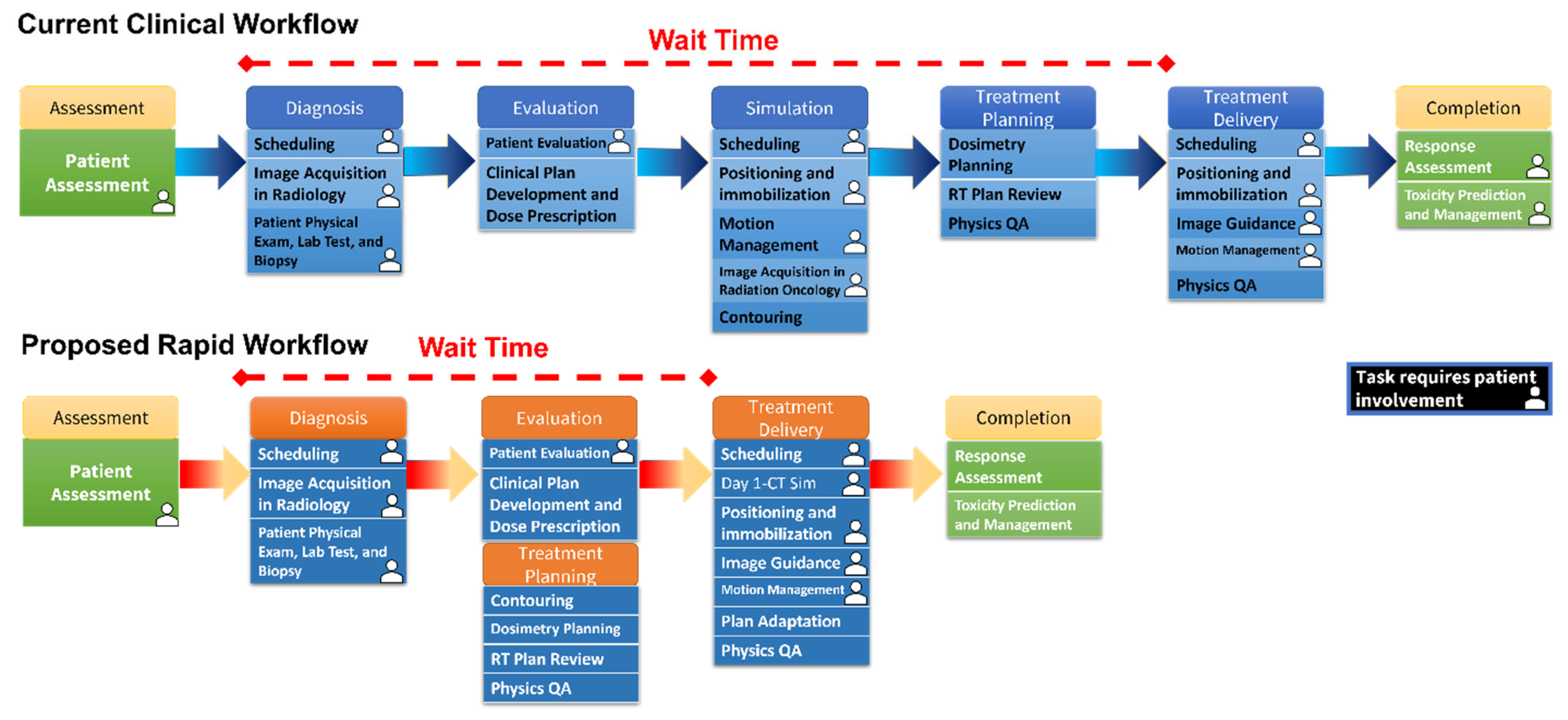
1. Introduction
2. Materials and Methods
2.1. Data Preparation
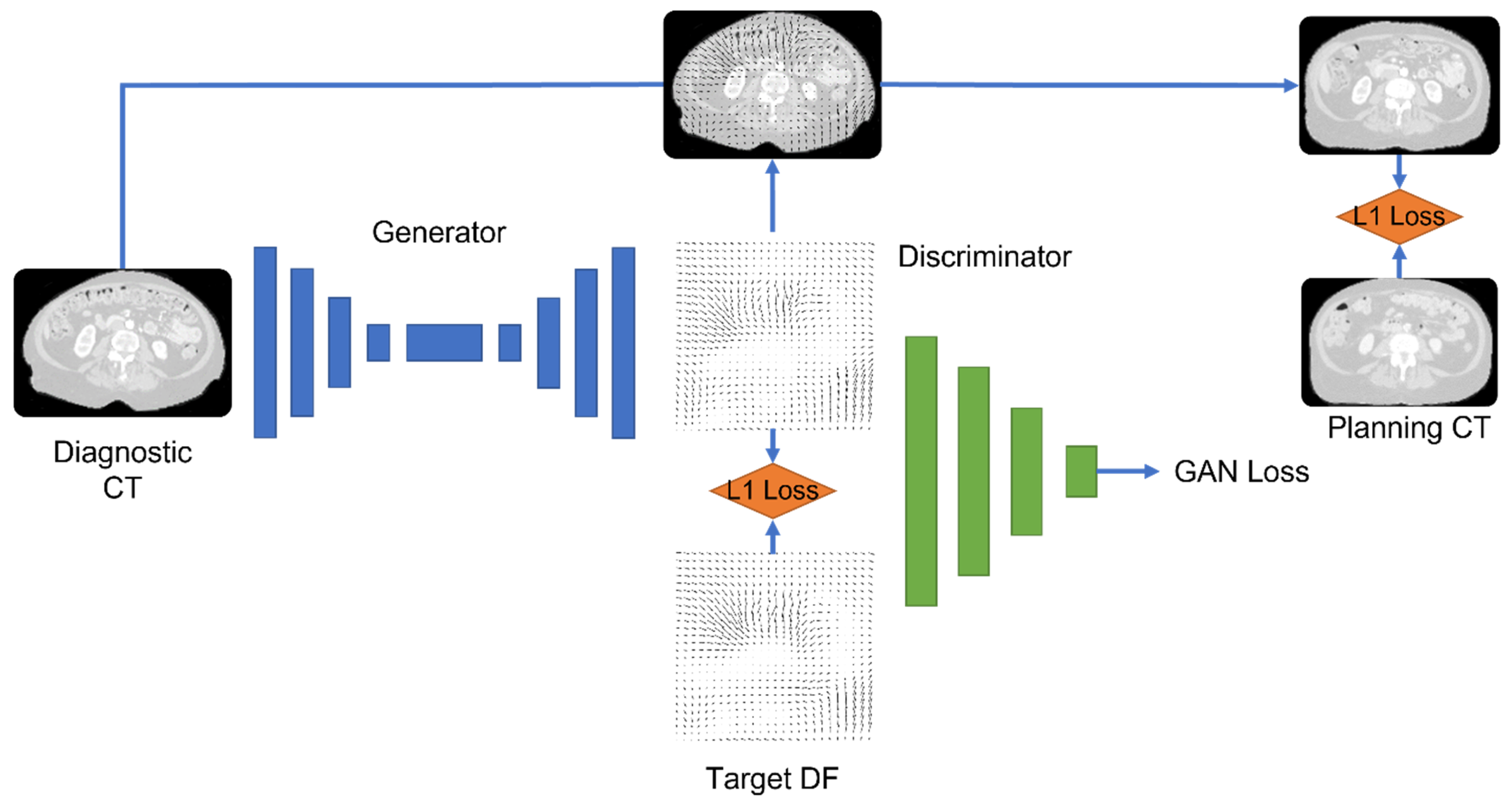
2.2. Deep Learning Model
2.3. Training and Testing of the Model
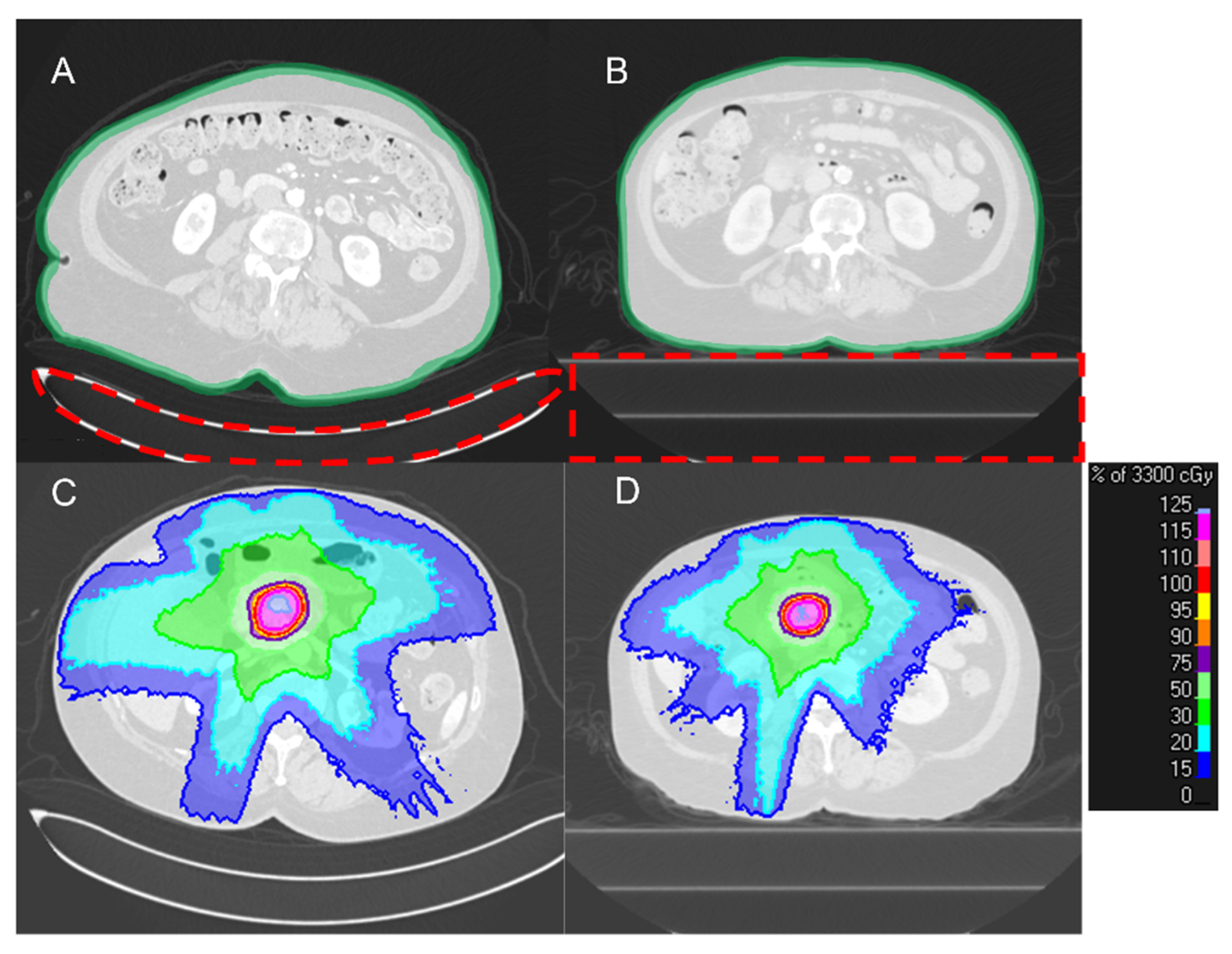
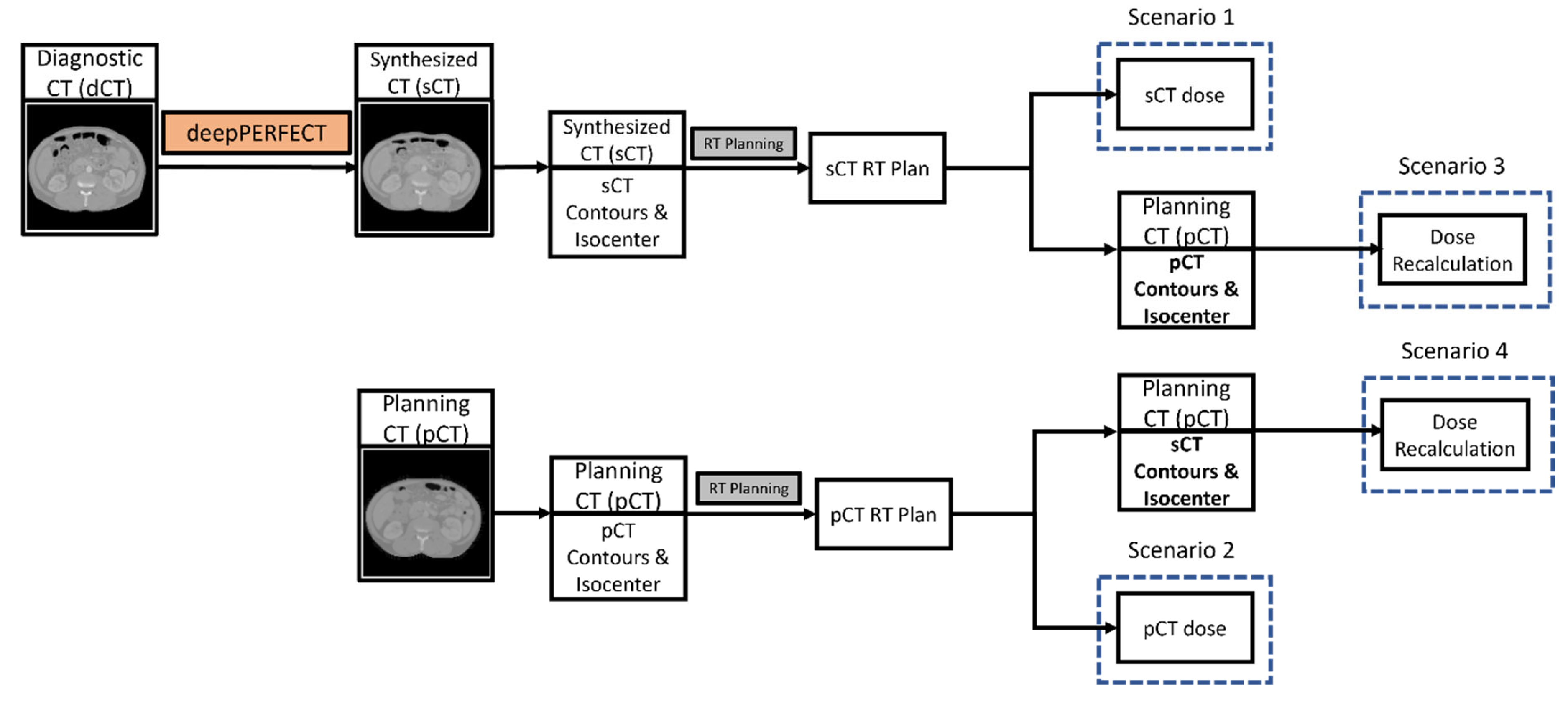
2.4. Radiation Therapy Planning
2.5. Evaluation Metrics
2.6. Statistical Analysis
2.7. Optimization Parameters
2.8. Placement of the Virtual Couch
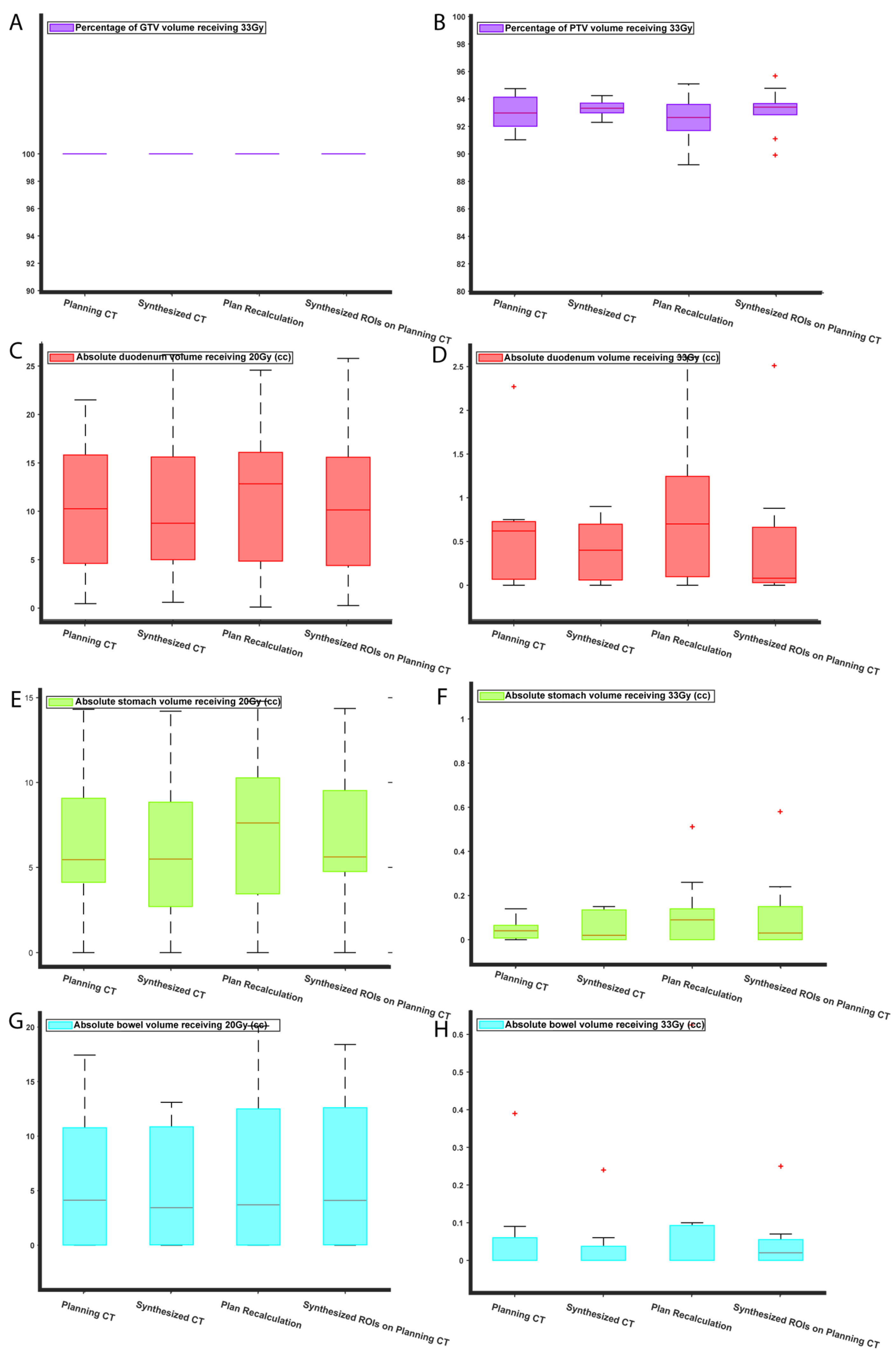
3. Results
4. Discussion
5. Conclusions
Supplementary Materials
Author Contributions
Funding
Institutional Review Board Statement
Informed Consent Statement
Data Availability Statement
Conflicts of Interest
References
- Siegel, R.L.; Miller, K.D.; Fuchs, H.E.; Jemal, A. Cancer Statistics, 2022. CA Cancer J. Clin. 2022, 72, 7–33. [Google Scholar] [CrossRef]
- Sener, S.F.; Fremgen, A.; Menck, H.R.; Winchester, D.P. Pancreatic Cancer: A Report of Treatment and Survival Trends for 100,313 Patients Diagnosed from 1985–1995, Using the National Cancer Database. J. Am. Coll. Surg. 1999, 189, 1–7. [Google Scholar] [CrossRef] [PubMed]
- Shin, S.H.; Kim, H.J.; Hwang, D.W.; Lee, J.H.; Song, K.B.; Jun, E.; Shim, I.K.; Hong, S.M.; Kim, H.J.; Park, K.M.; et al. The DPC4/SMAD4 Genetic Status Determines Recurrence Patterns and Treatment Outcomes in Resected Pancreatic Ductal Adenocarcinoma: A Prospective Cohort Study. Oncotarget 2017, 8, 17945–17959. [Google Scholar] [CrossRef] [PubMed] [Green Version]
- Hooshangnejad, H.; Han-Oh, S.; Shin, E.J.; Narang, A.; Rao, A.D.; Lee, J.; McNutt, T.; Hu, C.; Wong, J.; Ding, K. Demonstrating the Benefits of Corrective Intra-Operative Feedback in Improving the Quality of Duodenal Hydrogel Spacer Placement. Med. Phys. 2022, 49, 4794–4803. [Google Scholar] [CrossRef] [PubMed]
- Hooshangnejad, H.; Youssefian, S.; Narang, A.; Shin, E.J.; Rao, A.D.; Han-Oh, S.; McNutt, T.; Lee, J.; Hu, C.; Wong, J.; et al. Finite Element-Based Personalized Simulation of Duodenal Hydrogel Spacer: Spacer Location Dependent Duodenal Sparing and a Decision Support System for Spacer-Enabled Pancreatic Cancer Radiation Therapy. Front. Oncol. 2022, 12, 833231. [Google Scholar] [CrossRef] [PubMed]
- Hooshangnejad, H.; Youssefian, S.; Guest, J.K.; Ding, K. FEMOSSA: Patient-Specific Finite Element Simulation of the Prostate–Rectum Spacer Placement, a Predictive Model for Prostate Cancer Radiotherapy. Med. Phys. 2021, 48, 3438–3452. [Google Scholar] [CrossRef] [PubMed]
- Bruynzeel, A.M.E.; Lagerwaard, F.J. The Role of Biological Dose-Escalation for Pancreatic Cancer. Clin. Transl. Radiat. Oncol. 2019, 18, 128–130. [Google Scholar] [CrossRef] [Green Version]
- Ben-Josef, E.; Shields, A.F.; Vaishampayan, U.; Vaitkevicius, V.; El-Rayes, B.F.; McDermott, P.; Burmeister, J.; Bossenberger, T.; Philip, P.A. Intensity-Modulated Radiotherapy (IMRT) and Concurrent Capecitabine for Pancreatic Cancer. Int. J. Radiat. Oncol. Biol. Phys. 2004, 59, 454–459. [Google Scholar] [CrossRef]
- Li, J.; Ng, J.; Allendorf, J.; Saif, M.W. Locally Advanced Pancreatic Adenocarcinoma: Are We Making Progress? Highlights from the “2011 ASCO Annual Meeting”. Chicago, IL, USA, 3–7 June 2011. JOP J. Pancreas 2011, 12, 347–350. [Google Scholar]
- Machtay, M.; Paulus, R.; Moughan, J.; Komaki, R.; Bradley, J.E.; Choy, H.; Albain, K.; Movsas, B.; Sause, W.T.; Curran, W.J. Defining Local-Regional Control and Its Importance in Locally Advanced Non-Small Cell Lung Carcinoma. J. Thorac. Oncol. 2012, 7, 716–722. [Google Scholar] [CrossRef] [Green Version]
- International Atomic Energy Agency. Radiotherapy in Palliative Cancer Care: Development and Implementation; Human Health Reports; International Atomic Energy Agency: Vienna, Austria, 2012; ISBN 978-92-0-109009-6. [Google Scholar]
- Buss, E.J.; Kachnic, L.A.; Horowitz, D.P. Radiotherapy for Locally Advanced Pancreatic Ductal Adenocarcinoma. Semin. Oncol. 2021, 48, 106–110. [Google Scholar] [CrossRef] [PubMed]
- Wong, S.; Roderick, S.; Kejda, A.; Atyeo, J.; Grimberg, K.; Porter, B.; Booth, J.; Hruby, G.; Eade, T. Diagnostic Computed Tomography Enabled Planning for Palliative Radiation Therapy: Removing the Need for a Planning Computed Tomography Scan. Pract. Radiat. Oncol. 2021, 11, e146–e153. [Google Scholar] [CrossRef] [PubMed]
- Hooshangnejad, H.; Ding, K. Feasibility of Planning-CT-Free Rapid Workflow for Stereotactic Body Radiotherapy: Removing the Need for Planning CT by AI-Driven, Intelligent Prediction of Body Deformation. In Proceedings of the Medical Imaging 2022: Image-Guided Procedures, Robotic Interventions, and Modeling, Virtual, 21–27 March 2022; Volume 12034. [Google Scholar]
- Salomaa, E.-R.; Sällinen, S.; Hiekkanen, H.; Liippo, K. Delays in the Diagnosis and Treatment of Lung Cancer. Chest 2005, 128, 2282–2288. [Google Scholar] [CrossRef] [PubMed] [Green Version]
- Gamboa, A.C.; Rupji, M.; Switchenko, J.M.; Lee, R.M.; Turgeon, M.K.; Meyer, B.I.; Russell, M.C.; Cardona, K.; Kooby, D.A.; Maithel, S.K.; et al. Optimal Timing and Treatment Strategy for Pancreatic Cancer. J. Surg. Oncol. 2020, 122, 457–468. [Google Scholar] [CrossRef]
- Khorana, A.A.; Tullio, K.; Elson, P.; Pennell, N.A.; Grobmyer, S.R.; Kalady, M.F.; Raymond, D.; Abraham, J.; Klein, E.A.; Walsh, R.M.; et al. Time to Initial Cancer Treatment in the United States and Association with Survival over Time: An Observational Study. PLoS ONE 2019, 14, e0213209. [Google Scholar]
- Kenny, L.; Lehman, M. Sequential Audits of Unacceptable Delays in Radiation Therapy in Australia and New Zealand. Australas Radiol. 2004, 48, 29–34. [Google Scholar] [CrossRef]
- Schuler, T.; Back, M.; Hruby, G.; Carroll, S.; Jayamanne, D.; Kneebone, A.; Stevens, M.; Lamoury, G.; Morgia, M.; Wong, S.; et al. Introducing Computed Tomography Simulation—Free and Electronic Patient-Reported Outcomes—Monitored Palliative Radiation Therapy into Routine Care: Clinical Outcomes and Implementation Experience. Adv. Radiat. Oncol. 2021, 6, 100632. [Google Scholar] [CrossRef]
- Wilson, D.D.; Alonso, C.E.; Sim, A.J.; Peck, T.; Handsfield, L.L.; Chen, Q.; Blackhall, L.; Showalter, T.N.; Reardon, K.A.; Read, P.W. STAT RT: A Prospective Pilot Clinical Trial of Scan-Plan-QA-Treat Stereotactic Body Radiation Therapy for Painful Osseous Metastases. Ann. Palliat. Med. 2019, 8, 221–230. [Google Scholar] [CrossRef]
- Han-Oh, S.; Hill, C.; Wang, K.K.-H.; Ding, K.; Wright, J.L.; Alcorn, S.; Meyer, J.; Herman, J.; Narang, A. Geometric Reproducibility of Fiducial Markers and Efficacy of a Patient-Specific Margin Design Using Deep Inspiration Breath Hold for Stereotactic Body Radiation Therapy for Pancreatic Cancer. Adv. Radiat. Oncol. 2021, 6, 100655. [Google Scholar] [CrossRef]
- Su, L.; Iordachita, I.; Zhang, Y.; Lee, J.; Ng, S.K.; Jackson, J.; Hooker, T.; Wong, J.; Herman, J.M.; Sen, H.T.; et al. Feasibility Study of Ultrasound Imaging for Stereotactic Body Radiation Therapy with Active Breathing Coordinator in Pancreatic Cancer. J. Appl. Clin. Med. Phys. 2017, 18, 84–96. [Google Scholar] [CrossRef] [Green Version]
- Hooshangnejad, H.; Ding, K. Predicting the Efficacy of Spacer Placement for Pancreatic Cancer Using a Novel Patient-Specific Virtual Spacer. In Medical Physics; Wiley: Hoboken, NJ, USA, 2021; Volume 48. [Google Scholar]
- Jarrett, D.; Stride, E.; Vallis, K.; Gooding, M.J. Applications and Limitations of Machine Learning in Radiation Oncology. Br. J. Radiol. 2019, 92, 20190001. [Google Scholar] [CrossRef]
- Hooshangnejad, H.; Chen, Q.; Feng, X.; Zhang, R.; Ding, K. DAART: A Deep Learning Platform for Deeply Accelerated Adaptive Radiation Therapy for Lung Cancer. Johns Hopkins Med. 2023; in press. [Google Scholar]
- Lustberg, T.; van Soest, J.; Gooding, M.; Peressutti, D.; Aljabar, P.; van der Stoep, J.; van Elmpt, W.; Dekker, A. Clinical Evaluation of Atlas and Deep Learning Based Automatic Contouring for Lung Cancer. Radiother. Oncol. 2018, 126, 312–317. [Google Scholar] [CrossRef] [PubMed] [Green Version]
- Oberije, C.; Nalbantov, G.; Dekker, A.; Boersma, L.; Borger, J.; Reymen, B.; van Baardwijk, A.; Wanders, R.; De Ruysscher, D.; Steyerberg, E. A Prospective Study Comparing the Predictions of Doctors versus Models for Treatment Outcome of Lung Cancer Patients: A Step toward Individualized Care and Shared Decision Making. Radiother. Oncol. 2014, 112, 37–43. [Google Scholar] [CrossRef] [PubMed] [Green Version]
- Field, M.; Hardcastle, N.; Jameson, M.; Aherne, N.; Holloway, L. Machine Learning Applications in Radiation Oncology. Phys. Imaging Radiat. Oncol. 2021, 19, 13–24. [Google Scholar] [CrossRef] [PubMed]
- Sahiner, B.; Pezeshk, A.; Hadjiiski, L.M.; Wang, X.; Drukker, K.; Cha, K.H.; Summers, R.M.; Giger, M.L. Deep Learning in Medical Imaging and Radiation Therapy. Med. Phys. 2019, 46, e1–e36. [Google Scholar] [CrossRef] [PubMed] [Green Version]
- Peng, Z.; Shan, H.; Liu, T.; Pei, X.; Wang, G.; Xu, X.G. MCDNet—A Denoising Convolutional Neural Network to Accelerate Monte Carlo Radiation Transport Simulations: A Proof of Principle with Patient Dose From X-Ray CT Imaging. IEEE Access 2019, 7, 76680–76689. [Google Scholar] [CrossRef]
- Balakrishnan, G.; Zhao, A.; Sabuncu, M.R.; Dalca, A.V.; Guttag, J. An Unsupervised Learning Model for Deformable Medical Image Registration. In Proceedings of the 2018 IEEE/CVF Conference on Computer Vision and Pattern Recognition, Salt Lake City, UT, USA, 18–23 June 2018; pp. 9252–9260. [Google Scholar]
- Markham, M.J.; Wachter, K.; Agarwal, N.; Bertagnolli, M.M.; Chang, S.M.; Dale, W.; Diefenbach, C.S.M.; Rodriguez-Galindo, C.; George, D.J.; Gilligan, T.D. Clinical Cancer Advances 2020: Annual Report on Progress against Cancer from the American Society of Clinical Oncology. J. Clin. Oncol. 2020, 38, 1081. [Google Scholar] [CrossRef] [PubMed] [Green Version]
- Yang, Y.; Ford, E.C.; Wu, B.; Pinkawa, M.; van Triest, B.; Campbell, P.; Song, D.Y.; McNutt, T.R. An Overlap-Volume-Histogram Based Method for Rectal Dose Prediction and Automated Treatment Planning in the External Beam Prostate Radiotherapy Following Hydrogel Injection. Med. Phys. 2013, 40, 11709. [Google Scholar] [CrossRef]
- Cilla, S.; Ianiro, A.; Romano, C.; Deodato, F.; Macchia, G.; Buwenge, M.; Dinapoli, N.; Boldrini, L.; Morganti, A.G.; Valentini, V. Template-Based Automation of Treatment Planning in Advanced Radiotherapy: A Comprehensive Dosimetric and Clinical Evaluation. Sci. Rep. 2020, 10, 423. [Google Scholar] [CrossRef] [Green Version]
- Greenspan, H.; van Ginneken, B.; Summers, R.M. Guest Editorial Deep Learning in Medical Imaging: Overview and Future Promise of an Exciting New Technique. IEEE Trans. Med. Imaging 2016, 35, 1153–1159. [Google Scholar] [CrossRef]
- Liu, Y.; Lei, Y.; Wang, T.; Fu, Y.; Tang, X.; Curran, W.J.; Liu, T.; Patel, P.; Yang, X. CBCT-Based Synthetic CT Generation Using Deep-Attention CycleGAN for Pancreatic Adaptive Radiotherapy. Med. Phys. 2020, 47, 2472–2483. [Google Scholar] [CrossRef] [PubMed]
- Chen, L.; Liang, X.; Shen, C.; Jiang, S.; Wang, J. Synthetic CT Generation from CBCT Images via Deep Learning. Med. Phys. 2020, 47, 1115–1125. [Google Scholar] [CrossRef] [PubMed]
- Liu, Y.; Chen, A.; Shi, H.; Huang, S.; Zheng, W.; Liu, Z.; Zhang, Q.; Yang, X. CT Synthesis from MRI Using Multi-Cycle GAN for Head-and-Neck Radiation Therapy. Comput. Med. Imaging Graph. 2021, 91, 101953. [Google Scholar] [CrossRef] [PubMed]
- Hanna, T.P.; King, W.D.; Thibodeau, S.; Jalink, M.; Paulin, G.A.; Harvey-Jones, E.; O’Sullivan, D.E.; Booth, C.M.; Sullivan, R.; Aggarwal, A. Mortality Due to Cancer Treatment Delay: Systematic Review and Meta-Analysis. BMJ 2020, 371, m4087. [Google Scholar] [CrossRef] [PubMed]
- Han, D.; Hooshangnejad, H.; Chen, C.-C.; Ding, K. A Beam-Specific Optimization Target Volume for Stereotactic Proton Pencil Beam Scanning Therapy for Locally Advanced Pancreatic Cancer. Adv. Radiat. Oncol. 2021, 6, 100757. [Google Scholar] [CrossRef] [PubMed]
- Hooshangnejad, H.; Han, D.; Feng, Z.; Dong, L.; Sun, E.; Du, K.; Ding, K. Systematic Study of the Iodinated Rectal Hydrogel Spacer Material Discrepancy on Accuracy of Proton Dosimetry. J. Appl. Clin. Med. Phys. 2022, 23, e13774. [Google Scholar] [CrossRef]
- Feng, Z.; Hooshangnejad, H.; Shin, E.J.; Narang, A.; Lediju Bell, M.A.; Ding, K. The Feasibility of Haar Feature-Based Endoscopic Ultrasound Probe Tracking for Implanting Hydrogel Spacer in Radiation Therapy for Pancreatic Cancer. Front. Oncol. 2021, 11, 759811. [Google Scholar] [CrossRef] [PubMed]
- Shamonin, D.P.; Bron, E.E.; Lelieveldt, B.P.F.; Smits, M.; Klein, S.; Staring, M.; Initiative, A.D.N. Fast Parallel Image Registration on CPU and GPU for Diagnostic Classification of Alzheimer’s Disease. Front. Neuroinform. 2014, 7, 50. [Google Scholar] [CrossRef] [Green Version]
- Klein, S.; Staring, M.; Murphy, K.; Viergever, M.A.; Pluim, J.P.W. Elastix: A Toolbox for Intensity-Based Medical Image Registration. IEEE Trans Med. Imaging 2009, 29, 196–205. [Google Scholar] [CrossRef]
- Reinhardt, J.M.; Christensen, G.E.; Hoffman, E.A.; Ding, K.; Cao, K. Registration-Derived Estimates of Local Lung Expansion as Surrogates for Regional Ventilation. Lect. Notes Comput. Sci. 2007, 4584, 763. [Google Scholar]
- Du, K.; Reinhardt, J.M.; Christensen, G.E.; Ding, K.; Bayouth, J.E. Respiratory Effort Correction Strategies to Improve the Reproducibility of Lung Expansion Measurements. Med. Phys. 2013, 40, 123504. [Google Scholar] [CrossRef] [Green Version]
- Ding, K.; Cao, K.; Christensen, G.E.; Hoffman, E.A.; Reinhardt, J.M. Registration-Based Regional Lung Mechanical Analysis: Retrospectively Reconstructed Dynamic Imaging versus Static Breath-Hold Image Acquisition. In Medical Imaging 2009: Biomedical Applications in Molecular, Structural, and Functional Imaging; SPIE: Bellingham, WA, USA, 2009; pp. 101–109. [Google Scholar]
- Yin, Y.; Hoffman, E.A.; Ding, K.; Reinhardt, J.M.; Lin, C.-L. A Cubic B-Spline-Based Hybrid Registration of Lung CT Images for a Dynamic Airway Geometric Model with Large Deformation. Phys. Med. Biol. 2010, 56, 203. [Google Scholar] [CrossRef] [PubMed]
- Ding, K.; Bayouth, J.E.; Buatti, J.M.; Christensen, G.E.; Reinhardt, J.M. 4DCT-based Measurement of Changes in Pulmonary Function Following a Course of Radiation Therapy. Med. Phys. 2010, 37, 1261–1272. [Google Scholar] [CrossRef] [PubMed] [Green Version]
- Du, K.; Ding, K.; Cao, K.; Bayouth, J.E.; Christensen, G.E.; Reinhardt, J.M. Registration-Based Measurement of Regional Expiration Volume Ratio Using Dynamic 4DCT Imaging. In Proceedings of the 2011 IEEE International Symposium on Biomedical Imaging: From Nano to Macro, Chicago, IL, USA, 30 March–2 April 2011; pp. 424–428. [Google Scholar]
- Du, K.; Bayouth, J.E.; Cao, K.; Christensen, G.E.; Ding, K.; Reinhardt, J.M. Reproducibility of Registration-Based Measures of Lung Tissue Expansion. Med. Phys. 2012, 39, 1595–1608. [Google Scholar] [CrossRef] [Green Version]
- Reinhardt, J.M.; Ding, K.; Cao, K.; Christensen, G.E.; Hoffman, E.A.; Bodas, S. V Registration-Based Estimates of Local Lung Tissue Expansion Compared to Xenon CT Measures of Specific Ventilation. Med. Image Anal. 2008, 12, 752–763. [Google Scholar] [CrossRef] [Green Version]
- Kingma, D.P.; Ba, J. Adam: A Method for Stochastic Optimization. arXiv 2014, arXiv:1412.6980. [Google Scholar]
- Yan, Y.; Lee, H.; Somer, E.; Grau, V. Generation of Amyloid PET Images via Conditional Adversarial Training for Predicting Progression to Alzheimer’s Disease. In PRedictive Intelligence in MEdicine; Rekik, I., Unal, G., Adeli, E., Park, S.H., Eds.; Springer International Publishing: Cham, Switzerland, 2018; pp. 26–33. [Google Scholar]
- Shin, H.-C.; Ihsani, A.; Mandava, S.; Sreenivas, S.T.; Forster, C.; Cha, J.; Initiative, A.D.N. Ganbert: Generative Adversarial Networks with Bidirectional Encoder Representations from Transformers for Mri to Pet Synthesis. arXiv 2020, arXiv:2008.04393. [Google Scholar]
- Ranjan, A.; Lalwani, D.; Misra, R. GAN for Synthesizing CT from T2-Weighted MRI Data towards MR-Guided Radiation Treatment. Magn. Reson. Mater. Phys. Biol. Med. 2022, 35, 449–457. [Google Scholar] [CrossRef] [PubMed]
- Shitrit, O.; Riklin Raviv, T. Accelerated Magnetic Resonance Imaging by Adversarial Neural Network. In Deep Learning in Medical Image Analysis and Multimodal Learning for Clinical Decision Support; Springer: Cham, Switzerland, 2017; pp. 30–38. [Google Scholar]
- Seitzer, M.; Yang, G.; Schlemper, J.; Oktay, O.; Würfl, T.; Christlein, V.; Wong, T.; Mohiaddin, R.; Firmin, D.; Keegan, J.; et al. Adversarial and Perceptual Refinement for Compressed Sensing MRI Reconstruction. In Proceedings of the Medical Image Computing and Computer Assisted Intervention—MICCAI 2018, Granada, Spain, 16–20 September 2018; Frangi, A.F., Schnabel, J.A., Davatzikos, C., Alberola-López, C., Fichtinger, G., Eds.; Springer International Publishing: Cham, Switzerland, 2018; pp. 232–240. [Google Scholar]
- Ran, M.; Hu, J.; Chen, Y.; Chen, H.; Sun, H.; Zhou, J.; Zhang, Y. Denoising of 3D Magnetic Resonance Images Using a Residual Encoder–Decoder Wasserstein Generative Adversarial Network. Med. Image Anal. 2019, 55, 165–180. [Google Scholar] [CrossRef] [Green Version]
- Kim, K.H.; Do, W.-J.; Park, S.-H. Improving Resolution of MR Images with an Adversarial Network Incorporating Images with Different Contrast. Med. Phys. 2018, 45, 3120–3131. [Google Scholar] [CrossRef]
- Wani, N.; Raza, K. Chapter 3—Multiple Kernel-Learning Approach for Medical Image Analysis. In Soft Computing Based Medical Image Analysis; Dey, N., Ashour, A.S., Shi, F., Balas, V.E., Eds.; Academic Press: Cambridge, MA, USA, 2018; pp. 31–47. ISBN 9780128130872. [Google Scholar]
- Yi, X.; Babyn, P. Sharpness-Aware Low-Dose CT Denoising Using Conditional Generative Adversarial Network. J. Digit. Imaging 2018, 31, 655–669. [Google Scholar] [CrossRef]
- Jaffray, D.A.; Siewerdsen, J.H.; Wong, J.W.; Martinez, A.A. Flat-Panel Cone-Beam Computed Tomography for Image-Guided Radiation Therapy. Int. J. Radiat. Oncol. Biol. Phys. 2002, 53, 1337–1349. [Google Scholar] [CrossRef] [PubMed]
- Wong, J.W. How Good Are You? In True Tales of Medical Physics; Springer: Cham, Switzerland, 2022; pp. 153–171. [Google Scholar]
- Netherton, T.J.; Cardenas, C.E.; Rhee, D.J.; Court, L.E.; Beadle, B.M. The Emergence of Artificial Intelligence within Radiation Oncology Treatment Planning. Oncology 2021, 99, 124–134. [Google Scholar] [CrossRef]
- Jia, X.; Ziegenhein, P.; Jiang, S.B. GPU-Based High-Performance Computing for Radiation Therapy. Phys. Med. Biol. 2014, 59, R151. [Google Scholar] [CrossRef] [PubMed] [Green Version]
- Spalding, M.; Walsh, A.; Aland, T. Evaluation of a New GPU-Enabled VMAT Multi-Criteria Optimisation Plan Generation Algorithm. Med. Dosim. 2020, 45, 368–373. [Google Scholar] [CrossRef]
- Aland, T.; Walsh, A.; Jones, M.; Piccini, A.; Devlin, A. Accuracy and Efficiency of Graphics Processing Unit (GPU) Based Acuros XB Dose Calculation within the Varian Eclipse Treatment Planning System. Med. Dosim. 2019, 44, 219–225. [Google Scholar] [CrossRef] [PubMed]
- Byrne, M.; Archibald-Heeren, B.; Hu, Y.; Teh, A.; Beserminji, R.; Cai, E.; Liu, G.; Yates, A.; Rijken, J.; Collett, N.; et al. Varian Ethos Online Adaptive Radiotherapy for Prostate Cancer: Early Results of Contouring Accuracy, Treatment Plan Quality, and Treatment Time. J. Appl. Clin. Med. Phys. 2022, 23, e13479. [Google Scholar] [CrossRef] [PubMed]
- Sibolt, P.; Andersson, L.M.; Calmels, L.; Sjöström, D.; Bjelkengren, U.; Geertsen, P.; Behrens, C.F. Clinical Implementation of Artificial Intelligence-Driven Cone-Beam Computed Tomography-Guided Online Adaptive Radiotherapy in the Pelvic Region. Phys. Imaging Radiat. Oncol. 2021, 17, 4. [Google Scholar] [CrossRef]
- Yoon, S.W.; Lin, H.; Alonso-Basanta, M.; Anderson, N.; Apinorasethkul, O.; Cooper, K.; Dong, L.; Kempsey, B.; Marcel, J.; Metz, J. Initial Evaluation of a Novel Cone-Beam CT-Based Semi-Automated Online Adaptive Radiotherapy System for Head and Neck Cancer Treatment—A Timing and Automation Quality Study. Cureus 2020, 12, e9660. [Google Scholar] [CrossRef] [PubMed]
- Archambault, Y.; Boylan, C.; Bullock, D.; Morgas, T.; Peltola, J.; Ruokokoski, E.; Genghi, A.; Haas, B.; Suhonen, P.; Thompson, S. Making On-Line Adaptive Radiotherapy Possible Using Artificial Intelligence and Machine Learning for Efficient Daily Re-Planning. Med. Phys. Int. J. 2020, 8, 77–86. [Google Scholar]
- Güngör, G.; Serbez, İ.; Temur, B.; Gür, G.; Kayalılar, N.; Mustafayev, T.Z.; Korkmaz, L.; Aydın, G.; Yapıcı, B.; Atalar, B.; et al. Time Analysis of Online Adaptive Magnetic Resonance–Guided Radiation Therapy Workflow According to Anatomical Sites. Pract. Radiat. Oncol. 2021, 11, e11–e21. [Google Scholar] [CrossRef] [PubMed]
- Olson, S.H.; Xu, Y.; Herzog, K.; Saldia, A.; DeFilippis, E.M.; Li, P.; Allen, P.J.; O’Reilly, E.M.; Kurtz, R.C. Weight Loss, Diabetes, Fatigue, and Depression Preceding Pancreatic Cancer. Pancreas 2016, 45, 986–991. [Google Scholar] [CrossRef] [PubMed] [Green Version]
- Hendifar, A.E.; Petzel, M.Q.B.; Zimmers, T.A.; Denlinger, C.S.; Matrisian, L.M.; Picozzi, V.J.; Rahib, L.; on behalf of the Precision Promise Consortium. Pancreas Cancer-Associated Weight Loss. Oncologist 2019, 24, 691–701. [Google Scholar] [CrossRef] [PubMed] [Green Version]


| Model Architecture | Pix2Pix 3D Large Patches | Pix2Pix 3D Small Patches | Pix2Pix 2.5D | U-Net |
|---|---|---|---|---|
| Metric | Average ± STD | |||
| RASSD (HU) | 334 ± 65 | 541 ± 83 | 874 ± 156 | 1242 ± 132 |
| DSC body contour | 0.93 ± 0.04 | 0.82 ± 0.08 | 0.60 ± 0.09 | 0.56 ± 0.03 |
| HD body contour (mm) | 4.6 ± 2.1 | 14.6 ± 6.1 | 29.8 ± 5.7 | 37.2 ± 8.1 |
| DSC GTV | 0.82 ± 0.12 | 0.71 ± 0.16 | 0.61 ± 0.19 | 0.59 ± 0.09 |
| HD GTV (mm) | 7.12 ± 3.1 | 13.1 ± 8.4 | 20.4 ± 7.8 | 28.4 ± 6.1 |
Disclaimer/Publisher’s Note: The statements, opinions and data contained in all publications are solely those of the individual author(s) and contributor(s) and not of MDPI and/or the editor(s). MDPI and/or the editor(s) disclaim responsibility for any injury to people or property resulting from any ideas, methods, instructions or products referred to in the content. |
© 2023 by the authors. Licensee MDPI, Basel, Switzerland. This article is an open access article distributed under the terms and conditions of the Creative Commons Attribution (CC BY) license (https://creativecommons.org/licenses/by/4.0/).
Share and Cite
Hooshangnejad, H.; Chen, Q.; Feng, X.; Zhang, R.; Ding, K. deepPERFECT: Novel Deep Learning CT Synthesis Method for Expeditious Pancreatic Cancer Radiotherapy. Cancers 2023, 15, 3061. https://doi.org/10.3390/cancers15113061
Hooshangnejad H, Chen Q, Feng X, Zhang R, Ding K. deepPERFECT: Novel Deep Learning CT Synthesis Method for Expeditious Pancreatic Cancer Radiotherapy. Cancers. 2023; 15(11):3061. https://doi.org/10.3390/cancers15113061
Chicago/Turabian StyleHooshangnejad, Hamed, Quan Chen, Xue Feng, Rui Zhang, and Kai Ding. 2023. "deepPERFECT: Novel Deep Learning CT Synthesis Method for Expeditious Pancreatic Cancer Radiotherapy" Cancers 15, no. 11: 3061. https://doi.org/10.3390/cancers15113061