
Raman Spectroscopy: A Personalized Decision-Making Tool on Clinicians’ Hands for In Situ Cancer Diagnosis and Surgery Guidance


 ,
,  , , and
Abstract
:Simple Summary
Abstract
1. Introduction
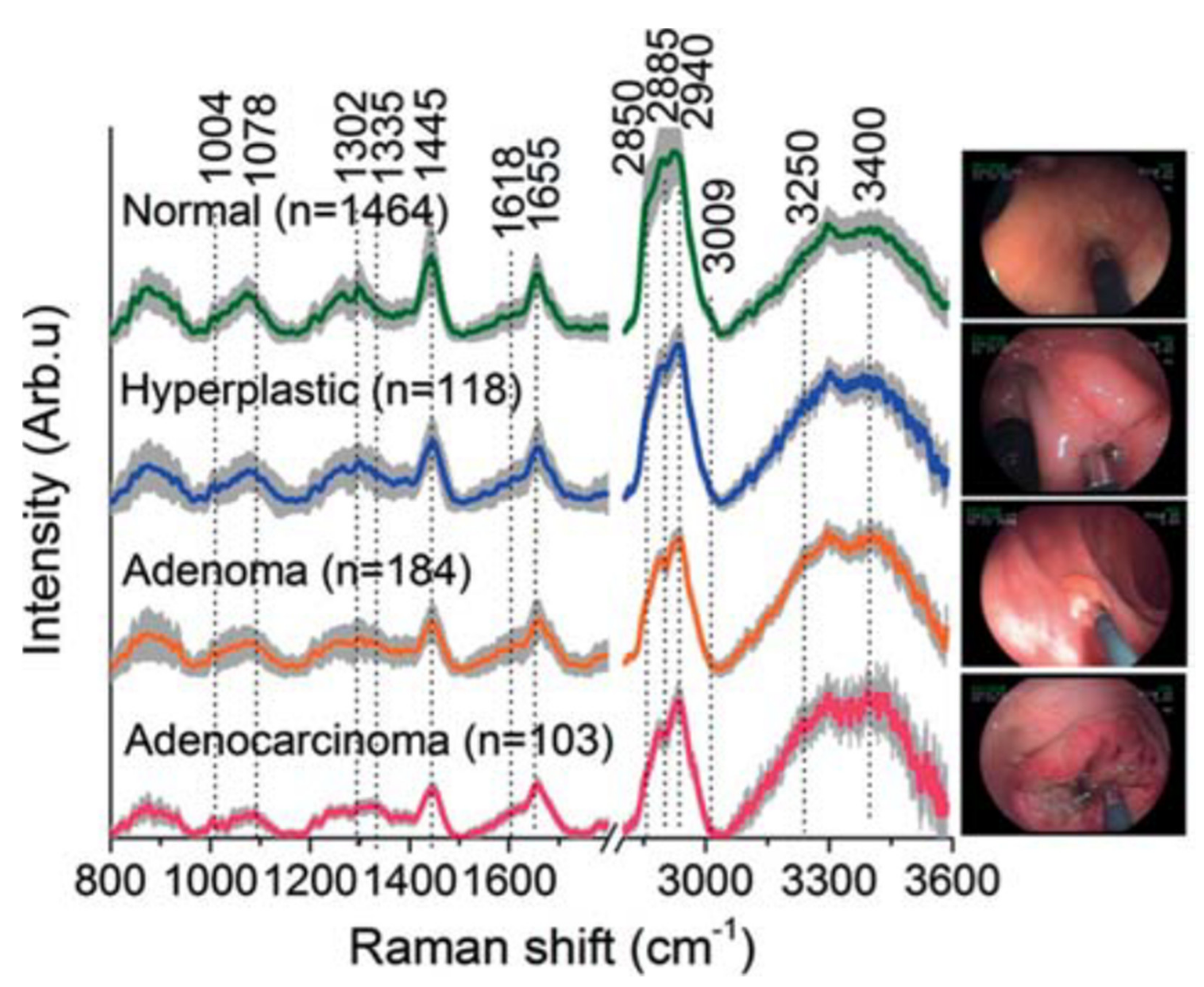
2. Raman Spectra Analysis for Tissue Characterization
3. Machine Learning and Deep Learning as Tools towards Raman Spectra Analysis
4. Advanced Raman Systems in Clinical Praxis
4.1. Raman Systems for Early Diagnosis
4.2. Raman Systems for Guided Surgery
5. Challenges and Future Perspectives
6. Conclusions
Author Contributions
Funding
Acknowledgments
Conflicts of Interest
References
- Global Initiative for Cancer Registry Development. Available online: https://gicr.iarc.fr/about-the-gicr/the-value-of-cancer-data/ (accessed on 5 February 2021).
- Lyon: International Agency for Research on Cancer. Global Initiative for Cancer Registry Development. Available online: https://gco.iarc.fr/today (accessed on 5 February 2021).
- Global Health Data Exchange. Available online: http://ghdx.healthdata.org/gbd-results-tool (accessed on 5 February 2021).
- Martel, C.; Georges, D.; Bray, F.; Ferlay, J.; Clifford, G.M. Global burden of cancer attributable to infections in 2018: A worldwide incidence analysis. Lancet Glob. Health 2020, 8, 180–190. [Google Scholar] [CrossRef] [Green Version]
- Wild, C.P.; Weiderpass, E.; Stewart, B.W. World Cancer Report: Cancer Research for Cancer Prevention; IARC Publications: Lyon, France, 2020. [Google Scholar]
- World Health Organization. Available online: https://www.who.int/publications/i/item/ncd-ccs-2019 (accessed on 5 March 2021).
- Geboes, K.; Geboes, K.; Jouret-Mourin, A. Endoscopy and Histopathology. In Endoscopy; IntechOpen: London, UK, 2013; Volume 1, pp. 3–32. [Google Scholar]
- Bokhorst, L.P.; Zhu, X.; Bul, M.; Bangma, C.H.; Schröder, F.H.; Roobol, M.J. Positive predictive value of prostate biopsy indicated by prostate-specific-antigen-based prostate cancer screening: Trends over time in a European randomized trial. BJU Int. 2012, 110, 1654. [Google Scholar] [CrossRef] [PubMed]
- Rominger, M.; Wisgickl, C.; Timmesfeld, N. Breast microcalcifications as type descriptors to stratify risk of malignancy: A systematic review and meta-analysis of 10665 cases with special focus on round/punctate microcalcifications. Rofo 2012, 184, 1144–1152. [Google Scholar] [CrossRef]
- McWilliams, A.; Tammemagi, M.C.; Mayo, J.R.; Roberts, H.; Liu, G.; Soghrati, K.; Yasufuku, K.; Martel, S.; Laberge, F.; Gingras, M.; et al. Probability of cancer in pulmonary noduls detected on first screening CT. N. Engl. J. Med. 2013, 369, 910–919. [Google Scholar] [CrossRef] [Green Version]
- Carli, P.; Mannone, F.; De Giorgi, V.; Nardini, P.; Chiarugi, A.; Giannoti, B. The problem of false-positive diagnosis in melanoma screening: The impact of dermoscopy. Melanoma Res. 2003, 13, 179–182. [Google Scholar] [CrossRef] [PubMed]
- Gambhir, S.S. Molecular imaging of cancer with positron emission tomography. Nat. Rev. Cancer 2002, 2, 683–693. [Google Scholar] [CrossRef]
- Khalil, M.M.; Tremoleda, J.L.; Bayomy, T.B.; Gsell, W. Molecular SPECT Imaging: An Overview. Int. J. Mol. Imaging 2011, 2011, 1–15. [Google Scholar] [CrossRef] [PubMed] [Green Version]
- Power, S.P.; Moloney, F.; Twomey, M.; James, K.; O’Connor, O.J.; Maher, M.M. Computed tomography and patient risk: Facts, perceptions and uncertainties. World J. Radiol. 2016, 8, 902–915. [Google Scholar] [CrossRef] [PubMed]
- Kuo, W.-C.; Kim, J.; Shemonski, N.D.; Chaney, E.J.; Spillman, D.R.; Boppart, A.B. Real-time three-dimensional optical coherence tomography image-guided core-needle biopsy system. Biomed. Opt. Express 2012, 3, 1149–2012. [Google Scholar] [CrossRef]
- Song, L.-M.W.K.; Banerjee, S.; Desilets, D.; Diehl, D.L.; Farraye, F.A.; Kaul, V.; Kethu, S.R.; Kwon, R.S.; Mamula, P.; Pedrosa, M.C.; et al. Autofluorescence imaging. Gastrointest. Endosc. 2011, 73, 647–650. [Google Scholar] [CrossRef]
- Shin, D.; Vigneswaran, N.; Gillenwater, A.; Richards-Kortum, R. Advances in fluorescence imaging techniques to detect oral cancer and its precursors. Future Oncol. 2010, 6, 1143–1154. [Google Scholar] [CrossRef] [PubMed] [Green Version]
- Haris, M.; Yadav, S.K.; Rizwan, A.; Singh, A.; Wang, E.; Hariharan, H.; Reddy, R.; Marincola, F.M. Molecular magnetic resonance imaging in cancer. J. Transl. Med. 2015, 13, 313. [Google Scholar] [CrossRef] [PubMed] [Green Version]
- Keren, S.; Zavaleta, C.; Cheng, Z.; de la Zerda, A.; Gheysens, O.; Gambhir, S.S. Noninvasive molecular imaging of small living subjects using Raman spectroscopy. Proc. Natl. Acad. Sci. USA 2008, 105, 5844–5849. [Google Scholar] [CrossRef] [PubMed] [Green Version]
- Blasberg, R.G. Molecular Imaging and Cancer. Mol. Cancer Ther. 2003, 2, 335. [Google Scholar]
- Wang, W.; Zhao, J.; Short, M.; Zeng, H. Real-time in vivo cancer diagnosis using raman spectroscopy. J. Biophotonics 2015, 8, 527–545. [Google Scholar] [CrossRef]
- Nijssen, A.; Koljenović, S.; Schut, T.C.B.; Caspers, P.J.; Puppels, G.J. Towards oncological application of Raman spectroscopy. J. Biophotonics 2009, 2, 29–36. [Google Scholar] [CrossRef]
- Kallaway, C.; Almond, L.M.; Barr, H.; Wood, J.; Hutchings, J.; Kendall, C.; Stone, N. Advances in the clinical application of Raman spectroscopy for cancer diagnostics. Photodiagnosis Photodyn. Ther. 2013, 10, 207–219. [Google Scholar] [CrossRef]
- Santos, I.P.; Barroso, E.M.; Bakker Schut, T.C.; Caspers, P.J.; van Lanschot, C.G.F.; Choi, D.-H.; van der Kamp, M.F.; Smits, R.W.H.; van Doorn, R.; Verdijk, R.M.; et al. Raman spectroscopy for cancer detection and cancer surgery guidance: Translation to the clinics. Analyst 2017, 142, 3025–3047. [Google Scholar] [CrossRef]
- Hanlon, E.B.; Manoharan, R.; Koo, T.W.; Shafer, K.E.; Motz, J.T.; Fitzmaurice, M.; Kramer, J.R.; Itzkan, I.; Dasari, R.R.; Feld, M.S. Prospects for in vivo Raman spectroscopy. Phys. Med. Biol. 2000, 45, R1–R59. [Google Scholar] [CrossRef] [Green Version]
- Santos, I.P.; Caspers, P.J.; Schut, T.B.; van Doorn, R.; Koljenović, S.; Puppels, G.J. Implementation of a novel low-noise InGaAs detector enabling rapid near-infrared multichannel Raman spectroscopy of pigmented biological samples. J. Raman Spectrosc. 2015, 46, 652–660. [Google Scholar] [CrossRef]
- Wachsmann-Hogiu, S.; Weeks, T.; Huser, T. Chemical analysis in vivo and in vitro by Raman spectroscopy—from single cells to humans. Curr. Opin. Biotechnol. 2009, 20, 63–73. [Google Scholar] [CrossRef] [PubMed] [Green Version]
- Krafft, C.; Schie, I.W.; Meyer, T.; Schmitt, M.; Popp, J. Developments in spontaneous and coherent Raman scattering microscopic imaging for biomedical applications. Chem. Soc. Rev. 2016, 45, 1819–1849. [Google Scholar] [CrossRef] [PubMed]
- Orringer, D.A.; Pandian, B.; Niknafs, Y.S.; Hollon, T.C.; Boyle, J.; Lewis, S.; Garrard, M.; Hervey-Jumper, S.L.; Garton, H.J.L.; Maher, C.O.; et al. Rapid intraoperative histology of unprocessed surgical specimens via fibre-laser-based stimulated Raman scattering microscopy. Nat. Biomed. Eng. 2017, 1, 27. [Google Scholar] [CrossRef] [PubMed]
- Çulha, M. Raman spectroscopy for cancer diagnosis: How far have we come? Bioanalysis 2015, 7, 2813–2824. [Google Scholar] [CrossRef]
- Wang, W.; McGregor, H.; Short, M.; Zeng, H. Clinical utility of Raman spectroscopy: Current applications and ongoing developments. Adv. Health Care Technol. 2016, 2, 13–29. [Google Scholar] [CrossRef] [Green Version]
- Camp, C.H., Jr.; Cicerone, M.T. Chemically sensitive bioimaging with coherent Raman scattering. Nat. Photonics 2015, 9, 295–305. [Google Scholar] [CrossRef]
- Barman, I.; Dingari, N.C.; Saha, A.; McGee, S.; Galindo, L.H.; Liu, W.; Plecha, D.; Klein, N.; Dasari, R.R.; Fitzmaurice, M. Application of Raman Spectroscopy to Identify Microcalcifications and Underlying Breast Lesions at Stereotactic Core Needle Biopsy. Cancer Res. 2013, 73, 3206–3215. [Google Scholar] [CrossRef] [Green Version]
- Bratchenko, I.A.; Artemyev, D.A.; Myakinin, O.O.; Khristoforova, Y.A.; Moryatov, A.A.; Kozlov, S.V.; Zakharov, V.P. Combined Raman and autofluorescence ex vivo diagnostics of skin cancer in near-infrared and visible regions. J. Biomed. Opt. 2017, 22, 27005. [Google Scholar] [CrossRef]
- Kourkoumelis, N.; Balatsoukas, I.; Moulia, V.; Elka, A.; Gaitanis, G.; Bassukas, I. Advances in the in Vivo Raman Spectroscopy of Malignant Skin Tumors Using Portable Instrumentation. Int. J. Mol. Sci. 2015, 16, 14554–14570. [Google Scholar] [CrossRef] [Green Version]
- Austin, L.A.; Osseiran, S.; Evans, C.L. Raman technologies in cancer diagnostics. Analyst 2016, 141, 476–503. [Google Scholar] [CrossRef]
- Pence, I.; Mahadevan-Jansen, A. Clinical instrumentation and applications of Raman spectroscopy. Chem. Soc. Rev. 2016, 45, 1958–1979. [Google Scholar] [CrossRef] [PubMed] [Green Version]
- Shipp, D.W.; Sinjab, F.; Notingher, I. Raman spectroscopy: Techniques and applications in the life sciences. Adv. Opt. Photonics 2017, 9, 315. [Google Scholar] [CrossRef] [Green Version]
- Jermyn, M.; Desroches, J.; Aubertin, K.; St-Arnaud, K.; Madore, W.-J.; De Montigny, E.; Guiot, M.-C.; Trudel, D.; Wilson, B.C.; Petrecca, K.; et al. A review of Raman spectroscopy advances with an emphasis on clinical translation challenges in oncology. Phys. Med. 2016, 61, 370–400. [Google Scholar] [CrossRef]
- Devpura, S.; Barton, K.N.; Brown, S.L.; Palyvoda, O.; Kalkanis, S.; Naik, V.M.; Siddiqui, F.; Naik, R.; Chetty, I.J. Vision 20/20: The role of Raman spectroscopy in early stage cancer detection and feasibility for application in radiation therapy response assessment. Med. Phys. 2014, 41, 50901. [Google Scholar] [CrossRef] [Green Version]
- Duraipandian, S.; Zheng, W.; Ng, J.; Low, J.J.H.; Ilancheran, A.; Huang, Z. In vivo diagnosis of cervical precancer using Raman spectroscopy and genetic algorithm techniques. Analyst 2011, 136, 4328. [Google Scholar] [CrossRef] [PubMed]
- Kong, K.; Rowlands, C.J.; Varma, S.; Perkins, W.; Leach, I.H.; Koloydenko, A.A.; Williams, H.C.; Notingher, I. Diagnosis of tumors during tissue-conserving surgery with integrated autofluorescence and Raman scattering microscopy. Proc. Natl. Acad. Sci. USA 2013, 110, 15189–15194. [Google Scholar] [CrossRef] [Green Version]
- McGregor, H.C.; Short, M.A.; McWilliams, A.; Shaipanich, T.; Ionescu, D.N.; Zhao, J.; Wang, W.; Chen, G.; Lam, S.; Zeng, H. Real-time endoscopic Raman spectroscopy for in vivo early lung cancer detection. J. Biophot. 2017, 10, 98–110. [Google Scholar] [CrossRef] [PubMed]
- Qi, D.; Berger, A.J. Chemical concentration measurement in blood serum and urine samples using liquid-core optical fiber Raman spectroscopy. Appl. Opt. 2007, 46, 1726. [Google Scholar] [CrossRef] [PubMed] [Green Version]
- Jones, R.R.; Hooper, D.C.; Zhang, L.; Wolverson, D.; Valev, V.K. Raman Techniques: Fundamentals and Frontiers. Nanoscale Res. Lett. 2019, 14, 231. [Google Scholar] [CrossRef] [Green Version]
- Woo, M.-A.; Lee, S.-M.; Kim, G.; Baek, J.H.; Noh, M.S.; Kim, J.E.; Park, S.J.; Minai-Tehrani, A.; Park, S.-C.; Seo, Y.T.; et al. Multiplex Immunoassay Using Fluorescent-Surface Enhanced Raman Spectroscopic Dots for the Detection of Bronchioalveolar Stem Cells in Murine Lung. Anal. Chem. 2009, 81, 1008–1015. [Google Scholar] [CrossRef]
- Nijssen, A.; Maquelin, K.; Santos, L.F.; Caspers, P.J.; Bakker Schut, T.C.; den Hollander, J.C.; Neumann, M.H.A.; Puppels, G.J. Discriminating basal cell carcinoma from perilesional skin using high wave-number Raman spectroscopy. J. Biomed. Opt. 2007, 12, 34004. [Google Scholar] [CrossRef] [PubMed]
- Schwartzberg, A.M.; Oshiro, T.Y.; Zhang, J.Z.; Huser, T.; Talley, C.E. Improving Nanoprobes Using Surface-Enhanced Raman Scattering from 30-nm Hollow Gold Particles. Anal. Chem. 2006, 78, 4732–4736. [Google Scholar] [CrossRef] [PubMed]
- Qian, X.; Peng, X.-H.; Ansari, D.O.; Yin-Goen, Q.; Chen, G.Z.; Shin, D.M.; Yang, L.; Young, A.N.; Wang, M.D.; Nie, S. In vivo tumor targeting and spectroscopic detection with surface-enhanced Raman nanoparticle tags. Nat. Biotechnol. 2008, 26, 83–90. [Google Scholar] [CrossRef] [PubMed]
- Kim, J.H.; Kim, J.S.; Choi, H.; Lee, S.M.; Jun, B.H.; Yu, K.N.; Kuk, E.; Kim, Y.K.; Jeong, D.H.; Cho, M.H.; et al. Nanoparticle Probes with Surface Enhanced Raman Spectroscopic Tags for Cellular Cancer Targeting. Anal. Chem. 2006, 78, 6967–6973. [Google Scholar] [CrossRef]
- Wang, H.; Huff, T.B.; Cheng, J.-X. Coherent anti-Stokes Raman scattering imaging with a laser source delivered by a photonic crystal fiber. Opt. Lett. 2006, 31, 1417–1419. [Google Scholar] [CrossRef] [Green Version]
- Okuno, M.; Kano, H.; Leproux, P.; Couderc, V.; Hamaguchi, H. Ultrabroadband multiplex CARS microspectroscopy and imaging using a subnanosecond supercontinuum light source in the deep near infrared. Opt. Lett. 2008, 33, 923–925. [Google Scholar] [CrossRef]
- Potma, E.O.; Evans, C.L.; Xie, X.S. Heterodyne coherent anti-Stokes Raman scattering (CARS) imaging. Opt. Lett. 2006, 31, 241–243. [Google Scholar] [CrossRef] [Green Version]
- Ly, S.; McNerney, G.; Fore, S.; Chan, J.; Huser, T. Time-gated single photon counting enables separation of CARS microscopy data from multiphoton-excited tissue autofluorescence. Opt. Express 2007, 15, 16839–16851. [Google Scholar] [CrossRef] [Green Version]
- Bergholt, M.S.; Lin, K.; Wang, J.; Zheng, W.; Xu, H.; Huang, Q.; Ren, J.L.; Ho, K.Y.; Teh, M.; Srivastava, S.; et al. Simultaneous fingerprint and high-wavenumber fiber-optic Raman spectroscopy enhances real-time in vivo diagnosis of adenomatous polyps during colonoscopy. J. Biophotonics 2015, 9, 333–342. [Google Scholar] [CrossRef]
- Brozek-Pluska, B.; Musial, J.; Kordek, R.; Abramczyk, H. Analysis of Human Colon by Raman Spectroscopy and Imaging-Elucidation of Biochemical Changes in Carcinogenesis. Int. J. Mol. Sci. 2019, 20, 3398. [Google Scholar] [CrossRef] [Green Version]
- He, H.; Yan, S.; Lyu, D.; Xu, M.; Ye, R.; Zheng, P.; Lu, X.; Wang, L.; Ren, B. Deep Learning for Biospectroscopy and Biospectral Imaging: State-of-the-Art and Perspectives. Anal. Chem. 2021, 93, 3653–3665. [Google Scholar] [CrossRef] [PubMed]
- Ian, G.; Yoshua, B.; Aaron, C. Deep Learning; MIT Press: Cambridge, MA, USA, 2016. [Google Scholar]
- Hinton, G.; Deng, L.; Yu, D.; Dahl, E.G.; Mohamed, A.; Jaitly, N.; Senior, A.; Vanhoucke, V.; Nguyen, P.; Sainath, N.T.; et al. Deep Neural Networks for Acoustic Modeling in Speech Recognition: The Shared Views of Four Research Groups. IEEE Signal Process. Mag. 2012, 29, 82–97. [Google Scholar] [CrossRef]
- Sainath, T.N.; Mohamed, A.; Kingsbury, B.; Ramabhadran, B. Deep convolutional neural networks for LVCSR. In Proceedings of the 2013 IEEE International Conference on Acoustics, Speech and Signal Processing, Vancouver, BC, Canada, 26 May 2013; pp. 8614–8618. [Google Scholar] [CrossRef]
- Mor-Yosef, S.; Samueloff, A.; Modan, B.; Navot, D.; Schenker, J.G. Ranking the risk factors for cesarean: Logistic regression analysis of a nationwide study. Obstet. Gynecol. 1990, 75, 944–947. [Google Scholar] [PubMed]
- Krizhevsky, A.; Sutskever, I.; Hinton, G.E. ImageNet classification with deep convolutional neural networks. Commun. ACM 2017, 60, 84–90. [Google Scholar] [CrossRef]
- Farabet, C.; Couprie, C.; Najman, L.; LeCun, Y. Learning Hierarchical Features for Scene Labeling. IEEE Trans. Pattern Anal. Mach. Intell. 2012, 35, 1915–1929. [Google Scholar] [CrossRef] [PubMed] [Green Version]
- Cornell University. Available online: http://arxiv.org/abs/1409.4842 (accessed on 17 September 2014).
- Tolstik, T.; Marquardt, C.; Matthäus, C.; Bergner, N.; Bielecki, C.; Krafft, C.; Stallmach, A.; Popp, J. Discrimination and classification of liver cancer cells and proliferation states by Raman spectroscopic imaging. Analyst 2014, 139, 6036–6043. [Google Scholar] [CrossRef] [PubMed]
- Jermyn, M.; Mok, K.; Mercier, J.; Desroches, J.; Pichette, J.; Saint-Arnaud, K.; Bernstein, L.; Guiot, M.C.; Petrecca, K.; Leblond, F. Intraoperative brain cancer detection with Raman spectroscopy in humans. Sci. Transl. Med. 2015, 7, 274ra19. [Google Scholar] [CrossRef] [PubMed]
- Shu, C.; Yan, H.; Lin, K.; Lim, C.M.; Zheng, W.; Feng, J.; Huang, Z. Biomedical Vibrational Spectroscopy 2020: Advances in Research and Industry; SPIE: San Francisco, CA, USA, 2020; Volume 11236. [Google Scholar]
- Chen, H.; Li, X.; Broderick, N.; Liu, Y.; Zhou, Y.; Han, J.; Xu, W.J. Identification and characterization of bladder cancer by low-resolution fiber-optic Raman spectroscopy. J. Biophoton. 2018, 11, e201800016. [Google Scholar] [CrossRef]
- Kourou, K.; Exarchos, T.P.; Exarchos, K.P.; Karamouzis, V.M.; Fotiadis, D.I. Machine learning applications in cancer prognosis and prediction. Comput. Struct. Biotechnol. J. 2015, 13, 8–17. [Google Scholar] [CrossRef] [Green Version]
- Li, X.; Yang, T.; Li, S.; Wang, D.; Song, Y.; Zhang, S. Raman spectroscopy combined with principal component analysis and k nearest neighbour analysis for non-invasive detection of colon cancer. Laser Phys. 2016, 26, 35702. [Google Scholar] [CrossRef] [Green Version]
- Kothari, R.; Jones, V.; Mena, D.; Bermúdez Reyes, V.; Shon, Y.; Smith, J.P.; Schmolze, D.; Cha, P.D.; Lai, L.; Fong, Y.; et al. Raman spectroscopy and artificial intelligence to predict the Bayesian probability of breast cancer. Sci. Rep. 2021, 11, 6482. [Google Scholar] [CrossRef] [PubMed]
- Li, Q.; Li, W.; Zhang, J.; Xu, Z. An improved k-nearest neighbour method to diagnose breast cancer. Analyst 2018, 143, 2807–2811. [Google Scholar] [CrossRef]
- Li, S.; Chen, G.; Zhang, Y.; Guo, Z.; Liu, Z.; Xu, J.; Li, X.; Lin, L. Identification and characterization of colorectal cancer using Raman spectroscopy and feature selection techniques. Opt. Express 2014, 22, 25895–25908. [Google Scholar] [CrossRef] [PubMed]
- Yu, M.; Yan, H.; Xia, J.; Zhu, L.; Zhang, T.; Zhu, Z.; Lou, X.; Sun, G.; Dong, M. Deep convolutional neural networks for tongue squamous cell carcinoma classification using Raman spectroscopy. Photodiagnosis Photodyn. Ther. 2019, 26, 430–435. [Google Scholar] [CrossRef] [PubMed]
- Short, M.A.; Lam, S.; McWilliams, A.M.; Ionescu, D.N.; Zeng, H. Using Laser Raman Spectroscopy to Reduce False Positives of Autofluorescence Bronchoscopies: A Pilot Study. J. Thorac. Oncol. 2011, 6, 1206–1214. [Google Scholar] [CrossRef] [Green Version]
- Krishna, H.; Majumder, S.K.; Chaturvedi, P.; Sidramesh, M.; Gupta, P.K. In vivo Raman spectroscopy for detection of oral neoplasia: A pilot clinical study. J. Biophotonics 2014, 7, 690–702. [Google Scholar] [CrossRef]
- Short, M.A.; Tai, I.T.; Owen, D.; Zeng, H. Using high frequency Raman spectra for colonic neoplasia detection. Opt. Express 2013, 21, 5025–5034. [Google Scholar] [CrossRef]
- Kwak, J.; Lee, W.; Kim, J.B.; Bae, S.I.; Jeong, K.H. Fiber-optic plasmonic probe with nanogap-rich Au nanoislands for on-site surface-enhanced Raman spectroscopy using repeated solid-state dewetting. J. Biomed. Opt. 2019, 24, 1–6. [Google Scholar] [CrossRef]
- Garai, E.; Sensarn, S.; Zavaleta, C.L.; Loewke, N.O.; Rogalla, S.; Mandella, M.J.; Flet, S.A.; Friedland, S.; Liu, J.T.C.; Contag, C.H.; et al. A Real-Time Clinical Endoscopic System for Intraluminal, Multiplexed Imaging of Surface-Enhanced Raman Scattering Nanoparticles. PLoS ONE 2015, 10, e0123185. [Google Scholar] [CrossRef]
- Matousek, P.; Clark, I.P.; Draper, E.R.C.; Morris, M.D.; Goodship, A.E.; Everall, N.; Towrie, M.; Finney, W.F.; Parker, A.W. Subsurface probing in diffusely scattering media using spatially offset Raman spectroscopy. Appl. Spectrosc. 2005, 59, 393–400. [Google Scholar] [CrossRef]
- Stone, N.; Kerssens, M.; Lloyd, G.R.; Faulds, K.; Graham, D.; Matousek, P. Surface enhanced spatially offset Raman spectroscopic (SESORS) imaging—the next dimension. Chem. Sci. 2011, 2, 776–780. [Google Scholar] [CrossRef]
- Yumin, Z.; Li, L.; Jing, H.Y. Optical penetration of surface-enhanced micro-scale spatial offset Raman spectroscopy in turbid gel and biological tissue. J. Innov. Opt. Health Sci. 2021, 14, 2141001. [Google Scholar] [CrossRef]
- Stevens, O.; Petterson, I.E.I.; Day, J.C.C.; Stone, N. Developing fiber optic Raman probes for applications in clinical spectroscopy. Chem. Soc. Rev. 2016, 45, 1919–1934. [Google Scholar] [CrossRef]
- Wang, J.; Lin, K.; Zheng, W.; Ho, K.; Teh, M.; Yeoh, K.; Huang, Z. Comparative study of the endoscope-based bevelled and volume fiber-optic Raman probes for optical diagnosis of gastric dysplasia in vivo at endoscopy. Anal. Bioanal. Chem. 2015, 407, 8303–8310. [Google Scholar] [CrossRef] [PubMed]
- Duraipandian, C.; Bergholt, M.D.; Zheng, W.; Ho, K.Y.; Teh, M.; Yeoh, K.G.; So, J.B.Y.; Shabbir, A.; Huang, Z. Real-time Raman spectroscopy for in vivo, online gastric cancer diagnosis during clinical endoscopic examination. J. Biomed. Opt. 2012, 17, 81418. [Google Scholar] [CrossRef] [PubMed]
- Agenant, M.; Grimbergen, M.; Draga, R.; Marple, E.; Bosch, R.; Van Swol, C. Clinical superficial Raman probe aimed for epithelial tumor detection: Phantom model results. Biomed. Opt. Express 2014, .5, 1203–1216. [Google Scholar] [CrossRef]
- Cordero, E.; Latka, I.; Matthäus, C.; Schie, I.; Popp, J. In-vivo Raman spectroscopy: From basics to applications. J. Biomed. Opt. 2018, 23, 1–23. [Google Scholar] [CrossRef]
- Latka, I.; Dochow, S.; Krafft, C.; Dietzek, B.; Popp, J. Fiber optic probes for linear and nonlinear Raman applications—Current trends and future development. Laser and Photonics Rev. 2013, 7, 698–731. [Google Scholar] [CrossRef]
- Motz, J.T.; Gandhi, S.J.; Scepanovic, O.R.; Haka, A.S.; Kramer, J.R.; Dasari, R.R.; Feld, M.S. Real-time Raman system for in vivo disease diagnosis. J. Biomed. Opt. 2005, 10, 31113. [Google Scholar] [CrossRef]
- Chen, K.; Qin, Y.; Zheng, F.; Sun, M.; Shi, D. Diagnosis of colorectal cancer using Raman spectroscopy of laser-trapped single living epithelial cells. Opt. Lett. 2006, 31, 2015–2017. [Google Scholar] [CrossRef]
- Denson, S.C.; Pommier, C.J.S.; Denton, M.B. The Impact of Array Detectors on Raman Spectroscopy. J. Chem. Educ. 2007, 84, 67–74. [Google Scholar] [CrossRef]
- Sharma, S.K.; Egan, M.J. Raman Spectroscopy, Remote Compositional Analysis; Cambridge University Press: Cambridge, UK, 2019; pp. 120–146. [Google Scholar] [CrossRef]
- Okhotnikov, O.G. Semiconductor Disk Lasers: Physics and Technology; WILEY-VCH: Weinheim, Germany, 2010. [Google Scholar]
- Pallmann, W.P.; Zaugg, C.A.; Mangold, M.; Wittwer, V.J.; Moench, H.; Gronenborn, S.; Miller, M.; Tilma, B.W.; Südmeyer, T.; Keller, U. Gain characterization and passive modelocking of electrically pumped VECSELs. Opt. Express 2012, 20, 24791. [Google Scholar] [CrossRef] [PubMed] [Green Version]
- Yan, Y.; Zheng, Y.; Sun, H.; Duan, J. Review of Issues and Solutions in High-Power Semiconductor Laser Packaging Technology. Front. Phys. 2021, 9, 669591. [Google Scholar] [CrossRef]
- Smits, R.W.H.; Koljenović, S.; Hardillo, J.A.; Hove, I.T.; Meeuwis, C.A.; Sewnaik, A.; Dronkers, E.A.; Schut, T.C.B.; Langeveld, T.P.M.; Molenaar, J.; et al. Resection margins in oral cancer surgery: Room for improvement. Head Neck 2016, 38 (Suppl. S1), E2197–E2203. [Google Scholar] [CrossRef]
- Binahmed, A.; Nason, R.W.; Abdoh, A.A. The clinical significance of the positive surgical margin in oral cancer. Oral Oncol. 2007, 43, 780–784. [Google Scholar] [CrossRef] [Green Version]
- Neuzillet, Y.; Soulie, M.; Larre, S.; Roupret, M.; Defortescu, G.; Murez, T.; Pignot, G.; Descazeaud, A.; Patard, J.J.; Bigot, P.; et al. Positive surgical margins and their locations in specimens are adverse prognosis features after radical cystectomy in non-metastatic carcinoma invading bladder muscle: Results from a nationwide case-control study. BJU Int. 2013, 111, 1253–1260. [Google Scholar] [CrossRef]
- Wong, J.M.; Panchmatia, J.R.; Ziewacz, J.E.; Bader, A.M.; Dunn, I.F.; Laws, E.R.; Gawande, A.A. Patterns in neurosurgical adverse events: Intracranial neoplasm surgery. Neurosurg. Focus 2012, 33, E16. [Google Scholar] [CrossRef] [Green Version]
- Auner, G.W.; Koya, S.K.; Huang, C.; Broadbent, B.; Trexler, M.; Auner, Z.; Elias, A.; Curtin Mehne, K.; Brusatori, M.A. Applications of Raman spectroscopy in cancer diagnosis. Cancer Metastasis Rev. 2018, 37, 691–717. [Google Scholar] [CrossRef] [Green Version]
- Haka, A.S.; Volynskaya, Z.; Gardecki, J.A.; Nazemi, J.; Shenk, R.; Wang, N.; Dasari, R.R.; Fitzmaurice, M.; Feld, M.S. Diagnosing breast cancer using Raman spectroscopy: Prospective analysis. J. Biomed. Opt. 2009, 14, 54023. [Google Scholar] [CrossRef] [Green Version]
- World Health Organization. Available online: http://globocan.iarc.fr (accessed on 22 March 2017).
- Saha, A.; Barman, I.; Dingari, N.C.; McGee, S.; Volynskaya, Z.; Galindo, L.H.; Liu, W.; Plecha, D.; Klein, N.; Dasari, R.R. Raman spectroscopy: A real-time tool for identifying microcalcifications during stereotactic breast core needle biopsies. Biomed. Opt. Express 2011, 2, 2792–2803. [Google Scholar] [CrossRef] [Green Version]
- Wang, M.; He, X.; Chang, Y.; Sun, G.; Thabane, L. A sensitivity and specificity comparison of fine needle aspiration cytology and core needle biopsy in evaluation of suspicious breast lesions: A systematic review and meta-analysis. The Breast 2017, 31, 157–166. [Google Scholar] [CrossRef]
- Johnson, J.M.; Dalton, R.R.; Wester, S.M.; Landercasper, J.; Lambert, P.J. Histological Correlation of Microcalcifications in Breast Biopsy Specimens. Arch Surg. 1999, 134, 712–716. [Google Scholar] [CrossRef] [PubMed] [Green Version]
- Kittler, H.; Pehamberger, H.; Wolff, K.; Binder, M. Diagnostic accuracy of dermoscopy. Lancet Oncol. 2002, 3, 159–165. [Google Scholar] [CrossRef]
- Morton, C.A.; Mackie, R.M. Clinical accuracy of the diagnosis of cutaneous malignant melanoma. Br. J. Dermatol. 1998, 138, 283–287. [Google Scholar] [CrossRef] [PubMed]
- Van der Rhee, J.I.; Bergman, W.; Kukutsch, N.A. Impact of Dermoscopy on the Management of High-risk Patients from Melanoma Families: A Prospective Study. Acta Derm.-Venereol. 2011, 91, 428–431. [Google Scholar] [CrossRef] [PubMed] [Green Version]
- Santos, I.P.; Caspers, P.J.; Bakker Schut, T.C.; Van Doorn, R.; Noordhoek Hegt, V.; Koljenović, S.; Puppels, G.J. Raman Spectroscopic Characterization of Melanoma and Benign Melanocytic Lesions Suspected of Melanoma Using High-Wavenumber Raman Spectroscopy. Anal. Chem. 2016, 88, 7683–7688. [Google Scholar] [CrossRef] [PubMed]
- Lui, H.; Zhao, J.; McLean, D.; Zeng, H. Real-time Raman Spectroscopy for In Vivo Skin Cancer Diagnosis. Cancer Res. 2012, 72, 2491–2500. [Google Scholar] [CrossRef] [Green Version]
- Schleusener, J.; Gluszczynska, P.; Reble, C.; Gersonde, I.; Helfmann, J.; Fluhr, J.W.; Lademann, J.; Röwert-Huber, J.; Patzelt, A.; Meinke, M.C. In vivo study for the discrimination of cancerous and normal skin using fibre probe-based Raman spectroscopy. Exp. Dermatol. 2015, 24, 767–772. [Google Scholar] [CrossRef]
- Bodanese, B.; Silveira, F.L.; Zângaro, R.A.; Pacheco, M.T.; Pasqualucci, C.A.; Silveira, L., Jr. Discrimination of basal cell carcinoma and melanoma from normal skin biopsies in vitro through Raman spectroscopy and principal component analysis. Photomed. Laser Surg. 2012, 30, 381–387. [Google Scholar] [CrossRef] [Green Version]
- O’Brien, C.M.; Vargis, E.; Rudin, A.; Slaughter, J.C.; Thomas, G.; Newton, J.M.; Reese, J.; Bennett, K.A.; Mahadevan-Jansen, A. In vivo Raman spectroscopy for biochemical monitoring of the human cervix throughout pregnancy. Am. J. Obs. Gynecol. 2018, 218, 528.e1–528.e18. [Google Scholar] [CrossRef] [Green Version]
- Lieber, C.A.; Majumder, S.K.; Ellis, D.L.; Billheimer, D.D.; Mahadevan-Jansen, A. In vivo nonmelanoma skin cancer diagnosis using Raman microspectroscopy. Lasers Surg. Med. 2008, 40, 461–467. [Google Scholar] [CrossRef] [PubMed] [Green Version]
- Sun, J.; Garfield, D.H.; Lam, B.; Yan, J.; Gu, A.; Shen, J.; Han, B. The Value of Autofluorescence Bronchoscopy Combined with White Light Bronchoscopy Compared with White Light Alone in the Diagnosis of Intraepithelial Neoplasia and Invasive Lung Cancer: A Meta-Analysis. J. Thorac. Oncol. 2011, 6, 1336–1344. [Google Scholar] [CrossRef] [Green Version]
- Criner, G.J.; Eberhardt, R.; Fernandez-Bussy, S.; Gompelmann, D.; Maldonado, F.; Patel, N.; Shah, P.L.; Slebos, D.J.; Valipour, A.; Wahidi, M.M.; et al. Interventional Bronchoscopy. Am. J. Respir. Crit. Care Med. 2020, 202, 29–50. [Google Scholar] [CrossRef]
- Idowu, M.O.; Powers, C.N. Lung cancer cytology: Potential pitfalls and mimics—A review. Int. J. Clin. Exp. Pathol. 2010, 3, 367–385. [Google Scholar] [PubMed]
- Short, M.A.; Lam, S.; McWilliams, A.; Zhao, J.; Lui, H.; Zeng, H. Development and preliminary results of an endoscopic Raman probe for potential in vivo diagnosis of lung cancers. Opt. Lett. 2008, 33, 711–713. [Google Scholar] [CrossRef]
- Ginsberg, R.J.; Rubinstein, L.V. Randomized trial of lobectomy versus limited resection for T1 No non-small cell lung cancer. Lung Cancer Study Group. Ann. Thorac. Surg. 1995, 60, 615–623. [Google Scholar] [CrossRef]
- Blasberg, J.D.; Pass, H.I.; Donington, J.S. Sublobar resection: A movement from the Lung Cancer Study Group. J. Thorac. Oncol. 2010, 5, 1583–1593. [Google Scholar] [CrossRef] [Green Version]
- Hoffmannová, J.; Foltán, R.; Vlk, M.; Šipoš, M.; Horká, E.; Pavlíková, G.; Kufa, R.; Bulík, O.; Šedý, J. Hemimandibulectomy and therapeutic neck dissection with radiotherapy in the treatment of oral squamous cell carcinoma involving mandible: A critical review of treatment protocol in the years 1994–2004. Int. J. Oral Maxillofac. Surg. 2010, 39, 561–567. [Google Scholar] [CrossRef]
- Aaboubout, Y.; Hove, I.T.; Smits, R.W.H.; Hardillo, J.A.; Puppels, G.J.; Koljenovic, S. Specimen-driven intraoperative assessment of resection margins should be standard of care for oral cancer patients. Oral Dis. 2020, 27, 111–116. [Google Scholar] [CrossRef]
- Hanif, F.; Muzaffar, K.; Perveen, K.; Malhi, S.M.; Simjee, S. Glioblastoma Multiforme: A Review of its Epidemiology and Pathogenesis through Clinical Presentation and Treatment. Asian Pac. J. Cancer Prev. APJCP 2017, 18, 3–9. [Google Scholar]
- Hollon, T.; Lewis, S.; Freudiger, C.W.; Sunney Xie, X.; Orringer, D.A. Improving the accuracy of brain tumor surgery via Raman-based technology. Neurosurg. Focus 2016, 40, E9. [Google Scholar] [CrossRef]
- Koljenović, S.; Choo-Smith, L.-P.; Bakker Schut, T.C.; Kros, J.M.; Van den Berge, H.J.; Puppels, G.J. Discriminating vital tumor from necrotic tissue in human glioblastoma tissue samples by Raman spectroscopy. Lab. Invest. 2002, 82, 1265–1277. [Google Scholar] [CrossRef] [PubMed]
- Lu, F.K.; Calligaris, D.; Olubiyi, O.I.; Norton, I.; Yang, W.; Santagata, S.; Xie, X.S.; Golby, A.J.; Agar, N.Y.R. Label-Free Neurosurgical Pathology with Stimulated Raman Imaging. Cancer Res. 2016, 76, 3451–3462. [Google Scholar] [CrossRef] [PubMed] [Green Version]
- Desroches, J.; Jermyn, M.; Pinto, M. A new method using Raman spectroscopy for in vivo targeted brain cancer tissue biopsy. Sci. Rep. 2018, 8, 1–10. [Google Scholar] [CrossRef] [PubMed]
- Labianca, R.; Merelli, B. Screening and Diagnosis for Colorectal Cancer: Present and Future. Tumori J. 2010, 96, 889–901. [Google Scholar] [CrossRef]
- Bressler, B.; Paszat, L.F.; Chen, Z.; Rothwell, D.M.; Vinden, C.; Rabeneck, L. Rates of new or missed colorectal cancers after colonoscopy and their risk factors: A population-based analysis. Gastroenterology 2007, 132, 96–102. [Google Scholar] [CrossRef]
- Quintero, E.; Hassan, C.; Senore, C.; Saito, Y. Progress and Challenges in Colorectal Cancer Screening. Gastroenterol. Res. Pract. 2012, 2012, 846985. [Google Scholar] [CrossRef]
- Amri, R.; Bordeianou, L.G.; Sylla, P.; Berger, D.L. Association of Radial Margin Positivity with Colon Cancer. JAMA Surg. 2015, 150, 890–898. [Google Scholar] [CrossRef]
- Kim, Y.I.; Jeong, S.; Jung, K.O.; Song, M.G.; Lee, C.H.; Chung, S.J.; Park, J.Y.; Cha, M.G.; Lee, S.G.; Jun, B.H.; et al. Simultaneous Detection of EGFR and VEGF in Colorectal Cancer using Fluorescence-Raman Endoscopy. Sci. Rep. 2017, 7, 1035. [Google Scholar] [CrossRef]
- Arimoto, J.; Higurashi, T.; Chiba, H.; Misawa, N.; Yoshihara, T.; Kato, T.; Kanoshima, K.; Fuyuki, A.; Ohkubo, H.; Nonaka, T.; et al. Investigation of the Local Recurrence Rate after Colorectal Endoscopic Mucosal Resection: Is Incomplete Polyp Resection Really a Clinically Important Problem? Analysis of the Rationale for the “Resect and Discard” Strategy. Can. J. Gastroenterol. Hepatol. 2019, 2019, 72435151-7. [Google Scholar] [CrossRef]
- Lynch, M.L.; Brand, M.I. Preoperative evaluation and oncologic principles of colon cancer surgery. Clin. Colon Rectal Surg. 2005, 18, 163–173. [Google Scholar] [CrossRef] [PubMed] [Green Version]
- O’Connell, J.B.; Maggard, M.A.; Ko, C.Y. Colon cancer survival rates with the new American Joint Committee on Cancer sixth edition staging. J. Natl Cancer Inst. 2004, 96, 1420–1425. [Google Scholar] [CrossRef] [PubMed]
- Peirson, L.; Fitzpatrick-Lewis, D.; Ciliska, D.; Warren, R. Screening for cervical cancer: A systematic review and meta-analysis. Syst. Rev. 2013, 2, 35. [Google Scholar] [CrossRef] [PubMed] [Green Version]
- Canfell, K.; Barnabas, R.; Patnick, J.; Beral, V. The predicted effect of changes in cervical screening practice in the UK: Results from a modelling study. Br. J. Cancer 2004, 91, 530–536. [Google Scholar] [CrossRef] [PubMed] [Green Version]
- Cuzick, J.; Clavel, C.; Petry, K.U.; Meijer, C.J.; Hoyer, H.; Ratnam, S.; Szarewski, A.; Birembaut, P.; Kulasingam, S.; Sasieni, P.; et al. Overview of the European and North American studies on HPV testing in primary cervical cancer screening. Int. J. Cancer 2006, 119, 1095–1101. [Google Scholar] [CrossRef]
- Arbyn, M.; Castellsague, X.; de Sanjose, S.; Bruni, L.; Saraiya, M.; Bray, F.; Ferlay, J. Worldwide burden of cervical cancer in 2008. Ann. Oncol. 2011, 22, 2675–2686. [Google Scholar] [CrossRef]
- Duraipandian, S.; Zheng, W.; Ng, J.; Low, J.J.H.; Ilancheran, A.; Huang, Z. Non-invasive analysis of hormonal variations and effect of postmenopausal Vagifem treatment on women using in vivo high wavenumber confocal Raman spectroscopy. Anal. Chem. 2012, 84, 5913–5919. [Google Scholar] [CrossRef]
- Petignat, P.; Roy, M. Diagnosis and management of cervical cancer. BMJ 2007, 335, 765. [Google Scholar] [CrossRef] [Green Version]
- Cornelio, D.B.; Meurer, L.; Schwartsmann, G.; Roesler, R. The gastrin-releasing peptide receptor as a marker of dysplastic alterations in cervical epithelial cells. Oncology 2012, 82, 90–97. [Google Scholar] [CrossRef]
- Ruijter, E.; van de Kaa, C.; Miller, G.; Ruiter, D.; Debruyne, F.; Schalken, J. Molecular genetics and epidemiology of prostate carcinoma. Endocr. Rev. 1999, 20, 22–45. [Google Scholar] [CrossRef]
- Park, J.J.; Kim, D.K.; Lee, S.; Choi, Y.; Kim, Y.H.; Lee, J.H.; Kim, K.H.; Kim, J.H. Diagnostic accuracy of Raman spectroscopy for prostate cancer: A systematic review and meta-analysis. Transl. Androl. Urol. 2021, 10, 574–583. [Google Scholar] [CrossRef] [PubMed]
- Aubertin, K.; Desroches, J.; Jermyn, M.; Trinh, V.Q.; Saad, F.; Trudel, D.; Leblond, F. Combining high wavenumber and fingerprint Raman spectroscopy for the detection of prostate cancer during radical prostatectomy. Biomed. Opt. Express 2018, 9, 4294–4305. [Google Scholar] [CrossRef] [PubMed]
- Kast, R.E.; Tucker, S.C.; Killian, K.; Trexler, M.V.; Honn, K.; Auner, G.W. Emerging technology: Applications of Raman spectroscopy for prostate cancer. Cancer Metastasis 2014, 33, 673–693. [Google Scholar] [CrossRef] [PubMed]
- Eberhardt, K.; Stiebing, C.; Matthäus, C.; Schmitt, M.; Popp, J. Advantages and limitations of Raman spectroscopy for molecular diagnostics: An update. Expert Rev. Mol. Diagn. 2015, 15, 773–787. [Google Scholar] [CrossRef] [PubMed]


| Cancer Type | Current Practice (CP) | Accuracy (CP) | Raman Applications (RA) | Accuracy (RA) |
|---|---|---|---|---|
| Breast | Diagnosis Screening mammography [102] | *s: 72% *sp: 47% [102] | early diagnosis *RS tool for microcalcifications detection in breast tissue [103] | *ppv: 97% [103] |
| Histopathological diagnosis: 1. Fine-needle aspiration cytology [104] | s: 82–99.7% [104] | surgery guidance *RS guided tool for mastectomy [33] | s: 62.5% sp: 100% *atd: 82.2% [33] | |
| 2. Biopsy [105] | s: 90.1–93% [104] | |||
| Skin | Diagnosis Visual inspection of morphologic characteristics with a dermoscope [106] | s: 68–96% [107] *fpsl: 30% [108] ppv: 7–23% [109] | early diagnosis Single fiber *Rp (in vivo): 1. Differentiation of malignant/benign lesions [110] | s: 91% sp: 75% [110] |
| 2. Distinguish of malignant melanoma/pigmented benign lesions [110]. | s: 97% sp: 78% [110] | |||
| 3. Malignant/pre-malignant lesions separation from benign skin [110]. | s: 90% sp: 64% [110] | |||
| Portable R. system with handheld probe for non-melanoma skin/cancerous tissue [111]. | s: 100% sp: 91% *ca: 95% [111] | |||
| Multi-fiber R.p. (in vivo) for lesions clinically suspected of being skin cancer [112]. | s: 52% sp: 67% [112] | |||
| RS with auto-fluorescence for melanoma and BCC diagnosis [34,113]. | *a: 97.3% [34,113] | |||
| Surgery guidance RS (with auto-fluorescence) for intraoperative detection of BCC in skin [42]. | s: 95% sp: 94% a: 85% [42,114] | |||
| Lung | Diagnosis White light bronchoscopy with tissue biopsy and cytologic evaluation [115,116,117]. | s: low sp: low [115,116,117] | early diagnosis Multi-fiber RS probe (in vivo) for lesions with bronchoscopy [77,118]. | s: 90% sp: 90% [77,118] |
| Treatment Surgery (in early-stage disease) [119,120]. | - | |||
| Head and Neck | Diagnosis Screening and Biopsies [121] | - | early diagnosis Multi-fibre R.p. (in vivo) for malignant oral lesions classification [78]. | s: 100% sp: 77% [78] |
| Treatment 1. Surgery (early stage) [121,122] | *ss: 30–85% [121,122] | surgery guidance High-wavenumber R. spectra (ex vivo) for tumour identification [78] | - [78] | |
| 2. Multimodality treatment: surgery, radiation, chemo/biotherapy, immunotherapy (advanced stage) [121,122] | *ss: 30–85% [121,122] | |||
| RS (ex vivo) for the borders of malignant/healthy tissue [78]. | - [78] | |||
| Brain | Diagnosis 1. Neuroradiology [123,124,125,126] | - | surgery guidance Hand-held R. system probe (in vivo) for brain tumour resection- distinguishing normal brain tissue/dense cancer [127]. | s: 93% sp: 91% [127] |
| 2. Stereotactic biopsy [123,124,125,126] | - | |||
| Treatment 1. Surgery [123,124,125,126] | - | |||
| 2. Three-dimensional stereotactic navigation (5-ALA-fluorescence, MRI for surgical guidance [123,124,125,126] | - | |||
| Colorectal | Diagnosis Screening by colonoscopy [128]. | CRS *m.r: 2–6% [129,130] | early diagnosis RS (ex vivo) on colon tissue for non malignant/malignant group classification [131]. | sp: 89% [131] |
| adenomas *m.r: 20–26% [129,130] | ||||
| Treatment Surgery for localized colon cancer [132,133,134] | - | Endoscopic multi-fibre R.p. (in vivo) for the separation of adenomatous polyps/hyperplastic polyps [135]. | s: 91% sp: 83% [135] | |
| Cervical | Diagnosis Screening by cervical cytology (test PAP) [136,137,138] | s: <50% sp: 95–98% [136,137,138] | early diagnosis High-wavenumber ball-lens fiber-optic RS probe (in vivo) for cervical pre-cancer diagnosis [139]. | s: 94% sp: 98% [139] |
| histopathology (colposcopy guided biopsy) [136,137,138] | s: 92% sp: 67% [136,137,138] | Portable fiber-optic R.p. for colposcopy-guided biopsy to investigate dysplasia [140]. | s: 86% sp: 97% ca: 88% [140] | |
| Treatment 1. surgery (small tumours) 2. chemoradiation (higher stages)[141,142] | - | |||
| Prostate | Diagnosis 1. transrectal ultrasound 2. guided prostatic biopsy [143] | - | Raman applications in real clinical area present difficulties due to limitations in research [86,144,145,146]. | |
| Treatment 1. radical prostatectomy 2. radiation therapy [143] | - |
Publisher’s Note: MDPI stays neutral with regard to jurisdictional claims in published maps and institutional affiliations. |
© 2022 by the authors. Licensee MDPI, Basel, Switzerland. This article is an open access article distributed under the terms and conditions of the Creative Commons Attribution (CC BY) license (https://creativecommons.org/licenses/by/4.0/).
Share and Cite
Kouri, M.A.; Spyratou, E.; Karnachoriti, M.; Kalatzis, D.; Danias, N.; Arkadopoulos, N.; Seimenis, I.; Raptis, Y.S.; Kontos, A.G.; Efstathopoulos, E.P. Raman Spectroscopy: A Personalized Decision-Making Tool on Clinicians’ Hands for In Situ Cancer Diagnosis and Surgery Guidance. Cancers 2022, 14, 1144. https://doi.org/10.3390/cancers14051144
Kouri MA, Spyratou E, Karnachoriti M, Kalatzis D, Danias N, Arkadopoulos N, Seimenis I, Raptis YS, Kontos AG, Efstathopoulos EP. Raman Spectroscopy: A Personalized Decision-Making Tool on Clinicians’ Hands for In Situ Cancer Diagnosis and Surgery Guidance. Cancers. 2022; 14(5):1144. https://doi.org/10.3390/cancers14051144
Chicago/Turabian StyleKouri, Maria Anthi, Ellas Spyratou, Maria Karnachoriti, Dimitris Kalatzis, Nikolaos Danias, Nikolaos Arkadopoulos, Ioannis Seimenis, Yannis S. Raptis, Athanassios G. Kontos, and Efstathios P. Efstathopoulos. 2022. "Raman Spectroscopy: A Personalized Decision-Making Tool on Clinicians’ Hands for In Situ Cancer Diagnosis and Surgery Guidance" Cancers 14, no. 5: 1144. https://doi.org/10.3390/cancers14051144