Review

Update on Management Recommendations for Advanced Cutaneous Squamous Cell Carcinoma

by
Jesús García-Foncillas
1,2,*,
Antonio Tejera-Vaquerizo
3,4,
Onofre Sanmartín
5,
Federico Rojo
1,
Javier Mestre
6,
Salvador Martín
7,
Ignacio Azinovic
1 and
Ricard Mesía
8
1
Departamento de Oncología, Hospital Universitario Fundación Jiménez Díaz, 28040 Madrid, Spain
2
Department of Medicine, Faculty of Medicine, Universidad Autónoma de Madrid, 28040 Madrid, Spain
3
Instituto Dermatológico GlobalDerm, Palma del Río, 14700 Cordoba, Spain
4
Unidad de Oncología Cutánea, Hospital San Juan de Dios, 14012 Cordoba, Spain
5
Instituto Valenciano de Oncología, 46009 Valencia, Spain
6
Hospital Universitario Miguel Servet, 50009 Zaragoza, Spain
7
Clínica Universitaria de Navarra, 28027 Madrid, Spain
8
B-ARGO Group, Medical Oncology Department, Institut Català d’Oncologia (ICO), Badalona, 08908 Barcelona, Spain
*
Author to whom correspondence should be addressed.
Cancers 2022, 14(3), 629; https://doi.org/10.3390/cancers14030629
Submission received: 2 December 2021 / Revised: 14 January 2022 / Accepted: 21 January 2022 / Published: 27 January 2022
(This article belongs to the Special Issue Prevention, Diagnosis and Treatment of Skin Cancer)

Abstract

:

Simple Summary

Cutaneous squamous cell carcinoma (cSCC) is the second most common form of skin cancer, which predominantly occurs on the head and neck. Early detection and treatment of primary tumours is crucial to limit progression and local invasion of deep tissues. While high-risk markers of poor prognosis have been identified, factors predicting regional control or survival remain uncertain. Therefore, diagnosis and management of cSCC should be performed individually, considering patient’s clinicopathological profile and the best available treatment options. Surgical excision, radiotherapy, and/or systemic treatments can be selected depending on patient’s status and tumour stage. Considering that a more comprehensive assessment will be provided by a multidisciplinary team, we aimed to generate a practical document that may assist oncologists and dermatologists on the prognosis, diagnosis, management, and follow-up of patients with advanced cSCC.

Abstract

Cutaneous squamous cell carcinoma (cSCC) is the second most common form of skin cancer, the incidence of which has risen over the last years. Although cSCC rarely metastasizes, early detection and treatment of primary tumours are critical to limit progression and local invasion. Several prognostic factors related to patients’ clinicopathologic profile and tumour features have been identified as high-risk markers and included in the stratification scales, but their association with regional control or survival is uncertain. Therefore, decision-making on the diagnosis and management of cSCC should be made based on each individual patient’s characteristics. Recent advances in non-invasive imaging techniques and molecular testing have enhanced clinical diagnostic accuracy. Surgical excision is the mainstay of local treatment, whereas radiotherapy (RT) is recommended for patients with inoperable disease or in specific circumstances. Novel systemic treatments including immunotherapies and targeted therapies have changed the therapeutic landscape for cSCC. The anti-PD-1 agent cemiplimab is currently the only FDA/EMA-approved first-line therapy for patients with locally advanced or metastatic cSCC who are not candidates for curative surgery or RT. Given the likelihood of recurrence and the increased risk of developing multiple cSCC, close follow-up should be performed during the first years of treatment and continued long-term surveillance is warranted.

1. Introduction

Cutaneous squamous cell carcinoma (cSCC) is one of the most relevant non-melanoma skin cancers (NMSC), along with basal cell carcinoma (BCC). With more than 700,000 cases diagnosed each year in the U.S. [1], the likelihood of patients developing at least a second cSCC is 13% [2]. In Spain, cSCC is the second cause of skin cancer-related mortality, with an estimated incidence of 40 cases per 100,000 person-years [3]. It predominantly occurs on the head and neck (cSCCHN). Sun exposure, particularly chronic UV exposure, is the most relevant risk factor for cSCC [4]. Artificial ultraviolet radiation, including PUVA therapy with a load of more than 350 sessions, has also been associated with a higher incidence of cSCC [5]. Immunosuppression, especially in the context of solid organ transplants and patients with chronic lymphocytic leukaemia, is associated with a higher frequency among younger patients and more aggressive carcinomas [6].
Diagnosis of advanced cSCC should involve experts from different specialties, including dermatologists, anatomical pathologists, surgeons, radiation oncologists, and medical oncologists, thereby enabling a more comprehensive patient assessment. Likewise, decision-making for patient management and follow-up should be guided by this multidisciplinary approach, selecting the best available therapeutic options from an individualized perspective [7,8]. In recent years, increased knowledge of molecular biology and the implementation of novel techniques for surgery and radiotherapy (RT) have led to new treatment lines which have significantly improved the chance of local control and survival. Moreover, a broader therapeutic arsenal beyond surgery and RT is currently available that includes novel systemic treatments, such as new chemotherapeutic agents, targeted therapies, or immunotherapies [8,9].
This review was prompted by several considerations. First, while the recent update of tumour classification system has improved its prognostic value, the most relevant factors predicting patient outcomes remain unclear. Second, standardized protocols are required to ensure the consistency of clinical and pathological reports, thus allowing the appropriate assessment of clinical risk from a multidisciplinary approach. Finally, a practical document that includes the characteristics, usefulness, and role of diagnostic tests and available treatments may assist both oncologists and dermatologists. To further support the evidence herein addressed and its applicability in clinical practice, a questionnaire was conducted among a multidisciplinary panel of specialists with the aim of providing consensus recommendations on the prognosis, diagnosis, treatment, and follow-up of cSCC (References [10,11,12] are cited in the Supplementary Material).

2. Tumour Staging and Prognostic Factors

Tumour staging of cSCC arising from the head and neck region is based on the American Joint Committee on Cancer (AJCC) classification, 8th ed. [13], which additionally considers tumour thickness [14], perineural infiltration diameter, and tumour invasion depth to classify primary tumours [15] (Table 1). Alternatively, the Brigham and Women’s Hospital (BWH) classification has also been proposed as a simpler and more intuitive classification system for localized stage disease. The BWH system may be a better predictor of regional nodal relapse or disease-related mortality than AJCC 8th ed. [16], although no differences have been observed for immunosuppressed patients [17]. In a recent systematic review, the BWH and AJCC systems similarly predicted the presence of metastasis after selective sentinel node biopsy [18].
Tumour staging according to the TNM classification, which is applicable for cSCC from all regions, is shown in Table 2. The N category referring to regional metastatic affectation is based on a scheme shared with other head and neck tumours. It should be noted that the AJCC classification system is only valid for these tumours. Other prognostic factors not included in this classification, such as degree of differentiation, growth rate [21], and the presence of budding, have also been described [22].

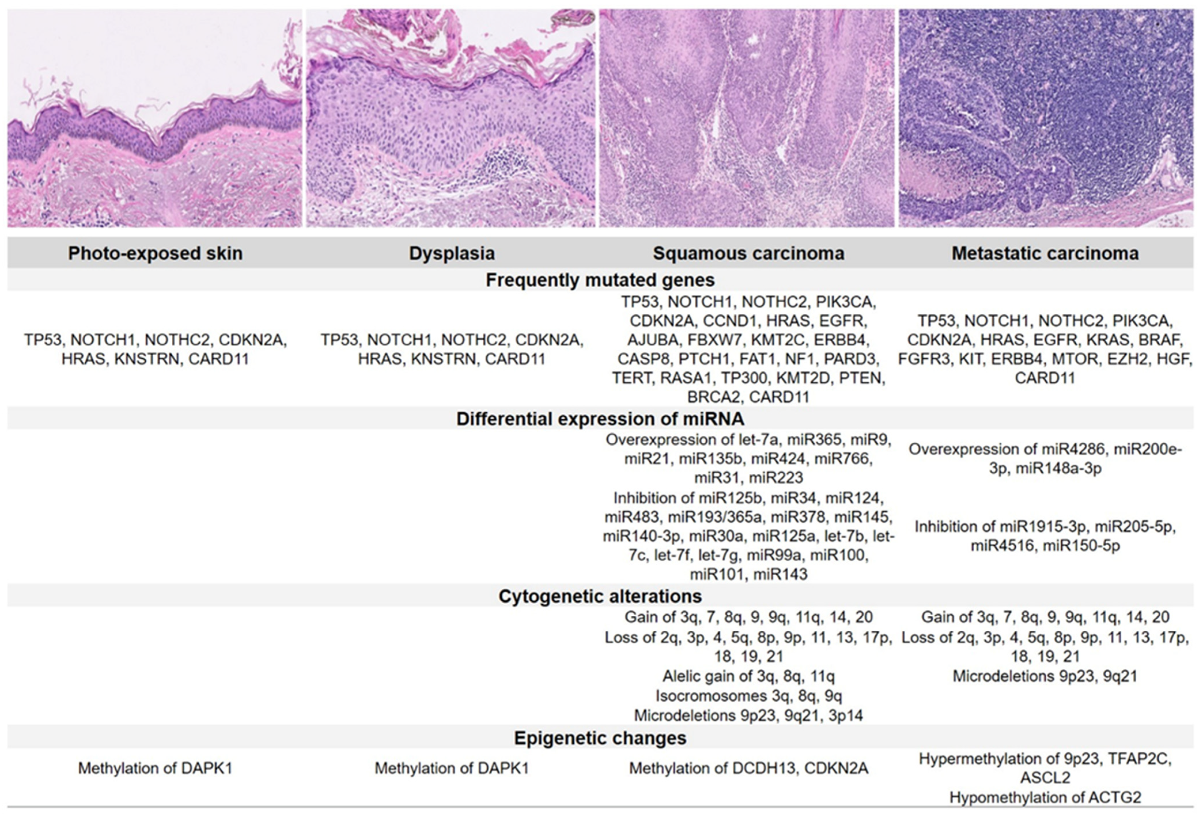
3. Molecular Pathology and Emerging Biomarkers

cSCC presents a higher number of mutations than other tumours, i.e., up to 5 times that of lung cancer or 4 times that of melanoma [23]. By accumulating these mutations, an area of skin can—usually in response to UV light damage—progress through increased levels of dysplasia to become a cSCC, and in fact, areas of photo-exposed skin share many of the same carcinoma features [24]. Although there is a large range of tumour mutational burden (TMB) in cSCC, the median TMB is the highest compared with other tumour types. Exome sequencing in the cSCC has revealed approximately 1300 nucleotide somatic variations per exome. This observation has led to the hypothesis that constant damage to basal keratinocytes by UV radiation may be responsible for multiple cSCC mutational events [25] (Figure 1).
The most frequent mutations in cSCC are associated with ultraviolet radiation and affect TP53 and NOTCH signalling pathway genes. While mutations in the TP53 family decrease the expression of other tumour suppressors, such as IFR6 [26], modifications in NOTCH signalling may have an oncogenic or suppressor role depending on the cellular microenvironment [27]. Activating mutations on genes of the RAS family, mainly HRAS, have been identified in up to 9% of SCC [25], with increasing incidence in up to 60% of tumours developed in patients treated with BRAF/MEK inhibitors [28]. CDKN2A, which is mutated in 31% of primary and metastatic SCC, is associated with tumour aggressiveness [29] and epigenetic changes due to hypermethylation of the CpG islands of the FRZB, TFAP2C, ASCL2 [30], and KMT2C [23] genes. Less frequently, alterations in STAT3, PIK3CA, KIT, RIPK4, and RAS1 genes have been also described [23,31]. Evidence also suggests that phenotypic changes caused by tumour–stromal interaction and the secretion of VEGF-C by tumour-associated fibroblasts might be involved in the cSCC metastatic process [32,33].

4. Diagnosis

4.1. Dermatology

In recent years, optical non-invasive diagnostic techniques have been increasingly applied in clinical practice to improve diagnostic accuracy and to characterise the tumour in vivo before surgery or biopsy [9,34]. Dermoscopy can be used to identify the main characteristics of cSCC, such as clustered vascular pattern, glomerular vessels, and hyperkeratosis. Several additional features can be observed using reflectance confocal microscopy, such as atypical honeycomb or disarranged pattern of the spinous-granular layer of the epidermis, round nucleated bright cells in the epidermis, and round vessels in the dermis [35].
While dermoscopy is performed more often in pigmented than in non-pigmented lesions, it is also useful in the differential diagnosis of equivocal cases. For instance, the identification of glomerular-like, clustered or hairpin vessels, and the scale and alignment of dots and vessels can be useful in particular scenarios, such as in minimally invasive cSCC or in pigmented forms [19]. In situ, cSCC frequently presents clinically as an erythematous scaly patch or slightly elevated plaque, whereas invasive cSCC is usually ulcerated and can be patchy, papulonodular, papillomatous, or exophytic [9]. Progression of in situ cSCC to microinvasive cSCC can be detected on dermoscopy examination with the appearance of a thicker lesion, hairpin and/or linear-irregular vessels, and a keratotic centre and/or ulceration (Figure 2). In invasive cSCC, a vertical growth phase reflecting dermal invasion is typically characterized by an increased number of polymorphic vessels, such as linear irregular, hairpin, and grouped glomerular/dotted vessels over a whitish background with a central mass of keratin or ulceration [9,35,36].

4.2. Histopathology

The histological diagnosis of cSCC is made using haematoxylin–eosin staining, although in cases of uncertain diagnosis, especially in non-keratinizing tumours, additional immunohistochemical studies for cytokeratins or stratified epithelia may be necessary. The morphological image shows bundles of atypical keratinocytes originating in the epidermis and infiltrating the dermis. Morphological features of differentiation can include the formation of corneal pearls, parakeratosis, and dyskeratosis. cSCC ranges from well-differentiated tumours to poorly differentiated neoplasms that show pleomorphic nuclei with a high degree of atypia, frequent mitosis, and very little keratin formation, if any [37].
Well-differentiated histological subtypes with low metastatic potential have been described, including keratoacanthoma and verrucous and fusocellular carcinoma, as well as other aggressive subtypes, such as acantholytic squamous, desmoplastic, and adenosquamous carcinomas, that have a risk of metastasis of 16.1% (95% CI 6.6–39.5) [14]. Other relevant morphological prognostic factors include the degree of dermal infiltration, tumour diameter (greater than 2.0 cm), the presence of perineural (especially in nerves greater than 0.1 mm) and lymphovascular invasion, and resection margins [38].

4.3. Medical Imaging

Radiological investigations are indicated when extensive disease is suspected, to determine bone or soft tissue involvement, invasion of surrounding areas, and the development of metastasis [7]. Main characteristics of imaging modalities used in cSCC are summarized in Table 3.
Computed tomography (CT) scans evaluate cartilage and bone involvement, and three-dimensional imaging is very useful in planning the surgical approach and subsequent reconstruction. Magnetic resonance imaging (MRI) is preferred to assess the involvement of deep soft tissue and structures such as bone, parotid glands, and major nerves [19,40]. Ultrasound (US) is a very sensitive method for identifying lymph node metastases and serves to guide the radiologist during needle biopsy [7,19]. Positron emission tomography (PET) is useful for detecting metastases in sites where other studies are inconclusive, as in the case of fibrosis, necrosis, and previously radiated tissues. However, it should be noted that the presence of infectious and inflammatory processes may lead to false positives [40]. The use of combined PET-CT increases CT sensitivity [40], thus permitting the accurate detection of metastasis in distant organs [19].

4.4. Assessment of Comorbidities

In NMSC patients, three comorbidity assessment tools are frequently used in clinical trials and could be considered in clinical practice: the Charlson Comorbidity Index (CCI), the American Society of Anesthesiologists (ASA) risk classification system, and the Adult Comorbidity Evaluation-27 (ACE-27) [41]. In a systematic review of 22 studies, the CCI was used most often to assess comorbidities (82% of cases), probably because of its extensive use among skin cancer patients and validation in other cancer populations [41]. While higher CCI scores were significantly associated with age > 80 years in patients with head and neck tumours [42], correlation between CCI and life expectancy in nonagenarians undergoing Mohs micrographic surgery is still unclear [43,44].
The ASA risk classification has been traditionally used as a predictor of risk in the preoperative screening of patients undergoing general anaesthesia [45]. Although it was not formally developed as a comorbidity index, its widespread use enables data retrieval from medical records of surgical skin cancer patients [41]. The ACE-27, which is a modification of the Kaplan–Feinstein Index, performs better than a standard medical interview in the identification of comorbidities among skin cancer patients [46]. Since it includes more comorbid conditions and enables further grading than the CCI score, the ACE-27 could potentially predict the prognosis of patients more accurately. However, larger studies are warranted [41].

5. Risk Stratification

Current prognostic factors for recurrence, which help determine the role of definitive or adjuvant treatments, are based on clinicopathologic features described in single-centre or large-scale clinical studies or consensus meetings. These factors have been established taking into account local staging, location, depth, and pathological features. The NCCN guidelines establish low- and high-risk features for cSCC (Table 4) [7]. High-risk features are observed in approximately 5% of all NMSC and include locally advanced disease (stages T3-4), nodal involvement, perineural invasion (PNI), local and regional recurrence, or immunosuppression [47].
PNI is a well-known high-risk factor for local or distant relapse that affects 5% of patients, mostly in cSCC [48]. In a systematic review, patients with clinical PNI (CPNI) showed a significantly increased risk of local recurrence (37% vs. 17%) and disease-specific death (27% vs. 6%) compared with patients with incidental PNI (IPNI), whereas nodal and distant metastasis were similar regardless of PNI classification. In addition, CPNI had significantly poorer mean 5-year recurrence-free survival (61% vs. 76%) and disease-specific survival (70% vs. 88%) than IPNI patients [49].

6. Treatment

6.1. Role of Surgery in Primary Tumours

Whenever possible, and taking into account the patient’s status, surgical excision of the tumour is the first-line treatment for cSCC patients, regardless of age and anatomical location [8]. The choice among available modalities, which include conventional surgery and micrographic-controlled surgery, should be based on the patient’s risk factors for poor prognosis (Figure 3).
Mohs micrographic surgery (MMS) may be the preferred technique for high-risk patients based on the high cure rates and low recurrence rates reported in retrospective analyses of primary and recurrent cSCC [7]. Alternatively, patients may undergo conventional surgery and intraoperative biopsies. Wide local excision may be more appropriate for large and invasive lesions, as it allows for complete assessment of peripheral and deep margins. To achieve histologically clean margins after surgical excision, the width of the margins should be adapted to the risk of cSCC extension and recurrence. For low-risk lesions, clinical practice guidelines recommend performing an excision with 4–6 mm margins [8]. However, larger margins are needed for larger tumours and when high-risk features, such as perineural or lymphatic invasion, are present. While margins > 6 mm would be required for lesions with a diameter > 1 cm [7], the most appropriate width should be determined by individual risk assessment [8]. From the oncological perspective, a surgical defect should be restored using techniques that do not mobilize surrounding tissues, such as direct closure or grafting [7], while reconstruction with skin flaps may be only advised after ensuring that the tumour has been completely removed.
Particular scenarios have been also described, such as cSCC that develop in association with scars or chronic wounds (Marjolin’s ulcer) or previously radiated skin, those that invade deep structures, such as bone, parotid or nerve trunks, and those located in the labial vermilion and the ear [7]. In these cases, amputation of the affected limb, parotidectomy, and prophylactic lymph node drainage may be necessary.

6.2. Locoregional Assessment and Sentinel Node Biopsy

Sentinel lymph node biopsy (SLNB) in patients with cSCC is aimed at the early detection and management of occult nodal metastasis. According to two systematic reviews that included 16 and 23 studies, the positive SLNB rate among cSCC patients ranged between 14% and 8%, respectively [50,51]. A meta-analysis of 19 studies identified microscopic sentinel node involvement in 12.3% of patients who had a tumour diameter greater than 2 cm in all cases. Using the AJCC criteria, a higher risk of positive SLNB was associated with advanced tumour stages, reaching 29.4% in T2b and 50.0% in T3 lesions [52].
The utility of SLNB in detecting cSCC micrometastases not identified by non-invasive examination methods (i.e., instrumental tests) has been discussed. Notably, the rate of micrometastases increased from 3.4% in the overall population to 6.5% in the high-risk group; this rate directly correlated with the depth and diameter of the tumour [53]. In a retrospective analysis of cSCC patients who underwent SLNB and did not present micrometastasis, no relapse event nor local/distant metastases were reported during a mean follow-up of 27.5 months [54].
Nonetheless, given the lack of high-level evidence supporting a real prognostic impact and a well-defined profile of patients who could benefit the most, SLNB is not currently recommended in patients with invasive cSCC outside of a clinical trial setting [19].

6.3. Local Treatment

Surgical excision should be always considered as first-line treatment option for patients with either high- or low-risk cSCC. Nonetheless, based on the risk stratification, local approaches may be considered in patients for whom surgery is not feasible. For low-risk cSCC, several non-surgical treatment modalities are currently available to treat the tumour and field cancerization. Photodynamic therapy consists of a 2-step method that involves the topical application of a photosensitizer, such as 5-aminolevulinic acid or methyl aminolevulinate, followed by an incubation period with light irradiation. Alternatively, a topical therapy with imiquimod up to 5% or 5-fluorouracil may be also applied [9].

6.4. Role of Radiotherapy

RT may be used in first line in patients unable to undergo surgical resection, such as cosmetic or frail patients, to avoid significantly disfiguring surgery or orbital exenteration or in patients too frail to undergo general anaesthesia. A recent meta-analysis involving 21,000 patients showed comparable, extremely low one-year recurrence rates for both surgery and RT: 0.8%, 0.2%, 2%, and 0% for surgical excision, MMS, external beam irradiation, and brachytherapy (BT), respectively, and excellent cosmesis [55]. Different RT techniques, such as external beam RT, BT or electronic low energy sources (ELS), and schedules (i.e., normal fractionation or hypofractionation) can be selected depending on the expertise of the hospital. More recently, newer technologies such as volumetric arc therapy (VMAT) have proven useful in the treatment of scalp lesions or extensive field cancerization in trunk or extremities unamenable to other radiation modalities [56].
The role of RT in NMSC, mainly cSCC, has been defined in a recently published clinical practice guideline, following the ASTRO recommendation grading system [57]. Definitive RT is: (a) advised for patients who cannot undergo or who decline surgery, (b) conditionally recommended in anatomic locations where surgery can compromise function or cosmesis, and (c) not recommended (conditionally) in genetic diseases predisposing to higher radiosensitivity (ataxia-telangiectasia, Gorlin syndrome, Li–Fraumeni syndrome). Definitive RT is only recommended for inoperable patients, whereas elective node irradiation is conditionally recommended in lesions (thickness > 6 mm) in which there is an overlap of the primary tumour with the adjacent nodal basin.
Adjuvant RT is strongly recommended in NMSC patients with gross PNI as well as for cSCC patients with any of the following characteristics: (a) close or positive margins not amenable to re-resection, (b) relapse after prior margin-free surgery, (c) stage T3–T4, (d) desmoplastic reaction or lymphocyte infiltration in chronic immunosuppression. In patients with regional node metastasis, adjuvant RT after therapeutic lymphadenectomy is strongly recommended except for single, <3 cm nodes without extracapsular invasion. The addition of concurrent carboplatin chemotherapy to postoperative RT has not been shown to improve locoregional control in patients with high-risk cSCCHN [47].
For adjuvant RT, conventional doses of 60–66 Gy (2 Gy/day) are the standard, most frequently used fractionation. In case of elective RT, doses of 50–54 Gy are recommended [57]. Hypofractionation (HF) is usually delivered for brachytherapy or ELS, most often at doses of 50 Gy (20 fractions, 4 weeks) or 45 Gy (15 fractions, 3 weeks). Alternatively, doses of 30–40 Gy (5–7 Gy fractions, 1–3 fractions per week) result in excellent local control rates and acceptable toxicity. HF is most advantageous in smaller lesions, frail patients, elderly patients, or anyone with problems coming into the clinic on a daily visit [58,59]. Zaorsky et al. recommend schedules of 50 Gy/15 fractions, 36.75 Gy/7 fractions, or 35 Gy/5 fractions, as cosmesis outcomes are “good” in 80% of patients, depending on frailty [59]. Suggested treatments with RT using external beam irradiation are shown in Table 5.

6.5. Systemic Treatment

6.5.1. Chemotherapy

Systemic treatment is administered to patients with cSCC who have progressed locally and/or with metastases on previous local treatments. Although no chemotherapeutic agents have been specifically approved for cSCCHN, platinum, 5-fluorouracil, anthracycline, or bleomycin alone or in combination are frequently used [60,61]. Moreover, the combined administration of cisplatin, interferon alfa, and 13-cis-retinoic acid has shown to be clinically active in patients with advanced cSCC, resulting in 34% overall and 17% complete response (CR) rates, with a median survival of 14.6 months [62].

6.5.2. Targeted Therapy

Epidermal growth factor receptor (EGFR) expression is present in 90% of cSCC tumours, with overexpression in up to 35% [63]. Four phase II trials tested EGFR tyrosine kinase inhibitors (TKIs), erlotinib [64] and gefitinib [65], and monoclonal antibodies cetuximab [66] or panitumumab [67] in the recurrent/metastatic setting (Table 6). Treatment with TKIs resulted in lower response rates without CR compared with antibodies but with similar disease control and duration of response. Likewise, survival outcomes did not differ, with a median progression-free survival (PFS) of 4–8 months or overall survival (OS) of 8–13 months. The largest cohort reporting clinical outcomes of cetuximab alone in unselected patients with unresectable or metastatic tumours confirmed a high disease control rate of 87% (53% response rate) [68].
In contrast with panitumumab or TKIs, cetuximab demonstrated efficacy in cSCCHN [69,70], so it has been widely used in this setting in monotherapy or combined with chemotherapy [71]. Notably, the administration of cetuximab with chemotherapy and/or anti-EGFR has been associated with significant toxicity, mainly cutaneous. Comorbidities should be evaluated, and geriatric assessment should be performed in elderly patients with cSCC to identify the most effective and tolerable therapy for each patient.

6.5.3. Immunotherapy

Immune checkpoint inhibitors (ICIs) have drastically improved survival outcomes in patients with advanced melanoma, with a considerable proportion of long-term survivors [72]. As tumours with a high mutational burden are known to be more responsive to ICIs and this mutational burden is up to 4 times greater in cSCC than melanoma [33], clinical trials were conducted in patients with cSCC [73]. Currently, anti-programmed cell death-1 (anti-PD-1) antibodies constitute first-line systemic treatment for metastatic or locally advanced cSCC in which curative surgery or RT are not feasible [8]. All systemic treatments are off-label, except for the anti-PD-1 agent cemiplimab, approved by the FDA/EMA in patients with locally advanced or metastatic cSCC who are not candidates for curative surgery or curative radiation [8,74,75].
Cemiplimab is a high-affinity, highly potent human immunoglobulin G4 anti-PD-1 receptor monoclonal antibody [76] that has demonstrated efficacy (overall response rate (ORR) 46.1%) and long-term durable response with very effective disease control (disease control rate: 72.5%) in patients with advanced and metastatic cSCC [77,78]. With a median age of 72 (38–96) years, 67.9% had cSCCHN, 59.6% had metastatic disease, and 40.4% locally advanced cSCC. Up to 66.3% of patients received cemiplimab as first-line treatment, and median observed time to response was 2.1 (1.9–3.7) months. Overall, the CR rate was 16.1% and median time to CR was 11.2 months [78]. Patients had deepening responses over time as evidenced by increasing CR rates [77,78,79]. Estimated median PFS was 18.4 months (95% CI: 10.3–24.3), while median DOR and OS had not been reached after 15.7 months follow-up. In responding patients, the estimated proportion of patients with ongoing response was 87.8% (95% CI: 78.5–93.3) at 12 months and 69.4% (95% CI: 55.6–79.6) at 24 months [78]. Advanced cSCC patients treated with cemiplimab experienced clinically meaningful improvements in global health status/health-related quality of life, and pain and functional status, and they maintained a low symptom burden [80]. Recently, data from real-world practice have confirmed a similar benefit of cemiplimab to that observed in clinical trials in a cohort of 240 patients with advanced cSCC [81].
Along with cemiplimab, other anti-PD-1 agents are under evaluation in cSCC (Table 7). Pembrolizumab is being tested in a phase II study of patients with recurrent/metastatic or locally advanced unresectable cSCC (NCT03284424). Results of a first interim analysis showed effective antitumour activity with 34.3% ORR and clinically meaningful, durable responses [82]. Pembrolizumab is also being evaluated in patients with locally advanced cSCC after surgery and radiation (NCT03833167). The efficacy of nivolumab monotherapy in patients with locally advanced/metastatic cSCC is being investigated in phase II studies (NCT04204837, NCT03834233).

7. Immunosuppressed Patients

Immunosuppressed solid organ transplant recipients (SOTRs) present a 65- to 250-fold greater incidence of cSCC than the general population, more aggressive progression, and a higher risk of metastasis and death [83,84]. Moreover, certain transplant recipients are more likely to develop cSCC than others; an increased risk has been reported for patients who undergo heart transplant compared with kidney or liver recipients [85]. In this respect, the type, intensity, and duration of the immunosuppressive regimen seem related to the development of cSCC in SOTRs. A lower incidence of cSCC has been reported in patients who receive sirolimus compared with those treated with calcineurin inhibitors [83].
Immune status has been identified as a strong prognostic factor for disease outcomes in immunosuppressed patients. A multi-institutional study including patients with cSCCHN reported significantly lower 2-year locoregional recurrence-free survival and PFS in immunocompromised patients compared with immunocompetent patients. Moreover, immunosuppressed status, recurrent disease, poor differentiation, and PNI were significantly associated with locoregional recurrence in this population [86].
A close clinical follow-up, every 3–6 months lifelong, and tailored immunosuppressive treatments, with adjustment or reduction of maintenance post-transplant therapy, may be necessary to reduce the risk of new cSCC [8,19]. Dermatologists, transplant physicians, and patients must collaborate to ensure adherence to dermatologic surveillance recommendations and must monitor suspicious lesions, thereby reducing the risk of cSCC in SOTRs [87,88].
It should be noted that although immunosuppression of cSCC patients is often described in the setting of SOTRs, other conditions should not be disregarded. As such, patients immunosuppressed due to human immunodeficiency virus (HIV) infection, allogenic bone marrow transplant, or autoimmune diseases should be carefully monitored.

8. Follow-Up and Monitoring

Patients with cSCC should be closely followed up for recurrences and development of new keratinocyte cancers and melanoma, particularly if they have a history of cSCC [8]. Evidence from cohort studies and registries shows that patients with at least one cSCC are at risk for developing additional cSCC and other skin cancers [20]. Therefore, it is recommended that follow-up for all patients should include regular clinical assessment, including inspection and palpation of the excision site, the in-transit route, and the regional lymph nodes [7,8]. Patients who have had cSCC should receive counselling regarding the risk for new primary skin cancers, such as BCC, cSCC, and melanoma, and the benefits of in-office and self-screening for early detection should be made clear [7,20].
The frequency of visits should be adjusted individually, depending on patient-specific underlying risk characteristics for cSCC [7,8]. The recently updated interdisciplinary European guidelines for the management of invasive cSCC recommend scheduling follow-up examination based on low- or high-risk common primary, advanced, or regional disease and the immunosuppressive setting [8]. While patients with low-risk cSCC may be assessed every 6–12 months for 5 years, high-risk patients should be examined in the clinic every 3–6 months for 2 years (including lymph node US), every 6–12 months from year 3–5, and annually thereafter. Closer follow-up is advised in patients with locally advanced/metastatic cSCC or after surgery for locoregional metastases, including clinical and US evaluation every 3 months for 5 years and then once or twice per year [8]. Similarly, the National Comprehensive Cancer Network (NCCN) guidelines for cSCC recommend that the frequency of patients’ follow-up should depend on whether the tumour has spread to lymph nodes. Local cSCC should be monitored at least every 3–12 months during the first 2 years, every 6–12 months for another 3 years, and then annually for life. For regional cSCC, a more frequent history and clinical examination is advised: every 1–3 months for 1 year, every 2–4 months for 1 year, every 4–6 months for another 3 years, and then every 6–12 months for life [7].
Imaging tests (CT, MRI, or PET-CT) for non-palpable regional lymph nodes should also be performed in patients with regional cSCC [7] or locally advanced or metastatic disease [8]. The NCCN guidelines establish that the frequency of imaging scans should be tailored to individual clinical factors [7], whereas the European guidelines recommend examination every 3–6 months in the first 3 years and then based on individual symptoms and stage [8].
It should be noted that increased surveillance may be required for cSCC patients at very high risk of other primary tumours and recurrence, such as immunosuppressed patients or individuals with haematological comorbidities, genetic predisposition, or previous history of cSCC [7,8]. Their follow-up schedule, including clinical and imaging evaluation, should be modified depending on the characteristics of individual primary tumours (e.g., number and frequency of development) [8].

9. Summary and Conclusions

Tumour staging, management decisions, and monitoring of the cSCC are continuously improving thanks to the development of novel diagnostic tools and therapeutic agents. Multidisciplinary teams may ensure that the most appropriate strategy, tailored to the patient’s individual profile, is undertaken from the initial diagnosis to follow-up. Patient-specific characteristics for risk including comorbidities, clinical parameters, histopathology, and molecular biomarkers should be considered to provide an accurate diagnosis. While surgery remains the cornerstone of treatment and RT is often used in the management of cSCC, ICIs and targeted therapies have changed the therapeutic landscape, showing improved outcomes in patients with locally advanced or metastatic disease. Considering the likelihood of recurrence and the increased risk of developing multiple cSCC, particularly in immunosuppressed patients, patients should be closely followed up in the first two years after starting treatment and continued long-term surveillance by clinical and imaging examination is warranted.

Supplementary Materials

The following are available online at https://www.mdpi.com/article/10.3390/cancers14030629/s1, Table S1: Methods and results of the Delphi survey.

Author Contributions

Conceptualization, J.G.-F., R.M., O.S., F.R., J.M., A.T.-V., S.M. and I.A.; writing—original draft preparation, J.G.-F., R.M., O.S., F.R., J.M., A.T.-V., S.M. and I.A.; writing—review and editing, J.G.-F., R.M., O.S., F.R., J.M., A.T.-V., S.M. and I.A.; project administration, J.G.-F. All authors have read and agreed to the published version of the manuscript.

Funding

This work was supported by Sanofi.

Institutional Review Board Statement

Not applicable.

Informed Consent Statement

Not applicable.

Data Availability Statement

The authors confirm that all data supporting the findings of this study are available within the article and its Supplementary Materials.

Acknowledgments

The authors thank Celia Miguel-Blanco from Medical Statistics Consulting S.L. (Valencia, Spain) for editorial support in the writing of this manuscript, and the 15 panellists who participated in the survey: A. Rueda Domínguez (H.U. Carlos Haya, Málaga), A. Soria (H.U. Ramón y Cajal, Madrid), J.L. Rodríguez Peralto (H. 12 de Octubre, Madrid), M. Iglesias (H. del Mar, Barcelona), E. Muñoz (H. Vall d’Hebron, Barcelona), A. García-Castaño (H. Marqués de Valdecilla, Santander), I. Márquez (H. Gregorio Marañón, Madrid), P. Cerezuela (H. Virgen de la Arrixaca, Murcia), J. Cañueto (H.U. Salamanca, Salamanca), R. Botella (H.U. La Fe, Valencia), S. Puig (H. Clinic, Barcelona), Y. Escobar (H. Gregorio Marañón, Madrid), F. Almeida (H. Ramón y Cajal, Madrid), B. Bellosillo (H. de Mar, Barcelona), and E. Espinosa (H. La Paz, Madrid).

Conflicts of Interest

J. Garcia-Foncillas discloses consulting/advisory/honoraria speaker roles with Abbott, Amgen, Astellas, Astra Zeneca, Biocartis, Boehringer Ingelheim, BMS, Bayer, Celgene, Eisai, Foundation Medicine, GSK, Hospira, Janssen, Lilly, Merck Serono, MSD, Novartis, Pharmamar, Pfizer, Roche, Sanofi, Servier, Sysmex, and Tesaro. R. Mesía discloses consulting/advisory/honoraria speaker role with Amgen, BMS, Merck Serono, MSD, Roche, and Sanofi. O. Sanmartín discloses consulting/advisory/honoraria speaker role with Roche, ISDIN, and Sanofi. F. Rojo has received honorary for consultancy from Roche, BMS, MSD, Merck, Novartis, Pfizer, Astra Zeneca, Abbvie, Pierre Fabre, InCyte, and Bayer. J. Mestre discloses consulting/advisory/honoraria speaker roles with Lavigor, Roche, and Sanofi. A. Tejera has received honoraria for consultancy from Sanofi. S. Martín discloses consulting/advisory/honoraria speaker roles with MSD, Sanofi/Regeneron, BMS, Novartis, Roche, and Pierre-Fabre. I. Azinovic discloses no conflicts of interest. The funder had no role in the design of the study; in the collection, analyses, or interpretation of data; in the writing of the manuscript; or in the decision to publish the results.

References

  1. Karia, P.S.; Han, J.; Schmults, C.D. Cutaneous squamous cell carcinoma: Estimated incidence of disease, nodal metastasis, and deaths from disease in the United States, 2012. J. Am. Acad. Dermatol. 2013, 68, 957–966. [Google Scholar] [CrossRef] [PubMed]
  2. Flohil, S.C.; van der Leest, R.J.; Arends, L.R.; de Vries, E.; Nijsten, T. Risk of subsequent cutaneous malignancy in patients with prior keratinocyte carcinoma: A systematic review and meta-analysis. Eur. J. Cancer 2013, 49, 2365–2375. [Google Scholar] [CrossRef] [PubMed]
  3. Tejera-Vaquerizo, A.; Descalzo-Gallego, M.A.; Otero-Rivas, M.M.; Posada-García, C.; Rodríguez-Pazos, L.; Pastushenko, I.; Marcos-Gragera, R.; García-Doval, I. Skin Cancer Incidence and Mortality in Spain: A Systematic Review and Meta-Analysis. Actas Dermosifiliogr. 2016, 107, 318–328. [Google Scholar] [CrossRef] [PubMed]
  4. Armstrong, B.K.; Kricker, A. The epidemiology of UV induced skin cancer. J. Photochem. Photobiol. B 2001, 63, 8–18. [Google Scholar] [CrossRef]
  5. Stern, R.S. The risk of squamous cell and basal cell cancer associated with psoralen and ultraviolet A therapy: A 30-year prospective study. J. Am. Acad. Dermatol. 2012, 66, 553–562. [Google Scholar] [CrossRef]
  6. Lindelöf, B.; Sigurgeirsson, B.; Gäbel, H.; Stern, R.S. Incidence of skin cancer in 5356 patients following organ transplantation. Br. J. Dermatol. 2000, 143, 513–519. [Google Scholar]
  7. National Comprehensive Cancer Center. NCCN Clinical Practice Guidelines in Oncology; Squamous Cell Carcinoma (V2. 2018). Available online: https://oncolife.com.ua/doc/nccn/Squamous_Cell_Skin_Cancer.pdf (accessed on 13 September 2021).
  8. Stratigos, A.J.; Garbe, C.; Dessinioti, C.; Lebbe, C.; Bataille, V.; Bastholt, L.; Dreno, B.; Concetta Fargnoli, M.; Forsea, A.M.; Frenard, C.; et al. European interdisciplinary guideline on invasive squamous cell carcinoma of the skin: Part 2. Treatment. Eur. J. Cancer 2020, 128, 83–102. [Google Scholar] [CrossRef] [Green Version]
  9. Combalia, A.; Carrera, C. Squamous Cell Carcinoma: An Update on Diagnosis and Treatment. Dermatol. Pract. Concept 2020, 10, e2020066. [Google Scholar] [CrossRef]
  10. Diamond, I.R.; Grant, R.C.; Feldman, B.M.; Pencharz, P.B.; Ling, S.C.; Moore, A.M.; Wales, P.W. Defining consensus: A systematic review recommends methodologic criteria for reporting of Delphi studies. J. Clin. Epidemiol. 2014, 67, 401–409. [Google Scholar] [CrossRef]
  11. Norman, G. Likert scales, levels of measurement and the “laws” of statistics. Adv. Health Sci. Educ. Theory Pract. 2010, 15, 625–632. [Google Scholar] [CrossRef]
  12. Fitch, K.; Bernstein, S.J.; Aguilar, M.D.; Burnanrd, B.; LaCalle, J.R.; Lazaro, P.; van het Loo, M.; McDonell, J.; Vader, J.; Kahan, J.P. The Rand/UCLA Appropriateness Method User’s Manual; RAND Corporation: Santa Monica, CA, USA, 2001; Available online: https://www.rand.org/pubs/monograph_reports/MR1269.html (accessed on 3 March 2021).
  13. Armin, M.B.; Edge, S.B.; Greene, F.; Byrd, D.R.; Brookland, R.K.; Washington, M.K.; Gershenwald, J.E.; Compton, C.C.; Hess, K.R.; Sullivan, D.C.; et al. (Eds.) AJCC Cancer Staging Manual, 8th ed.; Springer: New York, NY, USA, 2017. [Google Scholar]
  14. Brantsch, K.D.; Meisner, C.; Schönfisch, B.; Trilling, B.; Wehner-Caroli, J.; Röcken, M.; Breuninger, H. Analysis of risk factors determining prognosis of cutaneous squamous-cell carcinoma: A prospective study. Lancet Oncol. 2008, 9, 713–720. [Google Scholar] [CrossRef]
  15. Karia, P.S.; Jambusaria-Pahlajani, A.; Harrington, D.P.; Murphy, G.F.; Qureshi, A.A.; Schmults, C.D. Evaluation of American JoInt. Committee on Cancer, International Union against Cancer, and Brigham and Women’s Hospital tumor staging for cutaneous squamous cell carcinoma. J. Clin. Oncol. 2014, 32, 327–334. [Google Scholar] [CrossRef] [Green Version]
  16. Ruiz, E.S.; Karia, P.S.; Besaw, R.; Schmults, C.D. Performance of the American Joint. Committee on Cancer Staging Manual, 8th Edition vs the Brigham and Women’s Hospital Tumor Classification System for Cutaneous Squamous Cell Carcinoma. JAMA Dermatol. 2019, 155, 819–825. [Google Scholar] [CrossRef] [PubMed]
  17. Blechman, A.B.; Carucci, J.A.; Stevenson, M.L. Stratification of Poor Outcomes for Cutaneous Squamous Cell Carcinoma in Immunosuppressed Patients Using the American JoInt. Committee on Cancer Eighth Edition and Brigham and Women’s Hospital Staging Systems. Dermatol. Surg. 2019, 45, 1117–1124. [Google Scholar] [CrossRef] [PubMed]
  18. Tejera-Vaquerizo, A.; Canueto, J.; Llombart, B.; Martorell-Calatayud, A.; Sanmartin, O. Predictive Value of Sentinel Lymph Node Biopsy in Cutaneous Squamous Cell Carcinoma Based on the AJCC-8 and Brigham and Women’s Hospital Staging Criteria. Dermatol. Surg. 2020, 46, 857–862. [Google Scholar] [CrossRef] [PubMed]
  19. Stratigos, A.J.; Garbe, C.; Dessinioti, C.; Lebbe, C.; Bataille, V.; Bastholt, L.; Dreno, B.; Fargnoli, M.C.; Forsea, A.M.; Frenard, C.; et al. European interdisciplinary guideline on invasive squamous cell carcinoma of the skin: Part 1. epidemiology, diagnostics and prevention. Eur. J. Cancer 2020, 128, 60–82. [Google Scholar] [CrossRef] [Green Version]
  20. Alam, M.; Armstrong, A.; Baum, C.; Bordeaux, J.S.; Brown, M.; Busam, K.J.; Eisen, D.B.; Iyengar, V.; Lober, C.; Margolis, D.J.; et al. Guidelines of care for the management of cutaneous squamous cell carcinoma. J. Am. Acad. Dermatol. 2018, 78, 560–578. [Google Scholar] [CrossRef] [Green Version]
  21. Cañueto, J.; Martín-Vallejo, J.; Cardeñoso-Álvarez, E.; Fernández-López, E.; Pérez-Losada, J.; Román-Curto, C. Rapid growth rate is associated with poor prognosis in cutaneous squamous cell carcinoma. Clin. Exp. Dermatol. 2018, 43, 876–882. [Google Scholar] [CrossRef]
  22. Gonzalez-Guerrero, M.; Martínez-Camblor, P.; Vivanco, B.; Fernández-Vega, I.; Munguía-Calzada, P.; Gonzalez-Gutierrez, M.P.; Rodrigo, J.P.; Galache, C.; Santos-Juanes, J. The adverse prognostic effect of tumor budding on the evolution of cutaneous head and neck squamous cell carcinoma. J. Am. Acad. Dermatol. 2017, 76, 1139–1145. [Google Scholar] [CrossRef]
  23. Pickering, C.R.; Zhou, J.H.; Lee, J.J.; Drummond, J.A.; Peng, S.A.; Saade, R.E.; Tsai, K.Y.; Curry, J.L.; Tetzlaff, M.T.; Lai, S.Y.; et al. Mutational landscape of aggressive cutaneous squamous cell carcinoma. Clin. Cancer Res. 2014, 20, 6582–6592. [Google Scholar] [CrossRef] [Green Version]
  24. Hussein, M.R. Ultraviolet radiation and skin cancer: Molecular mechanisms. J. Cutan. Pathol. 2005, 32, 191–205. [Google Scholar] [CrossRef]
  25. Durinck, S.; Ho, C.; Wang, N.J.; Liao, W.; Jakkula, L.R.; Collisson, E.A.; Pons, J.; Chan, S.W.; Lam, E.T.; Chu, C.; et al. Temporal dissection of tumorigenesis in primary cancers. Cancer Discov. 2011, 1, 137–143. [Google Scholar] [CrossRef] [Green Version]
  26. Botti, E.; Spallone, G.; Moretti, F.; Marinari, B.; Pinetti, V.; Galanti, S.; De Meo, P.D.; De Nicola, F.; Ganci, F.; Castrignanò, T.; et al. Developmental factor IRF6 exhibits tumor suppressor activity in squamous cell carcinomas. Proc. Natl. Acad. Sci. USA 2011, 108, 13710–13715. [Google Scholar] [CrossRef] [PubMed] [Green Version]
  27. Moriyama, M.; Durham, A.D.; Moriyama, H.; Hasegawa, K.; Nishikawa, S.; Radtke, F.; Osawa, M. Multiple roles of Notch signaling in the regulation of epidermal development. Dev. Cell 2008, 14, 594–604. [Google Scholar] [CrossRef] [PubMed] [Green Version]
  28. Su, F.; Viros, A.; Milagre, C.; Trunzer, K.; Bollag, G.; Spleiss, O.; Reis-Filho, J.S.; Kong, X.; Koya, R.C.; Flaherty, K.T.; et al. RAS Mutations in Cutaneous Squamous-Cell Carcinomas in Patients Treated with BRAF Inhibitors. N. Engl. J. Med. 2012, 366, 207–215. [Google Scholar] [CrossRef] [PubMed] [Green Version]
  29. Küsters-Vandevelde, H.V.; Van Leeuwen, A.; Verdijk, M.A.; de Koning, M.N.; Quint, W.G.; Melchers, W.J.; Ligtenberg, M.J.; Blokx, W.A. CDKN2A but not TP53 mutations nor HPV presence predict poor outcome in metastatic squamous cell carcinoma of the skin. Int. J. Cancer 2010, 126, 2123–2132. [Google Scholar] [CrossRef]
  30. Darr, O.A.; Colacino, J.A.; Tang, A.L.; McHugh, J.B.; Bellile, E.L.; Bradford, C.R.; Prince, M.P.; Chepeha, D.B.; Rozek, L.S.; Moyer, J.S. Epigenetic alterations in metastatic cutaneous carcinoma. Head Neck 2015, 37, 994–1001. [Google Scholar] [CrossRef] [Green Version]
  31. Lui, V.W.; Peyser, N.D.; Ng, P.K.; Hritz, J.; Zeng, Y.; Lu, Y.; Li, H.; Wang, L.; Gilbert, B.R.; General, I.J.; et al. Frequent mutation of receptor protein tyrosine phosphatases provides a mechanism for STAT3 hyperactivation in head and neck cancer. Proc. Natl. Acad. Sci. USA 2014, 111, 1114–1119. [Google Scholar] [CrossRef] [Green Version]
  32. Commandeur, S.; Ho, S.H.; de Gruijl, F.R.; Willemze, R.; Tensen, C.P.; El Ghalbzouri, A. Functional characterization of cancer-associated fibroblasts of human cutaneous squamous cell carcinoma. Exp. Dermatol. 2011, 20, 737–742. [Google Scholar] [CrossRef]
  33. Moussai, D.; Mitsui, H.; Pettersen, J.S.; Pierson, K.C.; Shah, K.R.; Suárez-Fariñas, M.; Cardinale, I.R.; Bluth, M.J.; Krueger, J.G.; Carucci, J.A. The human cutaneous squamous cell carcinoma microenvironment is characterized by increased lymphatic density and enhanced expression of macrophage-derived VEGF-C. J. Investig. Dermatol. 2011, 131, 229–236. [Google Scholar] [CrossRef] [Green Version]
  34. Ulrich, M.; Stockfleth, E.; Roewert-Huber, J.; Astner, S. Noninvasive diagnostic tools for nonmelanoma skin cancer. Br. J. Dermatol. 2007, 157, 56–58. [Google Scholar] [CrossRef]
  35. Warszawik-Hendzel, O.; Olszewska, M.; Maj, M.; Rakowska, A.; Czuwara, J.; Rudnicka, L. Non-invasive diagnostic techniques in the diagnosis of squamous cell carcinoma. J. Dermatol. Case Rep. 2015, 9, 89–97. [Google Scholar] [CrossRef] [Green Version]
  36. Zalaudek, I.; Giacomel, J.; Schmid, K.; Bondino, S.; Rosendahl, C.; Cavicchini, S.; Tourlaki, A.; Gasparini, S.; Bourne, P.; Keir, J.; et al. Dermatoscopy of facial actinic keratosis, intraepidermal carcinoma, and invasive squamous cell carcinoma: A progression model. J. Am. Acad. Dermatol. 2012, 66, 589–597. [Google Scholar] [CrossRef] [PubMed]
  37. Yanofsky, V.R.; Mercer, S.E.; Phelps, R.G. Histopathological variants of cutaneous squamous cell carcinoma: A review. J. Skin Cancer 2011, 2011, 210813. [Google Scholar] [CrossRef] [PubMed] [Green Version]
  38. Thompson, A.K.; Kelley, B.F.; Prokop, L.J.; Murad, M.H.; Baum, C.L. Risk Factors for Cutaneous Squamous Cell Carcinoma Recurrence, Metastasis, and Disease-Specific Death: A Systematic Review and Meta-analysis. JAMA Dermatol. 2016, 152, 419–428. [Google Scholar] [CrossRef] [PubMed]
  39. Liao, L.J.; Lo, W.C.; Hsu, W.L.; Wang, C.T.; Lai, M.S. Detection of cervical lymph node metastasis in head and neck cancer patients with clinically N0 neck-a meta-analysis comparing different imaging modalities. BMC Cancer 2012, 12, 236. [Google Scholar] [CrossRef]
  40. Jennings, L.; Schmults, C.D. Management of high-risk cutaneous squamous cell carcinoma. J. Clin. Aesthetic Dermatol. 2010, 3, 39–48. [Google Scholar]
  41. Connolly, K.L.; Jeong, J.M.; Barker, C.A.; Hernandez, M.; Lee, E.H. A systematic review of comorbidity indices used in the nonmelanoma skin cancer population. J. Am. Acad. Dermatol. 2017, 76, 344–346.e2. [Google Scholar] [CrossRef] [Green Version]
  42. Dhiwakar, M.; Khan, N.A.; McClymont, L.G. Surgery for head and neck skin tumors in the elderly. Head Neck 2007, 29, 851–856. [Google Scholar] [CrossRef]
  43. Charles, A.J., Jr.; Otley, C.C.; Pond, G.R. Prognostic factors for life expectancy in nonagenarians with nonmelanoma skin cancer: Implications for selecting surgical candidates. J. Am. Acad. Dermatol. 2002, 47, 419–422. [Google Scholar] [CrossRef]
  44. Delaney, A.; Shimizu, I.; Goldberg, L.H.; MacFarlane, D.F. Life expectancy after Mohs micrographic surgery in patients aged 90 years and older. J. Am. Acad. Dermatol. 2013, 68, 296–300. [Google Scholar] [CrossRef]
  45. Reid, B.C.; Alberg, A.J.; Klassen, A.C.; Koch, W.M.; Samet, J.M. The American Society of Anesthesiologists’ class as a comorbidity index in a cohort of head and neck cancer surgical patients. Head Neck 2001, 23, 985–994. [Google Scholar] [CrossRef]
  46. Lee, E.H.; Nijhawan, R.I.; Nehal, K.S.; Dusza, S.W.; Levine, A.; Hill, A.; Barker, C.A. Comorbidity Assessment in Skin Cancer Patients: A Pilot Study Comparing Medical Interview with a Patient-Reported Questionnaire. J. Skin Cancer 2015, 2015, 953479. [Google Scholar] [CrossRef] [PubMed]
  47. Porceddu, S.V.; Bressel, M.; Poulsen, M.G.; Stoneley, A.; Veness, M.J.; Kenny, L.M.; Wratten, C.; Corry, J.; Cooper, S.; Fogarty, G.B.; et al. Postoperative Concurrent Chemoradiotherapy Versus Postoperative Radiotherapy in High-Risk Cutaneous Squamous Cell Carcinoma of the Head and Neck: The Randomized Phase III TROG 05.01 Trial. J. Clin. Oncol. 2018, 36, 1275–1283. [Google Scholar] [CrossRef] [PubMed]
  48. Carter, J.B.; Johnson, M.M.; Chua, T.L.; Karia, P.S.; Schmults, C.D. Outcomes of Primary Cutaneous Squamous Cell Carcinoma with Perineural Invasion: An 11-Year Cohort Study. JAMA Dermatol. 2013, 149, 35–42. [Google Scholar] [CrossRef] [PubMed] [Green Version]
  49. Karia, P.S.; Morgan, F.C.; Ruiz, E.S.; Schmults, C.D. Clinical and Incidental Perineural Invasion of Cutaneous Squamous Cell Carcinoma: A Systematic Review and Pooled Analysis of Outcomes Data. JAMA Dermatol. 2017, 153, 781–788. [Google Scholar] [CrossRef] [PubMed]
  50. Navarrete-Dechent, C.; Veness, M.J.; Droppelmann, N.; Uribe, P. High-risk cutaneous squamous cell carcinoma and the emerging role of sentinel lymph node biopsy: A literature review. J. Am. Acad. Dermatol. 2015, 73, 127–137. [Google Scholar] [CrossRef]
  51. Tejera-Vaquerizo, A.; García-Doval, I.; Llombart, B.; Cañueto, J.; Martorell-Calatayud, A.; Descalzo-Gallego, M.A.; Sanmartín, O. Systematic review of the prevalence of nodal metastases and the prognostic utility of sentinel lymph node biopsy in cutaneous squamous cell carcinoma. J. Dermatol. 2018, 45, 781–790. [Google Scholar] [CrossRef]
  52. Schmitt, A.R.; Brewer, J.D.; Bordeaux, J.S.; Baum, C.L. Staging for Cutaneous Squamous Cell Carcinoma as a Predictor of Sentinel Lymph Node Biopsy Results: Meta-analysis of American JoInt. Committee on Cancer Criteria and a Proposed Alternative System. JAMA Dermatol. 2014, 150, 19–24. [Google Scholar] [CrossRef] [Green Version]
  53. Samsanavicius, D.; Kaikaris, V.; Cepas, A.; Ulrich, J.; Makstiene, J.; Rimdeika, R. Importance of sentinel lymphatic node biopsy in detection of early micrometastases in patients with cutaneous squamous cell carcinoma. J. Plast. Reconstr. Aesthetic Surg. 2018, 71, 597–603. [Google Scholar] [CrossRef]
  54. Samsanavičius, D.; Kaikaris, V.; Norvydas, S.J.; Liubauskas, R.; Valiukevičienė, S.; Makštienė, J.; Maslauskas, K.; Rimdeika, R. Sentinel lymph node biopsy for high-risk cutaneous squamous cell carcinoma: Analysis of recurrence-free survival. Medicina 2016, 52, 276–282. [Google Scholar] [CrossRef]
  55. Lee, C.T.; Lehrer, E.J.; Aphale, A.; Lango, M.; Galloway, T.J.; Zaorsky, N.G. Surgical excision, Mohs micrographic surgery, external-beam radiotherapy, or brachytherapy for indolent skin cancer: An international meta-analysis of 58 studies with 21,000 patients. Cancer 2019, 125, 3582–3594. [Google Scholar] [CrossRef]
  56. Martin, T.; Moutrie, Z.; Tighe, D.; Haady, H.; Fogarty, G. Volumetric modulated arc therapy (VMAT) for skin field cancerisation of the nose—A technique and case report. Int. J. Radiol. Radiat. Ther. 2018, 5, 142–148. [Google Scholar] [CrossRef] [Green Version]
  57. Likhacheva, A.; Awan, M.; Barker, C.A.; Bhatnagar, A.; Bradfield, L.; Brady, M.S.; Buzurovic, I.; Geiger, J.L.; Parvathaneni, U.; Zaky, S.; et al. Definitive and Postoperative Radiation Therapy for Basal and Squamous Cell Cancers of the Skin: Executive Summary of an American Society for Radiation Oncology Clinical Practice Guideline. Pract. Radiat. Oncol. 2020, 10, 8–20. [Google Scholar] [CrossRef] [PubMed] [Green Version]
  58. Gunaratne, D.A.; Veness, M.J. Efficacy of hypofractionated radiotherapy in patients with non-melanoma skin cancer: Results of a systematic review. J. Med. Imaging Radiat. Oncol. 2018, 62, 401–411. [Google Scholar] [CrossRef] [PubMed]
  59. Zaorsky, N.G.; Lee, C.T.; Zhang, E.; Keith, S.W.; Galloway, T.J. Hypofractionated radiation therapy for basal and squamous cell skin cancer: A meta-analysis. Radiother. Oncol. 2017, 125, 13–20. [Google Scholar] [CrossRef] [PubMed]
  60. Nakamura, K.; Okuyama, R.; Saida, T.; Uhara, H. Platinum and anthracycline therapy for advanced cutaneous squamous cell carcinoma. Int. J. Clin. Oncol. 2013, 18, 506–509. [Google Scholar] [CrossRef] [PubMed] [Green Version]
  61. Rogers, H.W.; Weinstock, M.A.; Feldman, S.R.; Coldiron, B.M. Incidence Estimate of Nonmelanoma Skin Cancer (Keratinocyte Carcinomas) in the U.S. Population, 2012. JAMA Dermatol. 2015, 151, 1081–1086. [Google Scholar] [CrossRef]
  62. Shin, D.M.; Glisson, B.S.; Khuri, F.R.; Clifford, J.L.; Clayman, G.; Benner, S.E.; Forastiere, A.A.; Ginsberg, L.; Liu, D.; Lee, J.J.; et al. Phase II and biologic study of interferon alfa, retinoic acid, and cisplatin in advanced squamous skin cancer. J. Clin. Oncol. 2002, 20, 364–370. [Google Scholar] [CrossRef]
  63. Cañueto, J.; Cardeñoso, E.; García, J.L.; Santos-Briz, Á.; Castellanos-Martín, A.; Fernández-López, E.; Blanco Gómez, A.; Pérez-Losada, J.; Román-Curto, C. Epidermal growth factor receptor expression is associated with poor outcome in cutaneous squamous cell carcinoma. Br. J. Dermatol. 2017, 176, 1279–1287. [Google Scholar] [CrossRef]
  64. Gold, K.A.; Kies, M.S.; William, W.N., Jr.; Johnson, F.M.; Lee, J.J.; Glisson, B.S. Erlotinib in the treatment of recurrent or metastatic cutaneous squamous cell carcinoma: A single-arm phase 2 clinical trial. Cancer 2018, 124, 2169–2173. [Google Scholar] [CrossRef]
  65. William, W.N., Jr.; Feng, L.; Ferrarotto, R.; Ginsberg, L.; Kies, M.; Lippman, S.; Glisson, B.; Kim, E.S. Gefitinib for patients with incurable cutaneous squamous cell carcinoma: A single-arm phase II clinical trial. J. Am. Acad. Dermatol. 2017, 77, 1110–1113.e2. [Google Scholar] [CrossRef]
  66. Maubec, E.; Petrow, P.; Scheer-Senyarich, I.; Duvillard, P.; Lacroix, L.; Gelly, J.; Certain, A.; Duval, X.; Crickx, B.; Buffard, V.; et al. Phase II study of cetuximab as first-line single-drug therapy in patients with unresectable squamous cell carcinoma of the skin. J. Clin. Oncol. 2011, 29, 3419–3426. [Google Scholar] [CrossRef] [PubMed]
  67. Foote, M.C.; McGrath, M.; Guminski, A.; Hughes, B.G.; Meakin, J.; Thomson, D.; Zarate, D.; Simpson, F.; Porceddu, S.V. Phase II study of single-agent panitumumab in patients with incurable cutaneous squamous cell carcinoma. Ann. Oncol. 2014, 25, 2047–2052. [Google Scholar] [CrossRef] [PubMed]
  68. Montaudié, H.; Viotti, J.; Combemale, P.; Dutriaux, C.; Dupin, N.; Robert, C.; Mortier, L.; Kaphan, R.; Duval-Modeste, A.B.; Dalle, S.; et al. Cetuximab is efficient and safe in patients with advanced cutaneous squamous cell carcinoma: A retrospective, multicentre study. Oncotarget 2020, 11, 378–385. [Google Scholar] [CrossRef] [PubMed]
  69. Vermorken, J.B.; Mesia, R.; Rivera, F.; Remenar, E.; Kawecki, A.; Rottey, S.; Erfan, J.; Zabolotnyy, D.; Kienzer, H.-R.; Cupissol, D.; et al. Platinum-Based Chemotherapy plus Cetuximab in Head and Neck Cancer. N. Engl. J. Med. 2008, 359, 1116–1127. [Google Scholar] [CrossRef] [PubMed] [Green Version]
  70. Vermorken, J.B.; Trigo, J.; Hitt, R.; Koralewski, P.; Diaz-Rubio, E.; Rolland, F.; Knecht, R.; Amellal, N.; Schueler, A.; Baselga, J. Open-label, uncontrolled, multicenter phase II study to evaluate the efficacy and toxicity of cetuximab as a single agent in patients with recurrent and/or metastatic squamous cell carcinoma of the head and neck who failed to respond to platinum-based therapy. J. Clin. Oncol. 2007, 25, 2171–2177. [Google Scholar] [CrossRef]
  71. Galbiati, D.; Cavalieri, S.; Alfieri, S.; Resteghini, C.; Bergamini, C.; Orlandi, E.; Platini, F.; Locati, L.; Giacomelli, L.; Licitra, L.; et al. Activity of platinum and cetuximab in cutaneous squamous cell cancer not amenable to curative treatment. Drugs Context 2019, 8, 212611. [Google Scholar] [CrossRef]
  72. Sahni, S.; Valecha, G.; Sahni, A. Role of Anti-PD-1 Antibodies in Advanced Melanoma: The Era of Immunotherapy. Cureus 2018, 10, e3700. [Google Scholar] [CrossRef] [Green Version]
  73. Goodman, A.M.; Kato, S.; Bazhenova, L.; Patel, S.P.; Frampton, G.M.; Miller, V.; Stephens, P.J.; Daniels, G.A.; Kurzrock, R. Tumor Mutational Burden as an Independent Predictor of Response to Immunotherapy in Diverse Cancers. Mol. Cancer Ther. 2017, 16, 2598–2608. [Google Scholar] [CrossRef] [Green Version]
  74. European Medicines Agency. Libtayo: EPAR—Product Information. Available online: https://www.ema.europa.eu/en/documents/productinformation/libtayo-epar-product-information_en.pdf (accessed on 4 October 2021).
  75. Regeneron Pharmaceuticals, Inc. LIBTAYO® [Cemiplimab-Rwlc] Injection Full US Prescribing Information. Available online: https://www.accessdata.fda.gov/drugsatfda_docs/label/2018/761097s000lbl.pdf (accessed on 14 September 2021).
  76. Burova, E.; Hermann, A.; Waite, J.; Potocky, T.; Lai, V.; Hong, S.; Liu, M.; Allbritton, O.; Woodruff, A.; Wu, Q.; et al. Characterization of the Anti-PD-1 Antibody REGN2810 and Its Antitumor Activity in Human PD-1 Knock-In Mice. Mol. Cancer Ther. 2017, 16, 861–870. [Google Scholar] [CrossRef] [Green Version]
  77. Migden, M.R.; Rischin, D.; Schmults, C.D.; Guminski, A.; Hauschild, A.; Lewis, K.D.; Chung, C.H.; Hernandez-Aya, L.; Lim, A.M.; Chang, A.L.S.; et al. PD-1 Blockade with Cemiplimab in Advanced Cutaneous Squamous-Cell Carcinoma. N. Engl. J. Med. 2018, 379, 341–351. [Google Scholar] [CrossRef] [Green Version]
  78. Rischin, D.; Khushalani, N.I.; Schmults, C.D.; Guminski, A.D.; Chang, A.L.S.; Lewis, K.D.; Lim, A.M.L.; Hernandez-Aya, L.F.; Hughes, B.G.M.; Schadendorf, D.; et al. Integrated analysis of a phase 2 study of cemiplimab in advanced cutaneous squamous cell carcinoma: Extended follow-up of outcomes and quality of life analysis. J. Immunother. Cancer 2021, 9, e002757. [Google Scholar] [CrossRef] [PubMed]
  79. Migden, M.R.; Khushalani, N.I.; Chang, A.L.S.; Lewis, K.D.; Schmults, C.D.; Hernandez-Aya, L.; Meier, F.; Schadendorf, D.; Guminski, A.; Hauschild, A.; et al. Cemiplimab in locally advanced cutaneous squamous cell carcinoma: Results from an open-label, phase 2, single-arm trial. Lancet Oncol. 2020, 21, 294–305. [Google Scholar] [CrossRef]
  80. Migden, M.R.; Rischin, D.; Sasane, M.; Mastey, V.; Pavlick, A.; Schmults, C.D.; Chen, Z.; Guminski, A.D.; Hauschild, A.; Bury, D.; et al. Health-related quality of life (HRQL) in patients with advanced cutaneous squamous cell carcinoma (CSCC) treated with cemiplimab: Post hoc exploratory analyses of a phase II clinical trial. J. Clin. Oncol. 2020, 38, 10033. [Google Scholar] [CrossRef]
  81. Hober, C.; Fredeau, L.; Ledard, A.P.; Boubaya, M.; Herms, F.; Aubin, F.; Benetton, N.; Dinulescu, M.; Jannic, A.; Cesaire, L.; et al. 1086P Cemiplimab for advanced cutaneous squamous cell carcinoma: Real life experience. Ann. Oncol. 2020, 31, S737. [Google Scholar] [CrossRef]
  82. Grob, J.-J.; Gonzalez, R.; Basset-Seguin, N.; Vornicova, O.; Schachter, J.; Joshi, A.; Meyer, N.; Grange, F.; Piulats, J.M.; Bauman, J.R.; et al. Pembrolizumab Monotherapy for Recurrent or Metastatic Cutaneous Squamous Cell Carcinoma: A Single-Arm Phase II Trial (KEYNOTE-629). J. Clin. Oncol. 2020, 38, 2916–2925. [Google Scholar] [CrossRef] [PubMed]
  83. Kim, C.; Cheng, J.; Colegio, O.R. Cutaneous squamous cell carcinomas in solid organ transplant recipients: Emerging strategies for surveillance, staging, and treatment. Semin Oncol. 2016, 43, 390–394. [Google Scholar] [CrossRef] [PubMed]
  84. Lanz, J.; Bouwes Bavinck, J.N.; Westhuis, M.; Quint, K.D.; Harwood, C.A.; Nasir, S.; Van-de-Velde, V.; Proby, C.M.; Ferrándiz, C.; Genders, R.E.; et al. Aggressive Squamous Cell Carcinoma in Organ Transplant Recipients. JAMA Dermatol. 2019, 155, 66–71. [Google Scholar] [CrossRef]
  85. Nuño-González, A.; Vicente-Martín, F.J.; Pinedo-Moraleda, F.; López-Estebaranz, J.L. High-risk cutaneous squamous cell carcinoma. Actas Dermosifiliogr. 2012, 103, 567–578. [Google Scholar] [CrossRef]
  86. Manyam, B.V.; Garsa, A.A.; Chin, R.I.; Reddy, C.A.; Gastman, B.; Thorstad, W.; Yom, S.S.; Nussenbaum, B.; Wang, S.J.; Vidimos, A.T.; et al. A multi-institutional comparison of outcomes of immunosuppressed and immunocompetent patients treated with surgery and radiation therapy for cutaneous squamous cell carcinoma of the head and neck. Cancer 2017, 123, 2054–2060. [Google Scholar] [CrossRef] [Green Version]
  87. Cheng, J.Y.; Li, F.Y.; Ko, C.J.; Colegio, O.R. Cutaneous Squamous Cell Carcinomas in Solid Organ Transplant Recipients Compared with Immunocompetent Patients. JAMA Dermatol. 2018, 154, 60–66. [Google Scholar] [CrossRef]
  88. Rizvi, S.M.H.; Aagnes, B.; Holdaas, H.; Gude, E.; Boberg, K.M.; Bjørtuft, Ø.; Helsing, P.; Leivestad, T.; Møller, B.; Gjersvik, P. Long-term Change in the Risk of Skin Cancer After Organ Transplantation: A Population-Based Nationwide Cohort Study. JAMA Dermatol. 2017, 153, 1270–1277. [Google Scholar] [CrossRef] [PubMed] [Green Version]
Figure 1. Histology and molecular pathology of photo-exposed skin, dysplasia, squamous carcinoma, and metastatic carcinoma.
Cancers 14 00629 g001
Figure 2. Dermoscopy of cutaneous squamous cell carcinoma. (A) Wart-like tumour lesion on the dorsum of the nose; (B) dermoscopy with polarised light showing a predominantly vascular pattern with serpentine, hairpin and irregular vessels (arrows), central ulceration and blood staining; (C) crateriform keratinising tumour lesion; (D) polarised light dermoscopy with central whitish crust and presence of irregular and comma-shaped vessels (arrows) in the periphery; (E) crateriform tumour lesion; (F) polarised light dermoscopy showing white unstructured areas with irregular groups of white perifollicular circles, central vascular pattern with hairpin and irregular vessels (arrows).
Cancers 14 00629 g002
Figure 3. Therapeutic algorithm for low- and high-risk SCC. cSCC, cutaneous squamous cell carcinoma; MMS, Mohs micrographic surgery; PNI, perineural invasion; RT, radiotherapy.
Cancers 14 00629 g003
Table 1. Summary of tumour classification systems AJCC 8th edition and BWH.
AJCC-8 ClassificationBWH Classification
Primary tumour (T)
T1Tumour < 2 cm in greatest dimensionT10 high-risk factors b
T2Tumour ≥ 2 cm and <4 cm in greatest dimensionT2a1 high-risk factor
T3Tumour ≥ 4 cm in greatest dimension or minor bone erosion or PNI or deep invasion aT2b2–3 high-risk factors
T4aTumour with gross cortical bone/marrow invasion T34 high-risk factors or bone invasion
T4bTumour with axial skeleton invasion including foraminal involvement and vertebral foramen involvement to the epidural space
Regional lymph nodes (N)
NxRegional lymph nodes cannot be assessed
N1Metastasis in a single ipsilateral lymph node ≤3 cm in greatest dimension and ENE (−)
N2aMetastasis in a single ipsilateral lymph node >3 cm and ≤6 cm in greatest dimension and ENE (−)
N2bMetastasis in multiple ipsilateral nodes all ≤6 cm in greatest dimension and ENE (−)
N2cMetastasis in bilateral or contralateral lymph node(s), all ≤6 cm in greatest dimension and ENE (−)
N3aMetastasis in a lymph node >6 cm in greatest dimension and ENE (−)
N3bMetastasis in any lymph node(s) and ENE (+)
Distant metastasis (M)
M0No distant metastasis
M1Distant metastasis
AJCC, American Joint Committee on Cancer; BWH, Brigham and Women’s Hospital; ENE, extranodal extension; PNI, perineural invasion. a Deep invasion defined as invasion beyond the subcutaneous fat or >6 mm (as measured from the granular layer of adjacent normal epidermis to the base of the tumour); perineural invasion for T3 classification is defined as tumour cells within the nerve sheath of a nerve lying deeper than the dermis or measuring 0.1 mm or larger in calibre or presenting with clinical or radiographic involvement of tumour named nerves without skull base invasion or transgression [19]. b Risk factors include tumour diameter 2 cm or larger, poorly differentiated histology, perineural invasion, and tumour invasion beyond the subcutaneous fat (excluding bone, which automatically upgrades to T3) [20].
Table 2. Staging based on AJCC TNM classification 8th edition for head and neck cSCC.
TNMStage
T1N0M0Stage I
T2N0M0Stage II
T3N0, N1M0Stage III
T1N1M0Stage III
T2N1M0Stage III
T1–3N2M0Stage IV
T1–4N3M0Stage IV
T4N0–3M0Stage IV
T1–4N0–3M1Stage IV
AJCC, American Joint Committee on Cancer; M, distant metastasis; N, regional lymph nodes; T, primary tumour.
Table 3. Main characteristics of imaging modalities used in cSCC.
Imaging ModalityOptimal Use in cSCCAdvantagesDisadvantagesSensitivity/Specificity for H&N Nodal Disease a
CTBone or lymph node diseaseLess expensive, more widely available, and faster image acquisition than MRIExposure to contrast dye and ionizing radiation52%/93%
MRIPerineural, CNS, deep soft tissue, BM, or lymph node diseaseNo exposure to ionizing radiationLess widely available, longer acquisition time, more expensive than CT65%/81%
USSuperficial lymph node disease and image-guided FNALeast expensive, no exposure to contrast dye or ionizing radiation, rapid image acquisition, global accessibilityOperator and technique-dependent, limited visualization of deep structures66%/78%
PET/CTDistant metastasesFunctional and anatomic information, distinguishes postoperative scar tissue from recurrenceMost expensive, lesions less than 10 mm are below resolution for FDG-PET66%/87%
BM, bone marrow; CNS, central nervous system; cSCC, cutaneous squamous cell carcinoma; CT, computed tomography; FDG, fluorodeoxyglucose; FNA, fine needle aspiration; H&N, head and neck; MRI, magnetic resonance imaging; PET, positron emission tomography; US, ultrasonography. a Adapted from Liao et al., 2012 [39].
Table 4. Clinical and pathological features for risk stratification of cSCC.
Low-Risk cSCCHigh-Risk cSCC
Clinical history and parameters
  • Location/size
Area L < 20 mm
Area M < 10 mm
Area L ≥ 20 mm
Area M ≥ 10 mm
Area H
  • Borders
Well-definedPoorly defined
  • Primary vs. recurrent
PrimaryRecurrent
  • Immunosuppression
-+
  • Prior RT or chronic inflammatory process
-+
  • Rapidly growing tumour
-+
  • Neurologic symptoms
-+
Pathology
  • Degree of differentiation
Well or moderately definedPoorly defined
  • Subtypes
  • Acantholytic (adenoid), adenosquamous, desmoplastic, or metaplastic (carcinosarcomatous) subtypes
-+
  • Depth: thickness or level of invasion
≤6 mm, no invasion beyond subcutaneous fat>6 mm or invasion of subcutaneous fat
  • Perineural, lymphatic, or vascular involvement
-+
cSCC, cutaneous squamous cell carcinoma; RT, radiotherapy. Area H: “mask areas” of face (central face, eyelids, eyebrows, periorbital, nose, lips (cutaneous and vermilion), chin, mandible, preauricular and postauricular skin/sulci, temple, and ear), genitalia, hands, and feet. Area M: cheeks, forehead, scalp, neck, and pretibial. Area L: trunk and extremities (excluding hands, nail units, pretibial, ankles, and feet). Adapted from NCCN guidelines for SCC, 2018 [7].
Table 5. Suggested treatments with RT using external beam irradiation.
Type RTObservationsDose (Gy)Sessions (n)Times/wkFractionation
DefinitiveStandard, GPS, size > 2 cm64–6632–335conventional
Neck, no surgery70355conventional
Size < 2 cm50205hypofractionation
Frail patients + inconvenience50155hypofractionation
Frail patients36,7575hypofractionation
Frail patients3553–5hypofractionation
AdjuvantPositive margins66335conventional
Negative margins60305conventional
50205hypofractionation
Elective50–5425–275conventional
GPS, good performance status; RT, radiotherapy; wk, week.
Table 6. Trials in loco-regional and/or metastatic unresectable disease with targeted therapies.
DrugPhase (N)Patient CharacteristicsRRDCmDoRmPFSmOSRef.
ErlotinibII (39)PS 0–2
Median age 68 y
10%
(no CR)
72%7.2 mo4.7 mo13 mo[64]
GefitinibII (40)PS 0–2
Median age 67 y
16%
(no CR)
51%31.4 mo3.8 mo12.9 mo[65]
CetuximabII (36)PS 0–2
Strong/moderate EGFR expression
Median age 79 y
28%
(6% CR)
68%6.8 mo4.1 moNR[66]
PanitumumabII (16)PS 0–2
Median age 68 y
31%
(12% CR)
69%6 mo8 mo11 mo[67]
CR, complete response; DC, disease control; DoR, duration of response; mo, months; m, median; N, patient number; NR, not reached; OS, overall survival; PFS, progression-free survival; PS, performance status; RR, response rate; y, years.
Table 7. Selected ongoing clinical trials of immunotherapy in cSCC.
ImmunotherapyTreatmentPatientsNCT Code
CemiplimabAlone, pre-operative therapy, intralesionalRecurrent cSCCNCT03889912
Adjuvant therapy, after surgery and radiotherapyHigh-risk cSCCNCT03969004
Alone or in combination with RP1Advanced or metastatic cSCCNCT04050436
AloneUnresectable locally recurrent and/or metastatic cSCCNCT04242173
Alone, neoadjuvant therapyStage II to IV cSCCNCT04154943
PembrolizumabAloneRecurrent/metastatic or locally advanced unresectable cSCCNCT03284424
AloneLocally advanced or metastatic cSCCNCT02964559
Adjuvant therapy, after surgery and radiotherapyHigh risk locally advanced cSCCNCT03833167
Combination with cetuximabRecurrent/metastatic cSCCNCT03082534
NivolumabAloneLocally advanced/metastatic cSCCNCT04204837
AloneAdvanced cSCCNCT03834233
Alone or in combination with ipilimumabMetastatic cSCC in immunosuppressed patientsNCT03816332
Publisher’s Note: MDPI stays neutral with regard to jurisdictional claims in published maps and institutional affiliations.

