Article

Genetic Inactivation of Notch1 Synergizes with Loss of Trp53 to Induce Tumor Formation in the Adult Mouse Forebrain

Embryology and Stem Cell Biology, Department of Biomedicine, University of Basel, Mattenstrasse 28, 4058 Basel, Switzerland
*
Author to whom correspondence should be addressed.
Cancers 2022, 14(21), 5409; https://doi.org/10.3390/cancers14215409
Submission received: 28 September 2022 / Revised: 26 October 2022 / Accepted: 31 October 2022 / Published: 2 November 2022

Abstract

:

Simple Summary

Notch signaling plays a context-dependent role in multiple cancer types by either promoting or suppressing tumor development. The role of the Notch receptors in the formation of brain tumors remains controversial. By exploiting conditional genetics and lineage tracing approaches to study unperturbed solid tumor growth in vivo, we uncover a tumor suppressor function for the Notch1 receptor in the forebrain and show that p53 and Notch1 cooperate to inhibit tumor formation.

Abstract

Simultaneous genetic inactivation of the key Notch signaling mediator RBP-Jk and p53 leads to the formation of forebrain tumors in mice, suggesting a tumor suppressor role of the Notch pathway in this context. However, the contribution of individual Notch receptors to the tumor-suppressive activity of Notch signaling in the brain remains elusive. Here, we show that simultaneous Notch1 and Notch2 deletion, similar to complete ablation of canonical Notch signaling by Rbpj inactivation, cooperates with Trp53 deletion to promote tumor growth in the adult forebrain. We also demonstrate that inactivation of Notch1 and Trp53 in cells with active Notch signaling is sufficient to induce brain tumor or hyperplasia formation. Analysis of tumor location suggests a multifocal origin and shows that ventral forebrain regions and olfactory bulbs are the most affected sites. Hence, Notch1 cooperates with p53 to repress malignant transformation in the adult mouse forebrain.

1. Introduction

The Notch signaling pathway plays pleiotropic roles during tissue development and tumor formation [1]. Depending on the cellular context and tissue type, Notch signaling can function either as an oncogene or a tumor suppressor [1,2]. This dual function of Notch in the regulation of tumorigenesis has, in some instances, been observed even within the same organ, such as in the lung or the hematopoietic system [3,4,5].
Opposing oncogenic and tumor-suppressive roles of Notch have also been described for the central nervous system (CNS) in mammals [4,6]. Notch signaling is fundamental for the maintenance of neural stem cell (NSC) identity and for the regulation of cell fate decisions in the CNS [7,8]. The Notch1 and Notch2 receptors, the indispensable mediator of canonical Notch signaling RBP-Jk, and the Notch downstream targets of the Hes/Hey gene family are all essential in NSCs [9,10,11,12,13,14,15,16,17,18,19]. Despite the stem cell-promoting activity of Notch, which certainly plays roles in Notch oncogenic function in various forms of primary CNS cancers, Notch signaling can also have a tumor-suppressive function linked to the regulation of mitotic quiescence and immune evasion in the brain [4,6,20]. This dual role of Notch is likely related to the vast genomic and epigenomic heterogeneity of CNS tumors [21,22,23,24,25]. Decoding the context dependent functions of Notch signaling is fundamental in order to develop more effective strategies to treat primary brain malignancies.
Reduced Notch signaling activity associated with increased aggressiveness and worse prognosis has been observed in subtypes of glioma and of CNS primitive neuroectodermal tumors [21,23,26,27,28,29,30,31]. Inactivating mutations in Notch pathway components, particularly in the NOTCH1 and NOTCH2 receptor and RBPJ genes, have been detected in isocitrate dehydrogenase (IDH) mutant gliomas [21,23,26,27,29,32]. In support of these observations in human brain tumor subtypes, simultaneous genetic inactivation of Notch1 and Notch2 or Rbpj accelerates the growth of PDGF-driven gliomas in mice [20,28]. TP53 mutations and elevated PDGF signaling are common in human proneural glioma, a transcriptional subtype where Notch signaling can act as a tumor suppressor [24,28]. Since deletion of Trp53 predisposes to hyperplasia formation but does not induce tumor development in the murine brain, it can be exploited in combination with inhibition of Notch signaling components to reveal their tumor suppressive activity [28]. Strikingly, genetic deletion of Rbpj together with Trp53 induces aggressive, de novo forebrain tumors even in the absence of PDGF signal activation [28]. However, it is not known whether inactivation of Notch1 and Notch2 with Trp53 can induce brain tumor formation. Moreover, the relative contribution of Notch1 and Notch2 receptors to the tumor-suppressive activity of Notch signaling in this context remains elusive. Here, by exploiting long-term genetic lineage tracing approaches to study unperturbed solid tumor growth in vivo, we show that the inactivation of Notch1 and Trp53 in Notch signaling-active cells is sufficient to induce tumor or hyperplasia formation in the ventral forebrain and olfactory bulbs (OBs).

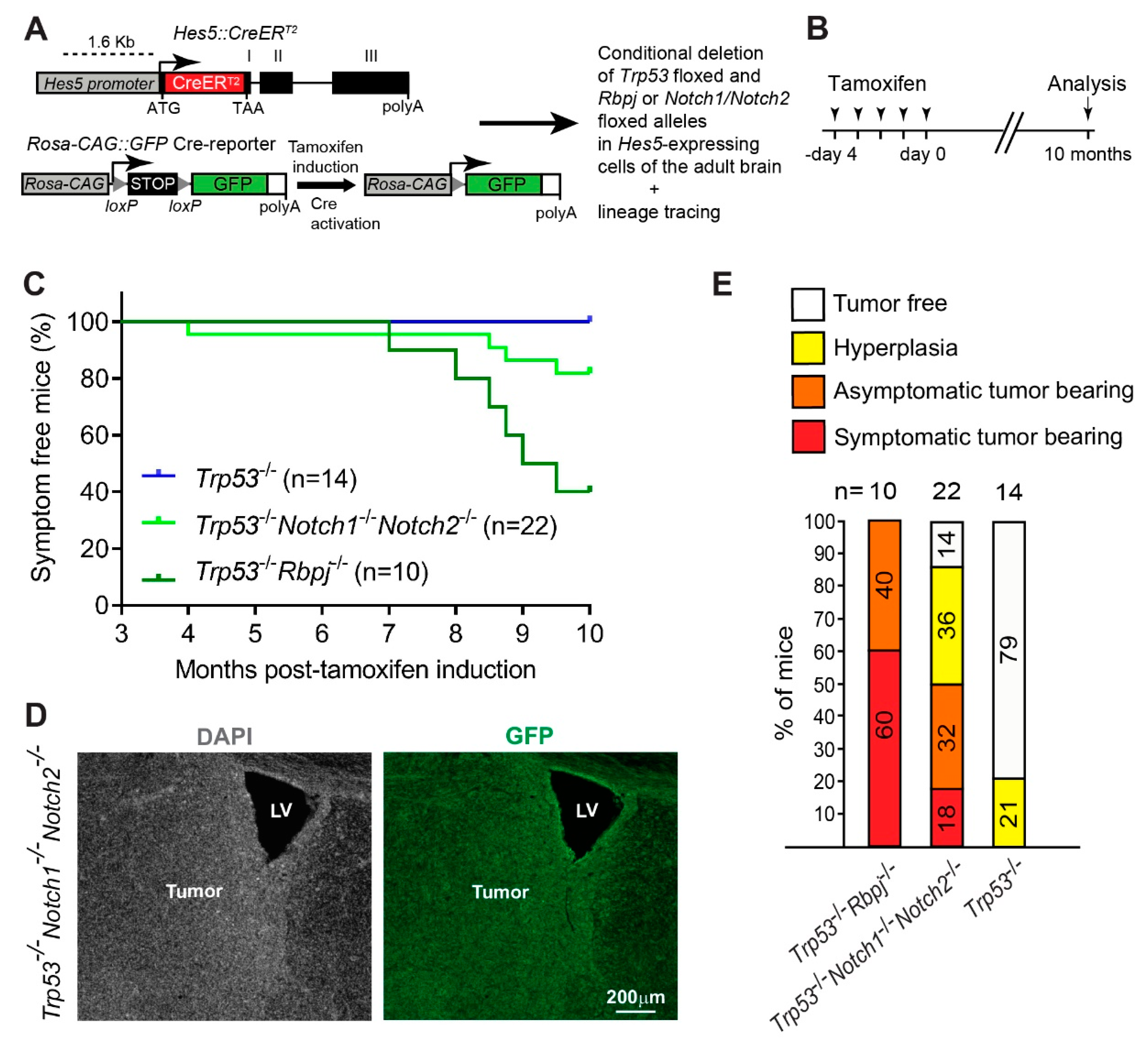
2. Materials and Methods

2.1. Animals

Hes5::CreERT2, floxed Rbpj, floxed Trp53, floxed Notch1, floxed Notch2, and Rosa-CAG::GFP mice have been described previously [16,33,34,35,36,37]. Young adult mice between 8 and 10 weeks of age were used for the experiments. Mice were maintained on a 12 h day/night cycle with adequate food and water under SPF conditions and according to institutional regulations under license numbers 2689, 2538, and 2537, and all experiments were approved by the ethics commission of the Kantonales Veterinäramt Basel-Stadt, Basel, Switzerland.

2.2. Tamoxifen Administration

Stock solutions of Tamoxifen (TAM, Sigma-Aldrich, St Louis, MO, USA ) were prepared at a concentration of 20 mg/mL in corn oil (Sigma-Aldrich). Adult mice (8–10 weeks of age) were injected intraperitoneal with TAM once per day for 5 consecutive days at a dose of 2 mg/day. Animals were killed 10 months after the last injection, or when they developed symptoms of brain tumor formation (lethargy, poor grooming, weight loss, or macrocephaly), and the brains were prepared for immunohistochemistry as described below.

2.3. Tissue Preparation and Immunohistochemistry

For histology, mice were deeply anaesthetized by injection of a ketamine/xylazine/acepromazine solution (130 mg, 26 mg and 4 mg/kg body weight, respectively) and perfused with ice-cold 0.9% saline solution followed by ice-cold 4% paraformaldehyde (PFA) solution in 0.1 M phosphate buffer (PB). Brains were post-fixed with 4% PFA overnight, washed in PB, cryoprotected in a 30% sucrose solution in 0.1 M PB for 48 h, embedded and frozen in OCT (TissueTEK). Free floating coronal sections (30 μm) were collected in multi-well dishes (Corning) and stored at −20 °C in anti-freeze solution until use.
For immunostaining, sections were incubated overnight at 4 °C with the primary antibody diluted in blocking solution of 2% normal donkey serum (Jackson ImmunoResearch), 0.5% Triton X-100 in phosphate-buffered saline (PBS). Sections were washed three times in PBS and incubated at room temperature for 1 h with the corresponding secondary antibodies in blocking solution. When necessary, sections were counter-stained with DAPI (1 μg/mL). For PCNA detection, antigen was recovered at 80 °C for 20 min in Sodium Citrate solution (10 mM, pH 6). Stained sections were mounted on Superfrost glass slides (Thermo Scientific, Waltham, MA, USA), embedded in mounting medium containing diazabicyclo-octane (DABCO, Sigma-Aldrich) as an anti-fading agent and visualized using a Zeiss Observer.Z1 equipped with Apotome.
Tumors were defined as discernible masses (increased cellularity based on DAPI staining) of proliferating (PCNA+) cells that were spanning at least 2 consecutive sections in a series of sections spaced 360 μm apart. Hyperplasias were defined as areas containing increased numbers of diffuse, highly proliferative PCNA+ cells, with only a limited increase in cellularity (DAPI staining).
The primary and secondary antibodies used are listed below: rabbit anti-PCNA (clone D3H8P, Cell Signaling, Cat# 13110); goat anti-SOX2 (R&D, Cat# AF2018); rabbit anti-OLIG2 (Merck Millipore, Cat# AB9610); rabbit anti-GFAP ( Sigma-Aldrich, Cat# G9269); goat anti-DCX (Santa Cruz Biotechnology, Cat# sc-8066); rabbit anti-RBP-Jk (Cell Signaling, Cat# 5313); chicken anti-GFP (Aves labs, Cat# GFP-1020); sheep anti-GFP (AbD Serotec/Biorad, Cat# 4745–1051); rabbit anti-GFP (Invitrogen, Cat# A11122); donkey Alexa488/Cy3/Alexa647 conjugated anti-rabbit, chicken, goat, and sheep secondary antibodies (Jackson ImmunoResearch). Anti-GFP antibodies were detected with appropriate Alexa488-conjugated secondary antibodies. All other primary antibodies were detected with Cy3- or Alexa647-conjugated secondary antibodies and the images were pseudo-colored in green in some of the figures for visualization purpose.

3. Results

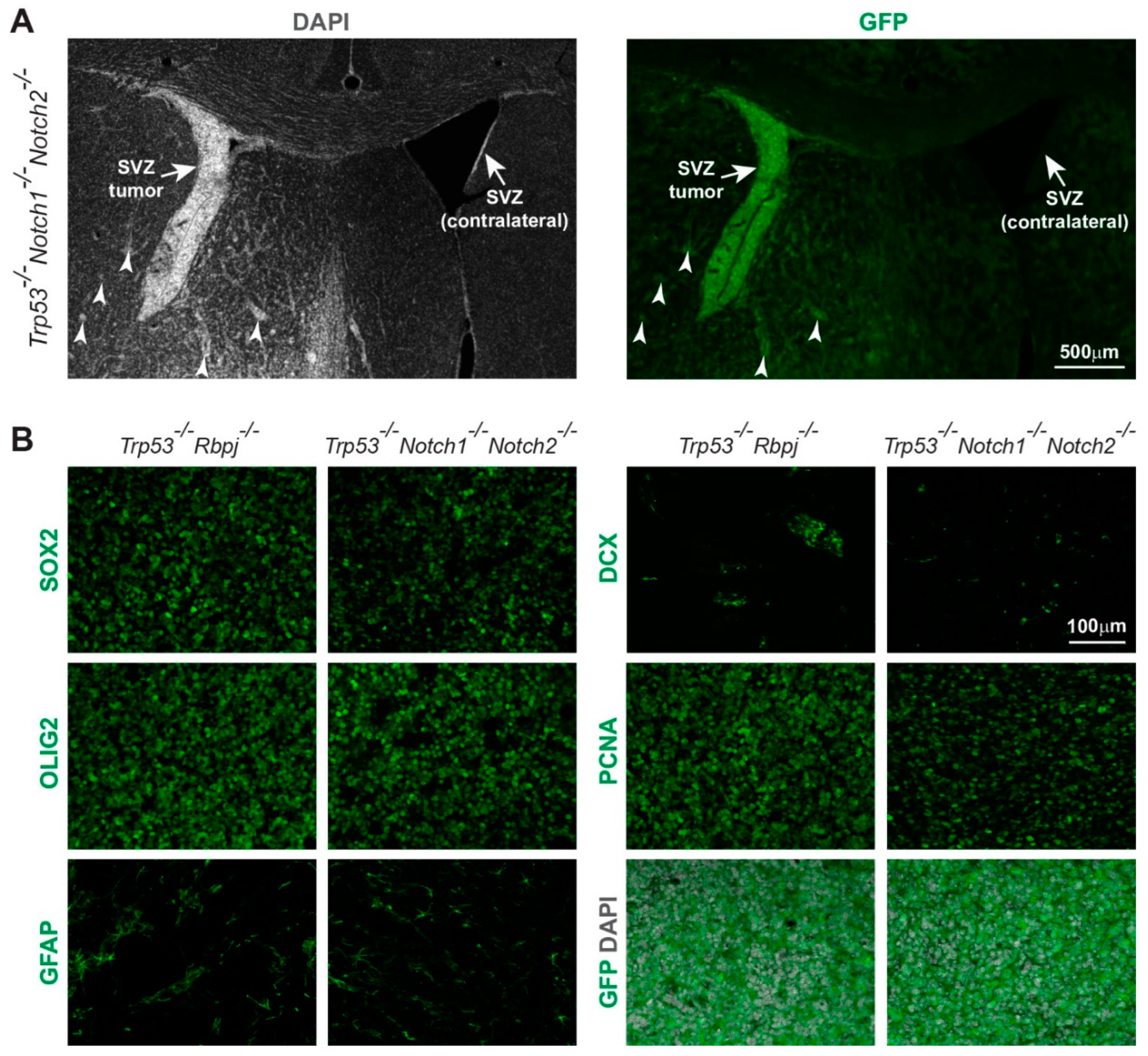
3.1. Combined Deletion of Notch1/Notch2 and Trp53 in Hes5+ Cells Leads to the Formation of Forebrain Tumors in Adult Mice

To address whether simultaneous inactivation of the Notch1 and Notch2 receptors cooperates with p53 inactivation to promote tumor growth in the adult brain, we combined gene-ablation and long-term genetic lineage tracing. We used the Hes5::CreERT2 transgenic mouse line to delete floxed Trp53 alleles, either alone or in combination with floxed Rbpj or Notch1/Notch2 alleles, specifically in Notch-active Hes5+ cells of the adult brain (Figure 1A) [13,16,28]. We have previously used the Hes5::CreERT2 line to efficiently delete conditional alleles [12,18]. A Cre-reporter allele (Rosa-CAG::GFP) was used to lineage trace the cells (Figure 1A) [37]. We induced Cre-activity by Tamoxifen (TAM) treatment of 2-months-old young adult mice (Figure 1B). We then followed the animals for 10 months after TAM administration and harvested their brains at 1 year of age (Figure 1B). None of the Trp53-/- mutant mice (n = 14) showed overt symptoms of tumor formation (Figure 1C). In contrast, and in agreement with our previous published data [28], 60% of the Trp53-/-Rbpj-/- animals (n = 10) became symptomatic starting 7 months after induction with TAM (Figure 1C). Moreover, also 18% of the Trp53-/-Notch1-/-Notch2-/- mutants (n = 22) developed symptoms and succumbed before 10 months to supratentorial brain tumors (Figure 1C,D). Histological examination showed that 79% of the Trp53-/- mutant brains were normal and 21% showed localized hyperproliferation/hyperplasia (Figure 1E). In contrast, none of the Trp53-/-Rbpj-/- animals and only 14% of the Trp53-/-Notch1-/-Notch2-/- mutants had brains that appeared normal (Figure 1E). All Trp53-/-Rbpj-/- mutants had brain tumors, and Trp53-/-Notch1-/-Notch2-/- mutants had either tumor (50%) or hyperplasia (36%) (Figure 1E).
The expression of GFP by the tumor cells indicated derivation from Hes5+ cells (Figure 1D, Figure 2A,B and Figure S1A). Trp53-/-Rbpj-/- and Trp53-/-Notch1-/-Notch2-/- tumors showed similar expression of progenitor markers as well as markers of the oligodendrocyte (OLIG2), astrocyte (GFAP), and neuronal (DCX) lineages (Figure 2B and Figure S1B). Thus, loss of RBP-Jk and p53 or simultaneous loss of Notch1, Notch2 and p53 induced supratentorial brain tumors and hyperplasia formation with high penetrance.

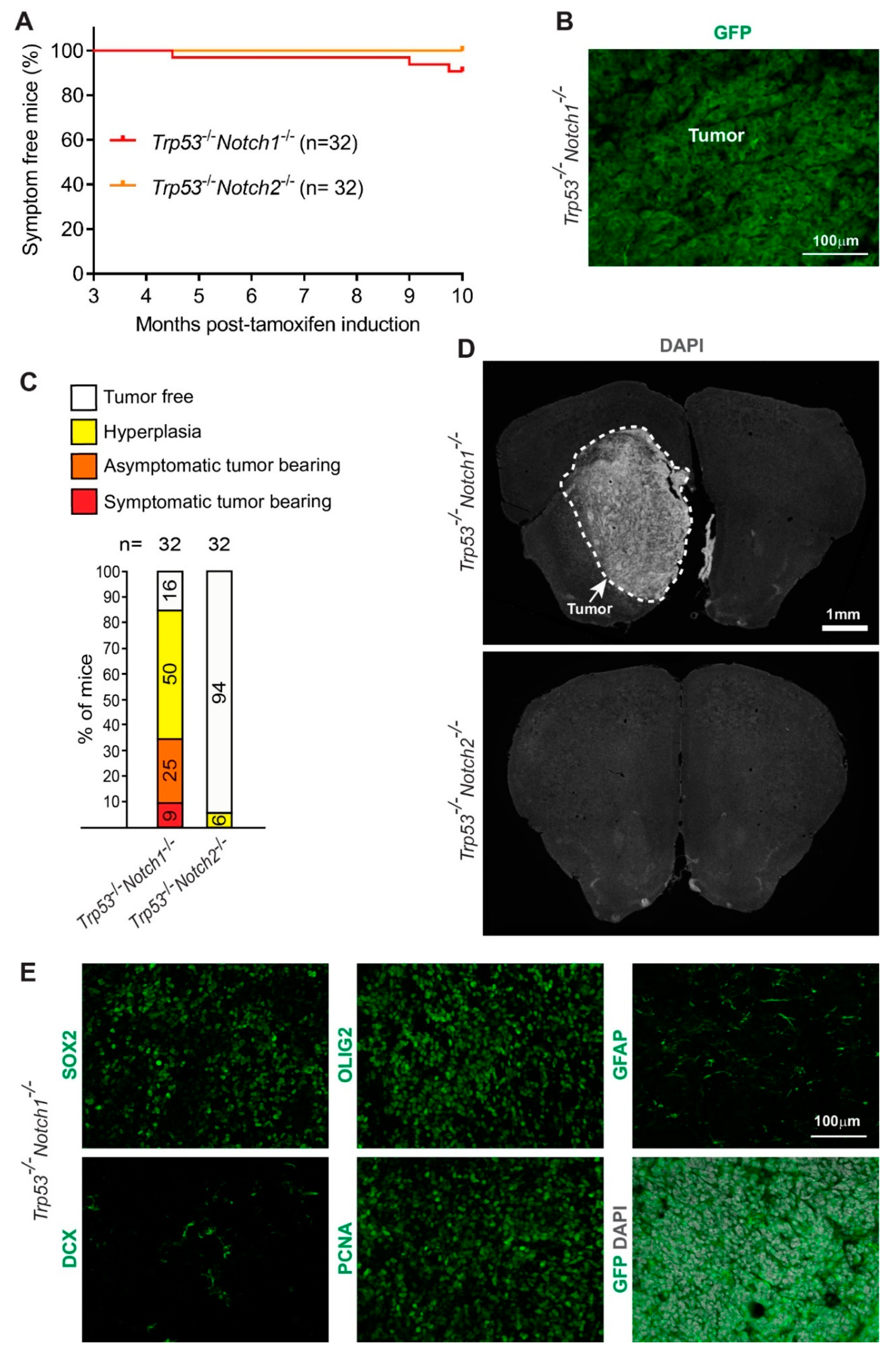
3.2. Deletion of Notch1 and Trp53 in Hes5+ Cells Leads to the Formation of Forebrain Tumors in Adult Mice

The relative contribution of Notch1 and Notch2 receptors to the tumor-suppressive activity of Notch signaling in this context is unknown. We addressed whether loss of p53 in combination with loss of either Notch1 or Notch2 is tumorigenic in the adult brain. We induced Cre-activity by TAM treatment of floxed Trp53/Notch1 and Trp53/Notch2 adult mice carrying the Hes5::CreERT2 transgene and followed the animals for 10 months (Figure 3). Overall, 9% of the Trp53-/-Notch1-/- mutants (n = 32) showed symptoms of tumor formation during the chase period (Figure 3A). However, none of the Trp53-/-Notch2-/- mice (n = 32) developed overt symptoms (Figure 3A). We examined the brains of Trp53-/-Notch1-/- and Trp53-/-Notch2-/- mice by histology. Overall, 34% of the Trp53-/-Notch1-/- mutants had tumors, 50% had hyperplasia, and only 16% appeared normal (Figure 3B–D). In contrast, only 6% of the Trp53-/-Notch2-/- mutant brains showed localized hyperproliferation/hyperplasia, and the remaining brains (94%) appeared normal (Figure 3C,D). Similar to the Trp53-/-Notch1-/-Notch2-/- tumors (Figure 2B), Trp53-/-Notch1-/- tumors expressed high levels of progenitor and oligodendrocyte lineage markers (SOX2, OLIG2), lower levels of astrocytic (GFAP) and neuronal (DCX) markers, and were highly proliferative (Figure 3E, Table 1). We concluded that Notch1 and Trp53 deletion in Hes5+ cells is sufficient to induce tumor or hyperplasia formation in the forebrains of adult mice.

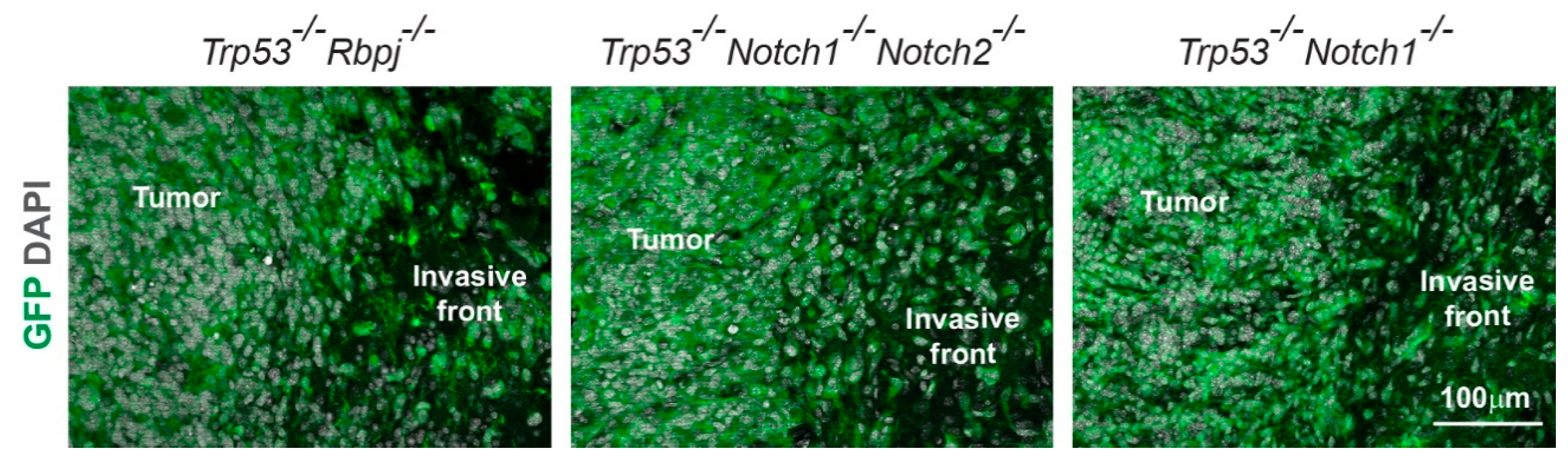
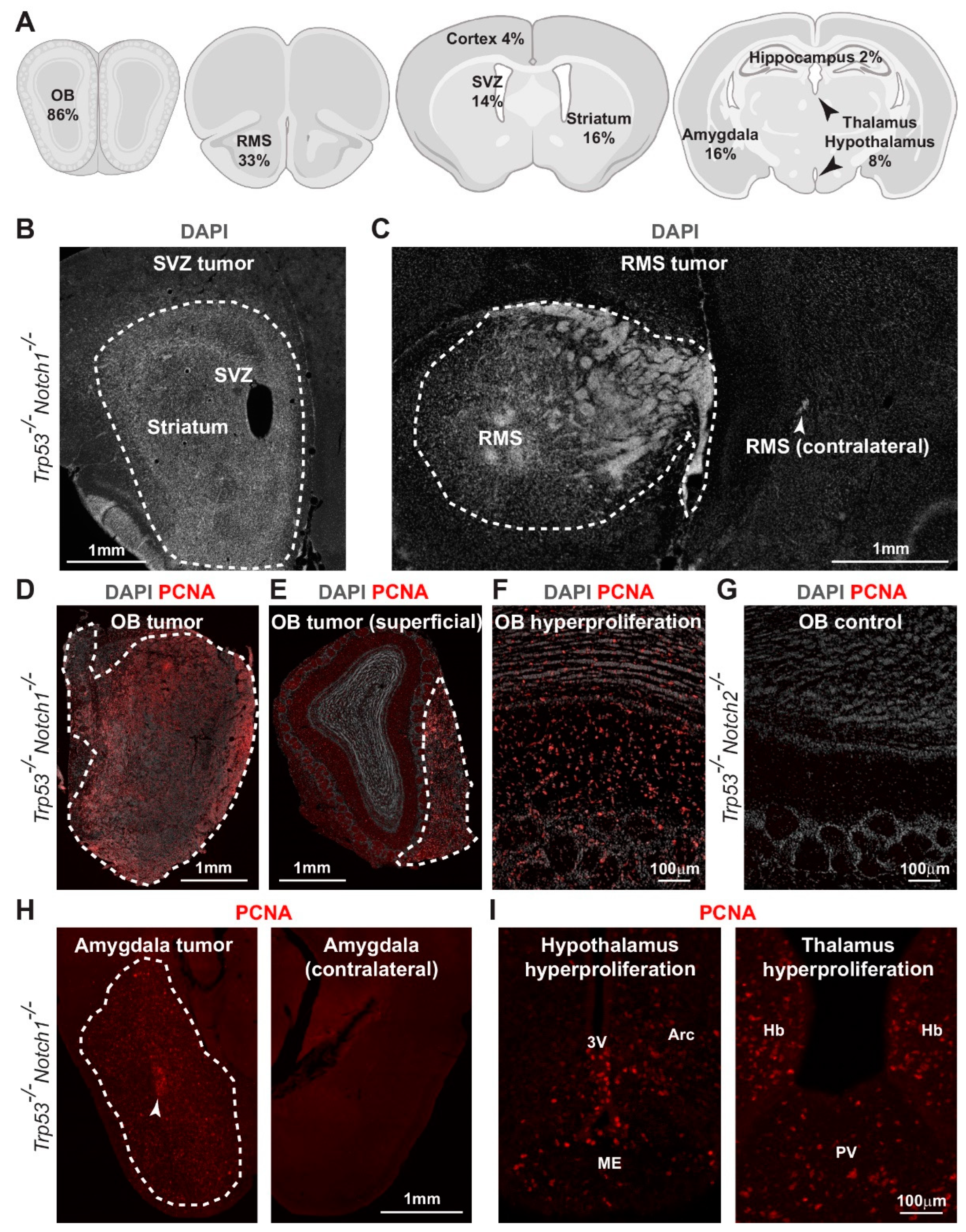
3.3. Hyperplasia and Tumors Are Preferentially Located in Ventral Forebrain Regions and OBs

Examination of the tumor border showed moderate invasion of Trp53-/-Rbpj-/-, Trp53-/-Notch1-/-Notch2-/-, and Trp53-/-Notch1-/- tumor cells into the surrounding brain parenchyma (Figure 4). All tumors that we identified had a supratentorial location, in agreement with our previous results from Trp53-/-Rbpj-/- mutants [28]. However, the large size of Trp53-/-Rbpj-/- tumors often precluded a more precise characterization of their location. We took advantage of the relatively smaller size of Trp53-/-Notch1-/-Notch2-/-, Trp53-/-Notch1-/-, and Trp53-/-Notch2-/- tumors and hyperplasias compared to Trp53-/-Rbpj-/- tumors to better define the prevalent sites of tumor formation within the forebrain after genetic inhibition of Notch and p53 (Figure 5, Table S1). We scored the number and location of tumors and hyperplasias in Trp53-/-Notch1-/-Notch2-/-, Trp53-/-Notch1-/-, Trp53-/-Notch2-/-, and Trp53-/- mutants. Tumors and hyperplasias were predominantly located in ventral and anterior forebrain regions, including the OBs, the rostral migratory stream (RMS) and surrounding areas, and the subventricular zone (SVZ) and adjacent striatum (Figure 5A–G, Table S1). The OB was the brain region where malignant cells were most frequently identified (Figure 5A, Table S1). OB tumors and hyperplasias in Trp53-/-Notch1-/-Notch2-/- and Trp53-/-Notch1-/- animals were either large and invaded all OB layers (Figure 5D,F) or were superficial and preferentially occupied the glomerular and nerve layers (Figure 5E). The OBs of most Trp53-/-Notch2-/- animals did not show signs of hyperproliferation and looked normal (Figure 5G). However, the rare, small hyperplasias that could be detected in Trp53-/-Notch2-/- and Trp53-/- mutants were also preferentially located in the OBs (Table S1). In contrast to the anterior and ventral forebrain, tumor formation very rarely affected dorsal forebrain regions such as the cerebral cortex and the hippocampus (Figure 5A, Table S1). Large tumors and hyperplasias were also relatively common in the amygdala region (Figure 5H, Table S1). In addition, hyperproliferation was observed in the hypothalamus and thalamus of a proportion of the mice (Figure 5I, Table S1). In some animals, multiple tumors and hyperplasias co-occurred at distant locations in the brain, for example OB and amygdala, OB and hypothalamus/thalamus, and OB and SVZ (Table S1).

4. Discussion

Our gene-ablation and long-term genetic lineage tracing experiments in mice indicate that Notch/RBP-Jk signaling, and particularly the Notch1 receptor, can inhibit brain tumor initiation and growth in the adult forebrain. Trp53 deletion alone in cells expressing the Notch target gene Hes5 predisposes to hyperplasia formation with very low penetrance. We have previously shown that deletion of Rbpj, Notch1, or Notch2 in the presence of an intact Trp53 gene alters NSC proliferation and differentiation, but does not induce formation of brain tumors [12,28]. In contrast, simultaneous deletion of Trp53 and Rbpj, Notch1/Notch2, or Notch1 induces tumor or hyperplasia formation in over 80% of the mice. Thus, Notch signaling cooperates with p53 to inhibit tumor formation in the adult murine brain. These data confirm and extend our previous findings from PDGF-driven gliomas and indicate that Notch signaling can behave as a tumor suppressor in the brain even in the absence of PDGF overexpression [28]. Although Trp53-/-Rbpj-/- tumors are clearly separable from PDGF+Trp53-/-Rbpj-/- tumors at the transcriptional level [28], it is conceivable that tumor cells in both models acquire oligodendrocyte progenitor cell (OPC)-like features, a cellular state that is associated with a proneural glioma transcriptional subtype and a tumor suppressor role of Notch signaling [20]. This scenario is in agreement with a previous study showing that the transcriptional profiles of murine glioma models driven by different oncogenic alterations can convergence to a proneural signature [38].
RBP-Jk can act as a transcriptional repressor and loss of the RBPJ gene promotes the growth of human breast cancer cells by de-repressing target gene promoters in a Notch-independent manner [39,40]. However, our data showing that not only Trp53-/-Rbpj-/- mutants, but also Trp53-/-Notch1-/-Notch2-/- and Trp53-/-Notch1-/- mutants develop tumors suggest that the tumor suppressor function of RBP-Jk in the brain is linked to the regulation of Notch signaling. This hypothesis is supported by an in vivo CRISPR screen that identified Notch1 among potential tumor suppressors in glioma [41], and is further in line with the occurrence of NOTCH1, NOTCH2, and RBPJ inactivating mutations and/or reduced Notch signaling activity in human glioma subtypes [21,23,26,27,28,29,30,31].
We found that Trp53-/-Notch2-/- mutants rarely develop hyperplasia, in contrast to Trp53-/-Notch1-/- and Trp53-/-Rbpj-/- mutants. We cannot exclude that differences in recombination efficiency of Notch1, Notch2, and Rbpj conditional alleles contribute to the different penetrance of the phenotypes. However, we have recently shown that glioma cells lacking Notch1 are more aggressive than glioma cells lacking Notch2, despite efficient deletion of both Notch1 and Notch2 floxed alleles [20]. Thus, our current finding confirms our observation that Notch1 is a stronger tumor suppressor than Notch2 in a PDGF-driven mouse model of glioma [20] and implies that Notch1 could partially compensate for Notch2 loss. Although Notch2 deletion does not accelerate hyperplasia formation in Trp53-/- mutants, it significantly accelerates the growth of PDGF+Trp53-/- gliomas [20], suggesting that a strong mitogenic stimulus, provided, for instance, by PDGF, is required to release the tumorigenic effects of Notch2 inhibition. Our data do not exclude the residual expression of Hes/Hey gene family members or other Notch downstream targets, particularly in cells deleted for one Notch receptor, as a result of compensatory mechanisms or direct induction of Hes genes expression by BMP signaling [42]. In agreement with this hypothesis, we have recently shown that Notch1 or Notch2 deletion in PDGF+Trp53-/- gliomas leads to changes in the expression of only partially overlapping sets of genes [20]. We observed tumor or hyperplasia formation in roughly 85% of Trp53-/-Notch1-/-Notch2-/- and Trp53-/-Notch1-/- mutants, but complete penetrance in Trp53-/-Rbpj-/- mutants. This suggests that other receptors beyond Notch1 and Notch2 might be involved. It would be interesting to determine whether Notch3, which represses the proliferation of adult NSCs [43,44], contributes to the tumor suppressor activity of RBP-Jk in cooperation with Notch1 and Notch2.
We found that a proportion of the tumors and hyperplasias were located in the proximity of neurogenic regions including the RMS and SVZ and adjacent striatum, implying that adult neural stem and progenitor cells are probable cells of origin of some of the tumors [45]. However, a significant proportion of the tumors and hyperplasias were confined to anterior and ventral brain regions such as the surface of the OB, the amygdala, the hypothalamus and the thalamus, raising the possibility that some of the tumors originate locally. Parenchymal OPCs express Notch receptors [46,47,48] and can be an important cell type of origin of tumors, notably in the anterior and ventral brain [49,50,51,52]. Further work is needed to explore whether the resilience of ventral OPCs to pathological stimuli can make them exceptionally vulnerable to malignant transformation [49,53]. It is also possible that the glycosylation status of the Notch receptors, which can affect ligand binding, as well as varying expression levels of the Notch ligands contribute to regional heterogeneity in tumorigenesis [54,55]. In the future, it would be interesting to determine whether the frequency of tumor formation correlates with domain specific expression of Jagged and Delta ligands in different brain areas.
We show that genetic inactivation of Notch signaling and p53 in a subpopulation of brain cells induces CNS tumors with long latency but high penetrance. Notch-regulated competitive interactions, triggered by dishomogeneus Notch inactivating mutations in adjacent cells, contribute to the initiation of epithelial cancers by favoring the slow and progressive expansion of mutant clones at the expense of wild-type cells [56]. Intriguingly, data suggest that transformed NSCs can outcompete neighboring NSCs by repressing their proliferation through Notch signaling [57]. Whether Notch inhibition in malignant NSCs exacerbates cell competition and fitness selection is unknown. Likewise, OPC populations compete for space in the developing brain [58], but whether a similar phenomenon occurs during glioma formation from OPCs remains unclear. We speculate that Notch-regulated cell competition mechanisms could be in place during tumor initiation and progression in the brain.

5. Conclusions

In conclusion, we show that Notch1 deletion cooperates with Trp53 deletion to promote tumor growth in the adult forebrain. Our data also suggest that tumors induced by Notch signaling inhibition have a multifocal origin. These findings shed light on the pleiotropic roles of Notch signaling in the development of primary brain malignancies.

Supplementary Materials

The following supporting information can be downloaded at: https://www.mdpi.com/article/10.3390/cancers14215409/s1, Table S1: Distribution of hyperplasia and tumors. Figure S1: Immunohistochemical analysis of Trp53-/-Rbpj-/- tumors and contralateral hemispheres.

Author Contributions

Conceptualization, E.P. and C.G.; methodology, E.P. and C.G.; software, E.P. and C.G.; validation, E.P. and C.G.; formal analysis, E.P. and C.G.; investigation, E.P. and C.G.; resources, E.P. and C.G.; data curation, E.P. and C.G.; writing—original draft preparation, C.G.; writing—review and editing, E.P. and C.G.; visualization, E.P. and C.G.; supervision, C.G.; project administration, C.G.; funding acquisition, E.P. and C.G. All authors have read and agreed to the published version of the manuscript.

Funding

This work was supported by the Swiss Cancer League (KFS-3600-02-2015 and KLS-4518-08-2018), the Wilhelm Sander-Stiftung (2021.144.1), the Stiftung zur Krebsbekämpfung (409), the Novartis foundation for medical-biological research (20A069), the Stiftung Tumorforschung Kopf-Hals and the Forschungsfonds Nachwuchsforschende of the University of Basel.

Institutional Review Board Statement

The animal study protocol was approved by the Ethics Committee of the Kantonales Veterinäramt Basel-Stadt, Basel, Switzerland (license numbers 2689, 2538, and 2537).

Informed Consent Statement

Not applicable.

Data Availability Statement

All data generated or analyzed during this study are included in this article and its Supplementary Material files. Further enquiries can be directed to the corresponding author.

Acknowledgments

We thank the members of the Taylor lab for critical reading of the manuscript and Karin Burger and Frank Sager for excellent technical assistance. We are grateful to Pascal Lorentz and the DBM Microscopy Core Facility for support with microscopy. Figure 5A was created with BioRender.com (accessed on 14 May 2022).

Conflicts of Interest

The authors declare no conflict of interest.

References

  1. Ntziachristos, P.; Lim, J.S.; Sage, J.; Aifantis, I. From fly wings to targeted cancer therapies: A centennial for notch signaling. Cancer Cell 2014, 25, 318–334. [Google Scholar] [CrossRef] [PubMed] [Green Version]
  2. Nowell, C.S.; Radtke, F. Notch as a tumour suppressor. Nat. Rev. Cancer 2017, 17, 145–159. [Google Scholar] [CrossRef] [PubMed]
  3. Lim, J.S.; Ibaseta, A.; Fischer, M.M.; Cancilla, B.; O’Young, G.; Cristea, S.; Luca, V.C.; Yang, D.; Jahchan, N.S.; Hamard, C.; et al. Intratumoural heterogeneity generated by Notch signalling promotes small-cell lung cancer. Nature 2017, 545, 360–364. [Google Scholar] [CrossRef] [PubMed] [Green Version]
  4. Parmigiani, E.; Taylor, V.; Giachino, C. Oncogenic and Tumor-Suppressive Functions of NOTCH Signaling in Glioma. Cells 2020, 9, e2304. [Google Scholar] [CrossRef]
  5. Klinakis, A.; Lobry, C.; Abdel-Wahab, O.; Oh, P.; Haeno, H.; Buonamici, S.; van De Walle, I.; Cathelin, S.; Trimarchi, T.; Araldi, E.; et al. A novel tumour-suppressor function for the Notch pathway in myeloid leukaemia. Nature 2011, 473, 230–233. [Google Scholar] [CrossRef] [Green Version]
  6. Jung, E.; Osswald, M.; Ratliff, M.; Dogan, H.; Xie, R.; Weil, S.; Hoffmann, D.C.; Kurz, F.T.; Kessler, T.; Heiland, S.; et al. Tumor cell plasticity, heterogeneity, and resistance in crucial microenvironmental niches in glioma. Nat. Commun. 2021, 12, 1014. [Google Scholar] [CrossRef]
  7. Koch, U.; Lehal, R.; Radtke, F. Stem cells living with a Notch. Development 2013, 140, 689–704. [Google Scholar] [CrossRef] [Green Version]
  8. Kobayashi, T.; Kageyama, R. Lysosomes and signaling pathways for maintenance of quiescence in adult neural stem cells. FEBS J. 2021, 288, 3082–3093. [Google Scholar] [CrossRef]
  9. Basak, O.; Giachino, C.; Fiorini, E.; Macdonald, H.R.; Taylor, V. Neurogenic subventricular zone stem/progenitor cells are Notch1-dependent in their active but not quiescent state. J. Neurosci. 2012, 32, 5654–5666. [Google Scholar] [CrossRef] [Green Version]
  10. Basak, O.; Taylor, V. Identification of self-replicating multipotent progenitors in the embryonic nervous system by high Notch activity and Hes5 expression. Eur. J. Neurosci. 2007, 25, 1006–1022. [Google Scholar] [CrossRef]
  11. Ehm, O.; Goritz, C.; Covic, M.; Schaffner, I.; Schwarz, T.J.; Karaca, E.; Kempkes, B.; Kremmer, E.; Pfrieger, F.W.; Espinosa, L.; et al. RBPJkappa-dependent signaling is essential for long-term maintenance of neural stem cells in the adult hippocampus. J. Neurosci. 2010, 30, 13794–13807. [Google Scholar] [CrossRef] [PubMed] [Green Version]
  12. Engler, A.; Rolando, C.; Giachino, C.; Saotome, I.; Erni, A.; Brien, C.; Zhang, R.; Zimber-Strobl, U.; Radtke, F.; Artavanis-Tsakonas, S.; et al. Notch2 Signaling Maintains NSC Quiescence in the Murine Ventricular-Subventricular Zone. Cell Rep. 2018, 22, 992–1002. [Google Scholar] [CrossRef] [PubMed] [Green Version]
  13. Giachino, C.; Basak, O.; Lugert, S.; Knuckles, P.; Obernier, K.; Fiorelli, R.; Frank, S.; Raineteau, O.; Alvarez-Buylla, A.; Taylor, V. Molecular diversity subdivides the adult forebrain neural stem cell population. Stem Cells 2014, 32, 70–84. [Google Scholar] [CrossRef] [PubMed] [Green Version]
  14. Imayoshi, I.; Sakamoto, M.; Yamaguchi, M.; Mori, K.; Kageyama, R. Essential roles of Notch signaling in maintenance of neural stem cells in developing and adult brains. J. Neurosci. 2010, 30, 3489–3498. [Google Scholar] [CrossRef] [Green Version]
  15. Lugert, S.; Basak, O.; Knuckles, P.; Haussler, U.; Fabel, K.; Gotz, M.; Haas, C.A.; Kempermann, G.; Taylor, V.; Giachino, C. Quiescent and active hippocampal neural stem cells with distinct morphologies respond selectively to physiological and pathological stimuli and aging. Cell Stem Cell 2010, 6, 445–456. [Google Scholar] [CrossRef] [Green Version]
  16. Lugert, S.; Vogt, M.; Tchorz, J.S.; Muller, M.; Giachino, C.; Taylor, V. Homeostatic neurogenesis in the adult hippocampus does not involve amplification of Ascl1(high) intermediate progenitors. Nat. Commun. 2012, 3, 670. [Google Scholar] [CrossRef] [Green Version]
  17. Sueda, R.; Imayoshi, I.; Harima, Y.; Kageyama, R. High Hes1 expression and resultant Ascl1 suppression regulate quiescent vs. active neural stem cells in the adult mouse brain. Genes Dev. 2019, 33, 511–523. [Google Scholar] [CrossRef] [Green Version]
  18. Zhang, R.; Boareto, M.; Engler, A.; Louvi, A.; Giachino, C.; Iber, D.; Taylor, V. Id4 Downstream of Notch2 Maintains Neural Stem Cell Quiescence in the Adult Hippocampus. Cell Rep. 2019, 28, 1485–1498.e6. [Google Scholar] [CrossRef]
  19. Imayoshi, I.; Isomura, A.; Harima, Y.; Kawaguchi, K.; Kori, H.; Miyachi, H.; Fujiwara, T.; Ishidate, F.; Kageyama, R. Oscillatory control of factors determining multipotency and fate in mouse neural progenitors. Science 2013, 342, 1203–1208. [Google Scholar] [CrossRef] [Green Version]
  20. Parmigiani, E.; Ivanek, R.; Rolando, C.; Hafen, K.; Turchinovich, G.; Lehmann, F.M.; Gerber, A.; Brkic, S.; Frank, S.; Meyer, S.C.; et al. Interferon-gamma resistance and immune evasion in glioma develop via Notch-regulated co-evolution of malignant and immune cells. Dev. Cell 2022, 57, 1847–1865. [Google Scholar] [CrossRef]
  21. Brat, D.J.; Verhaak, R.G.; Aldape, K.D.; Yung, W.K.; Salama, S.R.; Cooper, L.A.; Rheinbay, E.; Miller, C.R.; Vitucci, M.; Morozova, O.; et al. Comprehensive, Integrative Genomic Analysis of Diffuse Lower-Grade Gliomas. N. Engl. J. Med. 2015, 372, 2481–2498. [Google Scholar] [CrossRef] [PubMed] [Green Version]
  22. Capper, D.; Jones, D.T.W.; Sill, M.; Hovestadt, V.; Schrimpf, D.; Sturm, D.; Koelsche, C.; Sahm, F.; Chavez, L.; Reuss, D.E.; et al. DNA methylation-based classification of central nervous system tumours. Nature 2018, 555, 469–474. [Google Scholar] [CrossRef] [PubMed] [Green Version]
  23. Suzuki, H.; Aoki, K.; Chiba, K.; Sato, Y.; Shiozawa, Y.; Shiraishi, Y.; Shimamura, T.; Niida, A.; Motomura, K.; Ohka, F.; et al. Mutational landscape and clonal architecture in grade II and III gliomas. Nat. Genet. 2015, 47, 458–468. [Google Scholar] [CrossRef] [PubMed]
  24. Verhaak, R.G.; Hoadley, K.A.; Purdom, E.; Wang, V.; Qi, Y.; Wilkerson, M.D.; Miller, C.R.; Ding, L.; Golub, T.; Mesirov, J.P.; et al. Integrated genomic analysis identifies clinically relevant subtypes of glioblastoma characterized by abnormalities in PDGFRA, IDH1, EGFR, and NF1. Cancer Cell 2010, 17, 98–110. [Google Scholar] [CrossRef] [Green Version]
  25. Sturm, D.; Orr, B.A.; Toprak, U.H.; Hovestadt, V.; Jones, D.T.W.; Capper, D.; Sill, M.; Buchhalter, I.; Northcott, P.A.; Leis, I.; et al. New Brain Tumor Entities Emerge from Molecular Classification of CNS-PNETs. Cell 2016, 164, 1060–1072. [Google Scholar] [CrossRef] [Green Version]
  26. Aoki, K.; Nakamura, H.; Suzuki, H.; Matsuo, K.; Kataoka, K.; Shimamura, T.; Motomura, K.; Ohka, F.; Shiina, S.; Yamamoto, T.; et al. Prognostic relevance of genetic alterations in diffuse lower-grade gliomas. Neuro. Oncol. 2018, 20, 66–77. [Google Scholar] [CrossRef] [Green Version]
  27. Bai, H.; Harmanci, A.S.; Erson-Omay, E.Z.; Li, J.; Coskun, S.; Simon, M.; Krischek, B.; Ozduman, K.; Omay, S.B.; Sorensen, E.A.; et al. Integrated genomic characterization of IDH1-mutant glioma malignant progression. Nat. Genet. 2016, 48, 59–66. [Google Scholar] [CrossRef] [Green Version]
  28. Giachino, C.; Boulay, J.L.; Ivanek, R.; Alvarado, A.; Tostado, C.; Lugert, S.; Tchorz, J.; Coban, M.; Mariani, L.; Bettler, B.; et al. A Tumor Suppressor Function for Notch Signaling in Forebrain Tumor Subtypes. Cancer Cell 2015, 28, 730–742. [Google Scholar] [CrossRef] [Green Version]
  29. Halani, S.H.; Yousefi, S.; Velazquez Vega, J.; Rossi, M.R.; Zhao, Z.; Amrollahi, F.; Holder, C.A.; Baxter-Stoltzfus, A.; Eschbacher, J.; Griffith, B.; et al. Multi-faceted computational assessment of risk and progression in oligodendroglioma implicates NOTCH and PI3K pathways. NPJ Precis. Oncol. 2018, 2, 24. [Google Scholar] [CrossRef]
  30. Phillips, H.S.; Kharbanda, S.; Chen, R.; Forrest, W.F.; Soriano, R.H.; Wu, T.D.; Misra, A.; Nigro, J.M.; Colman, H.; Soroceanu, L.; et al. Molecular subclasses of high-grade glioma predict prognosis, delineate a pattern of disease progression, and resemble stages in neurogenesis. Cancer Cell 2006, 9, 157–173. [Google Scholar] [CrossRef]
  31. Somasundaram, K.; Reddy, S.P.; Vinnakota, K.; Britto, R.; Subbarayan, M.; Nambiar, S.; Hebbar, A.; Samuel, C.; Shetty, M.; Sreepathi, H.K.; et al. Upregulation of ASCL1 and inhibition of Notch signaling pathway characterize progressive astrocytoma. Oncogene 2005, 24, 7073–7083. [Google Scholar] [CrossRef] [PubMed] [Green Version]
  32. Bettegowda, C.; Agrawal, N.; Jiao, Y.; Sausen, M.; Wood, L.D.; Hruban, R.H.; Rodriguez, F.J.; Cahill, D.P.; McLendon, R.; Riggins, G.; et al. Mutations in CIC and FUBP1 contribute to human oligodendroglioma. Science 2011, 333, 1453–1455. [Google Scholar] [CrossRef] [PubMed] [Green Version]
  33. Besseyrias, V.; Fiorini, E.; Strobl, L.J.; Zimber-Strobl, U.; Dumortier, A.; Koch, U.; Arcangeli, M.L.; Ezine, S.; Macdonald, H.R.; Radtke, F. Hierarchy of Notch-Delta interactions promoting T cell lineage commitment and maturation. J. Exp. Med. 2007, 204, 331–343. [Google Scholar] [CrossRef] [PubMed] [Green Version]
  34. Han, H.; Tanigaki, K.; Yamamoto, N.; Kuroda, K.; Yoshimoto, M.; Nakahata, T.; Ikuta, K.; Honjo, T. Inducible gene knockout of transcription factor recombination signal binding protein-J reveals its essential role in T versus B lineage decision. Int. Immunol. 2002, 14, 637–645. [Google Scholar] [CrossRef] [PubMed]
  35. Jonkers, J.; Meuwissen, R.; van der Gulden, H.; Peterse, H.; van der Valk, M.; Berns, A. Synergistic tumor suppressor activity of BRCA2 and p53 in a conditional mouse model for breast cancer. Nat. Genet. 2001, 29, 418–425. [Google Scholar] [CrossRef] [PubMed]
  36. Radtke, F.; Wilson, A.; Stark, G.; Bauer, M.; van Meerwijk, J.; MacDonald, H.R.; Aguet, M. Deficient T cell fate specification in mice with an induced inactivation of Notch1. Immunity 1999, 10, 547–558. [Google Scholar] [CrossRef] [Green Version]
  37. Tchorz, J.S.; Suply, T.; Ksiazek, I.; Giachino, C.; Cloetta, D.; Danzer, C.P.; Doll, T.; Isken, A.; Lemaistre, M.; Taylor, V.; et al. A modified RMCE-compatible Rosa26 locus for the expression of transgenes from exogenous promoters. PLoS ONE 2012, 7, e30011. [Google Scholar] [CrossRef] [PubMed] [Green Version]
  38. Alessandrini, F.; Ceresa, D.; Appolloni, I.; Pagani, F.; Poliani, P.L.; Marubbi, D.; Malatesta, P. Glioblastoma models driven by different mutations converge to the proneural subtype. Cancer Lett. 2020, 469, 447–455. [Google Scholar] [CrossRef]
  39. Braune, E.B.; Tsoi, Y.L.; Phoon, Y.P.; Landor, S.; Silva Cascales, H.; Ramskold, D.; Deng, Q.; Lindqvist, A.; Lian, X.; Sahlgren, C.; et al. Loss of CSL Unlocks a Hypoxic Response and Enhanced Tumor Growth Potential in Breast Cancer Cells. Stem Cell Rep. 2016, 6, 643–651. [Google Scholar] [CrossRef] [Green Version]
  40. Kulic, I.; Robertson, G.; Chang, L.; Baker, J.H.; Lockwood, W.W.; Mok, W.; Fuller, M.; Fournier, M.; Wong, N.; Chou, V.; et al. Loss of the Notch effector RBPJ promotes tumorigenesis. J. Exp. Med. 2015, 212, 37–52. [Google Scholar] [CrossRef]
  41. Chow, R.D.; Guzman, C.D.; Wang, G.; Schmidt, F.; Youngblood, M.W.; Ye, L.; Errami, Y.; Dong, M.B.; Martinez, M.A.; Zhang, S.; et al. AAV-mediated direct in vivo CRISPR screen identifies functional suppressors in glioblastoma. Nat. Neurosci. 2017, 20, 1329–1341. [Google Scholar] [CrossRef] [PubMed] [Green Version]
  42. Kobayashi, T.; Mizuno, H.; Imayoshi, I.; Furusawa, C.; Shirahige, K.; Kageyama, R. The cyclic gene Hes1 contributes to diverse differentiation responses of embryonic stem cells. Genes Dev. 2009, 23, 1870–1875. [Google Scholar] [CrossRef] [PubMed] [Green Version]
  43. Kawai, H.; Kawaguchi, D.; Kuebrich, B.D.; Kitamoto, T.; Yamaguchi, M.; Gotoh, Y.; Furutachi, S. Area-Specific Regulation of Quiescent Neural Stem Cells by Notch3 in the Adult Mouse Subependymal Zone. J. Neurosci. 2017, 37, 11867–11880. [Google Scholar] [CrossRef] [PubMed] [Green Version]
  44. Alunni, A.; Krecsmarik, M.; Bosco, A.; Galant, S.; Pan, L.; Moens, C.B.; Bally-Cuif, L. Notch3 signaling gates cell cycle entry and limits neural stem cell amplification in the adult pallium. Development 2013, 140, 3335–3347. [Google Scholar] [CrossRef] [Green Version]
  45. Alcantara Llaguno, S.; Sun, D.; Pedraza, A.M.; Vera, E.; Wang, Z.; Burns, D.K.; Parada, L.F. Cell-of-origin susceptibility to glioblastoma formation declines with neural lineage restriction. Nat. Neurosci. 2019, 22, 545–555. [Google Scholar] [CrossRef]
  46. Hu, Q.D.; Ang, B.T.; Karsak, M.; Hu, W.P.; Cui, X.Y.; Duka, T.; Takeda, Y.; Chia, W.; Sankar, N.; Ng, Y.K.; et al. F3/contactin acts as a functional ligand for Notch during oligodendrocyte maturation. Cell 2003, 115, 163–175. [Google Scholar] [CrossRef] [Green Version]
  47. John, G.R.; Shankar, S.L.; Shafit-Zagardo, B.; Massimi, A.; Lee, S.C.; Raine, C.S.; Brosnan, C.F. Multiple sclerosis: Re-expression of a developmental pathway that restricts oligodendrocyte maturation. Nat. Med. 2002, 8, 1115–1121. [Google Scholar] [CrossRef]
  48. Wang, S.; Sdrulla, A.D.; diSibio, G.; Bush, G.; Nofziger, D.; Hicks, C.; Weinmaster, G.; Barres, B.A. Notch receptor activation inhibits oligodendrocyte differentiation. Neuron 1998, 21, 63–75. [Google Scholar] [CrossRef] [Green Version]
  49. Alcantara Llaguno, S.R.; Wang, Z.; Sun, D.; Chen, J.; Xu, J.; Kim, E.; Hatanpaa, K.J.; Raisanen, J.M.; Burns, D.K.; Johnson, J.E.; et al. Adult Lineage-Restricted CNS Progenitors Specify Distinct Glioblastoma Subtypes. Cancer Cell 2015, 28, 429–440. [Google Scholar] [CrossRef] [Green Version]
  50. Galvao, R.P.; Kasina, A.; McNeill, R.S.; Harbin, J.E.; Foreman, O.; Verhaak, R.G.; Nishiyama, A.; Miller, C.R.; Zong, H. Transformation of quiescent adult oligodendrocyte precursor cells into malignant glioma through a multistep reactivation process. Proc. Natl. Acad. Sci. USA 2014, 111, E4214–E4223. [Google Scholar] [CrossRef]
  51. Sutcliffe, M.D.; Galvao, R.P.; Wang, L.; Kim, J.; Rosenfeld, L.K.; Singh, S.; Zong, H.; Janes, K.A. Premalignant Oligodendrocyte Precursor Cells Stall in a Heterogeneous State of Replication Stress Prior to Gliomagenesis. Cancer Res. 2021, 81, 1868–1882. [Google Scholar] [CrossRef]
  52. Chen, P.; Wang, W.; Liu, R.; Lyu, J.; Zhang, L.; Li, B.; Qiu, B.; Tian, A.; Jiang, W.; Ying, H.; et al. Olfactory sensory experience regulates gliomagenesis via neuronal IGF1. Nature 2022, 606, 550–556. [Google Scholar] [CrossRef] [PubMed]
  53. Boda, E.; Lorenzati, M.; Parolisi, R.; Harding, B.; Pallavicini, G.; Bonfanti, L.; Moccia, A.; Bielas, S.; Di Cunto, F.; Buffo, A. Molecular and functional heterogeneity in dorsal and ventral oligodendrocyte progenitor cells of the mouse forebrain in response to DNA damage. Nat. Commun. 2022, 13, 2331. [Google Scholar] [CrossRef] [PubMed]
  54. Bigas, A.; Espinosa, L. The multiple usages of Notch signaling in development, cell differentiation and cancer. Curr. Opin. Cell Biol. 2018, 55, 1–7. [Google Scholar] [CrossRef] [PubMed] [Green Version]
  55. Henrique, D.; Schweisguth, F. Mechanisms of Notch signaling: A simple logic deployed in time and space. Development 2019, 146, dev172148. [Google Scholar] [CrossRef] [PubMed] [Green Version]
  56. Alcolea, M.P.; Greulich, P.; Wabik, A.; Frede, J.; Simons, B.D.; Jones, P.H. Differentiation imbalance in single oesophageal progenitor cells causes clonal immortalization and field change. Nat. Cell. Biol. 2014, 16, 615–622. [Google Scholar] [CrossRef] [Green Version]
  57. Lawlor, K.; Marques-Torrejon, M.A.; Dharmalingham, G.; El-Azhar, Y.; Schneider, M.D.; Pollard, S.M.; Rodriguez, T.A. Glioblastoma stem cells induce quiescence in surrounding neural stem cells via Notch signaling. Genes Dev. 2020, 34, 1599–1604. [Google Scholar] [CrossRef]
  58. Kessaris, N.; Fogarty, M.; Iannarelli, P.; Grist, M.; Wegner, M.; Richardson, W.D. Competing waves of oligodendrocytes in the forebrain and postnatal elimination of an embryonic lineage. Nat. Neurosci. 2006, 9, 173–179. [Google Scholar] [CrossRef]
Figure 1. Combined Notch1/Notch2 and Trp53 deletion in Hes5+ cells leads to formation of forebrain tumors in adult mice. (A,B) Conditional deletion of Notch1, Notch2 and Trp53 or Rbpj and Trp53 in brain cells expressing the Notch target gene Hes5. Adult Hes5::CreERT2 mice carrying floxed Trp53 alleles and either floxed Notch1 and Notch2 alleles or floxed Rbpj alleles and a GFP Cre-reporter for lineage tracing were treated with tamoxifen (A) and were analyzed 10 months later (B). (C) Kaplan–Meier curves showing survival of Hes5::CreERT2 Trp53-/-, Trp53-/-Notch1-/-Notch2-/- and Trp53-/-Rbpj-/- mutant mice. (D) A Trp53-/-Notch1-/-Notch2-/- tumor in the forebrain. The GFP expression from the Rosa-CAG::GFP Cre-reporter indicates derivation from Hes5+ cells. LV, lateral ventricle. (E) Percentages of Trp53-/-, Trp53-/-Notch1-/-Notch2-/- and Trp53-/-Rbpj-/- mice that developed hyperplasia or tumors in the brain.
Cancers 14 05409 g001
Figure 2. Immunohistochemical analysis of Trp53-/-Notch1-/-Notch2-/- and Trp53-/-Rbpj-/- tumors. (A) A Trp53-/-Notch1-/-Notch2-/- tumor in the SVZ. Streams of tumor cells invading the brain parenchyma are indicated by arrowheads. The GFP expression from the Rosa-CAG::GFP Cre-reporter indicates derivation from Hes5+ cells. (B) Expression of GFP, progenitor (SOX2) and glial markers (OLIG2, GFAP), as well as staining for mitotically active cells (PCNA) and immature neurons (DCX) in Trp53-/-Notch1-/-Notch2-/- and Trp53-/-Rbpj-/- tumors.
Cancers 14 05409 g002
Figure 3. Notch1 and Trp53 deletion in Hes5+ cells leads to formation of forebrain tumors in adult mice. (A) Kaplan–Meier curves showing survival of Hes5::CreERT2 Trp53-/-Notch1-/- and Trp53-/-Notch2-/- mutant mice. (B) A Trp53-/-Notch1-/- tumor in the forebrain. The GFP expression from the Rosa-CAG::GFP Cre-reporter indicates derivation from Hes5+ cells. (C) Percentages of Trp53-/-Notch1-/- and Trp53-/-Notch2-/- mice that developed hyperplasia or tumors in the brain. (D) A Trp53-/-Notch1-/- tumor (dashed line) in the anterior forebrain. In contrast, the forebrain of Trp53-/-Notch2-/- mice appears grossly normal. (E) Expression of GFP, progenitor (SOX2) and glial markers (OLIG2, GFAP), as well as staining for mitotically active cells (PCNA) and immature neurons (DCX) in Trp53-/-Notch1-/- tumors.
Cancers 14 05409 g003
Figure 4. Pictures of the invasive front of Trp53-/-Rbpj-/-, Trp53-/-Notch1-/-Notch2-/- and Trp53-/-Notch1-/- tumors. The GFP expression from the Rosa-CAG::GFP Cre-reporter indicates derivation from Hes5+ cells.
Cancers 14 05409 g004
Figure 5. Hyperplasia and tumors are preferentially located in ventral forebrain regions and olfactory bulbs. (A) Representative schemes of sections of the mouse brain at different anteroposterior levels summarizing the distribution of hyperplasia and tumors. Percentages of mice with hyperplasia or tumors in a given location is indicated (see also Supplementary Table S1). (B) A Trp53-/-Notch1-/- tumor (dashed line) in the SVZ and adjacent striatum. (C) A Trp53-/-Notch1-/- tumor (dashed line) in the RMS and adjacent brain parenchyma. The contralateral RMS is indicated by the arrowhead. (D) A Trp53-/-Notch1-/- tumor (dashed line) that entirely occupies the OB. (E) A Trp53-/-Notch1-/- tumor (dashed line) located on the surface of the OB. (F) A diffuse, proliferating (PCNA) Trp53-/-Notch1-/- hyperplasia that invades all OB layers. (G) No signs of hyperplasia in the OB of a Trp53-/-Notch2-/- mouse. (H) A Trp53-/-Notch1-/- tumor (dashed line) in the amygdala region. Note the denser core of the tumor located in the lateral nucleus of the amygdala (arrowhead). The contralateral, tumor-free amygdala is also shown. (I) PCNA+ hyperplasia in the hypothalamus and thalamus of a Trp53-/-Notch1-/- mouse. 3V, 3rd ventricle; Arc, arcuate nucleus; ME, median eminence; Hb, habenular nucleus; PV, periventricular thalamic nucleus.
Cancers 14 05409 g005
Table 1. PCNA+ cells (% of GFP+ cells) in hyperplasias and tumors. Data represent mean ± standard deviation.
Trp53-/-Trp53-/-Rbpj-/-Trp53-/-Notch1-/-Notch2-/-Trp53-/-Notch1-/-Trp53-/-Notch2-/-
Hyperplasia49.5 ± 10.5 (n = 3)n/a73.6 ± 4.3 (n = 5)63.7 ± 9.1 (n = 5)55.0 ± 3.8 (n = 2)
Tumorn/a89.6 ± 0.6 (n = 4)85.4 ± 4.9 (n = 4)76.7 ± 5.5 (n = 5)n/a
Publisher’s Note: MDPI stays neutral with regard to jurisdictional claims in published maps and institutional affiliations.

