
An Overview of Pituitary Incidentalomas: Diagnosis, Clinical Features, and Management
 , , ,
, , ,
Abstract
:Simple Summary
Abstract
1. Introduction
2. Frequency of Pituitary Incidentalomas
2.1. Frequency at Autopsy
2.2. Frequency during Imaging
3. Opportunity to Detect Pituitary Incidentalomas
4. Types of Pituitary Incidentalomas
5. Evaluation at the Time of Discovery and Natural History
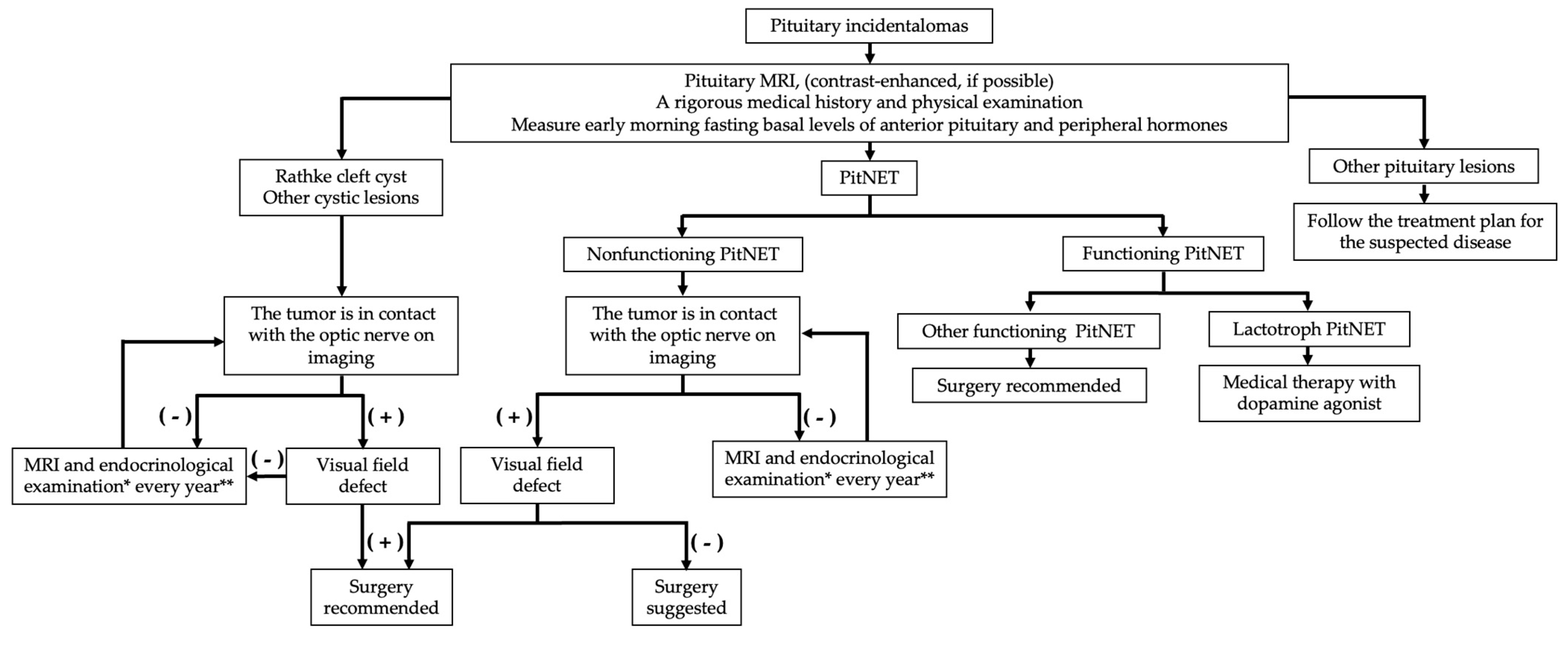
5.1. Evaluation at the Time of Detection
5.2. Natural History
6. Surgical Indications
7. Follow-Up
8. Treatment Outcome (Including Endocrine Function)
9. Conclusions
Author Contributions
Funding
Conflicts of Interest
References
- Feldkamp, J.; Santen, R.; Harms, E.; Aulich, A.; Mödder, U.; Scherbaum, W.A. Incidentally discovered pituitary lesions: High frequency of macroadenomas and hormone-secreting adenomas—Results of a prospective study. Clin. Endocrinol. 1999, 51, 109–113. [Google Scholar] [CrossRef]
- Arita, K.; Tominaga, A.; Sugiyama, K.; Eguchi, K.; Iida, K.; Sumida, M.; Migita, K.; Kurisu, K. Natural course of incidentally found nonfunctioning pituitary adenoma, with special reference to pituitary apoplexy during follow-up examination. J. Neurosurg. 2006, 104, 884–891. [Google Scholar] [CrossRef] [PubMed]
- Reincke, M.; Allolio, B.; Saeger, W.; Menzel, J.; Winkelmann, W. The ‘incidentaloma’ of the pituitary gland. Is neurosurgery required? JAMA 1990, 263, 2772–2776. [Google Scholar] [CrossRef] [PubMed]
- Donovan, L.E.; Corenblum, B. The natural history of the pituitary incidentaloma. Arch. Intern. Med. 1995, 155, 181–183. [Google Scholar] [CrossRef] [PubMed]
- Sanno, N.; Oyama, K.; Tahara, S.; Teramoto, A.; Kato, Y. A survey of pituitary incidentaloma in Japan. Eur. J. Endocrinol. 2003, 149, 123–127. [Google Scholar] [CrossRef] [PubMed]
- Day, P.F.; Guitelman, M.; Artese, R.; Fiszledjer, L.; Chervin, A.; Vitale, N.M.; Stalldecker, G.; De Miguel, V.; Cornaló, D.; Alfieri, A.; et al. Retrospective multicentric study of pituitary incidentalomas. Pituitary 2004, 7, 145–148. [Google Scholar] [CrossRef]
- Freda, P.U.; Beckers, A.M.; Katznelson, L.; Molitch, M.E.; Montori, V.M.; Post, K.D.; Vance, M.L.; Endocrine, S. Pituitary incidentaloma: An endocrine society clinical practice guideline. J. Clin. Endocrinol. Metab. 2011, 96, 894–904. [Google Scholar] [CrossRef]
- Losa, M.; Donofrio, C.A.; Barzaghi, R.; Mortini, P. Presentation and surgical results of incidentally discovered nonfunctioning pituitary adenomas: Evidence for a better outcome independently of other patients’ characteristics. Eur. J. Endocrinol. 2013, 169, 735–742. [Google Scholar] [CrossRef]
- Kreitschmann-Andermahr, I.; Siegel, S.; Weber Carneiro, R.; Maubach, J.M.; Harbeck, B.; Brabant, G. Headache and pituitary disease: A systematic review. Clin. Endocrinol. 2013, 79, 760–769. [Google Scholar] [CrossRef]
- Asa, S.L.; Mete, O.; Perry, A.; Osamura, R.Y. Overview of the 2022 WHO Classification of Pituitary Tumors. Endocr. Pathol. 2022, 33, 6–26. [Google Scholar] [CrossRef]
- Costello, R.T. Subclinical Adenoma of the Pituitary Gland. Am. J. Pathol. 1936, 12, 205–216.201. [Google Scholar] [PubMed]
- Sommers, S.C. Pituitary cell relations to body states. Lab. Investig. 1959, 8, 588–621. [Google Scholar] [PubMed]
- Hardy, J. Transphenoidal microsurgery of the normal and pathological pituitary. Clin. Neurosurg. 1969, 16, 185–217. [Google Scholar] [CrossRef] [PubMed]
- McCormick, W.F.; Halmi, N.S. Absence of chromophobe adenomas from a large series of pituitary tumors. Arch. Pathol. 1971, 92, 231–238. [Google Scholar] [PubMed]
- Haugen, O.A. Pituitary adenomas and the histology of the prostate in elderly men. An analysis in an autopsy series. Acta Pathol. Microbiol. Scand. A 1973, 81, 425–434. [Google Scholar]
- Kovacs, K.; Ryan, N.; Horvath, E.; Singer, W.; Ezrin, C. Pituitary adenomas in old age. J. Gerontol. 1980, 35, 16–22. [Google Scholar] [CrossRef]
- Burrow, G.N.; Wortzman, G.; Rewcastle, N.B.; Holgate, R.C.; Kovacs, K. Microadenomas of the pituitary and abnormal sellar tomograms in an unselected autopsy series. N. Engl. J. Med. 1981, 304, 156–158. [Google Scholar] [CrossRef]
- Max, M.B.; Deck, M.D.; Rottenberg, D.A. Pituitary metastasis: Incidence in cancer patients and clinical differentiation from pituitary adenoma. Neurology 1981, 31, 998–1002. [Google Scholar] [CrossRef]
- Muhr, C.; Bergström, K.; Grimelius, L.; Larsson, S.G. A parallel study of the roentgen anatomy of the sella turcica and the histopathology of the pituitary gland in 205 autopsy specimens. Neuroradiology 1981, 21, 55–65. [Google Scholar] [CrossRef]
- Parent, A.D.; Bebin, J.; Smith, R.R. Incidental pituitary adenomas. J. Neurosurg. 1981, 54, 228–231. [Google Scholar] [CrossRef]
- Chambers, E.F.; Turski, P.A.; LaMasters, D.; Newton, T.H. Regions of low density in the contrast-enhanced pituitary gland: Normal and pathologic processes. Radiology 1982, 144, 109–113. [Google Scholar] [CrossRef] [PubMed]
- Schwesinger, G.; Warzok, R. Hyperplasias and adenomas of the pituitary gland in an unselected autopsy material. Zentralbl. Allg. Pathol. 1982, 126, 495–498. [Google Scholar] [PubMed]
- Coulon, G.; Fellmann, D.; Arbez-Gindre, F.; Pageaut, G. Latent pituitary adenoma. Autopsy study. Sem. Hop. 1983, 59, 2747–2750. [Google Scholar] [PubMed]
- Siqueira, M.G.; Guembarovski, A.L. Subclinical pituitary microadenomas. Surg. Neurol. 1984, 22, 134–140. [Google Scholar] [CrossRef]
- Kontogeorgos, G.; Kovacs, K.; Horvath, E.; Scheithauer, B.W. Multiple adenomas of the human pituitary. A retrospective autopsy study with clinical implications. J. Neurosurg. 1991, 74, 243–247. [Google Scholar] [CrossRef]
- Marin, F.; Kovacs, K.T.; Scheithauer, B.W.; Young, W.F., Jr. The pituitary gland in patients with breast carcinoma: A histologic and immunocytochemical study of 125 cases. Mayo Clin. Proc. 1992, 67, 949–956. [Google Scholar] [CrossRef]
- Teramoto, A.; Hirakawa, K.; Sanno, N.; Osamura, Y. Incidental pituitary lesions in 1000 unselected autopsy specimens. Radiology 1994, 193, 161–164. [Google Scholar] [CrossRef]
- Camaris, C.; Balleine, R.; Little, D. Microadenomas of the human pituitary. Pathology 1995, 27, 8–11. [Google Scholar] [CrossRef]
- Tomita, T.; Gates, E. Pituitary adenomas and granular cell tumors. Incidence, cell type, and location of tumor in 100 pituitary glands at autopsy. Am. J. Clin. Pathol. 1999, 111, 817–825. [Google Scholar] [CrossRef]
- Kurosaki, M.; Saeger, W.; Lüdecke, D.K. Pituitary tumors in the elderly. Pathol. Res. Pract. 2001, 197, 493–497. [Google Scholar] [CrossRef]
- Ezzat, S.; Asa, S.L.; Couldwell, W.T.; Barr, C.E.; Dodge, W.E.; Vance, M.L.; McCutcheon, I.E. The prevalence of pituitary adenomas: A systematic review. Cancer 2004, 101, 613–619. [Google Scholar] [CrossRef] [PubMed]
- Buurman, H.; Saeger, W. Subclinical adenomas in postmortem pituitaries: Classification and correlations to clinical data. Eur. J. Endocrinol. 2006, 154, 753–758. [Google Scholar] [CrossRef] [PubMed]
- Furgal-Borzych, A.; Lis, G.J.; Litwin, J.A.; Rzepecka-Wozniak, E.; Trela, F.; Cichocki, T. Increased incidence of pituitary microadenomas in suicide victims. Neuropsychobiology 2007, 55, 163–166. [Google Scholar] [CrossRef] [PubMed]
- Kim, J.H.; Seo, J.S.; Lee, B.W.; Lee, S.Y.; Jeon, S.H.; Lee, K.B. The characteristics of incidental pituitary microadenomas in 120 Korean forensic autopsy cases. J. Korean Med. Sci 2007, 22, S61–S65. [Google Scholar] [CrossRef]
- Rittierodt, M.; Hori, A. Pre-morbid morphological conditions of the human pituitary. Neuropathology 2007, 27, 43–48. [Google Scholar] [CrossRef] [PubMed]
- Aghakhani, K.; Kadivar, M.; Kazemi-Esfeh, S.; Zamani, N.; Moradi, M.; Sanaei-Zadeh, H. Prevalence of pituitary incidentaloma in the Iranian cadavers. Indian J. Pathol. Microbiol. 2011, 54, 692–694. [Google Scholar] [PubMed]
- Chong, B.W.; Kucharczyk, W.; Singer, W.; George, S. Pituitary gland MR: A comparative study of healthy volunteers and patients with microadenomas. AJNR Am. J. Neuroradiol. 1994, 15, 675–679. [Google Scholar]
- Hall, W.A.; Luciano, M.G.; Doppman, J.L.; Patronas, N.J.; Oldfield, E.H. Pituitary magnetic resonance imaging in normal human volunteers: Occult adenomas in the general population. Ann. Intern. Med. 1994, 120, 817–820. [Google Scholar] [CrossRef]
- Nammour, G.M.; Ybarra, J.; Naheedy, M.H.; Romeo, J.H.; Aron, D.C. Incidental pituitary macroadenoma: A population-based study. Am. J. Med. Sci. 1997, 314, 287–291. [Google Scholar]
- Yue, N.C.; Longstreth, W.T., Jr.; Elster, A.D.; Jungreis, C.A.; O’Leary, D.H.; Poirier, V.C. Clinically serious abnormalities found incidentally at MR imaging of the brain: Data from the Cardiovascular Health Study. Radiology 1997, 202, 41–46. [Google Scholar] [CrossRef]
- Vernooij, M.W.; Ikram, M.A.; Tanghe, H.L.; Vincent, A.J.; Hofman, A.; Krestin, G.P.; Niessen, W.J.; Breteler, M.M.; van der Lugt, A. Incidental findings on brain MRI in the general population. N. Engl. J. Med. 2007, 357, 1821–1828. [Google Scholar] [CrossRef]
- Jeong, S.Y.; Lee, S.W.; Lee, H.J.; Kang, S.; Seo, J.H.; Chun, K.A.; Cho, I.H.; Won, K.S.; Zeon, S.K.; Ahn, B.C.; et al. Incidental pituitary uptake on whole-body 18F-FDG PET/CT: A multicentre study. Eur. J. Nucl Med. Mol. Imaging 2010, 37, 2334–2343. [Google Scholar] [CrossRef] [PubMed]
- Pette, G.A.; Norkin, F.J.; Ganeles, J.; Hardigan, P.; Lask, E.; Zfaz, S.; Parker, W. Incidental findings from a retrospective study of 318 cone beam computed tomography consultation reports. Int. J. Oral Maxillofac. Implants 2012, 27, 595–603. [Google Scholar] [PubMed]
- Sandeman, E.M.; Hernandez Mdel, C.; Morris, Z.; Bastin, M.E.; Murray, C.; Gow, A.J.; Corley, J.; Henderson, R.; Deary, I.J.; Starr, J.M.; et al. Incidental findings on brain MR imaging in older community-dwelling subjects are common but serious medical consequences are rare: A cohort study. PLoS ONE 2013, 8, e71467. [Google Scholar] [CrossRef] [PubMed]
- Hegenscheid, K.; Seipel, R.; Schmidt, C.O.; Völzke, H.; Kühn, J.P.; Biffar, R.; Kroemer, H.K.; Hosten, N.; Puls, R. Potentially relevant incidental findings on research whole-body MRI in the general adult population: Frequencies and management. Eur. Radiol. 2013, 23, 816–826. [Google Scholar] [CrossRef]
- Bos, D.; Poels, M.M.; Adams, H.H.; Akoudad, S.; Cremers, L.G.; Zonneveld, H.I.; Hoogendam, Y.Y.; Verhaaren, B.F.; Verlinden, V.J.; Verbruggen, J.G.; et al. Prevalence, Clinical Management, and Natural Course of Incidental Findings on Brain MR Images: The Population-based Rotterdam Scan Study. Radiology 2016, 281, 507–515. [Google Scholar] [CrossRef]
- Håberg, A.K.; Hammer, T.A.; Kvistad, K.A.; Rydland, J.; Müller, T.B.; Eikenes, L.; Gårseth, M.; Stovner, L.J. Incidental Intracranial Findings and Their Clinical Impact; The HUNT MRI Study in a General Population of 1006 Participants between 50–66 Years. PLoS ONE 2016, 11, e0151080. [Google Scholar] [CrossRef]
- Boutet, C.; Vassal, F.; Celle, S.; Schneider, F.C.; Barthélémy, J.C.; Laurent, B.; Barral, F.G.; Roche, F. Incidental findings on brain magnetic resonance imaging in the elderly:the PROOF study. Brain Imaging Behav. 2017, 11, 293–299. [Google Scholar] [CrossRef]
- Hoang, J.K.; Hoffman, A.R.; González, R.G.; Wintermark, M.; Glenn, B.J.; Pandharipande, P.V.; Berland, L.L.; Seidenwurm, D.J. Management of Incidental Pituitary Findings on CT, MRI, and (18)F-Fluorodeoxyglucose PET: A White Paper of the ACR Incidental Findings Committee. J. Am. Coll. Radiol. 2018, 15, 966–972. [Google Scholar] [CrossRef]
- Yoo, H.W.; Kim, J.R.; Yu, J.S. Prevalence and MRI findings of incidentally detected pituitary non-enhancing lesion on brain MRI in children. J. Pediatr. Endocrinol. Metab. 2021, 34, 591–598. [Google Scholar] [CrossRef]
- Lohner, V.; Lu, R.; Enkirch, S.J.; Stöcker, T.; Hattingen, E.; Breteler, M.M.B. Incidental findings on 3 T neuroimaging: Cross-sectional observations from the population-based Rhineland Study. Neuroradiology 2022, 64, 503–512. [Google Scholar] [CrossRef] [PubMed]
- Kuo, M.; Maya, M.M.; Bonert, V.; Melmed, S. Prospective Evaluation of Incidental Pituitary Imaging Findings in the Sella Turcica. J. Endocr. Soc. 2021, 5, bvaa186. [Google Scholar] [CrossRef]
- Anagnostis, P.; Adamidou, F.; Polyzos, S.A.; Efstathiadou, Z.; Panagiotou, A.; Kita, M. Pituitary incidentalomas: A single-centre experience. Int. J. Clin. Pract. 2011, 65, 172–177. [Google Scholar] [CrossRef]
- Esteves, C.; Neves, C.; Augusto, L.; Menezes, J.; Pereira, J.; Bernardes, I.; Fonseca, J.; Carvalho, D. Pituitary incidentalomas: Analysis of a neuroradiological cohort. Pituitary 2015, 18, 777–781. [Google Scholar] [CrossRef] [PubMed]
- Imran, S.A.; Yip, C.E.; Papneja, N.; Aldahmani, K.; Mohammad, S.; Imran, F.; Zwicker, D.A.; Theriault, C.; Thompson, K.; Clarke, D.B.; et al. Analysis and natural history of pituitary incidentalomas. Eur. J. Endocrinol. 2016, 175, 1–9. [Google Scholar] [CrossRef] [PubMed]
- Ishii, K.; Abe, I.; Kameda, W.; Sugimoto, K.; Morinaga, Y.; Ito, M.; Takashi, Y.; Abe, M.; Hada, Y.; Takase, K.; et al. Clinical investigation of pituitary incidentalomas: A two-center study. Intractable Rare Dis. Res. 2019, 8, 239–244. [Google Scholar] [CrossRef]
- Freda, P.U.; Bruce, J.N.; Khandji, A.G.; Jin, Z.; Hickman, R.A.; Frey, E.; Reyes-Vidal, C.; Otten, M.; Wardlaw, S.L.; Post, K.D. Presenting Features in 269 Patients With Clinically Nonfunctioning Pituitary Adenomas Enrolled in a Prospective Study. J. Endocr. Soc. 2020, 4, bvaa021. [Google Scholar] [CrossRef]
- Ono, M.; Fukuda, I.; Soga, A.; Tahara, S.; Morita, A.; Sugihara, H. A survey of surgically resected pituitary incidentalomas and a comparison of the clinical features and surgical outcomes of non-functioning pituitary adenomas discovered incidentally versus symptomatically. Endocr. J. 2021, 68, 561–571. [Google Scholar] [CrossRef]
- Donckier, J.E.; Gustin, T. Pituitary incidentaloma: To operate or not to operate? Acta Chir. Belg. 2012, 112, 255–260. [Google Scholar] [CrossRef]
- Hattori, Y.; Tahara, S.; Aso, S.; Matsui, H.; Fushimi, K.; Yasunaga, H.; Morita, A. Pituitary surgery’s epidemiology using a national inpatient database in Japan. Acta Neurochir. 2020, 126, 1317–1323. [Google Scholar] [CrossRef]
- Freda, P.U.; Post, K.D. Differential diagnosis of sellar masses. Endocrinol. Metab. Clin. N. Am. 1999, 28, 81–117. [Google Scholar] [CrossRef]
- Nishioka, H.; Inoshita, N.; Mete, O.; Asa, S.L.; Hayashi, K.; Takeshita, A.; Fukuhara, N.; Yamaguchi-Okada, M.; Takeuchi, Y.; Yamada, S. The Complementary Role of Transcription Factors in the Accurate Diagnosis of Clinically Nonfunctioning Pituitary Adenomas. Endocr. Pathol. 2015, 26, 349–355. [Google Scholar] [CrossRef] [PubMed]
- Daly, A.F.; Rixhon, M.; Adam, C.; Dempegioti, A.; Tichomirowa, M.A.; Beckers, A. High prevalence of pituitary adenomas: A cross-sectional study in the province of Liege, Belgium. J. Clin. Endocrinol. Metab. 2006, 91, 4769–4775. [Google Scholar] [CrossRef] [Green Version]
- Fontana, E.; Gaillard, R. Epidemiology of pituitary adenoma: Results of the first Swiss study. Rev. Med. Suisse 2009, 5, 2172–2174. [Google Scholar] [PubMed]
- Fernandez, A.; Karavitaki, N.; Wass, J.A. Prevalence of pituitary adenomas: A community-based, cross-sectional study in Banbury (Oxfordshire, UK). Clin. Endocrinol. 2010, 72, 377–382. [Google Scholar] [CrossRef]
- Agustsson, T.T.; Baldvinsdottir, T.; Jonasson, J.G.; Olafsdottir, E.; Steinthorsdottir, V.; Sigurdsson, G.; Thorsson, A.V.; Carroll, P.V.; Korbonits, M.; Benediktsson, R. The epidemiology of pituitary adenomas in Iceland, 1955–2012: A nationwide population-based study. Eur. J. Endocrinol. 2015, 173, 655–664. [Google Scholar] [CrossRef] [PubMed]
- Raappana, A.; Koivukangas, J.; Ebeling, T.; Pirilä, T. Incidence of pituitary adenomas in Northern Finland in 1992–2007. J. Clin. Endocrinol. Metab. 2010, 95, 4268–4275. [Google Scholar] [CrossRef]
- Tjörnstrand, A.; Gunnarsson, K.; Evert, M.; Holmberg, E.; Ragnarsson, O.; Rosén, T.; Filipsson Nyström, H. The incidence rate of pituitary adenomas in western Sweden for the period 2001–2011. Eur. J. Endocrinol. 2014, 171, 519–526. [Google Scholar] [CrossRef]
- Day, P.F.; Loto, M.G.; Glerean, M.; Picasso, M.F.; Lovazzano, S.; Giunta, D.H. Incidence and prevalence of clinically relevant pituitary adenomas: Retrospective cohort study in a Health Management Organization in Buenos Aires, Argentina. Arch. Endocrinol. Metab. 2016, 60, 554–561. [Google Scholar] [CrossRef]
- Matsubayashi, K.; Kawakami, K. Prevalence, incidence, comorbidities, and treatment patterns among Japanese patients with acromegaly: A descriptive study using a nationwide claims database. Endocr. J. 2020, 67, 997–1006. [Google Scholar] [CrossRef]
- Ekhzaimy, A.A.; Mujammami, M.; Tharkar, S.; Alansary, M.A.; Al Otaibi, D. Clinical presentation, evaluation and case management of primary empty sella syndrome: A retrospective analysis of 10-year single-center patient data. BMC Endocr. Disord. 2020, 20, 142. [Google Scholar] [CrossRef]
- Carosi, G.; Brunetti, A.; Mangone, A.; Baldelli, R.; Tresoldi, A.; Del Sindaco, G.; Lavezzi, E.; Sala, E.; Mungari, R.; Fatti, L.M.; et al. A Multicenter Cohort Study in Patients With Primary Empty Sella: Hormonal and Neuroradiological Features Over a Long Follow-Up. Front. Endocrinol. 2022, 13, 925378. [Google Scholar] [CrossRef] [PubMed]
- Han, L.; Wang, J.; Shu, K.; Lei, T. Pituitary tumorous hyperplasia due to primary hypothyroidism. Acta Neurochir. 2012, 154, 1489–1492; discussion 1492. [Google Scholar] [CrossRef] [PubMed] [Green Version]
- Galland, F.; Vantyghem, M.C.; Cazabat, L.; Boulin, A.; Cotton, F.; Bonneville, J.F.; Jouanneau, E.; Vidal-Trécan, G.; Chanson, P. Management of nonfunctioning pituitary incidentaloma. Ann. Endocrinol. 2015, 76, 191–200. [Google Scholar] [CrossRef] [PubMed]
- Molitch, M.E. Nonfunctioning pituitary tumors and pituitary incidentalomas. Endocrinol. Metab. Clin. N. Am. 2008, 37, 151–171. [Google Scholar] [CrossRef] [PubMed]
- Molitch, M.E. Pituitary tumours: Pituitary incidentalomas. Best Pract. Res. Clin. Endocrinol. Metab. 2009, 23, 667–675. [Google Scholar] [CrossRef]
- Molitch, M.E. Management of incidentally found nonfunctional pituitary tumors. Neurosurg. Clin. N. Am. 2012, 23, 543–553. [Google Scholar] [CrossRef]
- Molitch, M.E. Nonfunctioning pituitary tumors. Handb. Clin. Neurol. 2014, 124, 167–184. [Google Scholar]
- Boguszewski, C.L.; de Castro Musolino, N.R.; Kasuki, L. Management of pituitary incidentaloma. Best Pract. Res. Clin. Endocrinol. Metab. 2019, 33, 101268. [Google Scholar] [CrossRef]
- Bevan, J.S. Pituitary incidentaloma. Clin. Med. 2013, 13, 296–298. [Google Scholar] [CrossRef]
- Vasilev, V.; Rostomyan, L.; Daly, A.F.; Potorac, I.; Zacharieva, S.; Bonneville, J.F.; Beckers, A. Management of Endocrine Disease: Pituitary ‘incidentaloma’: Neuroradiological assessment and differential diagnosis. Eur. J. Endocrinol. 2016, 175, R171–R184. [Google Scholar] [CrossRef] [PubMed]
- Hitzeman, N.; Cotton, E. Incidentalomas: Initial management. Am. Fam. Physician 2014, 90, 784–789. [Google Scholar] [PubMed]
- Giraldi, E.A.; Veledar, E.; Oyesiku, N.M.; Ioachimescu, A.G. Incidentally detected acromegaly: Single-center study of surgically treated patients over 22 years. J. Investig. Med. 2021, 69, 351–357. [Google Scholar] [CrossRef] [PubMed]
- Tresoldi, A.S.; Carosi, G.; Betella, N.; Del Sindaco, G.; Indirli, R.; Ferrante, E.; Sala, E.; Giavoli, C.; Morenghi, E.; Locatelli, M.; et al. Clinically Nonfunctioning Pituitary Incidentalomas: Characteristics and Natural History. Neuroendocrinology 2020, 110, 595–603. [Google Scholar] [CrossRef] [PubMed]
- Iglesias, P.; Arcano, K.; Trivino, V.; Garcia-Sancho, P.; Diez, J.J.; Villabona, C.; Cordido, F. Prevalence, Clinical Features, and Natural History of Incidental Clinically Non-Functioning Pituitary Adenomas. Horm. Metab. Res. 2017, 49, 654–659. [Google Scholar] [CrossRef] [PubMed]
- Briet, C.; Salenave, S.; Bonneville, J.F.; Laws, E.R.; Chanson, P. Pituitary Apoplexy. Endocr. Rev. 2015, 36, 622–645. [Google Scholar] [CrossRef]
- Yamamoto, T.; Yano, S.; Kuroda, J.; Hasegawa, Y.; Hide, T.; Kuratsu, J. Pituitary apoplexy associated with endocrine stimulation test: Endocrine stimulation test, treatment, and outcome. Case Rep. Endocrinol. 2012, 2012, 826901. [Google Scholar] [CrossRef]
- Toini, A.; Dolci, A.; Ferrante, E.; Verrua, E.; Malchiodi, E.; Sala, E.; Lania, A.G.; Chiodini, I.; Beck-Peccoz, P.; Arosio, M.; et al. Screening for ACTH-dependent hypercortisolism in patients affected with pituitary incidentaloma. Eur. J. Endocrinol. 2015, 172, 363–369. [Google Scholar] [CrossRef]
- Torpy, D.J. Screening for ACTH-dependent hypercortisolism in patients with pituitary incidentaloma. Eur. J. Endocrinol. 2015, 172, C1–C4. [Google Scholar] [CrossRef]
- Bonomo, G.; Bertani, G.A.; Carrabba, G.G.; Ferrante, E.; Pluderi, M.; Guastella, C.; Pignataro, L.; Rampini, P.; Mantovani, G.; Locatelli, M. The suprasellar volume of nonfunctioning pituitary adenomas: A useful tool for predicting visual field deficits. Pituitary 2020, 23, 552–557. [Google Scholar] [CrossRef]
- Nishizawa, S.; Ohta, S.; Yokoyama, T.; Uemura, K. Therapeutic strategy for incidentally found pituitary tumors (“pituitary incidentalomas”). Neurosurgery 1998, 43, 1344–1348; discussion 1348–1350. [Google Scholar] [PubMed]
- Karavitaki, N.; Collison, K.; Halliday, J.; Byrne, J.V.; Price, P.; Cudlip, S.; Wass, J.A. What is the natural history of nonoperated nonfunctioning pituitary adenomas? Clin. Endocrinol. 2007, 67, 938–943. [Google Scholar] [CrossRef] [PubMed]
- Fernández-Balsells, M.M.; Murad, M.H.; Barwise, A.; Gallegos-Orozco, J.F.; Paul, A.; Lane, M.A.; Lampropulos, J.F.; Natividad, I.; Perestelo-Pérez, L.; Ponce de León-Lovatón, P.G.; et al. Natural history of nonfunctioning pituitary adenomas and incidentalomas: A systematic review and metaanalysis. J. Clin. Endocrinol. Metab. 2011, 96, 905–912. [Google Scholar] [CrossRef] [PubMed]
- Igarashi, T.; Saeki, N.; Yamaura, A. Long-term magnetic resonance imaging follow-up of asymptomatic sellar tumors.—Their natural history and surgical indications. Neurol. Med. Chir. 1999, 39, 592–598; discussion 598–599. [Google Scholar] [CrossRef] [PubMed] [Green Version]
- Möller-Goede, D.L.; Brändle, M.; Landau, K.; Bernays, R.L.; Schmid, C. Pituitary apoplexy: Re-evaluation of risk factors for bleeding into pituitary adenomas and impact on outcome. Eur. J. Endocrinol. 2011, 164, 37–43. [Google Scholar] [CrossRef]
- Ciric, I.; Ragin, A.; Baumgartner, C.; Pierce, D. Complications of transsphenoidal surgery: Results of a national survey, review of the literature, and personal experience. Neurosurgery 1997, 40, 225–236; discussion 236–237. [Google Scholar] [CrossRef]
- Morinaga, Y.; Abe, I.; Nii, K.; Hanada, H.; Takemura, Y.; Takashi, Y.; Sakamoto, K.; Inoue, R.; Mitsutake, T.; Kobayashi, K.; et al. Characteristics and clinical outcomes in pituitary incidentalomas and non-incidental pituitary tumors treated with endoscopic transsphenoidal surgery. Medicine 2020, 99, e22713. [Google Scholar] [CrossRef]
- Messerer, M.; Dubourg, J.; Raverot, G.; Bervini, D.; Berhouma, M.; George, I.; Chacko, A.G.; Perrin, G.; Levivier, M.; Daniel, R.T.; et al. Non-functioning pituitary macro-incidentalomas benefit from early surgery before becoming symptomatic. Clin. Neurol. Neurosurg. 2013, 115, 2514–2520. [Google Scholar] [CrossRef]
- Seltzer, J.; Wedemeyer, M.A.; Bonney, P.A.; Carmichael, J.D.; Weiss, M.; Zada, G. Outcomes following transsphenoidal surgical management of incidental pituitary adenomas: A series of 52 patients over a 17-year period. J. Neurosurg. 2018, 130, 1584–1592. [Google Scholar] [CrossRef]
- Suzuki, M.; Minematsu, T.; Oyama, K.; Tahara, S.; Miyai, S.; Sanno, N.; Osamura, R.Y.; Teramoto, A. Expression of proliferation markers in human pituitary incidentalomas. Endocr. Pathol. 2006, 17, 263–275. [Google Scholar] [CrossRef]
- Mete, O.; Gomez-Hernandez, K.; Kucharczyk, W.; Ridout, R.; Zadeh, G.; Gentili, F.; Ezzat, S.; Asa, S.L. Silent subtype 3 pituitary adenomas are not always silent and represent poorly differentiated monomorphous plurihormonal Pit-1 lineage adenomas. Mod. Pathol. 2016, 29, 131–142. [Google Scholar] [CrossRef] [PubMed]
- George, D.H.; Scheithauer, B.W.; Kovacs, K.; Horvath, E.; Young, W.F., Jr.; Lloyd, R.V.; Meyer, F.B. Crooke’s cell adenoma of the pituitary: An aggressive variant of corticotroph adenoma. Am. J. Surg. Pathol. 2003, 27, 1330–1336. [Google Scholar] [CrossRef] [PubMed]
- Di Ieva, A.; Davidson, J.M.; Syro, L.V.; Rotondo, F.; Montoya, J.F.; Horvath, E.; Cusimano, M.D.; Kovacs, K. Crooke’s cell tumors of the pituitary. Neurosurgery 2015, 76, 616–622. [Google Scholar] [CrossRef]
- Almeida, J.P.; Stephens, C.C.; Eschbacher, J.M.; Felicella, M.M.; Yuen, K.C.J.; White, W.L.; Mooney, M.A.; Bernat, A.L.; Mete, O.; Zadeh, G.; et al. Clinical, pathologic, and imaging characteristics of pituitary null cell adenomas as defined according to the 2017 World Health Organization criteria: A case series from two pituitary centers. Pituitary 2019, 22, 514–519. [Google Scholar] [CrossRef] [PubMed]
- Balogun, J.A.; Monsalves, E.; Juraschka, K.; Parvez, K.; Kucharczyk, W.; Mete, O.; Gentili, F.; Zadeh, G. Null cell adenomas of the pituitary gland: An institutional review of their clinical imaging and behavioral characteristics. Endocr. Pathol. 2015, 26, 63–70. [Google Scholar] [CrossRef] [PubMed]
- Haddad, A.F.; Young, J.S.; Oh, T.; Pereira, M.P.; Joshi, R.S.; Pereira, K.M.; Osorio, R.C.; Donohue, K.C.; Peeran, Z.; Sudhir, S.; et al. Clinical characteristics and outcomes of null-cell versus silent gonadotroph adenomas in a series of 1166 pituitary adenomas from a single institution. Neurosurg. Focus 2020, 48, E13. [Google Scholar] [CrossRef]
- Scheithauer, B.W.; Jaap, A.J.; Horvath, E.; Kovacs, K.; Lloyd, R.V.; Meyer, F.B.; Laws, E.R., Jr.; Young, W.F., Jr. Clinically silent corticotroph tumors of the pituitary gland. Neurosurgery 2000, 47, 723–729; discussion 729–730. [Google Scholar] [PubMed]
- Xu, Z.; Ellis, S.; Lee, C.C.; Starke, R.M.; Schlesinger, D.; Lee Vance, M.; Lopes, M.B.; Sheehan, J. Silent corticotroph adenomas after stereotactic radiosurgery: A case-control study. Int. J. Radiat. Oncol. Biol. Phys. 2014, 90, 903–910. [Google Scholar] [CrossRef]
- Randall, B.R.; Kraus, K.L.; Simard, M.F.; Couldwell, W.T. Cost of evaluation of patients with pituitary incidentaloma. Pituitary 2010, 13, 383–384. [Google Scholar] [CrossRef]


| Year | Authors | Number of Pituitaries Studied | Number of PitNETs Found | Frequency (%) |
|---|---|---|---|---|
| 1936 | Costello [11] | 1000 | 225 | 22.5 |
| 1959 | Sommers [12] | 400 | 26 | 6.5 |
| 1969 | Hardy [13] | 1000 | 27 | 2.7 |
| 1971 | McCormick [14] | 1600 | 140 | 8.8 |
| 1973 | Haugen [15] | 170 | 33 | 19.4 |
| 1980 | Kovacs [16] | 152 | 20 | 13.2 |
| 1981 | Burrow [17] | 120 | 32 | 26.7 |
| 1981 | Max [18] | 500 | 9 | 1.8 |
| 1981 | Muhr [19] | 205 | 3 | 1.5 |
| 1981 | Parent [20] | 500 | 42 | 8.4 |
| 1982 | Chambers [21] | 100 | 14 | 14.0 |
| 1982 | Schwesinger [22] | 5100 | 485 | 9.5 |
| 1983 | Coulon [23] | 100 | 10 | 10.0 |
| 1984 | Siqueira [24] | 450 | 39 | 9.5 |
| 1991 | Kontogeorgos [25] | 470 | 49 | 10.4 |
| 1992 | Marin [26] | 210 | 35 | 16.7 |
| 1994 | Teramoto [27] | 1000 | 51 | 5.1 |
| 1995 | Camaris [28] | 423 | 14 | 3.2 |
| 1999 | Tomita [29] | 100 | 24 | 24.0 |
| 2001 | Kurosaki [30] | 692 | 79 | 11.4 |
| 2006 | Buurman [32] | 3048 | 334 | 11.0 |
| 2007 | Furgal-Borzych [33] | 151 | 47 | 31.1 |
| 2007 | Kim [34] | 120 | 7 | 5.8 |
| 2007 | Rittierodt [35] | 228 | 7 | 3.0 |
| 2011 | Aghakhani [36] | 485 | 61 | 12.6 |
| Year | Authors | Subject | Age (y/o) | Number of Pituitaries Studied | Number of PIs Found | Frequency (%) | Remarks |
|---|---|---|---|---|---|---|---|
| Examination using the CT | |||||||
| 1982 | Chambers [21] | Patients with orbital symptoms | NC | 50 | 10 | 20.0 | Contrast-enhanced high-resolution CT |
| 1997 | Nammour [39] | Consecutive patients undergoing head CT at a single institution | 57 | 3550 | 7 | 0.2 | Macro PitNETs only |
| 2012 | Pette [43] | Patients examined for dental implant therapy | 10–91 Ave. 64.73 | 318 | 2 | 0.63 | Cone beam CT |
| Examination using the MRI | |||||||
| 1994 | Chong [37] | Healthy adult volunteers | 22–68 Ave. 34 | 52 | 20 | 38.5 | Local low signal on T1-weighted image |
| 1994 | Hall [38] | Healthy adult volunteers | 18–60 | 100 | 10 | 10.0 | Contrast-enhanced MRI of the pituitary gland |
| 1997 | Yue [40] | Patients with cardiovascular and cerebrovascular disease | ≥65 | 3672 | 6 | 0.16 | |
| 2007 | Vernooij [41] | Health checkups for residents near Rotterdam, the Netherlands | 45.7–96.7 Ave. 63.3 | 2000 | 6 | 0.3 | Macro PitNETs only |
| 2013 | Sandeman [44] | Residents of Edinburgh | Ave. 72.5 | 700 | 2 | 0.28 | |
| 2013 | Hegenscheid [45] | Residents of northwest Germany | 21–88 Ave. 53 | 2500 | 9 | 0.36 | |
| 2016 | Bos [46] | Residents of the Netherlands over 45 y/o | Ave. 64.9 | 5800 | 67 | 1.2 | |
| 2016 | Håberg [47] | Healthy adult volunteers | 50–66 | 1006 | 3 | 0.3 | |
| 2017 | Boutet [48] | French retirees over 65 y/o | Ave. 75.3 | 503 | 11 | 2.2 | |
| 2021 | Yoo [50] | Patients without endocrine abnormalities | <18 | 365 | 76 | 20.8 | Contrast-enhanced MRI |
| 2022 | Lohner [51] | Residents of Bonn, Germany | 55 | 3589 | 3 | 0.08 | Using 3Tesla MRI, macro PitNETs only |
| Examination using FDG PET/CT | |||||||
| 2010 | Jeong [42] | Patients with malignancy or screened for cancer | NC | 40,967 | 30 | 0.073 | |
| Neoplastic Lesion |
|---|
| PitNET, craniopharyngioma, pituicyte tumor, meningioma, chordoma, neuroblastoma, germ cell tumor, lymphoma, metastatic tumor, Langerhans cell histiocytosis |
| Cystic lesion |
| Rathke’s cleft cyst, arachnoid cyst |
| Non-neoplastic lesion |
| Hypophysitis, IgG4-related disease, sarcoidosis, granulomatosis with polyangiitis, infective granuloma (tuberculosis, fungus, bacterial), abscess, pituitary hyperplasia, empty sella, cerebral aneurysm |
| Year | Authors | Total | Increased (%) | Decreased (%) | No Change (%) | Follow-Up Period (Months) |
|---|---|---|---|---|---|---|
| Micro PitNETs | ||||||
| 1990 | Reincke [3] | 7 | 1 (14.3) | 1 (14.3) | 5 (71.4) | 22 |
| 1995 | Donovan [4] | 15 | 0 | 4 (26.7) | 11 (73.3) | 76.8 |
| 1999 | Feldkamp [1] | 31 | 1 (3.2) | 1 (3.2) | 29 (93.6) | 32.4 |
| 2003 | Sanno [5] | 74 | 10 (13.5) | 7 (9.5) | 57 (77) | 27 |
| 2004 | Day [6] | 11 | 1 (9.1) | 0 | 10 (90.9) | 38.4 |
| 2006 | Arita [2] | 5 | 2 (40) | 0 | 3 (60) | 61.9 |
| 2007 | Karavitaki [92] | 16 | 2 (12.5) | 1(6.3) | 13 (81.2) | 42 |
| 2011 | Anagnostis [53] | 6 | 0 | 1 (16.7) | 5 (83.3) | 48 |
| 2020 | Tresoldi [84] | 132 | 12 (9.1) | 28 (21.2) | 92 (69.7) | 36 |
| Macro PitNETs | ||||||
| 1990 | Reincke [3] | 7 | 2 (28.6) | 0 | 5 (71.4) | 22 |
| 1995 | Donovan [4] | 16 | 5 (31.3) | 0 | 11 (68.7) | 76.8 |
| 1998 | Nishizawa [91] | 28 | 2 (7.1) | 0 | 26 (92.9) | 67.2 |
| 1999 | Feldkamp [1] | 19 | 5 (26.3) | 1 (5.3) | 13 (68.4) | 32.4 |
| 2003 | Sanno [5] | 165 | 20 (12.1) | 22 (13.3) | 123 (74.6) | 27 |
| 2004 | Day [6] | 7 | 1 (14.3) | 0 | 6 (85.7) | 38.4 |
| 2006 | Arita [2] | 37 | 19 (51.4) | 0 | 18 (48.6) | 61.9 |
| 2007 | Karavitaki [92] | 24 | 12 (50) | 4 (16.7) | 8 (33.3) | 42 |
| 2011 | Anagnostis [53] | 3 | 1 (33.3) | 0 | 2 (66.7) | 48 |
| 2020 | Tresoldi [84] | 71 | 19 (26.8) | 4 (5.6) | 48 (67.6) | 36 |
| No distinction between the micro PitNET and the macro PitNET | ||||||
| 2016 | Imran [55] | 113 | 21 (18.6) | 2 (1.8) | 90 (79.6) | 36 |
| 2017 | Iglesias [85] | 26 | 1 (3.8) | 2 (7.7) | 23 (88.5) | 15.5 |
Publisher’s Note: MDPI stays neutral with regard to jurisdictional claims in published maps and institutional affiliations. |
© 2022 by the authors. Licensee MDPI, Basel, Switzerland. This article is an open access article distributed under the terms and conditions of the Creative Commons Attribution (CC BY) license (https://creativecommons.org/licenses/by/4.0/).
Share and Cite
Tahara, S.; Hattori, Y.; Suzuki, K.; Ishisaka, E.; Teramoto, S.; Morita, A. An Overview of Pituitary Incidentalomas: Diagnosis, Clinical Features, and Management. Cancers 2022, 14, 4324. https://doi.org/10.3390/cancers14174324
Tahara S, Hattori Y, Suzuki K, Ishisaka E, Teramoto S, Morita A. An Overview of Pituitary Incidentalomas: Diagnosis, Clinical Features, and Management. Cancers. 2022; 14(17):4324. https://doi.org/10.3390/cancers14174324
Chicago/Turabian StyleTahara, Shigeyuki, Yujiro Hattori, Koji Suzuki, Eitaro Ishisaka, Shinichiro Teramoto, and Akio Morita. 2022. "An Overview of Pituitary Incidentalomas: Diagnosis, Clinical Features, and Management" Cancers 14, no. 17: 4324. https://doi.org/10.3390/cancers14174324