
Metastasectomy in Leiomyosarcoma: A Systematic Review and Pooled Survival Analysis

 , , , ,
Abstract
:Simple Summary
Abstract
1. Introduction
2. Materials and Methods
2.1. Search Strategy
2.2. Selection Process
2.3. Data Collection
2.4. Data Synthesis and Analysis
2.5. Risk of Bias Assessment and Certainty of Evidence
3. Results
3.1. Study Characteristics
3.2. Sociodemographic and Clinical Characteristics of Patients Undergoing Metastasectomy for LMS
3.3. Management of Patients Undergoing Metastasectomy for LMS
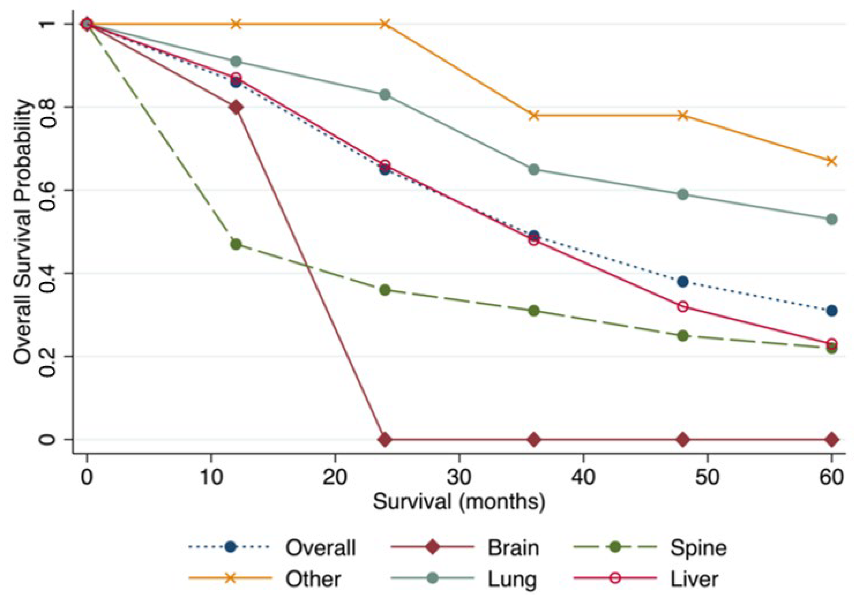
3.4. Post-Metastasectomy Outcomes
3.5. Prognostic Factors Associated with Post-Metastasectomy Outcomes
3.5.1. Lung
3.5.2. Liver
3.5.3. Spine
3.6. Recurrence Post-Metastasectomy
3.7. Risk of Bias and Certainty of Evidence
4. Discussion
Limitations
5. Conclusions
Supplementary Materials
Author Contributions
Funding
Institutional Review Board Statement
Informed Consent Statement
Data Availability Statement
Acknowledgments
Conflicts of Interest
Abbreviations
| ASPS | Alveolar soft part sarcoma |
| CI | Confidence interval |
| CSS | Cancer specific survival |
| DFI | Disease free interval |
| ECOG | Eastern Cooperative Oncology Group |
| GI | Gastrointestinal |
| GIST | Gastrointestinal stromal tumor |
| GTR | Gross total removal |
| GU | Genitourinary |
| IQR | Interquartile range |
| JBI | Joanna Briggs Institute |
| LMS | Leiomyosarcoma |
| LPS | Liposarcoma |
| MFH | Malignant fibrous histiocytoma |
| MPNST | Malignant peripheral nerve sheath tumor |
| NOS | Newcastle-Ottawa Quality Assessment Scale |
| NR | Not reported |
| OS | Overall survival |
| RMS | Rhabdomyosarcoma |
| SD | Standard deviation |
| SFT | Solitary fibrous tumor |
| SS | Synovial sarcoma |
| STR | Subtotal removal |
| STS | Soft tissue sarcoma |
| UPS | Undifferentiated pleomorphic sarcoma |
| WBRT | Whole brain radiation therapy |
References
- George, S.; Serrano, C.; Hensley, M.L.; Ray-Coquard, I. Soft Tissue and Uterine Leiomyosarcoma. J. Clin. Oncol. 2017, 36, 144–150. [Google Scholar] [CrossRef] [PubMed]
- Bathan, A.J.; Constantinidou, A.; Pollack, S.M.; Jones, R.L. Diagnosis, Prognosis, and Management of Leiomyosarcoma: Recognition of Anatomic Variants. Curr. Opin. Oncol. 2013, 25, 384–389. [Google Scholar] [CrossRef] [PubMed]
- Goldblum, J.; Volpe, A.; Weiss, S. Leiomyosarcoma. In Enzinger & Weiss’s Soft Tissue Tumors; Goldblum, J., Folpe, A., Weiss, S., Eds.; Elsevier: Philadelphia, PA, USA, 2020; pp. 591–613. [Google Scholar]
- Brennan, M.F.; Antonescu, C.R.; Moraco, N.; Singer, S. Lessons Learned from the Study of 10,000 Patients with Soft Tissue Sarcoma. Ann. Surg. 2014, 260, 416–422. [Google Scholar] [CrossRef] [PubMed] [Green Version]
- Yadav, U.; Mangla, A. Leiomyosarcoma. Available online: https://www.ncbi.nlm.nih.gov/books/NBK551667/ (accessed on 19 February 2022).
- Pisters, P.W.; Leung, D.H.; Woodruff, J.; Shi, W.; Brennan, M.F. Analysis of Prognostic Factors in 1,041 Patients with Localized Soft Tissue Sarcomas of the Extremities. J. Clin. Oncol. 1996, 14, 1679–1689. [Google Scholar] [CrossRef]
- Gronchi, A.; Strauss, D.C.; Miceli, R.; Bonvalot, S.; Swallow, C.J.; Hohenberger, P.; van Coevorden, F.; Rutkowski, P.; Callegaro, D.; Hayes, A.J.; et al. Variability in Patterns of Recurrence after Resection of Primary Retroperitoneal Sarcoma (RPS). A Report on 1007 Patients from the Multi-Institutional Collaborative RPS Working Group. Ann. Surg. 2016, 263, 1002–1009. [Google Scholar] [CrossRef]
- Yang, J.C.; Chang, A.E.; Baker, A.R.; Sindelar, W.F.; Danforth, D.N.; Topalian, S.L.; DeLaney, T.; Glatstein, E.; Steinberg, S.M.; Merino, M.J.; et al. Randomized Prospective Study of the Benefit of Adjuvant Radiation Therapy in the Treatment of Soft Tissue Sarcomas of the Extremity. J. Clin. Oncol. 1998, 16, 197–203. [Google Scholar] [CrossRef] [Green Version]
- Bonvalot, S.; Gronchi, A.; le Péchoux, C.; Swallow, C.J.; Strauss, D.; Meeus, P.; van Coevorden, F.; Stoldt, S.; Stoeckle, E.; Rutkowski, P.; et al. Preoperative Radiotherapy plus Surgery versus Surgery Alone for Patients with Primary Retroperitoneal Sarcoma (EORTC-62092: STRASS): A Multicentre, Open-Label, Randomised, Phase 3 Trial. Lancet Oncol. 2020, 21, 1366–1377. [Google Scholar] [CrossRef]
- Gamboa, A.C.; Gronchi, A.; Cardona, K. Soft-tissue Sarcoma in Adults: An Update on the Current State of Histiotype-specific Management in an Era of Personalized Medicine. CA Cancer J. Clin. 2020, 70, 200–229. [Google Scholar] [CrossRef] [Green Version]
- Van Cann, T.; Cornillie, J.; Wozniak, A.; Debiec-Rychter, M.; Sciot, R.; Hompes, D.; Vergote, I.; Schöffski, P. Retrospective Analysis of Outcome of Patients with Metastatic Leiomyosarcoma in a Tertiary Referral Center. Oncol. Res. Treat. 2018, 41, 206–213. [Google Scholar] [CrossRef]
- Tirotta, F.; Hodson, J.; Parente, A.; Pasquali, S.; Sutcliffe, R.; Desai, A.; Muiesan, P.; Ford, S.J.; Fiore, M.; Gronchi, A.; et al. Liver Resection for Sarcoma Metastases: A Systematic Review and Experience from Two European Centres. Eur. J. Surg. Oncol. 2020, 46, 1807–1813. [Google Scholar] [CrossRef]
- Deguchi, S.; Nakasu, Y.; Sakaida, T.; Akimoto, J.; Tanahashi, K.; Natsume, A.; Takahashi, M.; Okuda, T.; Asakura, H.; Mitsuya, K.; et al. Surgical Outcome and Graded Prognostic Assessment of Patients with Brain Metastasis from Adult Sarcoma: Multi-Institutional Retrospective Study in Japan. Int. J. Clin. Oncol. 2020, 25, 1995–2005. [Google Scholar] [CrossRef] [PubMed]
- Rao, G.; Suki, D.; Chakrabarti, I.; Feiz-Erfan, I.; Mody, M.G.; McCutcheon, I.E.; Gokaslan, Z.; Patel, S.; Rhines, L.D. Surgical Management of Primary and Metastatic Sarcoma of the Mobile Spine. J. Neurosurg. Spine 2008, 9, 120–128. [Google Scholar] [CrossRef] [PubMed] [Green Version]
- Wang, Y.; Delisle, M.; Smith, D.; Srikanthan, A. Survival by Histology among Patients with Bone and Soft Tissue Sarcoma Who Undergo Metastasectomy: Protocol for a Systematic Review and Meta-Analysis. Syst. Rev. 2020, 9, 189. [Google Scholar] [CrossRef]
- Page, M.J.; McKenzie, J.E.; Bossuyt, P.M.; Boutron, I.; Hoffmann, T.C.; Mulrow, C.D.; Shamseer, L.; Tetzlaff, J.M.; Akl, E.A.; Brennan, S.E.; et al. The PRISMA 2020 Statement: An Updated Guideline for Reporting Systematic Reviews. BMJ 2021, 372, n160. [Google Scholar] [CrossRef] [PubMed]
- Burt, B.M.; Ocejo, S.; Mery, C.M.; Dasilva, M.; Bueno, R.; Sugarbaker, D.J.; Jaklitsch, M.T. Repeated and Aggressive Pulmonary Resections for Leiomyosarcoma Metastases Extends Survival. Ann. Thorac. Surg. 2011, 92, 1202–1207. [Google Scholar] [CrossRef] [PubMed]
- Chen, H. Complete Hepatic Resection of Metastases from Leiomyosarcoma Prolongs Survival. J. Gastrointest. Surg. 1998, 2, 151–155. [Google Scholar] [CrossRef]
- Faraj, W.; El-Kehdy, J.; el Nounou, G.; Deeba, S.; Fakih, H.; Jabbour, M.; Haydar, A.; el Naaj, A.A.; Abou-Alfa, G.K.; O’Reilly, E.M.; et al. Liver Resection for Metastatic Colorectal Leiomyosarcoma: A Single Center Experience. J. Gastrointest. Oncol. 2015, 6, E70–E76. [Google Scholar] [CrossRef]
- Kato, S.; Demura, S.; Shinmura, K.; Yokogawa, N.; Yonezawa, N.; Shimizu, T.; Oku, N.; Kitagawa, R.; Murakami, H.; Kawahara, N.; et al. Clinical Outcomes and Survivals after Total En Bloc Spondylectomy for Metastatic Leiomyosarcoma in the Spine. Eur. Spine J. 2020, 29, 3237–3244. [Google Scholar] [CrossRef]
- Kim, Y.W.; Lee, J.H.; Kim, J.E.; Kang, J. Surgical Resection of Liver Metastasis of Leiomyosarcoma. Korean J. Clin. Oncol. 2017, 13, 143–146. [Google Scholar] [CrossRef]
- Lang, H.; Nußbaum, K.-T.; Kaudel, P.; Frü Hauf, N.; Flemming, P.; Raab, R. Hepatic Metastases from Leiomyosarcoma A Single-Center Experience with 34 Liver Resections During a 15-Year Period. Ann. Surg. 2000, 231, 500–505. [Google Scholar] [CrossRef]
- Paramanathan, A.; Wright, G. Pulmonary Metastasectomy for Sarcoma of Gynaecologic Origin. Heart Lung Circ. 2013, 22, 270–275. [Google Scholar] [CrossRef]
- Ziewacz, J.E.; Lau, D.; la Marca, F.; Park, P. Outcomes after Surgery for Spinal Metastatic Leiomyosarcoma. J. Neurosurg. Spine 2012, 17, 432–437. [Google Scholar] [CrossRef] [PubMed]
- Anraku, M.; Yokoi, K.; Nakagawa, K.; Fujisawa, T.; Nakajima, J.; Akiyama, H.; Nishimura, Y.; Kobayashi, K. Pulmonary Metastases from Uterine Malignancies: Results of Surgical Resection in 133 Patients. J. Thorac. Cardiovasc. Surg. 2004, 127, 1107–1112. [Google Scholar] [CrossRef] [PubMed] [Green Version]
- Blackmon, S.H.; Shah, N.; Roth, J.A.; Correa, A.M.; Vaporciyan, A.A.; Rice, D.C.; Hofstetter, W.; Walsh, G.L.; Benjamin, R.; Pollock, R.; et al. Resection of Pulmonary and Extrapulmonary Sarcomatous Metastases Is Associated with Long-Term Survival. Ann. Thorac. Surg. 2009, 88, 877–885. [Google Scholar] [CrossRef] [PubMed]
- Chudgar, N.P.; Brennan, M.F.; Munhoz, R.R.; Bucciarelli, P.R.; Tan, K.S.; D’Angelo, S.P.; Bains, M.S.; Bott, M.; Huang, J.; Park, B.J.; et al. Pulmonary Metastasectomy with Therapeutic Intent for Soft-Tissue Sarcoma. J. Thorac. Cardiovasc. Surg. 2017, 154, 319–330.e1. [Google Scholar] [CrossRef] [Green Version]
- Ercolani, G.; Grazi, G.L.; Ravaioli, M.; Ramacciato, G.; Cescon, M.; Varotti, G.; del Gaudio, M.; Vetrone, G.; Pinna, A.D. The Role of Liver Resections for Noncolorectal, Nonneuroendocrine Metastases: Experience with 142 Observed Cases. Ann. Surg. Oncol. 2005, 12, 459–466. [Google Scholar] [CrossRef]
- Goumard, C.; Marcal, L.P.; Wang, W.L.; Somaiah, N.; Okuno, M.; Roland, C.L.; Tzeng, C.W.D.; Chun, Y.S.; Feig, B.W.; Vauthey, J.N.; et al. Long-Term Survival According to Histology and Radiologic Response to Preoperative Chemotherapy in 126 Patients Undergoing Resection of Non-GIST Sarcoma Liver Metastases. Ann. Surg. Oncol. 2018, 25, 107–116. [Google Scholar] [CrossRef]
- Liebl, L.S.; Elson, F.; Quaas, A.; Gawad, K.A.; Izbicki, J.R. Value of Repeat Resection for Survival in Pulmonary Metastases from Soft Tissue Sarcoma. Anticancer Res. 2007, 27, 2897–2902. [Google Scholar]
- Lin, A.Y.; Kotova, S.; Yanagawa, J.; Elbuluk, O.; Wang, G.; Kar, N.; Elashoff, D.; Grogan, T.; Cameron, R.B.; Singh, A.; et al. Risk Stratification of Patients Undergoing Pulmonary Metastasectomy for Soft Tissue and Bone Sarcomas. J. Thorac. Cardiovasc. Surg. 2015, 149, 85–92. [Google Scholar] [CrossRef] [Green Version]
- Marudanayagam, R.; Sandhu, B.; Perera, M.T.P.R.; Bramhall, S.R.; Mayer, D.; Buckels, J.A.C.; Mirza, D.F. Liver Resection for Metastatic Soft Tissue Sarcoma: An Analysis of Prognostic Factors. Eur. J. Surg. Oncol. 2010, 37, 87–92. [Google Scholar] [CrossRef] [Green Version]
- Smith, R.; Pak, Y.; Kraybill, W.; Kane, J.M. Factors Associated with Actual Long-Term Survival Following Soft Tissue Sarcoma Pulmonary Metastasectomy. Eur. J. Surg. Oncol. 2009, 35, 356–361. [Google Scholar] [CrossRef] [PubMed]
- Zacherl, M.; Bernhardt, G.A.; Zacherl, J.; Gruber, G.; Kornprat, P.; Bacher, H.; Mischinger, H.J.; Windhager, R.; Jakesz, R.; Grünberger, T. Surgery for Liver Metastases Originating from Sarcoma-Case Series. Langenbeck’s Arch. Surg. 2011, 396, 1083–1091. [Google Scholar] [CrossRef]
- Zhang, F.; Wang, J. Clinical Features of Surgical Resection for Liver Metastasis from Extremity Soft Tissue Sarcoma. Hepatogastroenterology 2015, 62, 677–682. [Google Scholar]
- Farid, M.; Ong, W.S.; Tan, M.H.; Foo, L.S.S.; Lim, Y.K.; Chia, W.K.; Soh, L.T.; Poon, D.; Lee, M.J.F.; Ho, Z.C.; et al. The Influence of Primary Site on Outcomes in Leiomyosarcoma. Am. J. Clin. Oncol. 2013, 36, 368–374. [Google Scholar] [CrossRef] [PubMed]
- Rohatgi, A. WebPlotDigitizer; Version 4.5; 2021. [Google Scholar]
- Liu, N.; Zhou, Y.; Lee, J.J. IPDfromKM: Reconstruct Individual Patient Data from Published Kaplan-Meier Survival Curves. BMC Med. Res. Methodol. 2021, 21, 111. [Google Scholar] [CrossRef]
- Hozo, S.P.; Djulbegovic, B.; Hozo, I. Estimating the Mean and Variance from the Median, Range, and the Size of a Sample. BMC Med. Res. Methodol. 2005, 5, 13. [Google Scholar] [CrossRef] [Green Version]
- Deeks, J.; Higgins, J.; Altman, D. Random-Effects Methods for Meta-Analysis. In Cochrane Handbook for Systematic Reviews of Interventions; Thomas, J., Higgins, J., Eds.; Wiley-Blackwell: Chicester, UK, 2022. [Google Scholar]
- Mathes, T.; Pieper, D. Study Design Classification of Registry-Based Studies in Systematic Reviews. J. Clin. Epidemiol. 2018, 93, 84–87. [Google Scholar] [CrossRef] [PubMed]
- Moola, S.; Munn, Z.; Tufanaru, C.; Aromataris, E.; Sears, K.; Sfetcu, R.; Currie, M.; Qureshi, R.; Mattis, P.; Lisy, K.; et al. Systematic Reviews of Etiology and Risk. In Joanna Briggs Institute Reviewer’s Manual; Aromataris, E., Munn, Z., Eds.; 2017; Available online: https://synthesismanual.jbi.global (accessed on 20 February 2022).
- The Newcastle-Ottawa Scale (NOS) for Assessing the Quality of Nonrandomised Studies in Meta-Analyses. Available online: http://www.ohri.ca/programs/clinical_epidemiology/oxford.asp (accessed on 20 February 2022).
- Ma, L.-L.; Wang, Y.-Y.; Yang, Z.-H.; Huang, D.; Weng, H.; Zeng, X.-T. Methodological Quality (Risk of Bias) Assessment Tools for Primary and Secondary Medical Studies: What Are They and Which Is Better? Mil. Med. Res. 2020, 7, 7. [Google Scholar] [CrossRef] [Green Version]
- Santesso, N.; Glenton, C.; Dahm, P.; Garner, P.; Akl, E.A.; Alper, B.; Brignardello-Petersen, R.; Carrasco-Labra, A.; de Beer, H.; Hultcrantz, M.; et al. GRADE Guidelines 26: Informative Statements to Communicate the Findings of Systematic Reviews of Interventions. J. Clin. Epidemiol. 2020, 119, 126–135. [Google Scholar] [CrossRef] [Green Version]
- Murad, M.H.; Mustafa, R.A.; Schünemann, H.J.; Sultan, S.; Santesso, N. Rating the Certainty in Evidence in the Absence of a Single Estimate of Effect. Evid.-Based Med. 2017, 22, 85–87. [Google Scholar] [CrossRef] [Green Version]
- Pastorino, U.; Buyse, M.; Friedel, S.G.; Ginsberg, R.J.; Girard, P.; Goldstraw, P.; Johnston, M.; Mccormack, P.; Pass, H.; Putnam, J.B. Long-Term Results of Lung Metastasectomy: Prognostic Analyses Based on 5206 Cases. J. Thorac. Cardiovasc. Surg. 1997, 113, 37–49. [Google Scholar] [CrossRef] [Green Version]
- Van Geel, A.N.; Pastorino, U.; Jauch, K.; Judson, I.R.; van Coevorden, F.; Buesa, J.M.; Nieisen, S.; Boudinet, A.; Tursz, T.; Schmitz, P.I.M.; et al. Surgical Treatment of Lung Metastases the European Organization for Research and Treatment of Cancer-Soft Tissue and Bone Sarcoma Group Study of 255 Patients. Cancer 1996, 77, 675–682. [Google Scholar] [CrossRef]
- Choong, P.F.M.; Pritchard, D.J.; Rock, M.G.; Sim, F.H.; Frassica, F.J. Survival after Pulmonary Metastasectomy in Soft Tissue Sarcoma: Prognostic Factors in 214 Patients. Acta Orthop. Scand. 1995, 66, 561–568. [Google Scholar] [CrossRef]
- Maurel, J.; López-Pousa, A.; de Las Peñas, R.; Fra, J.; Martín, J.; Cruz, J.; Casado, A.; Poveda, A.; Martínez-Trufero, J.; Balañá, C.; et al. Efficacy of Sequential High-Dose Doxorubicin and Ifosfamide Compared with Standard-Dose Doxorubicin in Patients with Advanced Soft Tissue Sarcoma: An Open-Label Randomized Phase II Study of the Spanish Group for Research on Sarcomas. J. Clin. Oncol. 2009, 27, 1893–1898. [Google Scholar] [CrossRef] [PubMed]
- Tap, W.D.; Papai, Z.; van Tine, B.A.; Attia, S.; Ganjoo, K.N.; Jones, R.L.; Schuetze, S.; Reed, D.; Chawla, S.P.; Riedel, R.F.; et al. Doxorubicin plus Evofosfamide versus Doxorubicin Alone in Locally Advanced, Unresectable or Metastatic Soft-Tissue Sarcoma (TH CR-406/SARC021): An International, Multicentre, Open-Label, Randomised Phase 3 Trial. Lancet Oncol. 2017, 18, 1089–1103. [Google Scholar] [CrossRef]
- Judson, I.; Verweij, J.; Gelderblom, H.; Hartmann, J.T.; Schöffski, P.; Blay, J.-Y.; Kerst, J.M.; Sufliarsky, J.; Whelan, J.; Hohenberger, P.; et al. Doxorubicin Alone versus Intensified Doxorubicin plus Ifosfamide for First-Line Treatment of Advanced or Metastatic Soft-Tissue Sarcoma: A Randomised Controlled Phase 3 Trial. Lancet Oncol. 2014, 15, 415–423. [Google Scholar] [CrossRef]
- Edmonson, J.H.; Ryan, L.M.; Blum, R.H.; Brooks, J.S.; Shiraki, M.; Frytak, S.; Parkinson, D.R. Randomized Comparison of Doxorubicin Alone versus Ifosfamide plus Doxorubicin or Mitomycin, Doxorubicin, and Cisplatin against Advanced Soft Tissue Sarcomas. J. Clin. Oncol. 1993, 11, 1269–1275. [Google Scholar] [CrossRef] [Green Version]
- Oosten, A.W.; Seynaeve, C.; Schmitz, P.I.M.; den Bakker, M.A.; Verweij, J.; Sleijfer, S. Outcomes of First-Line Chemotherapy in Patients with Advanced or Metastatic Leiomyosarcoma of Uterine and Non-Uterine Origin. Sarcoma 2009, 2009, 348910. [Google Scholar] [CrossRef] [Green Version]
- Talbot, S.M.; Keohan, M.L.; Hesdorffer, M.; Orrico, R.; Bagiella, E.; Troxel, A.B.; Taub, R.N. A Phase II Trial of Temozolomide in Patients with Unresectable or Metastatic Soft Tissue Sarcoma. Cancer 2003, 98, 1942–1946. [Google Scholar] [CrossRef]
- D’Ambrosio, L.; Touati, N.; Blay, J.; Grignani, G.; Flippot, R.; Czarnecka, A.M.; Piperno-Neumann, S.; Martin-Broto, J.; Sanfilippo, R.; Katz, D.; et al. Doxorubicin plus Dacarbazine, Doxorubicin plus Ifosfamide, or Doxorubicin Alone as a First-line Treatment for Advanced Leiomyosarcoma: A Propensity Score Matching Analysis from the European Organization for Research and Treatment of Cancer Soft Tissue and Bone Sarcoma Group. Cancer 2020, 126, 2637–2647. [Google Scholar] [CrossRef]
- Hensley, M.L.; Blessing, J.A.; Mannel, R.; Rose, P.G. Fixed-Dose Rate Gemcitabine plus Docetaxel as First-Line Therapy for Metastatic Uterine Leiomyosarcoma: A Gynecologic Oncology Group Phase II Trial. Gynecol. Oncol. 2008, 109, 329–334. [Google Scholar] [CrossRef] [PubMed] [Green Version]
- Hensley, M.L.; Blessing, J.A.; DeGeest, K.; Abulafia, O.; Rose, P.G.; Homesley, H.D. Fixed-Dose Rate Gemcitabine plus Docetaxel as Second-Line Therapy for Metastatic Uterine Leiomyosarcoma: A Gynecologic Oncology Group Phase II Study. Gynecol. Oncol. 2008, 109, 323–328. [Google Scholar] [CrossRef] [PubMed] [Green Version]
- Schöffski, P.; Chawla, S.; Maki, R.G.; Italiano, A.; Gelderblom, H.; Choy, E.; Grignani, G.; Camargo, V.; Bauer, S.; Rha, S.Y.; et al. Eribulin versus Dacarbazine in Previously Treated Patients with Advanced Liposarcoma or Leiomyosarcoma: A Randomised, Open-Label, Multicentre, Phase 3 Trial. Lancet 2016, 387, 1629–1637. [Google Scholar] [CrossRef]
- Blay, J.-Y.; Schöffski, P.; Bauer, S.; Krarup-Hansen, A.; Benson, C.; D’Adamo, D.R.; Jia, Y.; Maki, R.G. Eribulin versus Dacarbazine in Patients with Leiomyosarcoma: Subgroup Analysis from a Phase 3, Open-Label, Randomised Study. Br. J. Cancer 2019, 120, 1026–1032. [Google Scholar] [CrossRef] [Green Version]
- Hirbe, A.C.; Eulo, V.; Moon, C.I.; Luo, J.; Myles, S.; Seetharam, M.; Toeniskoetter, J.; Kershner, T.; Haarberg, S.; Agulnik, M.; et al. A Phase II Study of Pazopanib as Front-Line Therapy in Patients with Non-Resectable or Metastatic Soft-Tissue Sarcomas Who Are Not Candidates for Chemotherapy. Eur. J. Cancer 2020, 137, P1–P9. [Google Scholar] [CrossRef] [PubMed]
- Grünwald, V.; Karch, A.; Schuler, M.; Schöffski, P.; Kopp, H.-G.; Bauer, S.; Kasper, B.; Lindner, L.H.; Chemnitz, J.-M.; Crysandt, M.; et al. Randomized Comparison of Pazopanib and Doxorubicin as First-Line Treatment in Patients with Metastatic Soft Tissue Sarcoma Age 60 Years or Older: Results of a German Intergroup Study. J. Clin. Oncol. 2020, 38, 3555–3564. [Google Scholar] [CrossRef] [PubMed]
- Grosso, F.; D’Ambrosio, L.; Zucchetti, M.; Ibrahim, T.; Tamberi, S.; Matteo, C.; Rulli, E.; Comandini, D.; Palmerini, E.; Baldi, G.G.; et al. Pharmacokinetics, Safety, and Activity of Trabectedin as First-Line Treatment in Elderly Patients Who Are Affected by Advanced Sarcoma and Are Unfit to Receive Standard Chemotherapy: A Phase 2 Study (TR1US Study) from the Italian Sarcoma Group. Cancer 2020, 126, 4726–4734. [Google Scholar] [CrossRef]
- Kawai, A.; Araki, N.; Sugiura, H.; Ueda, T.; Yonemoto, T.; Takahashi, M.; Morioka, H.; Hiraga, H.; Hiruma, T.; Kunisada, T.; et al. Trabectedin Monotherapy after Standard Chemotherapy versus Best Supportive Care in Patients with Advanced, Translocation-Related Sarcoma: A Randomised, Open-Label, Phase 2 Study. Lancet Oncol. 2015, 16, 406–416. [Google Scholar] [CrossRef]
- Garcia-Carbonero, R.; Supko, J.G.; Maki, R.G.; Manola, J.; Ryan, D.P.; Harmon, D.; Puchalski, T.A.; Goss, G.; Seiden, M.V.; Waxman, A.; et al. Ecteinascidin-743 (ET-743) for Chemotherapy-Naive Patients with Advanced Soft Tissue Sarcomas: Multicenter Phase II and Pharmacokinetic Study. J. Clin. Oncol. 2005, 23, 5484–5492. [Google Scholar] [CrossRef]
- Garcia-Carbonero, R.; Supko, J.G.; Manola, J.; Seiden, M.V.; Harmon, D.; Ryan, D.P.; Quigley, M.T.; Merriam, P.; Canniff, J.; Goss, G.; et al. Phase II and Pharmacokinetic Study of Ecteinascidin 743 in Patients with Progressive Sarcomas of Soft Tissues Refractory to Chemotherapy. J. Clin. Oncol. 2004, 22, 1480–1490. [Google Scholar] [CrossRef]
- Le Cesne, A.; Blay, J.Y.; Judson, I.; van Oosterom, A.; Verweij, J.; Radford, J.; Lorigan, P.; Rodenhuis, S.; Ray-Coquard, I.; Bonvalot, S.; et al. Phase II Study of ET-743 in Advanced Soft Tissue Sarcomas: A European Organisation for the Research and Treatment of Cancer (EORTC) Soft Tissue and Bone Sarcoma Group Trial. J. Clin. Oncol 2005, 23, 576–584. [Google Scholar] [CrossRef] [PubMed] [Green Version]
- Yovine, A.; Riofrio, M.; Blay, J.Y.; Brain, E.; Alexandre, J.; Kahatt, C.; Taamma, A.; Jimeno, J.; Martin, C.; Salhi, Y.; et al. Phase II Study of Ecteinascidin-743 in Advanced Pretreated Soft Tissue Sarcoma Patients. J. Clin. Oncol. 2004, 22, 890–899. [Google Scholar] [CrossRef] [PubMed]
- Martin-Broto, J.; Pousa, A.L.; de Las Peñas, R.; García Del Muro, X.; Gutierrez, A.; Martinez-Trufero, J.; Cruz, J.; Alvarez, R.; Cubedo, R.; Redondo, A.; et al. Randomized Phase II Study of Trabectedin and Doxorubicin Compared with Doxorubicin Alone as First-Line Treatment in Patients with Advanced Soft Tissue Sarcomas: A Spanish Group for Research on Sarcoma Study. J. Clin. Oncol. 2016, 34, 2294–2302. [Google Scholar] [CrossRef]
- Patel, S.; von Mehren, M.; Reed, D.R.; Kaiser, P.; Charlson, J.; Ryan, C.W.; Rushing, D.; Livingston, M.; Singh, A.; Seth, R.; et al. Overall Survival and Histology-Specific Subgroup Analyses from a Phase 3, Randomized Controlled Study of Trabectedin or Dacarbazine in Patients with Advanced Liposarcoma or Leiomyosarcoma. Cancer 2019, 125, 2610–2620. [Google Scholar] [CrossRef] [PubMed] [Green Version]
- Demetri, G.D.; von Mehren, M.; Jones, R.L.; Hensley, M.L.; Schuetze, S.M.; Staddon, A.; Milhem, M.; Elias, A.; Ganjoo, K.; Tawbi, H.; et al. Efficacy and Safety of Trabectedin or Dacarbazine for Metastatic Liposarcoma or Leiomyosarcoma After Failure of Conventional Chemotherapy: Results of a Phase III Randomized Multicenter Clinical Trial. J. Clin. Oncol. 2016, 34, 786–793. [Google Scholar] [CrossRef] [PubMed]
- Jones, R.L.; McCall, J.; Adam, A.; O’Donnell, D.; Ashley, S.; Al-Muderis, O.; Thway, K.; Fisher, C.; Judson, I.R. Radiofrequency Ablation Is a Feasible Therapeutic Option in the Multi Modality Management of Sarcoma. Eur. J. Surg. Oncol. 2010, 36, 477–482. [Google Scholar] [CrossRef] [PubMed] [Green Version]
- Berber, E.; Ari, E.; Herceg, N.; Siperstein, A. Laparoscopic Radiofrequency Thermal Ablation for Unusual Hepatic Tumors: Operative Indications and Outcomes. Surg. Endosc. 2005, 19, 1613–1617. [Google Scholar] [CrossRef]
- Dhakal, S.; Corbin, K.S.; Milano, M.T.; Philip, A.; Sahasrabudhe, D.; Jones, C.; Constine, L.S. Stereotactic Body Radiotherapy for Pulmonary Metastases from Soft-Tissue Sarcomas: Excellent Local Lesion Control and Improved Patient Survival. Int. J. Radiat. Oncol. Biol. Phys. 2012, 82, 940–945. [Google Scholar] [CrossRef]
- Navarria, P.; Ascolese, A.M.; Cozzi, L.; Tomatis, S.; D’Agostino, G.R.; de Rose, F.; de Sanctis, R.; Marrari, A.; Santoro, A.; Fogliata, A.; et al. Stereotactic Body Radiation Therapy for Lung Metastases from Soft Tissue Sarcoma. Eur. J. Cancer 2015, 51, 668–674. [Google Scholar] [CrossRef]
- Nakamura, T.; Matsumine, A.; Yamakado, K.; Matsubara, T.; Takaki, H.; Nakatsuka, A.; Takeda, K.; Abo, D.; Shimizu, T.; Uchida, A. Lung Radiofrequency Ablation in Patients with Pulmonary Metastases from Musculoskeletal Sarcomas. Cancer 2009, 115, 3774–3781. [Google Scholar] [CrossRef]
- Wigge, S.; Heißner, K.; Steger, V.; Ladurner, R.; Traub, F.; Sipos, B.; Bösmüller, H.; Kanz, L.; Mayer, F.; Kopp, H.-G. Impact of Surgery in Patients with Metastatic Soft Tissue Sarcoma: A Monocentric Retrospective Analysis. J. Surg. Oncol. 2018, 118, 167–176. [Google Scholar] [CrossRef] [PubMed] [Green Version]
- Weiser, M.R.; Downey, R.J.; Leung, D.H.; Brennan, M.F. Repeat Resection of Pulmonary Metastases in Patients with Soft-Tissue Sarcoma. J. Am. Coll. Surg. 2000, 191, 184–190. [Google Scholar] [CrossRef]
- Casson, A.G.; Putnam, J.B.; Natarajan, G.; Johnston, D.A.; Mountain, C.; McMurtrey, M.; Roth, J.A. Efficacy of Pulmonary Metastasectomy for Recurrent Soft Tissue Sarcoma. J. Surg. Oncol. 1991, 47, 1–4. [Google Scholar] [CrossRef] [PubMed]
- Pogrebniak, H.W.; Roth, J.A.; Steinberg, S.M.; Rosenberg, S.A.; Pass, H.I. Reoperative Pulmonary Resection in Patients with Metastatic Soft Tissue Sarcoma. Ann. Thorac. Surg. 1991, 52, 197–203. [Google Scholar] [CrossRef]
- Sterne, J.A.C.; Sutton, A.J.; Ioannidis, J.P.A.; Terrin, N.; Jones, D.R.; Lau, J.; Carpenter, J.; Rucker, G.; Harbord, R.M.; Schmid, C.H.; et al. Recommendations for Examining and Interpreting Funnel Plot Asymmetry in Meta-Analyses of Randomised Controlled Trials. BMJ 2011, 343, d4002. [Google Scholar] [CrossRef] [PubMed] [Green Version]
- Hirota, S.; Isozaki, K.; Moriyama, Y.; Hashimoto, K.; Nishida, T.; Ishiguro, S.; Kawano, K.; Hanada, M.; Kurata, A.; Takeda, M.; et al. Gain-of-Function Mutations of c- Kit in Human Gastrointestinal Stromal Tumors. Science 1998, 279, 577–580. [Google Scholar] [CrossRef]
- Newman, P.L.; Wadden, C.; Fletcher, C.D. Gastrointestinal Stromal Tumours: Correlation of Immunophenotype with Clinicopathological Features. J. Pathol. 1991, 164, 107–117. [Google Scholar] [CrossRef]
- Van Glabbeke, M.; van Oosterom, A.T.; Oosterhuis, J.W.; Mouridsen, H.; Crowther, D.; Somers, R.; Verweij, J.; Santoro, A.; Buesa, J.; Tursz, T. Prognostic Factors for the Outcome of Chemotherapy in Advanced Soft Tissue Sarcoma: An Analysis of 2,185 Patients Treated with Anthracycline-Containing First-Line Regimens—A European Organization for Research and Treatment of Cancer Soft Tissue and Bone Sarcoma Group Study. J. Clin. Oncol. 1999, 17, 150–157. [Google Scholar] [CrossRef]
- Van Houdt, W.J.; Raut, C.P.; Bonvalot, S.; Swallow, C.J.; Haas, R.; Gronchi, A. New Research Strategies in Retroperitoneal Sarcoma. The Case of TARPSWG, STRASS and RESAR: Making Progress through Collaboration. Curr. Opin. Oncol. 2019, 31, 310–316. [Google Scholar] [CrossRef]


| Study | Country | Center(s)/Registry | Inclusion Dates | Study Design | Inclusion Criteria |
|---|---|---|---|---|---|
| Anraku, 2004 | Japan | Metastatic lung tumor study group of Japan | 1984–2002 | Case series | Pulmonary metastasectomy for uterine malignancies |
| Blackmon, 2009 | USA | University of Texas M. D. Anderson Cancer Center | 1998–2006 | Case series | Pulmonary metastasectomy for STS and bone sarcoma |
| Burt, 2011 | USA | The Brigham and Women’s Hospital | 1989–2004 | Case series | Pulmonary metastasectomy for STS and bone sarcoma |
| Chen, 1998 | USA | The Johns Hopkins Hospital | 1984–1995 | Case series | Hepatic metastasectomy for LMS |
| Chudgar, 2017 | USA | Memorial Sloan Kettering Cancer Center | 1991–2014 | Case series | Pulmonary metastasectomy for STS |
| Deguchi, 2020 | Japan | Six institutes in Japan | 2002–2018 | Case series | Brain metastasectomy for STS and bone sarcoma |
| Ercolani, 2005 | Italy | University of Bologna | 1990–2003 | Case series | Hepatic metastasectomy for noncolorectal nonneuroendocrine tumors |
| Faraj, 2015 | Lebanon | American University of Beirut Medical Center | 1998–2009 | Case series | Hepatic metastasectomy for colorectal LMS |
| Farid, 2013 | Singapore | National University of Singapore | 2002–2010 | Cohort study | All LMS |
| Goumard, 2018 | USA | University of Texas M. D. Anderson Cancer Center | 1998–2015 | Case series | Hepatic metastasectomy for non-GIST sarcoma |
| Kato, 2020 | Japan | Kanazawa University | 2005–2016 | Case series | Spine metastasectomy for LMS |
| Kim, 2017 | Korea | Asian Medical Center | 2003–2015 | Case series | Hepatic metastasectomy for intra-abdominal LMS |
| Lang, 2000 | Germany | Hanover Medical School | 1982–1996 | Case series | Hepatic metastasectomy for LMS |
| Liebl, 2007 | Germany | University Medical Centre | 1990–2005 | Case series | Pulmonary metastasectomy for STS |
| Lin, 2015 | USA | University of California Los Angeles Medical Center | 1990–2010 | Case series | Pulmonary metastasectomy for STS and bone sarcoma |
| Marudanayagam, 2010 | UK | Queen Elizabeth University Hospital | 1997–2009 | Case series | Hepatic metastasectomy for STS |
| Paramanathan, 2013 | Australia | Peter MacCallum Cancer Center and St. Vincent’s Health | 2001–2011 | Case Series | Pulmonary metastasectomy for sarcoma of gynecologic origin and STS |
| Rao, 2008 | USA | University of Texas M. D. Anderson Cancer Center | 1993–2005 | Case series | Spine resection for primary or metastatic STS or bone sarcoma |
| Smith, 2009 | USA | Roswell Park Cancer Institute | 1976–2000 | Case series | Pulmonary metastasectomy for STS surviving longer than five years |
| Van Cann, 2018 | Belgium | University Hospitals Leuven | 2000–2014 | Cohort study | Metastatic LMS |
| Zacherl, 2011 | Austria | Medical University of Vienna and Medical University of Graz | 1987–2006 | Case series | Hepatic metastasectomy for STS |
| Zhang, 2015 | China | Central Hospital of PLA | 2000–2009 | Case series | Hepatic metastasectomy for extremity STS surviving longer than five years |
| Ziewacz, 2012 | USA | University of Michigan | 2005–2011 | Case series | Spine metastasectomy for LMS |
| |||||||||
| Study | Total # Undergoing Metastasectomy for LMS | Median Age Years (Range) | Male # | Primary Site Location # | Synchronous #/Metachronous # | DFI (Months) from Primary Tumor to Metastases | Site of Metastases #,a | ||
| Burt, 2011 | 31 | Mean 52 (SD ± 9.3) | 7 | Uterus 13; extremity 10; retroperitoneum 4; trunk 2; other 2 | NR | Mean 48 (SD ± 61) | Lung 31 | ||
| Chen, 1998 | 11 | 57 (30–69) | 2 | Retroperitoneum 5; gastric 3; small intestine 2; uterine/adnexal 1 | NR | Mean 16 (SD ± 4, range 0–40 months) | Liver 11 | ||
| Faraj, 2015 | 5 | 47 (24–69) | 2 | Colon 4; rectum 1 | 3/2 | NR | Liver 5; adrenal 1 | ||
| Kato, 2020 | 10 | Mean 53 (24–69) | 5 | Retroperitoneum 3; uterus 2; stomach 2; extremity 2; maxillary sinus 1 | 1/9 | Mean 50 (range 10–204) | Spine 10; liver 1; lymph nodes 1 peritoneum 3; lung 3 | ||
| Kim, 2017 | 10 | 48 (38–69) | 3 | Retroperitoneum 5; pancreas 1; small bowel 2; colon 1; stomach 1 | 2/8 | Median 15 (range 5–38) | Liver 10 | ||
| Lang, 2000 b | 26 | Mean 54 (23–67) | 18 | Stomach 8; small bowel 4; vena cava 1; kidney 1; colon 1; upper abdomen/stomach 5; retroperitoneum 5; not specified 1 | 8/15 c | Median 33 (range 0–164) | Liver 23; peritoneum 4; bone 1; lymph nodes 4 | ||
| Paramanathan, 2013 d | 12 | 58 (44–76) | 0 | Uterus 12; broad ligament/adnexal 1 | 0/13 | Median 26 (range 7–156) | Lung 13 | ||
| Ziewacz, 2012 | 8 | Mean 51 (25–66) | 3 | Uterus 4; chest wall 1; extremity 2; retroperitoneum 1 | NR | NR | Spine 8 | ||
| |||||||||
| Study | Total # Included | Total # Undergoing Metastasectomy for LMS | Median Age Years (Range) | Male # | Histology # | Primary Site Location # | Synchronous #/Metachronous # | DFI (Months) from Primary Tumor to Metastases | Site of Metastases #,a |
| Anraku, 2004 | 133 | 11 | Mean 56 (26–80) | 0 | Squamous cell carcinoma 58; adenocarcinoma 13; endometrial adenocarcinoma 23; choriocarcinoma 16; LMS 11; other 12 | Uterine 133 | 8/125 | Range 0–243 months (0 months 8; 1–11 months 23; 12–35 months 38; ≥36 months 60) | Lung 133; extra-pulmonary 8 |
| Blackmon, 2009 | 234 | 41 | Mean 43 (8–83) | 123 | Osteosarcoma 46; MFH 33; SS 29; LMS 41; other 85 | Extremity 136; NR 98 | NR | NR | Lung only 147; lung + extra-pulmonary metastases 87 |
| Chudgar, 2017 | 539 | 169 | 54 (15–90) | 227 | LMS 169; pleomorphic sarcoma/MFH 130; SS 81; other 81; fibrosarcoma 33; LPS 30; MPNST 15 | Extremity 249; trunk 65; retroperitoneum/abdomen/pelvis 65; Visceral/GU/gynecologic 136; head and neck 24 | 71/468 | Median 16 months (IQR 8–36) | Lung only 492; lung + extra-pulmonary metastases 47 |
| Deguchi, 2020 | 22 | 5 | 45 (18–76) | 11 | ASPS 6; RMS 1; LMS 5, MPNST 1; osteosarcoma 1; epithelioid cell tumor 1; pleomorphic sarcoma 2 SS 2; undifferentiated sarcoma 1; UPS 2 | NR | 2/20 | Median 20 months (range 0–267) | Brain 22; lung 19 |
| Ercolani, 2005 | 83 | 10 | Mean 55 (18–76) | 35 | NR | GI 18; breast 21; GU 15; soft tissue 10; other 19 | 11/72 | ≤1 year 34; >1 year 49 | Liver 83 |
| Farid, 2013 f,g | 97 | 11 | 51 (28–87) | 23 | LMS 97 | Uterine 51; extremity 16; retroperitoneum 9; pelvis 8; GI 6; GU 5; other 2 | 27/NR | NR | Uterine LMS h: liver 12.5%; lungs 81.3%; brain 6.3%; bones 12.5%; peritoneal 15.6%; lymph nodes 15.6%; others 25% Extrauterine LMS h: liver 38.5%; lungs 50%; bones 11.5%; peritoneal 19.2%; lymph nodes 19.2%; others 26.9% |
| Goumard, 2018 | 126 | 62 | 54 (4–79) | 56 | LMS 62; LPS 14; hemangiopericytoma/SFT 9; vascular 7 (hemangioendothelioma 4; angiosarcoma 3); osteosarcoma 2; RMS 1; unclassified 26; NR 4 | Abdominal 105; extra-abdominal 21 | 44/82 | Median 12 months (range 0–298); >24 months 45 | Liver 126; extra-hepatic metastases 26 |
| Liebl, 2007 | 42 | 13 | Mean 50 (17–73) | 25 | Alveolar sarcoma 2; extraskeletal chondrosarcoma 4; fibrosarcoma 2; LMS 13; MPNST 3; MFH 7; SS 4; spindle cell sarcoma 2; other 5 | NR | 10/32 | Median 12 months; >18 months 16; ≤18 months 26 | Lung 42 |
| Lin, 2015 | 155 | 26 | Mean 47 (11–92) | 87 | LMS 26; osteosarcoma 21; SS 19; chondrosarcoma 14; LPS 10; undifferentiated sarcoma/MFH 7; Ewing’s sarcoma 5; MPNST 5; alveolar soft part sarcoma 3; RMS 2; other 25; NR 18 | Extremity 87; non-extremity 52; Visceral-gynecologic 16 | 23/132 | Median 20 months (range 1–268) | Lung 155 |
| Marudanayagam, 2010 | 36 | 20 | 58 (23–81) | 13 | Spindle cell sarcoma 1; angiosarcoma 1; osteosarcoma 1; carcinosarcoma 2; LPS 2; sarcomatoid renal cell tumor 4; GIST 5; LMS 20 | Lung 1; vena cava 2; retroperitoneum 2; leg 3; skin 1; breast 1; ovary 1; uterus 3; kidney 4; colon 1; small bowel 5; mesentery 6; stomach 6 | 13/23 | Median 17 months (range 0–322) | Liver 36; extra-hepatic metastases 11 |
| Rao, 2008 | 80 | 21 | 53 (9–77) | NR | Chondrosarcoma 21; LMS 22; Osteosarcoma 10; LPS 9; RMS 1; SS 4; unclassified sarcoma 9; other 4 | NR 51 | NR/NR | Median 32 months (range 0–127) | Spine 51; active extraspinal disease 35 |
| Smith, 2009 | 94 | 22 | 49 (9–75) | 47 | MFH 16; SS 18; LMS 22; LPS 12; other 26 | Extremity 47; retroperitoneum 6; uterus 12; other 29 | 18/76 | Median 15 months (range 0–176) | Lung 94; extra-pulmonary metastases 34 |
| Van Cann, 2018 c | 122 | 28 | 60 (19–84) | 45 | LMS 122 | Extremity 43; uterine 24; abdominal 23; vascular 13; GI 12; thoracic 5; cutaneous 2 | 38/84 | Median 14 months (range 1–140) | Lung 78; liver 33; bone 9; lung only 47; liver only 10; bone only 3 |
| Zacherl, 2011 | 15 | 9 | Mean 62 (SD ± 12) | 5 | Pleiomorphic sarcoma 1; LMS 9; chondrosarcoma 1; GIST 2; malignant schwannoma 1; malignant GI autonomic nerve tumor 1 | Small intestine 4; bone 3; pancreas 1; stomach 1; kidney 1; uterus 1; retroperitoneum 1; unknown primary 3 | 5/10 | Median 33 months (range 15–124) | Liver 15 |
| Zhang, 2015 | 27 | 12 | 42 (16–64) | 15 | LMS 12; SS 4; LPS 5; MFH 3; spindle cell sarcoma 3 | Extremity 27 | 3/24 | Median 31 months (range 0–104) | Liver 27 |
| |||||||
| Study | Site of Metastasectomy #,a | Number of Resected Metastases # | Size of Resected Metastases | Completeness of Metastasectomy # | Type of Resection # | Perioperative Systemic Therapy # | Perioperative Radiotherapy # |
| Burt, 2011 | Lung 31 | Mean 1.9 +/− 1.5 (range 1–8) | 2.4 | R0 28; R1 3 | Wedge 22; segmentectomy 2; lobectomy 7 | Perioperative chemotherapy 20 | Perioperative 7 |
| Chen, 1998 | Liver 11 | Mean 2.6 (range 1–6) | Size of largest lesion mean 3.8 cm (range 1.1–10) | R0 6; R1/2 5 | Segmentectomy 5; lobectomy 4; complex resection 2 | Preoperative chemotherapy 1; postoperative chemotherapy 3 | Preoperative 1 |
| Faraj, 2015 | Liver 5; adrenal 1 | Multiple 5 | Sze of largest metastases median 12 cm (range 6–16) | R0 3; unknown 2 | Major hepatectomy 4; left adrenalectomy + right hepatectomy 1 | Postoperative chemotherapy 2 | NR |
| Kato, 2020 | Spine 10 | Solitary 10 | NR | NR | Single vertebral resection 5; two or three consecutive vertebral resections 5 | Preoperative chemotherapy 2; postoperative chemotherapy 6 | Preoperative 2; postoperative 1 |
| Kim, 2017 | Liver 10 | Solitary 6; multiple 4 | Maximum size of metastasis median 2.6 cm (range 0.9–3) | R0 9; R1 1 | Wedge 8; sectionectomy 1; right hepatectomy 1 | NR | NR |
| Lang, 2000 b | Liver 23 | Solitary 10; two metastases 3; three metastases 4; >three metastases 6 | Largest tumor diameter median 8 cm (range 2–25 cm) | R0 15; R1 3; R2 5 | Segmentectomies 12, major hepatectomies 7, extracorporeal resections 4 | NR | NR |
| Paramanathan, 2013 | Lung 13 | One metastasis 6; > one metastasis 7 | NR | R0 11; R1 1; unresectable at the time of surgery 1 | Wedge 7; segmentectomy 1; lobectomy 5; | Some patients had pre or postoperative chemotherapy c | NR |
| Ziewacz, 2012 | Spine 8 | NR | NR | NR | Intralesional 8 | Perioperative chemotherapy 7 | Perioperative 6 |
| |||||||
| Study | Site of Metastasectomy #,e | Number of Resected Metastases | Size of Resected Metastases | Completeness of Metastasectomy # | Type of Resection | Perioperative Systemic Therapy # | Perioperative Radiotherapy #,e |
| Anraku, 2004 | Lung 133 | 4 metastases resected 23; NR 2 | 3 cm 52; NR 10 | NR | Pneumonectomy 3; bilobectomy 3; lobectomy 61 f; wedge or segmentectomy 84 f Lung resection combined with mediastinal or hilar lymphadenectomy 45 | NR | NR |
| Blackmon, 2009 | Lung 234; abdomen 12; bone 16; brain 7; extra-pulmonary thoracic 3; pelvis 3; retroperitoneum 2; soft tissue/skin 7; scalp 5; spine 8 | Two 94; >2 132 | NR | R0 184; R1 21; R2 29 | For the first pulmonary resection only: Wedge 200; lobectomy, bilobectomy or sleeve 18; segmentectomy 15; pneumonectomy 1; Lung resection combined with lymph node dissection 7 | NR | NR |
| Chudgar, 2017 | Lung 539 | 5 metastases 138 | NR | R0 490; R1 18; R2 31 | Wedge 422; lobectomy 107; pneumonectomy 10 | Preoperative chemotherapy 160; postoperative chemotherapy 53 | NR |
| Deguchi, 2020 | Brain 22 | Single brain metastases 14; multiple brain metastases 8 | Maximum metastasis size median 39 mm (range 5–80) | GTR 21; STR 1 | NR | Postoperative chemotherapy 3; Postoperative tyrosine kinase inhibitor 3 | WBRT 10; Stereotactic 12 |
| Ercolani, 2005 | Liver 83 | Single metastases 58; multiple metastases 25 | <5 cm 50; >5 cm 33 | NR | Wedge resection 11; major hepatectomy 72 | Postoperative chemotherapy 26 | NR |
| Farid, 2013 | NR | NR | NR | NR | NR | NR | NR |
| Goumard, 2018 | Liver 126; resection of all extra-hepatic metastases 17 | 2 51 | Maximum metastasis size 38 mm (range 3–330) | R0 107 | Major liver resection 68; associated RFA 17; associated abdominal extrahepatic resection 37; associated thoracic extrahepatic resection 9 | Preoperative chemotherapy 65; postoperative chemotherapy 33 | Postoperative radiation 2 |
| Liebl, 2007 | Lung 42 | Solitary 16; multiple 26 | 2 cm 22; >2 cm 20 | NR | NR | Preoperative chemotherapy 12 | NR |
| Lin, 2015 | Lung 155 | Average 4 +/− 4; range 1–29 | Diameter of largest metastasis mean 2.9 cm +/− 3.0 (range 0.3–16) | R0 105; R1 13; R2 12; NR 25 | Wedge 102; segmentectomy 20; lobectomy 27; pneumonectomy 6 | Preoperative therapy not otherwise specified 93 | |
| Marudanayagam, 2010 | Liver 36; extra-hepatic metastases 11 | Median 1 (range 1–6) | Maximum diameter of metastasis 11 cm (range 1–26) | NR | Segmentectomy 6; wedge 8; hemihepatectomy 17; trisectionectomy 5 | NR | NR |
| Rao, 2008 | Spine 51 | NR | NR | NR | En bloc resection 6; intralesional resection 45 | NR | NR |
| Smith, 2009 | Lung 94; extra-pulmonary metastases 34 | One pulmonary metastasis 34; >1 pulmonary metastasis 60 | NR | R0 74; R1/2 20 | Wedge resection 74; lobectomy 17; pneumonectomy 3 | Postoperative chemotherapy 53 | Perioperative radiation 7; intraoperative radiation 7 |
| Van Cann, 2017 | Lung 28 | NR | NR | NR | NR | Perioperative systemic therapy 7 | Postoperative radiotherapy 1 |
| Zacherl, 2011 | Liver 15 | Solitary 5; multiple 10 | Median tumor diameter 60 mm (range 20–200) | R0 10; R1 3; R2 2 | Hemihepatectomy 9; Segmentectomy 4; wedge 3 | Postoperative chemotherapy 4 | NR |
| Zhang, 2015 | Liver 27 | 2 metastases 11 Median 3 (range 1–13) | NR | R0 21; R1 6 | Wedge 17; segmentectomy 8; Hemihepatectomy 2 | Postoperative chemotherapy 22 | NR |
| Study | Intent | Criteria |
|---|---|---|
| Anraku, 2004 | NR | NR |
| Blackmon, 2009 | Curative and palliative | Local control of the primary tumor. Immediate metastasectomy was recommended if there was a single or limited number of pulmonary metastases and a long DFI (minimum duration not specified) otherwise chemotherapy was recommended followed by metastasectomy if there was stable, responding, or slowly progressing disease. |
| Burt, 2011 | Curative | Control of all extra-thoracic disease and lack of a better alternative systemic therapy. |
| Chen, 1998 | NR | NR |
| Chudgar, 2017 | NR | NR |
| Deguchi, 2020 | Palliative | NR |
| Ercolani, 2005 | Curative | Metastatic disease limited to the liver. |
| Faraj, 2015 | Curative | NR |
| Farid, 2013 | NR | NR |
| Goumard, 2018 | NR | NR |
| Kato, 2020 | NR | Solitary metastasis of the spine involving three or fewer consecutive spinal levels, an Eastern Cooperative Oncology Group Performance Status (ECOG) equal to or less than three, stable disease, and three or fewer metastases in other organs. |
| Kim, 2017 | NR | NR |
| Lang, 2000 | NR | NR |
| Liebl, 2007 | NR | NR |
| Lin, 2015 | NR | Chemotherapy followed by metastasectomy was preferred in patients with a short disease-free interval, multiple lesions involving both lungs, high-grade sarcoma, or when preoperative chemotherapy was recommended for the primary tumor in synchronous disease. |
| Marudanayagam, 2010 | NR | Resectable with enough functional liver remanent, extrahepatic metastases a preclusion to hepatic resection. |
| Paramanathan, 2013 | Curative | Control of the primary tumor and no extra-thoracic disease. |
| Rao, 2008 | NR | NR |
| Smith, 2009 | Curative | NR |
| Van Cann, 2018 | Curative | NR |
| Zacherl, 2011 | NR | Resectable with enough functional liver remanent. |
| Zhang, 2015 | Curative | Metastatic disease limited to the liver. |
| Ziewacz, 2012 | Palliative | Life expectancy of at least three years and neurological deficits, refractory pain, radiographic instability, or tumor progression despite chemotherapy and radiation. |
| 1-Year Overall Survival | 2-Year Overall Survival | 3-Year Overall Survival | 4-Year Overall Survival | 5-Year Overall Survival | ||||||||
|---|---|---|---|---|---|---|---|---|---|---|---|---|
| Study | Site of Metastasectomy | Total # | # At Risk | Rate (%) | # At Risk | Rate (%) | # At Risk | Rate (%) | # At Risk | Rate (%) | # At Risk | Rate (%) |
| Anraku, 2003 | Lung | 11 | 7 | 64 | 5 | 55 | 4 | 38 | 3 | 38 | 2 | 38 |
| Burt, 2011 | Lung | 31 | 29 | 98 | 25 | 87 | 19 | 72 | 16 | 64 | 13 | 52 |
| Chen, 1998 | Liver | 11 | 11 | 100 | 7 | 72 | 4 | 52 | 1 | 35 | 0 | 0 |
| Deguchi, 2020 | Brain | 5 | 2 | 80 | 0 | 0 | 0 | 0 | 0 | 0 | 0 | 0 |
| Ercolani, 2005 | Liver | 10 | 8 | 80 | 6 | 60 | 6 | 60 | 5 | 50 | 3 | 30 |
| Faraj, 2015 | Liver | 5 | 3 | 60 | 2 | 40 | 1 | 20 | 0 | 0 | 0 | 0 |
| Farid, 2013 | Other | 11 | 11 | 100 | 9 | 100 | 7 | 78 | 7 | 78 | 6 | 67 |
| Goumard, 2018 | Liver | 55 | 52 | 98 | 36 | 89 | 26 | 69 | 19 | 58 | 17 | 52 |
| Kato, 2020 | Spine | 10 | 9 | 90 | 7 | 70 | 6 | 60 | 5 | 50 | 4 | 40 |
| Kim, 2017 | Liver | 10 | 8 | 100 | 2 | 58 | 2 | 58 | 1 | 58 | 1 | 58 |
| Lang, 2000 | Liver | 23 | 17 | 74 | 13 | 57 | 8 | 35 | 4 | 17 | 3 | 13 |
| Paramanathan, 2013 | Lung | 13 | 12 | 92 | 11 | 92 | 8 | 76 | 6 | 66 | 4 | 66 |
| Zacherl, 2011 | Liver | 9 | 5 | 56 | 5 | 56 | 3 | 33 | 1 | 11 | 1 | 11 |
| Ziewacz, 2012 | Spine | 8 | 3 | 57 | 0 | 0 | 0 | 0 | 0 | 0 | 0 | 0 |
| Pooled overall survival (95% CI) | 86 (78–94) | 65 (52–79) | 49 (36–62) | 38 (24–53) | 31 (18–44) | |||||||
Publisher’s Note: MDPI stays neutral with regard to jurisdictional claims in published maps and institutional affiliations. |
© 2022 by the authors. Licensee MDPI, Basel, Switzerland. This article is an open access article distributed under the terms and conditions of the Creative Commons Attribution (CC BY) license (https://creativecommons.org/licenses/by/4.0/).
Share and Cite
Delisle, M.; Alshamsan, B.; Nagaratnam, K.; Smith, D.; Wang, Y.; Srikanthan, A. Metastasectomy in Leiomyosarcoma: A Systematic Review and Pooled Survival Analysis. Cancers 2022, 14, 3055. https://doi.org/10.3390/cancers14133055
Delisle M, Alshamsan B, Nagaratnam K, Smith D, Wang Y, Srikanthan A. Metastasectomy in Leiomyosarcoma: A Systematic Review and Pooled Survival Analysis. Cancers. 2022; 14(13):3055. https://doi.org/10.3390/cancers14133055
Chicago/Turabian StyleDelisle, Megan, Bader Alshamsan, Kalki Nagaratnam, Denise Smith, Ying Wang, and Amirrtha Srikanthan. 2022. "Metastasectomy in Leiomyosarcoma: A Systematic Review and Pooled Survival Analysis" Cancers 14, no. 13: 3055. https://doi.org/10.3390/cancers14133055