Article

Epstein-Barr Virus (EBV) Is Mostly Latent and Clonal in Angioimmunoblastic T Cell Lymphoma (AITL)

1
Microbiology Department, Faculty of Pharmacy, 87025 Limoges, France
2
Unité Mixte de Recherche CNRS (Centre National de la Recherche Scientifique) 7276 and INSERM (Institut National de la Santé et de la Recherche Médicale) U1262, 87025 Limoges, France
3
Pathology Department, University Hospital Dupuytren, 87000 Limoges, France
4
Biological Hematology Department University Hospital Dupuytren, 87000 Limoges, France
5
Clinical Hematology Department, University Hospital Dupuytren, 87042 Limoges, France
6
Virology Department, University Hospital Dupuytren, 87000 Limoges, France
*
Author to whom correspondence should be addressed.
Cancers 2022, 14(12), 2899; https://doi.org/10.3390/cancers14122899
Submission received: 11 April 2022 / Revised: 1 June 2022 / Accepted: 6 June 2022 / Published: 12 June 2022
(This article belongs to the Special Issue Peripheral T-cell Lymphoma: From Biological Research to New Therapies)

Abstract

:

Simple Summary

Angioimmunoblastic T cell lymphoma (AITL) is the most common peripheral T cell lymphoma encountered in Europe. It is a non-Hodgkin’s lymphoma with a poor prognosis. The Epstein-Barr virus (EBV) virus is detected in more than 90% of biopsies, especially in large B lymphocytes. To date, the role of EBV in this pathology is still debated. The aim of our study was to analyze whole viral genomes in AITL compared to other EBV-associated lymphomas. We observed that two viral strains were mainly found in AITL, one of which appeared to be associated with poor post-diagnosis survival. Furthermore, the virus was found to be clonal and latent in all cases of AITL; for one biopsy, the virus was both latent and most likely replicative, depending on the cells. On the whole, these results support a role for EBV in AITL.

Abstract

The Epstein-Barr virus (EBV) is associated with angioimmunoblastic T cell lymphoma (AITL), a peripheral T lymphoma of poor prognosis in at least 90% of cases. The role of EBV in this pathology is unknown. Using next-generation sequencing, we sequenced the entire EBV genome in biopsies from 18 patients with AITL, 16 patients with another EBV-associated lymphoma, and 2 controls. We chose an EBV target capture method, given the high specificity of this technique, followed by a second capture to increase sensitivity. We identified two main viral strains in AITL, one of them associated with the mutations BNRF1 S542N and BZLF1 A206S and with mutations in the EBNA-3 and LMP-2 genes. This strain was characterized in patients with short post-diagnosis survival. The main mutations found during AITL on the most mutated latency or tegument genes were identified and discussed. We showed that the virus was clonal in all the AITL samples, suggesting that it may be involved in this pathology. Additionally, EBV was latent in all the AITL samples; for one sample only, the virus was found to be latent and probably replicative, depending on the cells. These various elements support the role of EBV in AITL.

1. Introduction

The Epstein-Barr virus (EBV) is a ubiquitous human γ-herpesvirus that infects more than 95% of the adult population. EBV primary infection corresponds to the infection of the oropharyngeal epithelial cells, where it actively replicates, and of B cells, where it remains as a lifelong latent infection, without production of new virions. Nine latent proteins (Epstein-Barr Nuclear Antigens, EBNA-1, -2, -3A, -3B, -3C, and –LP, and Latent Membrane Proteins, LMP-1, -2A, and -2B) are encoded in the unique regions of the 172 kb EBV genome. Four different latency programs can be identified in latently infected cells, based on which the latency proteins are expressed. The EBV genome also encodes for the non-coding small microRNAs (miRNAs) that map to the BHRF1 or BART (BamHI A rightward transcripts) regions, found during latency, and for the non-coding small RNAs, named EBV-encoded small RNA-1 and -2 (EBER-1 and EBER-2), which are highly abundant during all forms of EBV latency. The EBNA-1 protein is expressed in all forms of latency and is found in all EBV-associated malignancies. It is a homodimer and plays an essential role in maintaining the latency state and retaining the viral episome in latently infected cells [1]. The latency protein EBNA-2 acts principally as a transcription factor, whereas EBNA-LP, expressed at the same time as EBNA-2, is principally a coactivator of the transcriptional activator EBNA-2.
Reactivation, which corresponds to the resumption of the lytic cycle with infectious virus production, may occur in the context of the activation of latently infected cells. It begins with the activation of the very early transactivators BZLF1 and BRLF1, which are responsible for the expression of very early genes, followed by early then late gene expression, resulting in the sequential expression of more than 80 genes. Different genomic variations have been reported. Based on variations in the latent genes, principally in EBNA-2, -3A, -3B, and -3C genes, two types of EBV may be identified [2]: EBV type 1 (EBV-1) and EBV type 2 (EBV-2). Whereas EBV-1 is more prevalent in the developed world, EBV-2 is principally encountered in equatorial Africa. EBV1, because it transforms B cells more efficiently than EBV2 and is found more often in lymphomas, is considered to be more aggressive than EBV2 [3].
EBV plays an important role in human diseases. Primary infection, which often occurs in childhood, is commonly asymptomatic in children but may cause infectious mononucleosis (IM) when it occurs later, principally in Western countries. Moreover, EBV is involved in the development of malignancies of a lymphoid origin, where it is, respectively, detected in 80% of endemic Burkitt’s lymphoma (BL) cases, 71% of nasal NK/T lymphoma (NK/TL) cases, in about 30% of Hodgkin’s lymphomas (HL), and more rarely in B- or T-cell lymphoproliferations in immunocompromised patients. It is also associated with epithelial malignancies, such as undifferentiated nasopharyngeal carcinoma (NPC), where it is always found and seems to be implicated in 10% of gastric carcinomas (GC). Intriguingly, the incidence of pathologies involving EBV varies enormously across geographic regions [4]. For example, endemic BL is essentially found in tropical sub-Saharan African countries, where it may account for approximately half of all childhood cancers [5], while for NPC, the highest incidence and mortality rates are observed in Southeast Asia [6]. In addition, a recent study showed that, in immunocompromised patients, different EBV strains are able to induce the development of lymphoid tumors with variable efficacy. These different observations plus the fact that EBV genome variations have been identified among individuals with various types of cancers [7,8,9] lead to the hypothesis of a potentially disease-specific EBV strain [4,10,11,12,13].
Angioimmunoblastic T cell lymphoma (AITL) is one of the most common forms of nodal peripheral T cell lymphoma (PTCL) [14], but it represents a small percentage (1–2%) of all cases of non-Hodgkin’s lymphomas (NHL). It is less common in North America or Asia than in Europe, the prevalence being, respectively, 16–18% and 29% [15,16]. No racial predisposition or risk factors have been found. AITL mostly affects advanced-age people with a median of ≥62 years of age at diagnosis and has an equal male to female ratio. Clinically, AITL is characterized by a multitude of peculiar presentations with non-specific symptoms, and diagnosis is often made late. The main clinical presentation is a generalized lymphadenopathy, often accompanied by B-symptoms, and more rarely hepatosplenomegaly or skin rash [17]. These symptoms can be associated with immunologic abnormalities such as polyclonal hypergammaglobulinemia or Coombs positive hemolytic anemia [18]. Pathologic findings show collapsed lymph node architecture, including a polymorphous infiltrate of eosinophils and plasma cells, large immunoblasts, B cells, histiocytes, epithelioid cells, and atypical T cells with abundant clear cytoplasm. The perivascular expansion of follicular dendritic cells (FDCs) and abundant arborizing endothelial venules are observed. Neoplastic cells, often less abundant than the reactive background, are localized in close proximity to the endothelial venules. Molecular analysis revealed that the cell of origin belongs to an effector T-cell subset, the follicular T helper (TFH) cell, which plays a key role in B-cell activation and differentiation in the germinal center [19,20].
No characteristic karyotypic abnormality has been found in AITL. Gene expression profiling identified many aberrations, which unfortunately were not sufficient to explain lymphomagenesis. Genotype studies reported the clonal rearrangement of TCR genes in up to 80% of cases and IGH gene rearrangement in up to one third of cases [21,22]. Genome-wide sequencing revealed a recurrent somatic mutation G17V in the small GTPase ras homology family member A (RHOA) in 50 to 70% of AITL cases [23], together with genetic alterations in the epigenetic modifier genes, principally tet methylcytosine dioxygenase 2 (TET2) and, more rarely, DNA methyltransferase 3 alpha (DNMT3A) or isocitrate dehydrogenase 2 (NADP+), mitochondrial (IDH2), and signaling factors (e.g., FYN and CD28) [24,25,26]. The overall prognosis of AITL is poor; the 5-year median survival is only 32% [27,28,29].
EBV positive cells are detected in up to 85–95% of AITL biopsies, the virus being principally located in large B-cell blasts [30,31,32,33,34], which may resemble Hodgkin’s RS cells and are distributed throughout tissues [35]. In some cases, the neoplastic T cells may also be infected. The role of EBV in AITL is still uncertain and several hypotheses coexist. Some authors argue that EBV reactivation occurs as a consequence of the immunodeficient state created by AITL, thereby favoring the expansion of TFH and B cells and playing a role in the development of the tumor microenvironment. Others allege that EBV itself drives the development of AITL by activating TFH cells [36]. The presence of EBV positive cells detected early in the disease course and the fact that EBV-positive B-cell proliferation may occur during AITL progression, imply that this virus may play a role in the development of AITL [31].
Here, using-next generation sequencing (NGS), we characterized the viral genomic alterations in EBV-associated AITL. For these patients, we described the mutations on the most mutated latent and tegument genes, and we demonstrated that EBV was in a latent state in all the AITL tumor biopsy samples and in a clonal form in all samples, which is consistent with a direct role for EBV in AITL.

2. Materials and Methods

2.1. Cell Lines and Culture

Seven EBV positive cell lines were used principally for methodology validation by a comparison of the obtained sequences to the published sequences, when available. They were also used as controls to detect viral clonality. The prototypical B95-8 strain served as a reference for the type 1 virus. Four Burkitt lymphoma cell lines (Jijoye, Namalwa, P3HR1, and Raji) were chosen for their different characteristics. All cell lines are available at ATCC (catalog numbers CRL 1612—ECACC 85011419, CCL-87, CRL-1432, HTB-62, and CCL-86, respectively, Manassas, VA, USA). Two NK cell lines, SNK6 and MEC04, were obtained from Marion Travert (Inserm U955, Hôpital Henri Mondor, Créteil, France). The B cell lines were grown in RPMI-1640 with glutaMAX (ThermoFisher Scientific, Illkirch-Graffenstaden, France; catalog number 61870-010) supplemented with 10% (v/v) fetal bovine serum, (FBS; Eurobio Scientific, Les Ulis, France; catalog number CVFSVF00-0U) and 100 units/mL penicillin, 100 μg/mL streptomycin (Eurobio Scientific, Les Ulis, France), and 15 mM HEPES (pH 7.4) (Eurobio Scientific, Les Ulis, France) at 37 °C with 5% CO2. The NK cell lines were cultured under the same conditions in the presence of 100 U/mL interleukin-2 (Sigma-Aldrich, Saint-Quentin Fallavier, France; catalog number I7908).

2.2. Patients

This study was retrospectively conducted on 34 frozen EBV positive biopsies collected at the initial diagnosis from patients hospitalized at Limoges University Hospital, France, between 2000 and 2015. All the lymphoma cases were initially diagnosed after examination by a pathologist and reviewed independently by another pathologist using WHO criteria [14]. The median age was 64 ± 15.59 years, and the sex ratio was 0.49. Details of this patient cohort are provided in Table 1. Furthermore, one EBV-positive inflammatory reactive biopsy, belonging to a 73-year-old woman without hematological malignancy, and a serum collected from a 13-year-old boy hospitalized for symptomatic IM were studied as controls.
Informed consent was obtained from all the patients to analyze their samples, and the study was approved by the Ethics Committee of the Institutional Review Board.

2.3. EBER In Situ Hybridization

In order select EBV-positive AIL samples, the EBV was detected by in situ hybridization (ISH). The formalin-fixed paraffin-embedded (FFPE) tissue sections used for this detection were initially deparaffinized, then rehydrated in a graded solution of xylene and alcohol and deproteinized with proteinase K (ThermoFisher Scientific, Illkirch-Graffenstaden, France). They were subsequently incubated with the Ventana EBER 1 DNP Probe® (Roche Diagnostics, Meylan, France; catalog number 760-1209) used for EBER hybridization, followed by staining with the Ventana ISH iVIEW blue detection kit® (Roche Diagnostics; catalog number 760-097). Images of the EBER staining obtained for control 1 (absence of EBER detection) and for a patient with AITL are visible in the Figure S1.

2.4. B and T Clonality Determination

B-cell clonality was evaluated according to the van Dongen publication [37] after amplification of the VDJ region in heavy chains, using the consensus IGHJ primer and IGHV primer families (FR1, FR2, or FR3), as well as in light chains, using the IGkJ and IGkV primer families following the amplification protocol described by the authors. TCR clonality was determined by TCRβ and TCRγ gene amplification using, respectively, Vβ or Dβ family primers and the Jβ primer family for TCRβ or the Vγ and Jγ primer family) for TCRγ. All the primers were purchased from Sigma-Aldrich, Saint-Quentin Fallavier, France. The PCR products obtained from the Ig and TCR gene rearrangements were analyzed by heteroduplex and GeneScanning analysis (Applied Biosystmes, ThermoFisher Scientific, Illkirch-Graffenstaden, France). For heteroduplex analysis, PCR products obtained with unlabeled primers (Sigma-Aldrich) were denatured at a high temperature (95 °C for 5 min), followed by low-temperature rapid random renaturation (4 °C for 1 h). The products were then submitted to electrophoresis on a 6% polyacrylamide gel to distinguish between homo- and heteroduplexes. For GeneScanning analysis, the fluorochrome-labeled single-strand (denatured) PCR products were size separated in a denaturing polyacrylamide sequencing gel or a capillary sequencing polymer (Applied Biosystmes) and detected via automated scanning with a laser (Applied Biosystmes).

2.5. DNA Extractions and Generic Amplification of Serum DNA

DNA was extracted from the cell lines (106 cells) and frozen tissue samples (6 sections of 10 μm each) using the DNeasy Blood and Tissue Kit® (Qiagen, Les Ulis, France; catalog no. 69504) according to the manufacturer’s recommendations. The DNA concentration was determined by the Qubit 2.0 FluorometerTM (Life Technologies, Villebon-sur-Yvette, France). DNA was extracted from the serum using the NucliSENS EasyMAG platformTM (BioMérieux, Marcy-l’Etoile, France). Because the amount of viral DNA was too low in the serum samples, a generic amplification was conducted using the TruePrime WGA kit® (Ozyme, Saint-Cyr-l’Ecole, France, catalog no SYG351025) according to the manufacturer’s instructions. This technique, based on multiple displacement amplification, uses two enzymes: the newly discovered primase DNA TthPrimPol and the highly processive Phi29 DNA polymerase.

2.6. Probe Design for EBV Sequence Capture

Full-length EBV genomes of EBV type1 (NC_007605) and EBV type2 (NC_009334) prototypes were used to design the EBV probes by Roche (Madison, WI, USA). Overlapping 100–120 mer DNA probes were designed so as to cover the EBV genomes at least five times. The coverage was estimated at almost 99.7% and 99.9% of the EBV1 and EBV2 genomes, respectively. The probes did not match to the human hg19 genome (GRch38.p13) as determined by the SSAHA algorithm. A probe was considered to match to the genome if there were less than five single-base insertions, deletions, or substitutions between the probe and the genome. The vast majority of probes were unique, with a few probes that had a greater degree of multi-locus homology to increase the coverage in all regions.

2.7. Illumina Library Construction and Whole EBV Genome Sequencing

The overall experiment was conducted according to the NimbleGen Seqcap EZ library SR manufacturer’s protocol® (Roche). Two micrograms of each DNA sample were fragmented using the Bioruptor SonicatorTM (Diagenode, Liège, Belgium) with a size range between 100 and 400 bp fragments, using the following settings: volume, 100 μL, temperature, 4 °C, number of cycles, 13, for the cell lines and frozen biopsies. The fragmented samples were subsequently used to synthetize libraries with the KAPA Library Preparation Kit® for the Illumina NGS platform (KAPA Biosystems, Roche, catalog no 07137974001) according to the manufacturer’s recommendations. First, the fragments were submitted to the end-repair process to obtain blunt ends. Then, they were “A tailed” by the addition of nontemplated Adenine nucleobase, adaptor-ligated and index-tagged. The libraries were enriched with a PCR reaction for 8 cycles by ligated mediation PCR (pre LM-PCR). The final size selection of the library was achieved by a single AMPure XP Paramagnetic Bead® (Agencourt, Beckman Coulter Genomics, Villepinte, France, catalog no A63881) cleanup, targeting a final 300 to 500 bp library size. The libraries underwent a qualitative (final size distribution) and quantitative assay using the High Sensitivity DNA Labchip kit® (Agilent, Technologies, Les Ulis, France, catalog no 5067-626) on the 2100 Bioanalyzer (Agilent). The obtained libraries were pooled at equal molar quantities to a total of 1 microgram and hybridized with EBV biotinylated probes at 47 °C for 24 h. The hybridized fragments containing the targeted genes were adsorbed on magnetic streptavidin-beads, and the uncaptured DNA fragments were removed by washing. Enrichment of the eluted fragments was then performed by middle ligated mediation PCR (middle LMP-PCR) for 5 cycles. A second capture, a washing, and a post LM-PCR of 14 cycles were conducted to increase the hybridization yield, as AITL lymphomas are known to contain low copy numbers of EBV. The final concentration of each capture pool was verified through the 2100 Bioanalyzer, and sequencing was performed using Illumina technology with paired-end sequenced DNA libraries. According to the viral load before library synthesis, two Illumina sequencing platforms were used. The Illumina MiSeq Reagent Kit® V2 (2 × 250 bp pair-end sequencing) and the Illumina Nextseq Reagent Kit® (2 × 75 bp pair-end sequencing) (Illumina, ICM, Hôpital Pitié, Paris, France) were used, respectively, for samples with a viral load greater than and less than 100,000 copies/μg DNA. To validate the sequencing workflow, the MiSeq personal sequencer laser (Illumina, Evry, France) was used to resequence B95-8, Jijoye, P3HR1, and Raji whose sequences had been previously published.

2.8. Bioinformatics Analysis

The sequencing read pairs were first demultiplexed into fastq files using the MiSeq reporter (Illumina) by allowing one mismatch in the index sequence. The removing of the 3′/5′ adaptors was performed by using the Cutadapt tool implemented in the Trim Galore program (https://usegalaxy.org/?tool_id=toolshed.g2.bx.psu.edu%2Frepos%2Fbgruening%2Ftrim_galore%2Ftrim_galore%2F0.4.3.1&version=0.4.3.1&__identifer=77iq1zkls3t) accessed on 3 March 2021.

2.8.1. De Novo Assembly

VirAmp, a galaxy-based viral genome assembly pipeline, was used to generate scaffolds from the assembly of sequencing read pairs with a de Bruijin graph algorithm. Briefly, after stringent quality control and host decontamination steps, the Velvet de novo assembler is called to generate contigs. From these contigs, the Viramp pipeline assembles longer scaffolds, summarizes assembly statistics, and displays the contigs’ alignment to the reference genome in a Circos graph [38]. The scaffolds generated by Viramp were oriented to the reference genomes (NC_007605 for type 1 or NC_009334 for type 2) with BLAST (https://blast.ncbi.nlm.nih.gov/Blast.cgi, accessed on 10 June 2021) [39], and linear genome sequences were built accordingly by resolving overlaps with 5′ priority and by filling the gaps with Ns.

2.8.2. Read Mapping and Variant Calling

A variant calling pipeline was designed on the Galaxy main server [https://usegalaxy.org, accessed on 10 June 2021. Base quality control was performed with the sliding window trimming operation (number of base to average across: 4, average Phred quality required: 20 for NextSeq data, 25 for MiSeq data) from Trimmomatic (https://usegalaxy.org/?tool_id=toolshed.g2.bx.psu.edu%2Frepos%2Fpjbriggs%2Ftrimmomatic%2Ftrimmomatic%2F0.36.3&version=0.36.3&__identifer=21rzgzebycc, accessed on 9 March 2021 [40]. High-quality reads from each sample were then mapped to the human reference genome (NCBI build 37, HG19). Read pairs that aligned to the human genome were discarded while the unmapped reads were aligned to EBV reference genomes (NC_007605 and NC_009334). Both mapping steps were performed with Bowtie2 (https://usegalaxy.org/?tool_id=toolshed.g2.bx.psu.edu%2Frepos%2Fdevteam%2Fbowtie2%2Fbowtie2%2F2.3.2.2&version=2.3.2.2&__identifer=y4zlyajjmx, accessed on 10 March 2021 with default parameters [41]. Alignment statistics were retrieved with SAM tools [42], and position-wise read depths were computed with deepTools2 [43]. The resulting bam files were then converted to pileup with the Galaxy SAMtools wrapper “Generate pileup” (https://usegalaxy.org/?tool_id=toolshed.g2.bx.psu.edu%2Frepos%2Fdevteam%2Fsam_pileup%2Fsam_pileup%2F1.1.2&version=1.1.2&__identifer=ekt7zeimbgm, accessed on 16 March 2021). Nucleotide variants were called with VarScan (https://usegalaxy.org/?tool_id=toolshed.g2.bx.psu.edu%2Frepos%2Fdevteam%2Fvarscan_version_2%2Fvarscan%2F0.1&version=0.1&__identifer=8i8tfbb8q9l, accessed on 10 June 2021) with high-sensitivity parameters (min. read depth: 10, min. supporting reads: 5, min. variant allele freq.: 0.05, p-value: 0.99) [44] and reported in individual vcf files.

2.9. EBV Typing

As the type 1 and type 2 EBV sequences mostly diverge in EBNA-2 and EBNA-3 genes, the EBV type was determined by comparing the read mapping performance on these regions when the type 1 or type 2 sequences were used as a reference. For each sample, the ratio of the number of EBV1-mapped reads to the number of EBV2-mapped reads in the EBNA-2 and EBNA-3 regions was computed. A visual examination of read alignments was also performed in IGV Integrative Genomic viewers [45] to confirm the specificity of the read alignments over these regions.

2.10. Mutation Analysis and Clonality Assessment

The annotation of individual VCF files was performed using snpEff [46] with reference GenBank files NC_007605 (for type1) or NC_009334 (for type2). Variations found in major repeats (internal repeats and terminal repeats) were discarded. Custom Python scripts (available on request) were run to classify variants according to variation nature (synonymous or non-synonymous, substitution or insertion, or deletion), variant site homogeneity (heterogeneous if variant allele frequency is >5% or <95%), and affected protein function (according to the classification of Tabouriech et al., 2006) [47]. The proportion of heterogeneous sites was used to assess the clonality of each sample.

2.11. Phylogenetic Analysis of EBV Genomes

The reported homogeneous variations in the vcf files were parsed to generate whole genome or specific gene sequences for each sample. Multiple sequence alignments were obtained with MAFFT [48], and phylogenetic trees were built using the BioPython Phylo module functions to implement the UPGMA algorithm (Unweighted Pair Group Method with Arithmetic Mean).

2.12. Quantitative PCRs

The EBV viral load and albumin concentration were determined by TaqMan qPCR. The primer and probe sequences for EBV BMRF1 and human albumin gene amplification, designed by means of Primer Blast, were, respectively:
BMRF1s (forward), 5′-CCGGCCTGAATTTGTTAAGC-3′,
BMRF1as (reverse), 5′-CTTGGGCATCAACAGCACC-3′,
BMRF1p (probe), 5′-AATCATCTGCTCGTTCCTCAGCC-3′ and
AlbR (forward) 5′-AAACTCATGGGAGCTGCTGGTT-3′,
AlbS (reverse) 5′-GCTGTCATCTCTTGTGGGCTGT-3′, and
Albp (probe) 5′-CCTGTCATGCCCACACAAATCTCTCC-3′.
The samples (100 ng DNA) were analyzed in duplicate using the TaqMan masterMix® (Roche Life Science, catalog no 04535286001) on a Rotor-Gene QTM (Qiagen) at 95 °C for 10 min, followed by 45 cycles at 95 °C for 30 s and 60 °C for 1 min. Quantification was measured compared to the standards obtained by the insertion of the amplified sequence (BMRF1 or albumin) into the pCR2.1 TA cloning vector® (Invitrogen, Villebon-sur-Yvette, France). After purification of the two constructs by Qiagen Plasmid Maxi kit® (Qiagen), the copy number was calculated after concentration determination by OD260 measurement. Then, serial 10-fold dilutions were made to prepare the standard curves. Knowing that there are two copies of the albumin gene per cell, the albumin copy number obtained for each sample was used to relatively determine the number of viruses per cell.

3. Results

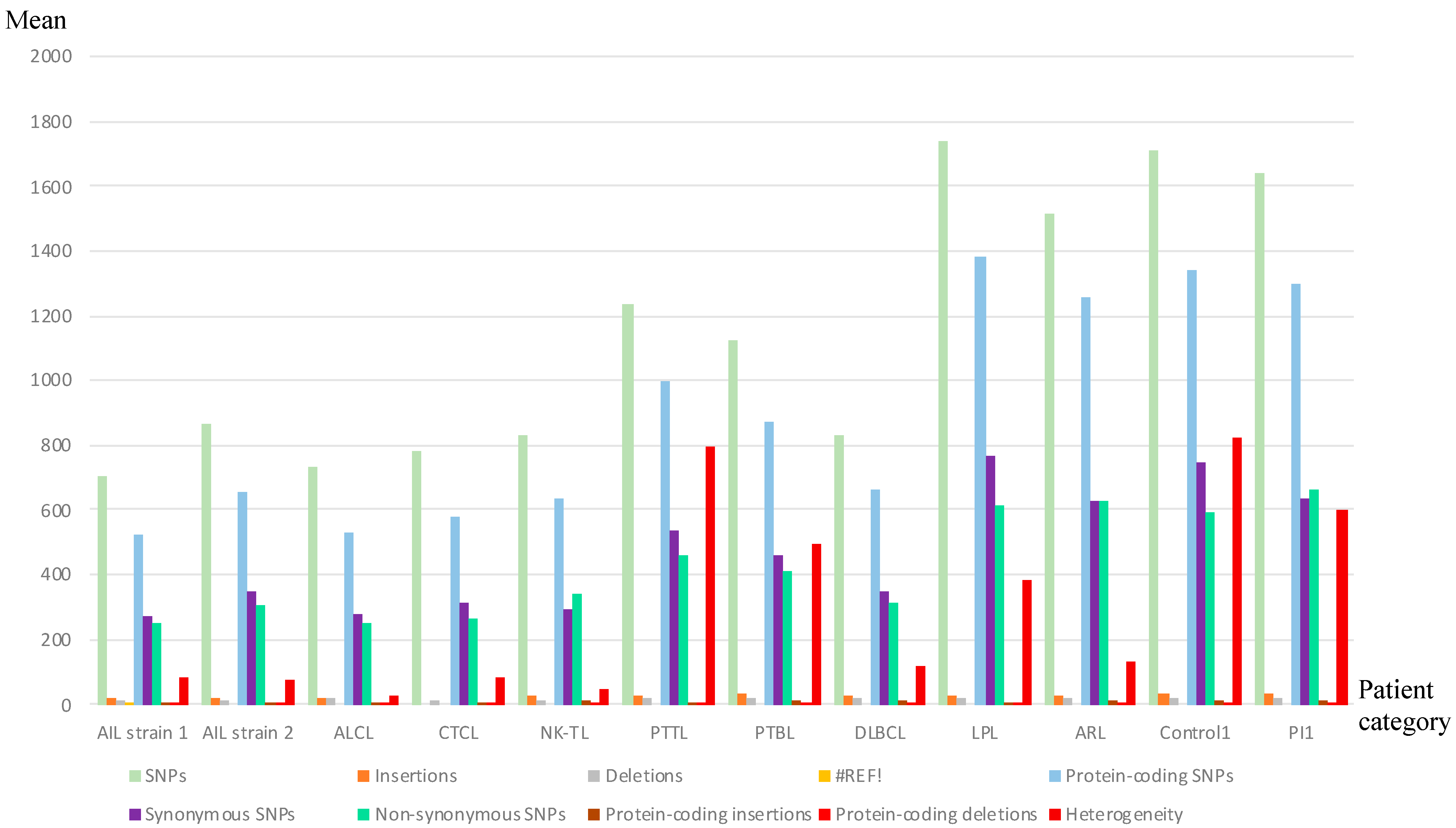
3.1. Analysis of Whole EBV Genome Sequences

The complete analysis concerned 36 patient samples (34 patients with lymphoproliferative disease, all being selected because of EBER RNA detection in their biopsies, one control with a reactive adenopathy without any EBV pathology, and one control with a primary infection) and seven cell lines. The patient description is shown in Table 1. To obtain complete EBV genome sequences, a target capture method was used because viral loads may be low and because the EBV genome shares several homologies with the human genome. For 10 samples, the viral load was <100,000 copies/μg DNA, and a very low depth was obtained by MiSeq sequencing. Therefore, these samples were sequenced using NextSeq sequencing. The use of two different approaches to analyze the results, namely reference mapping and de novo assembly, which are complementary, allowed us to obtain more complete and accurate whole EBV genomes. The overall results are reported in Table 2. The mean read number per sample was 10,889,606, with a mean depth of 4136. An average of 96% of the reads mapped to the EBV genome, and all the sequences were analyzed after removing the low-percentage reads mapping to the hg19 human genome. Using the de novo assembly, the genomes obtained on the MiSeq sequencer were assembled into contigs created by Viramp and aligned to the reference genome to generate scaffolds. Seventeen whole genome sequences, including eight AITL were determined and deposited in the NCBI GenBank (N° MH837512 to MH837528). The individual sample accession numbers are listed in Table 3. The coverage profile was similar for all samples and greater than 92%. The mean percent GC content was 58.99%. The Fastq reads obtained for the totality of the samples have been deposited in NCBI GeneBank (BioProject ID PRJNA505149).

3.2. Some AITL Strains Exhibited Similar Distribution Patterns of Mutations

The whole genome sequences obtained for the patients were mapped to the reference genomes NC_007605.1 for EBV-1 and NC_009334.1 for EBV-2. Given that the latent EBNA-2 and -3 genes are the most divergent genes between EBV-1 and -2, we used the ratio “number of reads mapping to EBV-1 EBNA-2, -3 genes/number of reads mapping to EBV-2 EBNA-2, -3 genes” to determine the EBV type of each sample (Table 2). All but one patient sample (AIL18, which belonged to a native North African woman) harbored EBV-1. The raw reads were then aligned to the EBV-1 reference genome, and genetic variations were detected using the Varscan tool (Figure 1 and Table S1). T Figure 2 illustrates the relative genetic variations among the genomes for the different strains compared to the reference. For the AITL patients, although no consensual sequence of the EBV genome was found, it is noticeable that AIL1/6/7/8/10/11/12/13/14/15/17 patients had very close strains. The same observation was true for the AIL3/4/5/9 patients. It is also noteworthy that two strains were particularly mutated: AIL2 and AIL16.

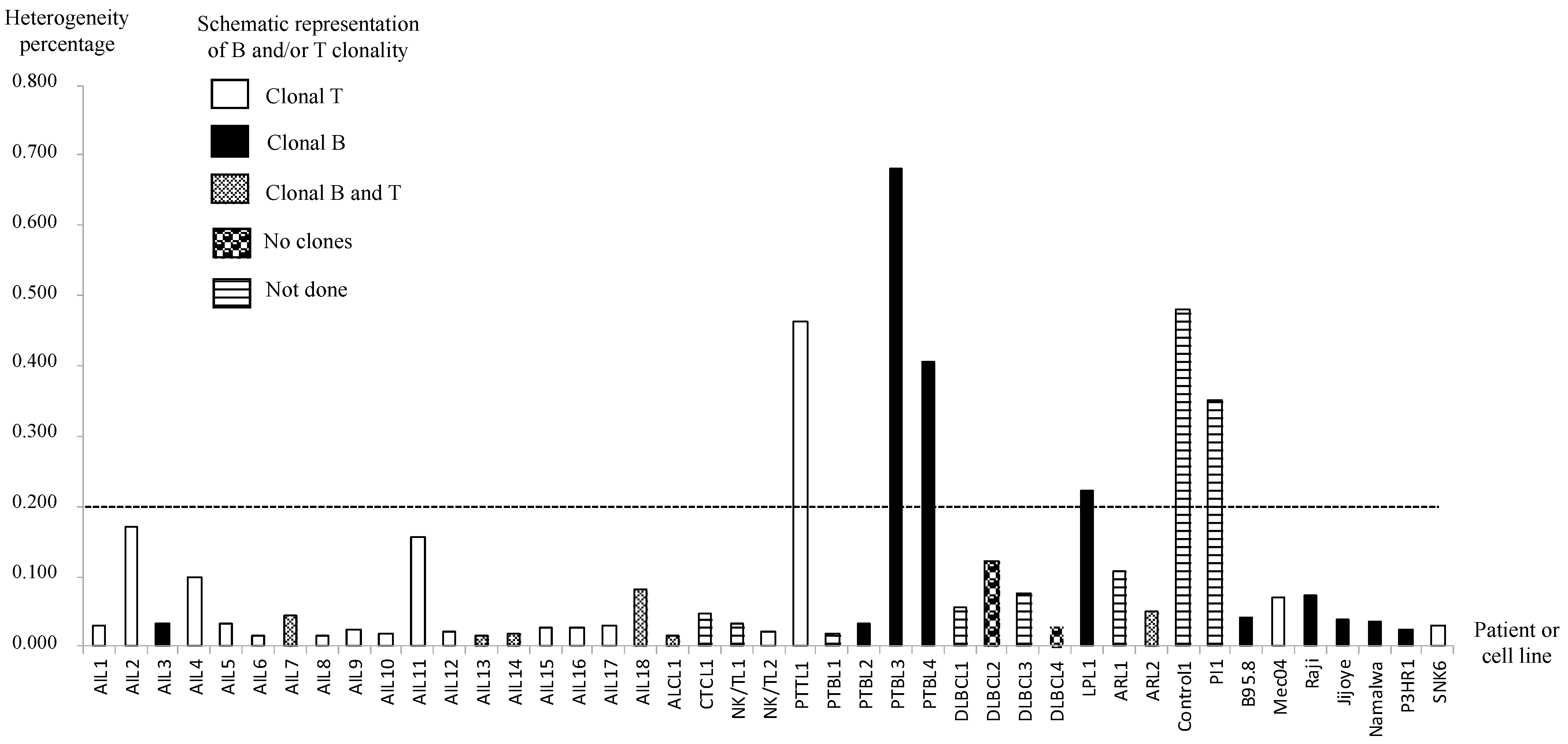
3.3. AITL Biopsies Revealed EBV in a Clonal Form

To determine whether the virus was clonal in biopsies, we calculated the heterogeneity, i.e., the number of heterogeneous positions, for each sample (Table S1). Because the sequence depth was high using deep sequencing, we considered a position as heterogeneous if the variant frequency was between 20 and 94%, as proposed by Kwok et al. [49]. A low level of heterogeneity supports the monoclonal origin of a strain, whereas high heterogeneity is due to the presence of various strains. Here, to determine clonality, we chose a cut-off of 0.2% heterogeneity, which is more stringent than the 0.5% cut-off chosen by Kwok et al. In this condition, Control1 and PI1 presented heterogeneous EBV strains, and the cell lines harbored monoclonal strains (Figure 3). Interestingly, all the AITL samples tested contained EBV in clonal form. Only four patient biopsies (PTTL1, PTBL3, PTBL4, and LPL1) showed heterogeneity in favor of a non-clonal virus. We then correlated these findings to the B and/or T clonality for each AITL sample and found that all but one sample (AIL3) had a T clonality (94.4%), while B clonality was found for five samples (27.7%) (Figure 3).

3.4. EBV Was Almost Always Latent in AITL Biopsies

In order to establish whether EBV was in a latent or a replicative state in the biopsies studied, we determined the viral load in each sample by quantitative PCR (qPCR), targeting the BMRF1 gene. The human albumin gene was also quantified in each sample using the same protocol in order to calculate the number of viruses per cell. Based on the publication of Hsieh et al. [50], a virus can be considered as latent if there are less than 20 copies per cell; a higher intracellular viral load is indicative of active replication. According to this calculation, only one AITL biopsy (AIL5) exhibited viral replication, while the others contained latent EBV (Table 4). Active replication was also found in other lymphoma samples (CTCL1, NK/TL2, PTBL1, DLBCL3, and ARL2).

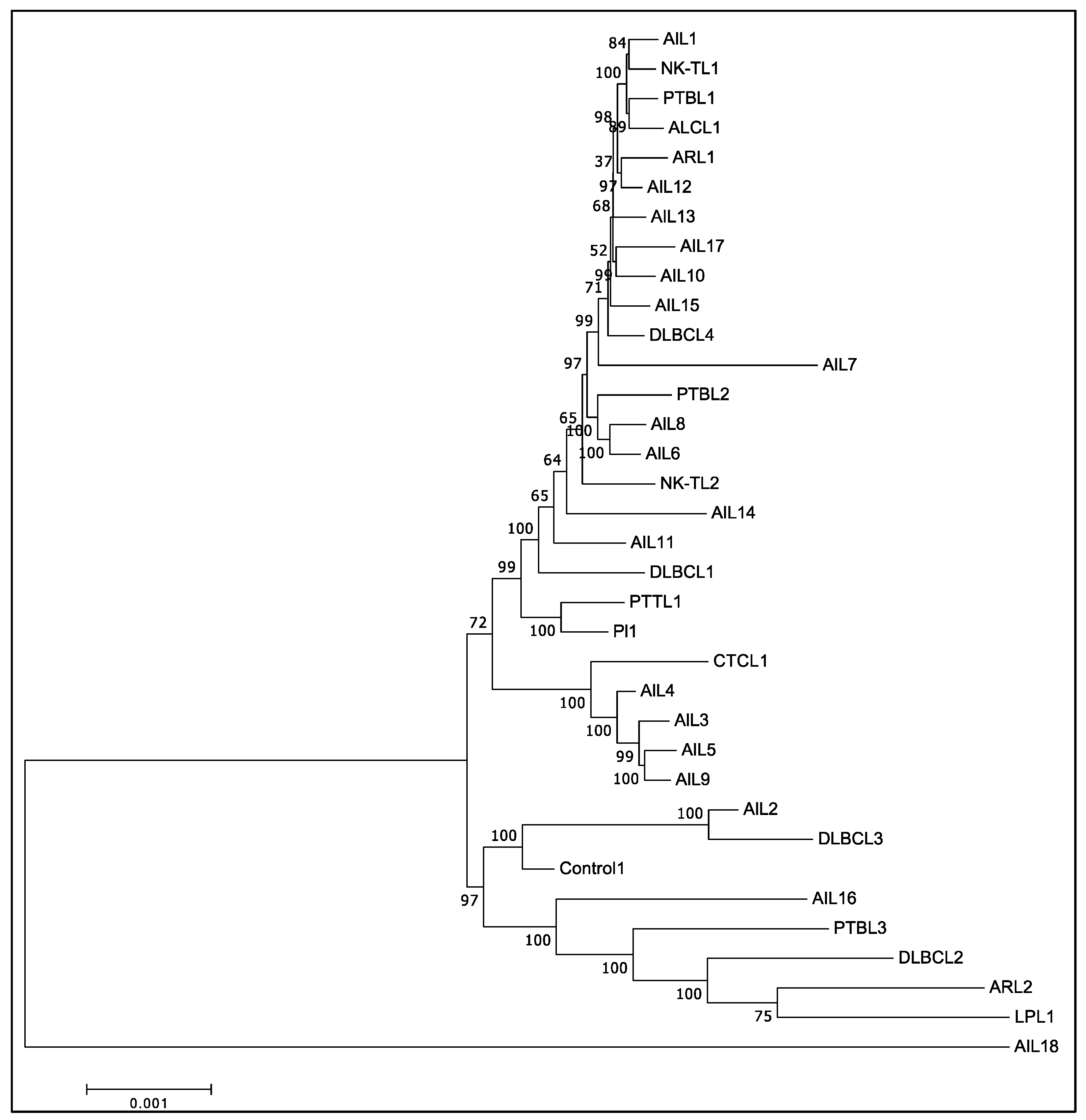
3.5. Phylogenic Analysis Confirmed the Existence of at Least Two Different Groups of EBV among AITL Biopsies

The phylogenic study was conducted for all the sequenced strains, based on the multiple nucleotide sequence alignment of whole genomes (Figure 4). Unsurprisingly, it clearly individualized AIL18, the only type 2 EBV of this series. Furthermore, the AIL2 and AIL16 strains were clearly different from the vast majority of the AITL strains, which were categorized into two different groups: strain 1 and strain 2. Strain 1 was found in patients AIL1/6/7/8/10/12/13/14/15/17 and strain 2 corresponded to AIL3/4/5 and -9 patients, whose proximity to each other and to CTCL1 was noticeable. AIL3/4/5/9 patients had an average survival of 2.4 years after their diagnosis, while it was 7.1 years for the AIL1/6/7/8/10/11/12/13/14/15/17 patients; this difference was significant, as demonstrated by the Kaplan-Meier curve (Table 1 and Figure 5). Overall, phylogenetic analysis showed that there was no EBV strain specific to AITL. Phylogenetic analysis was also conducted on each of the latency genes. Interestingly, the two groups already described were clearly individualized for the EBNA-3A, EBNA-3B, EBNA-3C, and LMP-2 genes (Figure 6). Figure S2 shows the phylogenetic trees obtained for the EBNA-1, EBNA-2, and EBNA-LP genes.

3.6. Mutations Occurred Mostly in Latency Genes and Secondarily in Tegument Genes

The means of the single nucleotide variations (SNVs) and the insertions and deletions (INDELs) were not higher in the AITL group compared to other lymphomas (respectively, 265 and 402 for SNVs and 5 and 7 for INDELs). The analysis of non-synonymous mutations according to nine main gene categories (namely latency, replication, membrane glycoprotein, tegument, capsid, transcription, metabolism, packaging, and unknown function) showed that the majority of changes were located in latency genes, though a large number of mutations also occurred in tegument genes (Figure 7). Conversely, capsid and transcription genes had the lowest number of variations. For each gene, we calculated the average number of non-synonymous mutations for the “AITL” group versus the “other lymphoma” group. The results showed a significant difference between these two groups and are reported in Table 5. In addition to the latency and tegument proteins, it can be noted that some replication proteins were more mutated in the AITL group, such as the BKRF3 protein for example. We also looked for variations in proteins implicated in the switch from latency to the lytic cycle, mainly Rta, a product of the BRLF1 gene, and Zta or ZEBRA, BZLF1 encoded protein. The patients AIL2/3/4/5/9/16 harbored the BRLF1 mutation S542N and, except for AIL2, the BZLF1 mutation A206S (Figure 8). These two mutations were positioned in CD8 epitope sites, while the other patients harbored BRLF1 A290D, V479I, and P486S. It is noteworthy that AIL2 exhibited nine BZLF1 mutations (not represented in Figure 8).

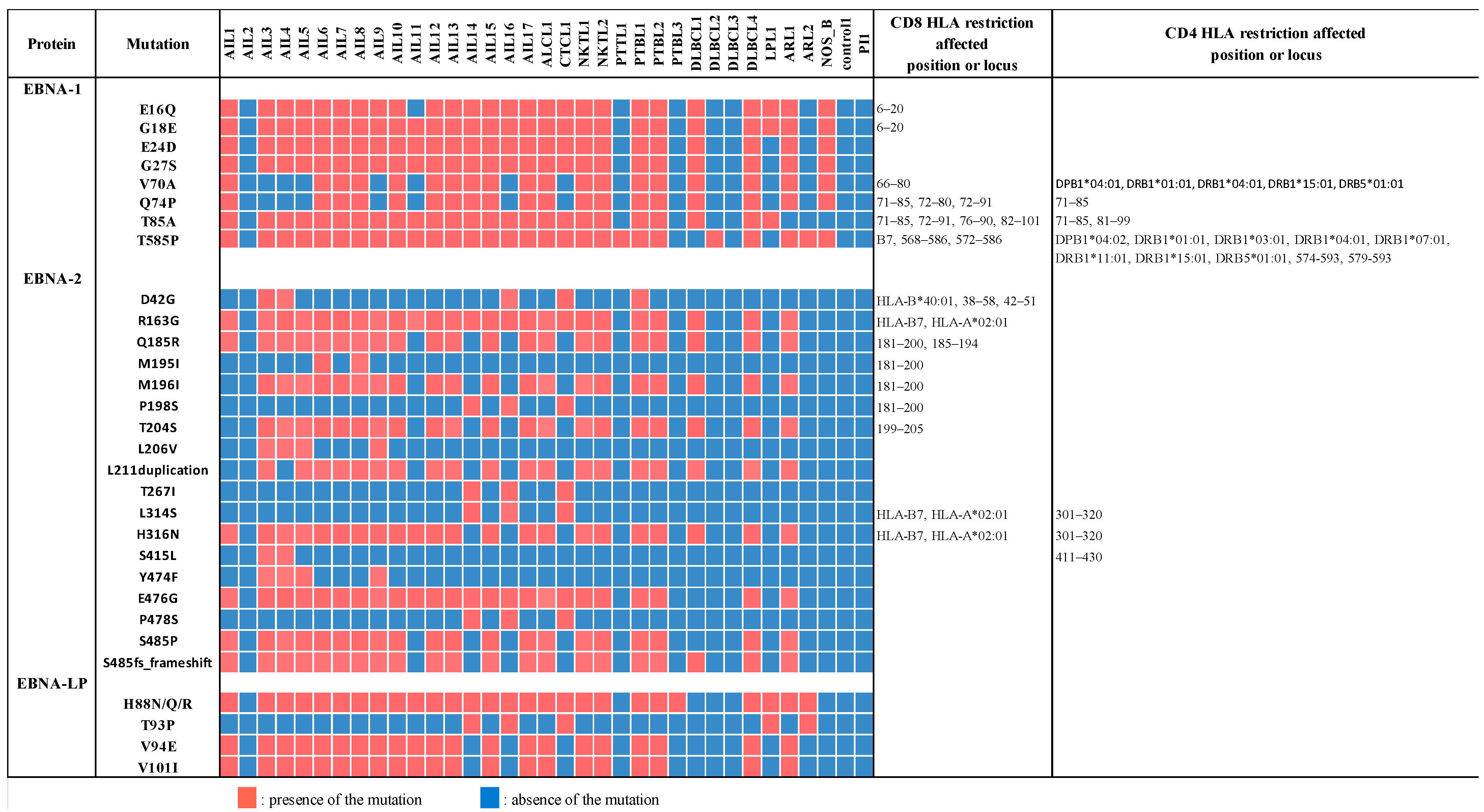
3.7. EBNA-1, EBNA-2, and Mainly EBNA-LP Genes Were the Most Mutated among Latency Genes

The majority of strains originating from the AITL biopsies contained four EBNA-1 mutations (E16Q, G18E, E24D, and G27S), located in the Gly-Arg domain (aa, amino acids, 8–67) and implicated in the EBNA-1-dependent DNA replication and partitioning of the EBV episomes in dividing cells (Figure 9). The region implicated in the other latency gene expression activation (aa 61–89) carried the mutation T85A present in almost all the strains derived from AITL and the mutations V70A and Q74P. Furthermore, the mutation T585P, which occurs in the dimerization domain of the protein, and which is located in important CD4 and CD8 recognition epitopes, was also present in all but one AITL strain. All AITL strains, except AIL2 and AIL11, presented a threonine at the signature codon 487 and therefore belonged to the P-thrV subtype, as described by Gutierrez at al. [51].
Among the three categories of EBNA-2 domains critical for its transcription regulation function, two are particularly mutated among the AITL strains: the self-association domain 3 (SAD3) and one of the nuclear localization signals (NLS). Indeed, SAD3 (aa 148–214), especially, carried the mutations R163G, Q185R, M196I, T204S, and the conservative duplication L211, which were present in a large number of AITL biopsies. Similarly, the highly represented mutations, H316N on one hand and E476G and P478S, as well as S485P or the S485 frameshift, on the other hand, were located, respectively, on NLS1 (aa 284–341) and NLS2 (aa 471–487). Some of these mutations affected the epitopes recognized mainly by CD8 cells.
EBNA-LP contained a variable number of 66 amino acid repeats corresponding to the W1 and W2 exons of IR1, followed by a C-terminal non-repetitive domain encoded by the exons Y1 and Y2. Conserved regions were determined in the C extremity (CR1 to CR3, implicated in EBNA-2 binding) and in the N-terminal region (CR4, CR5). Although one must be prudent with regard to results obtained for repeats with fragmentation sequencing, the sequences obtained here showed significantly more substitutions in AITL EBNA-LPs compared to the others. The mutations observed for the AITL biopsies (H88N/Q/R, V94E and V101I) were all localized at exon Y2 and concerned the majority of strains.
It is noticeable that for these three proteins the AIL2 strain had an identical profile to the PI or control1 strains without any of the described mutations.

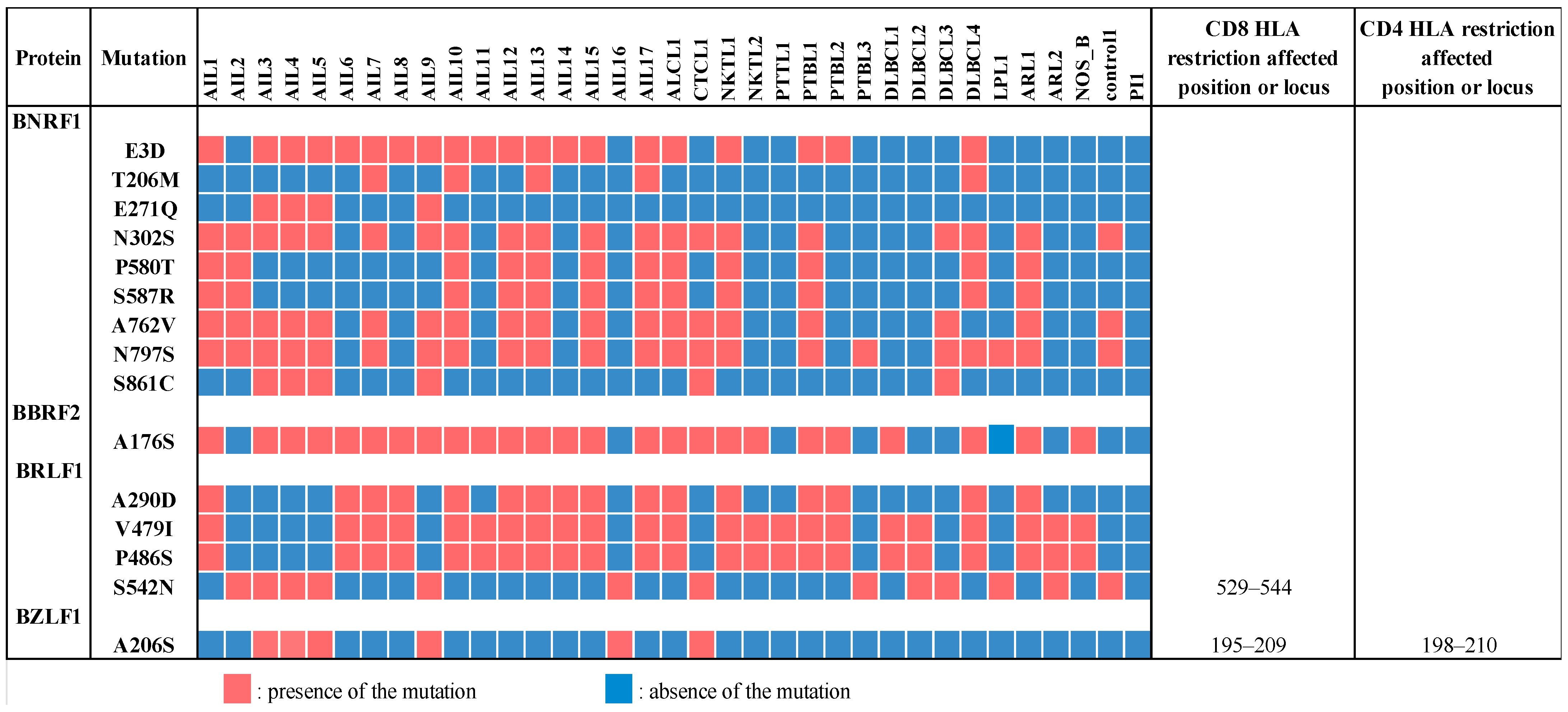
3.8. Two Tegument Genes, BNRF1 and BBRF2, Were Especially Mutated in AITL Biopsies

The major mutations concerning BNRF1 and BBRF2 are reported in Figure 8. The BBRF2 protein forms a hetero-complex with the tegument protein BSRF1, which mediates the viral envelopment [52]. For this protein, the AITL patients showed statistically more mutations than the other patients; A176S is particularly common in AILs.
The major tegument protein BNRF1 can bind to DAXX (death-domain associated protein-6) histone chaperone H3.3 and H4 to form a stable quaternary complex. BNRF1 also carries a PurM-like domain (aa 610–976) and a GATase domain (aa 1037–1318). Detected BNRF1 mutations were unevenly distributed according to the strains. For example, the two mutations, P580T and S587R, which occurred on the DAXX interaction domain (DID), aa 360–600, concerned seven AITL biopsies. Similarly, among the 3 mutations (A762V, N797S, and S861C) which affect the purM-like domain of the protein, 2 were found for 12 AITL biopsies. AIL3/4/5/9 exhibited a mutation profile different from the other strains. No mutations concerned the CD4 or CD8 epitopes.

4. Discussion

Although AITL is an uncommon, aggressive disease, it is one of the more frequent subtypes of PTCL in the western world. EBV is found in as high as 95% of cases [53], which highlights the close relationship between EBV and AITL. To date, however, whether EBV infection plays a role in AITL pathogenesis remains unclear, and diverse assumptions exist. For this reason, we decided to study the complete sequence of the EBV genome in AITL patients, compared to patients with other lymphomas.
Since EBV was discovered and characterized in BL [54], a B-cell lymphoma, it has been considered to be involved in other B-cell neoplasms. In these proliferations, EBV-encoded latent proteins have been shown to directly promote immortalization and proliferation through NF-kB pathway stimulation and increased anti-apoptotic gene expression. Concerning T cell lymphomas, and especially AITL, the role of EBV is probably much more complex and diverse. It has been shown that EBV is mainly present in AITL in B cells [32,55,56]; so, it is assumed that it plays an indirect role in the infected tumor cells through modification of the tumor microenvironment. In these conditions, it is essential to know whether the virus is latent or replicative as the proteins involved and the mechanisms of action are different. Unfortunately, for the biopsies we examined, there was not enough material to be able to determine which cells carried the virus. For these samples, with a particular interest in AITL biopsies, we explored the viral latent or replicative state, and we found that all but one AITL sample harbored only the latent virus (Table 4). To our knowledge, very few papers have examined whether the virus was latent in AITL. Unlike our results, Smith et al., arguing that EBV-positive cell numbers were greater than expected in reactive tissue and that infected cell nuclei were larger than average, reported an active replicative state of EBV [22]. Our method for determining whether the virus was latent [50] seems more accurate and is corroborated by the fact that small EBER RNAs were found in all the AITL cases. Interestingly, only one AITL patient (AIL5) had a high viral load, with an estimated copy number of 12,074 per cell, which is consistent with a replicative state of the virus. In this case, given the presence of EBERs, it can be assumed that some cells carry latent virus, while it is replicative in others. In addition to this work, we recently published a study of viral transcriptomes for seven of these patients (AIL2/3/7/11/14/15/16) and others, showing that the virus was in latency II and, more specifically, in latency IIc [57]. These results call into question the hypothesis that the virus could act in disease progression through cytokine and chemokine modulation. Indeed, in this hypothesis, the immunodeficient state resulting from the disease would lead to viral reactivation, thereby playing a role in the development of the tumor microenvironment [53]. It is more likely that EBV persists in a life-long latent state in infected cells and that this presence, in conjunction with other carcinogens, may promote the evolution to cancer [58].
For each sample, we then calculated the heterogeneity of the obtained sequence, namely the percentage of heterogeneous sites in relation to the genome size [59]. The variability threshold that we chose was 0.2%. This threshold, which is clearly below the threshold chosen by Kwok et al., was validated by the results obtained for the cell lines tested, which were considered as monoclonal controls, and for the primary infection sample and the inflammatory reactive biopsy, which were considered as polyclonal controls (Figure 3). The overall heterogeneity for AITL was between 0.015% (for AIL6 and AIL13) and 0.171% (for AIL2). The 18 AITL samples tested showed less than 0.2% variability, indicating that for these samples the heterogeneity was too low to be due to infection with various strains. As mentioned by others [49], rare mutations may spontaneously arise in viral genomes during clonal expansion. The low number of heterogeneous positions observed is consistent with this hypothesis, and the monoclonal origin of EBV can be attributed to it. This result, found for all AITL samples, makes it possible to consider a role for EBV in this pathology, which would here be more than a simple passenger, contrary to what is suggested by others [53,60]. As expected for this pathology, T lymphocytes were clonal in all but one sample, while B clonality was found in five samples. Unfortunately, due to a lack of material, we were unable to visualize in which cells the virus was present.
Overall, we did not observe any particular strain in the AITL biopsies. After alignment against the EBV-1 and EBV-2 reference strains, the overall results showed that only one patient (AIL18), a native of North Africa, had a type 2 EBV. The low number of EBV-2 in this series is not surprising given that the majority of the patients originate from France and, on the other hand, that EBV-2 is probably less pathogenic than EBV-1. Comparative analysis of all the mutations obtained for the AITL patients and the others does not reveal a strain characteristic of AITL (Figure 2). This figure shows that the mutation profiles obtained for the AIL1/6/7/8/10/11/12/13/14/15/17 strains are very close. Likewise, the mutation patterns of the AIL3/4/5/9 strains are quite similar; this is particularly visible in the EBNA-3A and -3B regions. It can also be noted that the strains of AIL16, and especially of AIL2, are particularly mutated. The phylogenic analysis carried out on entire sequences supports these different observations (Figure 4). Although many other factors are involved in disease progression, it is interesting to note that the AIL3/4/5/9/16 patients, whose viral strains are close, had an average survival of 2.1 years after their diagnosis, while it was 7.1 years for the AIL1/6/7/8/10/11/12/13/14/15/17 patients (Table 1). As demonstrated by the phylogeny and regarding the latency genes, the main differences between these two groups of strains relate to EBNA-3A, EBNA-3B, EBNA-3C, and LMP-2 (Figure 6). For the AIL2/3/4/5/9/16 strains, we showed that there was an S542N mutation on the BRLF1 gene encoding the Rta viral transcription factor. This mutation is positioned on the transactivation domain of the protein. This has also been described by Farrell P.J. (unpublished data) in T-cell disease in Paris. Similarly, the AIL3/4/5/9/16 strains carry the A206S mutation on the BZLF1 gene, a mutation already reported by other authors [61,62], located on the dimerization domain of the viral transcription factor Zta. These two mutations (BRLF1 S542N and BZLF1 A206S) affecting important domains can cause a change in viral behavior. Note that the other strains featured the mutations V479I, P486S on the activation accessory domain and A290D on the DNA-binding domain of BRLF1. These mutations have already been reported [62,63].
Our initial idea that there may be a viral strain specific to AITL led us to analyze the main mutations found on viral genomes. Analysis of genome variation locations showed that the coding regions constituted more than 70% of the mutations, while the EBER and microRNA regions were conserved. Consistent with previous reports [8,59,64], we observed a higher frequency of non-synonymous SNPs in latent genes (Figure 7), followed by the genes encoding tegument proteins. Although the mutation mean was not higher for AITL than for the other lymphomas, the calculation, for each gene, of the number of non-synonymous mutations for the AITL group versus that number for the other lymphomas, showed a significant difference for some genes, especially the EBNA-1, EBNA-2, and EBNA-LP latency genes and the tegument BBRF-2 and BNRF-1 genes (Table 5).
EBNA-1 is very interesting to consider because it is the only EBV antigen consistently expressed in EBV-associated malignancies [1], expressed in all forms of latency, and because of its major function in viral episome maintenance in latently infected cells. In this regard, it is intriguing to see that most AITL strains (15/17) carry four substitutions on the Gly-Arg domain aa 8–67, which is mainly involved in the replication and partitioning of the EBV episomes in the two dividing cells, which are thus able to modify these functions (Figure 9). Apart from these mutations, the T85A substitution, already described by Borozan et al. [62] and found in all our AITLs except AIL2, could play an important role in proliferation because it is positioned in a region involved in the transcriptional activation of other latency genes. The V70A and Q74P mutations present in 10 AITL, which are not described to our knowledge, may contribute to this effect, being positioned in the same region. Finally, the T585P mutation was found in all AITL except AIL2. Recently, the DNA-binding domain of EBNA-1 has been described to be in the form of an oligomeric hexameric ring, the oligomeric interface pivoting around residue T585 [65]. T585 is a position subject to substitutions. Mutations occurring on this residue had effects on EBNA-1-dependent DNA replication and episome maintenance [66]. Polymorphism at this position is often found in NPC tumors and Burkitt’s lymphoma [65]. We compared the sequences obtained for our patients to the sequences obtained for the four LCLs (DPL, KREB2, CoAN, and MLEB2) that we established from non-hematological subjects [57]. Among the different interesting mutations found in our present patients and concerning different genes, only EBNA-1 T85A and EBNA-1 T585P were found in LCL. These mutations seem to be widespread in the French population. It is noteworthy that the changes observed in the EBNA-1 sequences fell principally within the known T-cell epitopes: all mutations but E24D and G27S are located in CD8 epitopes and V70A, Q74P, T85A, and T585P are positioned in CD4 epitopes. These results imply that immune pressure could be in part responsible for these changes. Based on the amino acid present at EBNA-1 position 487, EBV has been classified into five subtypes: P-ala (wild type B95-8 subtype), P-thr, V-leu, V-val, and V-pro [51,67]. Considering these five subtypes, it is noteworthy that 15 among the 17 AITL EBV-1 positive patients harbored a P-thr subtype, which was also the most prevalent in the other samples. V-val is reported to be dominant in Asian regions, while the P-thr subtype is most commonly observed in the peripheral blood of European and Australian subjects, as well as in African tumors [63].
The latent protein EBNA-2 acts mainly as a transcription factor and therefore comprises the transactivation domains (TAD), auto-association domains (SAD), and nuclear location signals (NLS) important for its function. The main mutations affecting EBNA-2 in the AITL group impact the SAD3 domain or the NLS domains and are thus able to modify its regulatory function. Many of these mutations, particularly the E476G mutation, present among others in the E2A subtype [68], have been described in the French as well as English or Eurasian, American [62], or Asian [69] populations. To our knowledge, only the H316N mutation located on the NLS1 domain and the R163G positioned on the SAD3 domain have not been previously described, although position 163 was found to be very frequently mutated (R163M substitution) by Wang et al. [68]. These two mutations are highly represented in AIL.
EBNA-LP is a latent protein, acting principally as an EBNA-2 coactivator and therefore has an important role in B-cell immortalization. It is notable that this was the most significantly mutated protein in the AITL group compared to the other patients. The main substitutions observed for our patients (H88N/Q/R, T93P, V94E, and V101I) were grouped and located at the C-terminal extremity, between CR4 and CR5. Most published viral sequences do not carry these mutations, apart from V94E and V101I, which were described by Ba Abdullah et al. to characterize subgroup C [70].
Two tegument proteins were particularly more mutated in AITL patients than in other patients: BBRF2 and BNRF1 (Figure 8). Recently, it was shown that BBRF2 can associate with BSRF1, another tegument protein, and the heterocomplex formed has a role in the binding of EBV nucleocapsids to the Golgi membrane during secondary envelopment. Therefore, BBRF2 appears to play an important role in viral infectivity [71]. The main mutation found (A176S) is highly represented in AIL compared to other pathologies. However, it has already been described, including in healthy subjects [4,61].
The major tegument protein BNRF1, through its binding to the antiviral DAXX histone chaperone, H3.3 and H4, is implicated in the establishment of latency and cell immortalization [72]. The stable quaternary complex formed is responsible for its localization to the PML-nuclear bodies involved in the antiviral intrinsic resistance and the transcriptional repression of host cells [73]. The mutations that we have highlighted in AITL only concerned a subgroup of patients. Most of these mutations have been previously described [4,62]. However, some of them could alter the behavior of the protein, such as those located on DID, namely P580T and S587R, or those affecting the purM-like domain of the protein (A762V, N797S, and S861C).
Interestingly, BKRF3, a viral uracil-DNA glycosylase, participates in the repair of viral DNA and prevents its mutagenesis. Obviously, for this protein, the mutations that severely affect viral replication cannot be retained. In this study, E128D substitution was present for all AITL patients except AIL2. The presence of this mutation could modify the virus–cell relationships.
In the end, it is obvious that there is probably no specific viral mutation for AIL. However, on the other hand, it is possible that a combination of mutations affecting several genes, even if they have already been listed, could modify the behavior of the virus. With this in mind, it would be interesting to study how the virus found in AIL3/4/5/9 behaves within the cell.

5. Conclusions

In summary, the whole viral sequences obtained for the 18 AITL patients and compared to other patients identified a poorly represented strain within the AITLs, but which seemed to be associated with poor outcome. Furthermore, we demonstrated that the virus was clonal and latent in all the AITL biopsies analyzed. These various elements suggest that the virus is involved in this pathology.

Supplementary Materials

The following supporting information can be downloaded at: https://www.mdpi.com/article/10.3390/cancers14122899/s1, Figure S1: Results obtained for EBER ISH staining; Figure S2: Phylogenetic tree obtained after nucleotide sequence alignment of the different strains; Table S1: Genetic Variations and Heterogeneity Determination for All Samples.

Author Contributions

Conceptualization, S.R.-R.; data curation, F.B., A.C. and A.J.; formal analysis, R.B. and F.B.; funding acquisition, S.R.-R.; investigation, R.B. and M.A.H.; methodology, S.R.-R.; project administration, S.R.-R.; software, F.B.; supervision, J.F. and S.R.-R.; writing—original draft, R.B. and S.R.-R.; writing—review and editing, S.R.-R. All authors have read and agreed to the published version of the manuscript.

Funding

This work was supported by grants provided by the “Ligue contre le Cancer (Comité Haute-Vienne, Comité Corrèze and Comité Creuse)” and by grants provided by the National Health Service (Direction Générale de la Santé, Paris). It was promoted by the “Centre Hospitalier Universitaire”, Limoges, France.

Institutional Review Board Statement

For this study, biological samples from the biological collection of the Biological Resource Center of the Limoges University Hospital (CRBioLim) were used, declaration number of DC-2010-1074. The medical-scientific council gave its approval on 26 July 2016 under the transfer number CRB-103.

Informed Consent Statement

Informed consent was obtained from all subjects involved in the study.

Data Availability Statement

The seventeen generated whole genome sequences were deposited in the NCBI GenBank (N° MH837512 to MH837528). The totality of the Fastq reads obtained have been deposited in NCBI GeneBank (BioProject ID PRJNA505149).

Acknowledgments

We thank Manuella Delage-Corre from the Molecular Oncology Platform, Limoges for pathological diagnosis and for the in situ hybridization interpretation; the ICM platform, Paris, and especially Yannick Marie, for sequencing advice; Camille François and Stéphane Fénart (Roche) for advice on the capture technique; Philippe Vignoles from the Department of Mathematics and Statistics of the Faculty of Pharmacy for his assistance in the analysis of patient survival (Kaplan–Meier curve); Jeanne Cook-Moreau, from the Department of Microbiology of the Faculty of Pharmacy for her help in proofreading the English; Valentin Tilloy, from the Department of Bioinformatics of the CBRS, for his expertise in sequence analysis.

Conflicts of Interest

The authors declare no conflict of interest.

References

  1. Young, L.S.; Murray, P.G. Epstein-barr virus and oncogenesis: From latent genes to tumours. Oncogene 2003, 22, 5108–5121. [Google Scholar] [CrossRef] [PubMed] [Green Version]
  2. McGeoch, D.J.; Gatherer, D. Lineage structures in the genome sequences of three epstein-barr virus strains. Virology 2007, 359, 1–5. [Google Scholar] [CrossRef] [PubMed]
  3. Romero-Masters, J.C.; Huebner, S.M.; Ohashi, M.; Bristol, J.A.; Benner, B.E.; Barlow, E.A.; Turk, G.L.; Nelson, S.E.; Baiu, D.C.; Van Sciver, N.; et al. B Cells infected with type 2 epstein-barr virus (EBV) have increased NFATc1/NFATc2 activity and enhanced lytic gene expression in comparison to type 1 EBV infection. PLoS Pathog. 2020, 16, e1008365. [Google Scholar] [CrossRef] [PubMed] [Green Version]
  4. Correia, S.; Bridges, R.; Wegner, F.; Venturini, C.; Palser, A.; Middeldorp, J.M.; Cohen, J.I.; Lorenzetti, M.A.; Bassano, I.; White, R.E.; et al. Sequence variation of epstein-barr virus: Viral types, geography, codon usage, and diseases. J. Virol. 2018, 92, e01132-18. [Google Scholar] [CrossRef] [Green Version]
  5. Hämmerl, L.; Colombet, M.; Rochford, R.; Ogwang, D.M.; Parkin, D.M. The burden of burkitt lymphoma in Africa. Infect. Agents Cancer 2019, 14, 17. [Google Scholar] [CrossRef] [Green Version]
  6. Mahdavifar, N.; Ghoncheh, M.; Mohammadian-Hafshejani, A.; Khosravi, B.; Salehiniya, H. Epidemiology and inequality in the incidence and mortality of nasopharynx cancer in Asia. Osong Public Health Res. Perspect. 2016, 7, 360–372. [Google Scholar] [CrossRef] [Green Version]
  7. Kutok, J.L.; Wang, F. Spectrum of epstein-barr virus-associated diseases. Annu. Rev. Pathol. 2006, 1, 375–404. [Google Scholar] [CrossRef]
  8. Palser, A.L.; Grayson, N.E.; White, R.E.; Corton, C.; Correia, S.; Ba Abdullah, M.M.; Watson, S.J.; Cotten, M.; Arrand, J.R.; Murray, P.G.; et al. Genome diversity of epstein-barr virus from multiple tumor types and normal infection. J. Virol. 2015, 89, 5222–5237. [Google Scholar] [CrossRef] [Green Version]
  9. Chang, C.M.; Yu, K.J.; Mbulaiteye, S.M.; Hildesheim, A.; Bhatia, K. The extent of genetic diversity of epstein-barr virus and its geographic and disease patterns: A need for reappraisal. Virus Res. 2009, 143, 209–221. [Google Scholar] [CrossRef] [Green Version]
  10. Tsai, M.-H.; Lin, X.; Shumilov, A.; Bernhardt, K.; Feederle, R.; Poirey, R.; Kopp-Schneider, A.; Pereira, B.; Almeida, R.; Delecluse, H.-J. The biological properties of different epstein-barr virus strains explain their association with various types of cancers. Oncotarget 2017, 8, 10238–10254. [Google Scholar] [CrossRef] [Green Version]
  11. Hui, K.F.; Chan, T.F.; Yang, W.; Shen, J.J.; Lam, K.P.; Kwok, H.; Sham, P.C.; Tsao, S.W.; Kwong, D.L.; Lung, M.L.; et al. High risk epstein-barr virus variants characterized by distinct polymorphisms in the EBER locus are strongly associated with nasopharyngeal carcinoma. Int. J. Cancer 2019, 144, 3031–3042. [Google Scholar] [CrossRef] [PubMed]
  12. Kim, H.; Burassakarn, A.; Kang, Y.; Iizasa, H.; Yoshiyama, H. A single nucleotide polymorphism in the BART promoter region of epstein-barr virus isolated from nasopharyngeal cancer cells. Biochem. Biophys. Res. Commun. 2019, 520, 373–378. [Google Scholar] [CrossRef] [PubMed]
  13. Xu, M.; Yao, Y.; Chen, H.; Zhang, S.; Cao, S.-M.; Zhang, Z.; Luo, B.; Liu, Z.; Li, Z.; Xiang, T.; et al. Genome sequencing analysis identifies epstein-barr virus subtypes associated with high risk of nasopharyngeal carcinoma. Nat. Genet. 2019, 51, 1131–1136. [Google Scholar] [CrossRef] [PubMed]
  14. Swerdlow, S.H.; Campo, E.; Pileri, S.A.; Harris, N.L.; Stein, H.; Siebert, R.; Advani, R.; Ghielmini, M.; Salles, G.A.; Zelenetz, A.D.; et al. The 2016 revision of the world health organization classification of lymphoid neoplasms. Blood 2016, 127, 2375–2390. [Google Scholar] [CrossRef] [Green Version]
  15. Vose, J.; Armitage, J.; Weisenburger, D. International T-cell lymphoma project international peripheral T-cell and natural killer/T-cell lymphoma study: Pathology findings and clinical outcomes. J. Clin. Oncol. 2008, 26, 4124–4130. [Google Scholar] [CrossRef]
  16. Federico, M.; Rudiger, T.; Bellei, M.; Nathwani, B.N.; Luminari, S.; Coiffier, B.; Harris, N.L.; Jaffe, E.S.; Pileri, S.A.; Savage, K.J.; et al. Clinicopathologic characteristics of angioimmunoblastic T-cell lymphoma: Analysis of the international peripheral T-cell lymphoma project. J. Clin. Oncol. 2013, 31, 240–246. [Google Scholar] [CrossRef] [Green Version]
  17. Lunning, M.A.; Vose, J.M. Angioimmunoblastic T-cell lymphoma: The many-faced lymphoma. Blood 2017, 129, 1095–1102. [Google Scholar] [CrossRef]
  18. Mourad, N.; Mounier, N.; Brière, J.; Raffoux, E.; Delmer, A.; Feller, A.; Meijer, C.J.L.M.; Emile, J.-F.; Bouabdallah, R.; Bosly, A.; et al. Clinical, biologic, and pathologic features in 157 patients with angioimmunoblastic T-cell lymphoma treated within the groupe d’etude des lymphomes de l’adulte (GELA) trials. Blood 2008, 111, 4463–4470. [Google Scholar] [CrossRef]
  19. Piccaluga, P.P.; Agostinelli, C.; Califano, A.; Carbone, A.; Fantoni, L.; Ferrari, S.; Gazzola, A.; Gloghini, A.; Righi, S.; Rossi, M.; et al. Gene expression analysis of angioimmunoblastic lymphoma indicates derivation from T follicular helper cells and vascular endothelial growth factor deregulation. Cancer Res. 2007, 67, 10703–10710. [Google Scholar] [CrossRef] [Green Version]
  20. De Leval, L.; Rickman, D.S.; Thielen, C.; de Reynies, A.; Huang, Y.-L.; Delsol, G.; Lamant, L.; Leroy, K.; Brière, J.; Molina, T.; et al. The gene expression profile of nodal peripheral t-cell lymphoma demonstrates a molecular link between angioimmunoblastic T-cell lymphoma (AITL) and follicular helper T (TFH) cells. Blood 2007, 109, 4952–4963. [Google Scholar] [CrossRef]
  21. Tan, B.T.; Warnke, R.A.; Arber, D.A. The frequency of B- and T-cell gene rearrangements and epstein-barr virus in T-cell lymphomas: A comparison between angioimmunoblastic T-cell lymphoma and peripheral T-cell lymphoma, unspecified with and without associated B-cell proliferations. J. Mol. Diagn. 2006, 8, 466–475. [Google Scholar] [CrossRef] [PubMed] [Green Version]
  22. Smith, J.L.; Hodges, E.; Quin, C.T.; McCarthy, K.P.; Wright, D.H. Frequent T and B Cell oligoclones in histologically and immunophenotypically characterized angioimmunoblastic lymphadenopathy. Am. J. Pathol. 2000, 156, 661–669. [Google Scholar] [CrossRef] [Green Version]
  23. Nguyen, P.N.; Tran, N.T.B.; Nguyen, T.P.X.; Ngo, T.N.M.; Lai, D.V.; Deel, C.D.; Hassell, L.A.; Vuong, H.G. Clinicopathological implications of RHOA mutations in angioimmunoblastic T-cell lymphoma: A meta-analysis. Clin. Lymphoma Myeloma Leuk. 2021, 21, 431–438. [Google Scholar] [CrossRef]
  24. Willemsen, M.; Abdul Hamid, M.; Winkens, B.; Zur Hausen, A. Mutational heterogeneity of angioimmunoblastic T-cell lymphoma indicates distinct lymphomagenic pathways. Blood Cancer J. 2018, 8, 6. [Google Scholar] [CrossRef]
  25. Timmins, M.A.; Wagner, S.D.; Ahearne, M.J. The new biology of PTCL-NOS and AITL: Current status and future clinical impact. Br. J. Haematol. 2020, 189, 54–66. [Google Scholar] [CrossRef] [PubMed]
  26. Tari, G.; Lemonnier, F.; Morschhauser, F. Epigenetic focus on angioimmunoblastic T-cell lymphoma: Pathogenesis and treatment. Curr. Opin. Oncol. 2021, 33, 400–405. [Google Scholar] [CrossRef] [PubMed]
  27. Lemonnier, F.; Gaulard, P.; de Leval, L. New insights in the pathogenesis of T-cell lymphomas. Curr. Opin. Oncol. 2018, 30, 277–284. [Google Scholar] [CrossRef] [PubMed] [Green Version]
  28. Donner, I.; Katainen, R.; Kaasinen, E.; Aavikko, M.; Sipilä, L.J.; Pukkala, E.; Aaltonen, L.A. Candidate susceptibility variants in angioimmunoblastic T-cell lymphoma. Fam. Cancer 2019, 18, 113–119. [Google Scholar] [CrossRef] [Green Version]
  29. Xu, B.; Liu, P. No survival improvement for patients with angioimmunoblastic T-cell lymphoma over the past two decades: A population-based study of 1207 cases. PLoS ONE 2014, 9, e92585. [Google Scholar] [CrossRef] [Green Version]
  30. Anagnostopoulos, I.; Hummel, M.; Finn, T.; Tiemann, M.; Korbjuhn, P.; Dimmler, C.; Gatter, K.; Dallenbach, F.; Parwaresch, M.R.; Stein, H. Heterogeneous epstein-barr virus infection patterns in peripheral T-cell lymphoma of angioimmunoblastic lymphadenopathy type. Blood 1992, 80, 1804–1812. [Google Scholar] [CrossRef] [Green Version]
  31. Willenbrock, K.; Bräuninger, A.; Hansmann, M.-L. Frequent occurrence of B-cell lymphomas in angioimmunoblastic T-cell lymphoma and proliferation of epstein-barr virus-infected cells in early cases. Br. J. Haematol. 2007, 138, 733–739. [Google Scholar] [CrossRef] [PubMed]
  32. Beer, T.; Dorion, P. Angioimmunoblastic T-cell lymphoma presenting with an acute serologic epstein-barr virus profile. Hematol. Rep. 2015, 7, 5893. [Google Scholar] [CrossRef] [Green Version]
  33. Zettl, A.; Lee, S.-S.; Rüdiger, T.; Starostik, P.; Marino, M.; Kirchner, T.; Ott, M.; Müller-Hermelink, H.K.; Ott, G. Epstein-barr virus-associated B-cell lymphoproliferative disorders in angloimmunoblastic T-cell lymphoma and peripheral T-cell lymphoma, unspecified. Am. J. Clin. Pathol. 2002, 117, 368–379. [Google Scholar] [CrossRef] [PubMed]
  34. Xu, Y.; McKenna, R.W.; Hoang, M.P.; Collins, R.H.; Kroft, S.H. Composite angioimmunoblastic T-cell lymphoma and diffuse large B-cell lymphoma: A case report and review of the literature. Am. J. Clin. Pathol. 2002, 118, 848–854. [Google Scholar] [CrossRef] [PubMed] [Green Version]
  35. Quintanilla-Martinez, L.; Fend, F.; Moguel, L.R.; Spilove, L.; Beaty, M.W.; Kingma, D.W.; Raffeld, M.; Jaffe, E.S. Peripheral T-cell lymphoma with reed-sternberg-like cells of B-cell phenotype and genotype associated with epstein-barr virus infection. Am. J. Surg. Pathol. 1999, 23, 1233–1240. [Google Scholar] [CrossRef]
  36. Dunleavy, K.; Wilson, W.H. Angioimmunoblastic T-cell lymphoma: Immune modulation as a therapeutic strategy. Leuk. Lymphoma 2007, 48, 449–451. [Google Scholar] [CrossRef]
  37. Van Dongen, J.J.M.; Langerak, A.W.; Brüggemann, M.; Evans, P.A.S.; Hummel, M.; Lavender, F.L.; Delabesse, E.; Davi, F.; Schuuring, E.; García-Sanz, R.; et al. Design and standardization of PCR primers and protocols for detection of clonal immunoglobulin and T-cell receptor gene recombinations in suspect lymphoproliferations: Report of the BIOMED-2 concerted action BMH4-CT98-3936. Leukemia 2003, 17, 2257–2317. [Google Scholar] [CrossRef] [Green Version]
  38. Krzywinski, M.; Schein, J.; Birol, I.; Connors, J.; Gascoyne, R.; Horsman, D.; Jones, S.J.; Marra, M.A. Circos: An information aesthetic for comparative genomics. Genome Res. 2009, 19, 1639–1645. [Google Scholar] [CrossRef] [Green Version]
  39. Altschul, S.F.; Gish, W.; Miller, W.; Myers, E.W.; Lipman, D.J. Basic local alignment search tool. J. Mol. Biol. 1990, 215, 403–410. [Google Scholar] [CrossRef]
  40. Bolger, A.M.; Lohse, M.; Usadel, B. Trimmomatic: A flexible trimmer for illumina sequence data. Bioinformatics 2014, 30, 2114–2120. [Google Scholar] [CrossRef] [Green Version]
  41. Langmead, B.; Salzberg, S.L. Fast gapped-read alignment with bowtie 2. Nat. Methods 2012, 9, 357–359. [Google Scholar] [CrossRef] [PubMed] [Green Version]
  42. Li, H.; Handsaker, B.; Wysoker, A.; Fennell, T.; Ruan, J.; Homer, N.; Marth, G.; Abecasis, G.; Durbin, R. 1000 genome project data processing subgroup the sequence alignment/map format and SAMtools. Bioinformatics 2009, 25, 2078–2079. [Google Scholar] [CrossRef] [PubMed] [Green Version]
  43. Ramírez, F.; Ryan, D.P.; Grüning, B.; Bhardwaj, V.; Kilpert, F.; Richter, A.S.; Heyne, S.; Dündar, F.; Manke, T. DeepTools2: A next generation web server for deep-sequencing data analysis. Nucleic Acids Res. 2016, 44, W160–W165. [Google Scholar] [CrossRef] [PubMed]
  44. Koboldt, D.C.; Zhang, Q.; Larson, D.E.; Shen, D.; McLellan, M.D.; Lin, L.; Miller, C.A.; Mardis, E.R.; Ding, L.; Wilson, R.K. VarScan 2: Somatic mutation and copy number alteration discovery in cancer by exome sequencing. Genome Res. 2012, 22, 568–576. [Google Scholar] [CrossRef] [PubMed] [Green Version]
  45. Robinson, J.T.; Thorvaldsdóttir, H.; Winckler, W.; Guttman, M.; Lander, E.S.; Getz, G.; Mesirov, J.P. Integrative genomics viewer. Nat. Biotechnol. 2011, 29, 24–26. [Google Scholar] [CrossRef] [Green Version]
  46. Cingolani, P.; Platts, A.; Wang, L.L.; Coon, M.; Nguyen, T.; Wang, L.; Land, S.J.; Lu, X.; Ruden, D.M. A program for annotating and predicting the effects of single nucleotide polymorphisms, snpeff: SNPs in the genome of drosophila melanogaster strain W1118; Iso-2; Iso-3. Fly 2012, 6, 80–92. [Google Scholar] [CrossRef] [Green Version]
  47. Tarbouriech, N.; Buisson, M.; Géoui, T.; Daenke, S.; Cusack, S.; Burmeister, W.P. Structural genomics of the epstein-barr virus. Acta Crystallogr. D Biol. Crystallogr. 2006, 62, 1276–1285. [Google Scholar] [CrossRef] [Green Version]
  48. Katoh, K.; Standley, D.M. MAFFT multiple sequence alignment software version 7: Improvements in performance and usability. Mol. Biol. Evol. 2013, 30, 772–780. [Google Scholar] [CrossRef] [Green Version]
  49. Kwok, H.; Tong, A.H.Y.; Lin, C.H.; Lok, S.; Farrell, P.J.; Kwong, D.L.W.; Chiang, A.K.S. Genomic sequencing and comparative analysis of epstein-barr virus genome isolated from primary nasopharyngeal carcinoma biopsy. PLoS ONE 2012, 7, e36939. [Google Scholar] [CrossRef] [Green Version]
  50. Hsieh, P.-P.; Tung, C.-L.; Chan, A.B.-W.; Liao, J.-B.; Wang, J.-S.; Tseng, H.-H.; Su, H.-H.; Chang, K.-C.; Chang, C.-C. EBV viral load in tumor tissue is an important prognostic indicator for nasal NK/T-cell lymphoma. Am. J. Clin. Pathol. 2007, 128, 579–584. [Google Scholar] [CrossRef]
  51. Gutiérrez, M.I.; Raj, A.; Spangler, G.; Sharma, A.; Hussain, A.; Judde, J.G.; Tsao, S.W.; Yuen, P.W.; Joab, I.; Magrath, I.T.; et al. Sequence variations in EBNA-1 may dictate restriction of tissue distribution of epstein-barr virus in normal and tumour cells. J. Gen. Virol. 1997, 78 Pt 7, 1663–1670. [Google Scholar] [CrossRef] [PubMed]
  52. He, H.-P.; Luo, M.; Cao, Y.-L.; Lin, Y.-X.; Zhang, H.; Zhang, X.; Ou, J.-Y.; Yu, B.; Chen, X.; Xu, M.; et al. Structure of epstein-barr virus tegument protein complex BBRF2-BSRF1 reveals its potential role in viral envelopment. Nat. Commun. 2020, 11, 5405. [Google Scholar] [CrossRef] [PubMed]
  53. Zhou, Y.; Attygalle, A.D.; Chuang, S.-S.; Diss, T.; Ye, H.; Liu, H.; Hamoudi, R.A.; Munson, P.; Bacon, C.M.; Dogan, A.; et al. Angioimmunoblastic T-cell lymphoma: Histological progression associates with EBV and HHV6B viral load. Br. J. Haematol. 2007, 138, 44–53. [Google Scholar] [CrossRef] [PubMed]
  54. Epstein, M.A.; Achong, B.G.; Barr, Y.M.; Zajac, B.; Henle, G.; Henle, W. Morphological and virological investigations on cultured burkitt tumor lymphoblasts (Strain Raji). J. Natl. Cancer Inst. 1966, 37, 547–559. [Google Scholar]
  55. Gaulard, P.; de Leval, L. The microenvironment in T-cell lymphomas: Emerging themes. Semin. Cancer Biol. 2014, 24, 49–60. [Google Scholar] [CrossRef]
  56. Weiss, L.M.; Jaffe, E.S.; Liu, X.F.; Chen, Y.Y.; Shibata, D.; Medeiros, L.J. Detection and localization of epstein-barr viral genomes in angioimmunoblastic lymphadenopathy and angioimmunoblastic lymphadenopathy-like lymphoma. Blood 1992, 79, 1789–1795. [Google Scholar] [CrossRef] [Green Version]
  57. Bayda, N.; Tilloy, V.; Chaunavel, A.; Bahri, R.; Halabi, M.A.; Feuillard, J.; Jaccard, A.; Ranger-Rogez, S. Comprehensive epstein-barr virus transcriptome by RNA-sequencing in angioimmunoblastic t cell lymphoma (AITL) and other lymphomas. Cancers 2021, 13, 610. [Google Scholar] [CrossRef]
  58. Gru, A.A.; Haverkos, B.H.; Freud, A.G.; Hastings, J.; Nowacki, N.B.; Barrionuevo, C.; Vigil, C.E.; Rochford, R.; Natkunam, Y.; Baiocchi, R.A.; et al. The epstein-barr virus (EBV) in T cell and NK cell lymphomas: Time for a reassessment. Curr. Hematol. Malig. Rep. 2015, 10, 456–467. [Google Scholar] [CrossRef]
  59. Kwok, H.; Wu, C.W.; Palser, A.L.; Kellam, P.; Sham, P.C.; Kwong, D.L.W.; Chiang, A.K.S. Genomic diversity of epstein-barr virus genomes isolated from primary nasopharyngeal carcinoma biopsy samples. J. Virol. 2014, 88, 10662–10672. [Google Scholar] [CrossRef] [Green Version]
  60. Bräuninger, A.; Spieker, T.; Willenbrock, K.; Gaulard, P.; Wacker, H.H.; Rajewsky, K.; Hansmann, M.L.; Küppers, R. Survival and clonal expansion of mutating “forbidden” (immunoglobulin receptor-deficient) epstein-barr virus-infected b cells in angioimmunoblastic T cell lymphoma. J. Exp. Med. 2001, 194, 927–940. [Google Scholar] [CrossRef]
  61. Lei, H.; Li, T.; Hung, G.-C.; Li, B.; Tsai, S.; Lo, S.-C. Identification and characterization of EBV genomes in spontaneously immortalized human peripheral blood B lymphocytes by NGS technology. BMC Genomics 2013, 14, 804. [Google Scholar] [CrossRef] [PubMed] [Green Version]
  62. Borozan, I.; Zapatka, M.; Frappier, L.; Ferretti, V. Analysis of epstein-barr virus genomes and expression profiles in gastric adenocarcinoma. J. Virol. 2018, 92, e01239-17. [Google Scholar] [CrossRef] [PubMed] [Green Version]
  63. Correia, S.; Palser, A.; Elgueta Karstegl, C.; Middeldorp, J.M.; Ramayanti, O.; Cohen, J.I.; Hildesheim, A.; Fellner, M.D.; Wiels, J.; White, R.E.; et al. Natural variation of epstein-barr virus genes, proteins, and primary microRNA. J. Virol. 2017, 91, e00375-17. [Google Scholar] [CrossRef] [Green Version]
  64. Liu, Y.; Yang, W.; Pan, Y.; Ji, J.; Lu, Z.; Ke, Y. Genome-wide analysis of epstein-barr virus (EBV) isolated from EBV-associated gastric carcinoma (EBVaGC). Oncotarget 2016, 7, 4903–4914. [Google Scholar] [CrossRef] [Green Version]
  65. Deakyne, J.S.; Malecka, K.A.; Messick, T.E.; Lieberman, P.M. Structural and functional basis for an EBNA1 hexameric ring in epstein-barr virus episome maintenance. J. Virol. 2017, 91, e01046-17. [Google Scholar] [CrossRef] [PubMed] [Green Version]
  66. Wang, W.-Y.; Chien, Y.-C.; Jan, J.-S.; Chueh, C.-M.; Lin, J.-C. Consistent sequence variation of epstein-barr virus nuclear antigen 1 in primary tumor and peripheral blood cells of patients with nasopharyngeal carcinoma. Clin. Cancer Res. 2002, 8, 2586–2590. [Google Scholar] [PubMed]
  67. Bhatia, K.; Raj, A.; Guitierrez, M.I.; Judde, J.G.; Spangler, G.; Venkatesh, H.; Magrath, I.T. Variation in the sequence of epstein barr virus nuclear antigen 1 in normal peripheral blood lymphocytes and in burkitt’s lymphomas. Oncogene 1996, 13, 177–181. [Google Scholar] [PubMed]
  68. Wang, X.; Wang, Y.; Wu, G.; Chao, Y.; Sun, Z.; Luo, B. Sequence analysis of epstein-barr virus EBNA-2 gene coding amino acid 148–487 in nasopharyngeal and gastric carcinomas. Virol. J. 2012, 9, 49. [Google Scholar] [CrossRef] [Green Version]
  69. Lin, Z.; Wang, X.; Strong, M.J.; Concha, M.; Baddoo, M.; Xu, G.; Baribault, C.; Fewell, C.; Hulme, W.; Hedges, D.; et al. Whole-genome sequencing of the akata and mutu epstein-barr virus strains. J. Virol. 2013, 87, 1172–1182. [Google Scholar] [CrossRef] [Green Version]
  70. Ba Abdullah, M.M.; Palermo, R.D.; Palser, A.L.; Grayson, N.E.; Kellam, P.; Correia, S.; Szymula, A.; White, R.E. Heterogeneity of the epstein-barr virus (EBV) major internal repeat reveals evolutionary mechanisms of EBV and a functional defect in the prototype EBV strain B95-8. J. Virol. 2017, 91, e00920-17. [Google Scholar] [CrossRef] [Green Version]
  71. Masud, H.M.A.A.; Yanagi, Y.; Watanabe, T.; Sato, Y.; Kimura, H.; Murata, T. Epstein-barr virus BBRF2 is required for maximum infectivity. Microorganisms 2019, 7, 705. [Google Scholar] [CrossRef] [PubMed] [Green Version]
  72. Tsai, K.; Thikmyanova, N.; Wojcechowskyj, J.A.; Delecluse, H.-J.; Lieberman, P.M. EBV tegument protein BNRF1 disrupts DAXX-ATRX to activate viral early gene transcription. PLoS Pathog. 2011, 7, e1002376. [Google Scholar] [CrossRef] [PubMed] [Green Version]
  73. Huang, H.; Deng, Z.; Vladimirova, O.; Wiedmer, A.; Lu, F.; Lieberman, P.M.; Patel, D.J. Structural basis underlying viral hijacking of a histone chaperone complex. Nat. Commun. 2016, 7, 12707. [Google Scholar] [CrossRef] [PubMed] [Green Version]
Figure 1. Mean genetic variations and heterogeneity determination for all categories of patients.
Cancers 14 02899 g001
Figure 2. Genetic variations among the genome for the different viral strains compared to the reference. Regions shaded in gray correspond to internal and terminal repeats.
Cancers 14 02899 g002
Figure 3. EBV clonality in AITL and other lymphoma samples. Viral clonality was assessed based on the work of Kwok et al. [33]. B and/or T clonality is also represented for each sample.
Cancers 14 02899 g003
Figure 4. Phylogenetic tree obtained after whole genome nucleotide sequence alignment of the different strains.
Cancers 14 02899 g004
Figure 5. Kaplan-Meier curve showing significant differences in the survival of AITL patients carrying strain 1 compared to those carrying strain 2 (p = 0.048).
Cancers 14 02899 g005
Figure 6. Phylogenetic tree obtained after nucleotide sequence alignment of the different strains. (A) EBNA-3A, (B) EBNA-3B, (C) EBNA-3C, (D) LMP-2.
Cancers 14 02899 g006
Figure 7. EBV non-synonymous mutation analysis according to nine main gene categories: latency, replication, membrane glycoprotein, tegument, capsid, transcription, metabolism, packaging, and unknown function.
Cancers 14 02899 g007
Figure 8. Main mutations observed among the tegument proteins BNRF1 and BBRF2, and the transcriptional activators BRLF1 and BZLF1. The CD4 and CD8 HLA restriction positions affected by the mutations are reported. For each protein, some mutations (not mentioned here) were present in all samples, including Control1 and PI1 (BNRF1 E36Q, C736F, and F1110S, BRLF1 A377E). Such variations were considered as geographically restricted. Mutations of AIL18, which is an EBV-2, are not reported.
Cancers 14 02899 g008
Figure 9. Main mutations observed among the latency proteins EBNA-1, EBNA-2, and EBNA-LP. CD8- and CD4-HLA restriction-affected position or locus are mentioned. Mutations of AIL18, which is an EBV-2, are not reported.
Cancers 14 02899 g009
Table 1. Characteristics of the EBV-Associated Lymphoma Patients Included in this Study. Patients AIL10, AIL13, AIL18, NK/TL1, NK/TL2, PTBL1, PTBL3, PTBL4, LPL1 and ARL2 Are Still Alive Today.
PatientSexAge at DiagnosisPathology According to WHO Criteria (2016)Survival after Diagnosis (Years)
AIL1F73Angioimmunoblastic T-cell lymphoma1
AIL2M62Angioimmunoblastic T-cell lymphoma10
AIL3M80Angioimmunoblastic T-cell lymphoma2
AIL4M55Angioimmunoblastic T-cell lymphoma2
AIL5F66Angioimmunoblastic T-cell lymphoma4.5
AIL6F84Angioimmunoblastic T-cell lymphoma1
AIL7F59Angioimmunoblastic T-cell lymphoma4
AIL8F52Angioimmunoblastic T-cell lymphoma11
AIL9F76Angioimmunoblastic T-cell lymphoma1
AIL10F53Angioimmunoblastic T-cell lymphoma>16
AIL11M59Angioimmunoblastic T-cell lymphoma10
AIL12F56Angioimmunoblastic T-cell lymphoma11
AIL13F78Angioimmunoblastic T-cell lymphoma>10
AIL14M62Angioimmunoblastic T-cell lymphoma3
AIL15M67Angioimmunoblastic T-cell lymphoma3.5
AIL16M50Angioimmunoblastic T-cell lymphoma1
AIL17F84Angioimmunoblastic T-cell lymphoma8
AIL18F67Angioimmunoblastic T-cell lymphoma>10
ALCL1F73Anaplastic large T cell lymphoma3.5
CTCL1F73Cutaneous T cell lymphoma1
NK/TL1F51NK/T cell lymphoma>21
NK/TL2M64NK/T cell lymphoma>9
PTTL1M63Post-transplant T lymphoma1.5
PTBL1M28Post-transplant B lymphoma>5
PTBL2F63Post-transplant B lymphoma<1
PTBL3F65Post-transplant B lymphoma>10
PTBL4F52Post-transplant B lymphoma>15
DLBCL1F74Diffuse large B-cell lymphoma2.5
DLBCL2M77Diffuse large B-cell lymphoma<1
DLBCL3F59Diffuse large B-cell lymphoma4.5
DLBCL4F31Diffuse large B-cell lymphoma10
LPL1F18Lymphoplasmacytic lymphoma>7
ARL1F89Age related lymphoma<1
ARL2F76Age related lymphoma>8
Ct1F73Reactive adenopathy
PIM13EBV primary infection
Table 2. Overall Results Obtained for Whole EBV Genome Sequencing.
SampleNumber of ReadsMapping to EBV (%)Mapping to hg19 (%)Mean DepthGenome Coverage 10× (%)EBNA-2,3 EBV-1/EBNA-2,3 EBV-2 Read Numbers
AIL173,73097.866.766897.16.85
AIL2105,19697.037.248295.56.15
AIL323,145,28096.893.389,23599.96.99
AIL459,82298.1812.114696.87.07
AIL5128,64897.737.1310098.55.57
AIL613,342,51898.2822.044,42699.88.24
AIL7922,02698.735.477999.86.18
AIL89,076,71097.9929.912,70299.88.38
AIL934,991,16898.6711.4913,18099.98.59
AIL1027,435,25298.999.4610,60099.97.80
AIL112,827,22894.8350.355829812.48
AIL1238,416,25899.27.4615,19999.98.46
AIL1374,84696.9113.015896.86.03
AIL141,231,95898.895.031,05699.86.21
AIL151,047,87298.845.2887599.76.41
AIL16101,30898.1610.227798.35.78
AIL1753,864,512997.7221,19499.99.21
AIL1812,827,44697.9120.054,21099.80.39
DLBCL12,529,42494.253.5748593.58.52
DLBCL227,943,64898.35.3822,97598.35.57
DLBCL320,223,71698.065.5516,07698.26.28
DLBCL4393,99698.46.3431699.36.42
LPL1102,64296.710.267597.73.57
PTBL14,050,57098.345.023,18499.86.26
PTBL22,588,07498.484.642,13999.86.45
PTBL3828,392976.463099.814.44
PTLB4624,06298.425.0751399.76.08
ARL1328,09878.445.9515998.67.83
ARL21,772,59698.265.471,43599.52.21
PTTL11,777,55075.5739.2843799.47.05
ALCL11,021,93098.385.83116996.40
CTCL118,578,11898.635.3215,03996.15.60
NK/TL1342,06298.166.117098.97.12
NK/TL26,742,81898.434.745,68999.96.52
Control1841,76677.417.8649599.46.24
PI113,1045,84496.052.6160499.92.33
B95.817,718,57898.67.3315,28398.56.13
Mec04715,98097.996.8761999.86.60
Raji1,820,75897.636.661,46098.44.90
Jijoye3,813,61098.156.433,12498.30.39
Namalwa944,05497.565.5878096.20.39
P3HR199,17480.6111.6769920.30
SNK61,733,84098.057.041,48796.70.41
Table 3. Characteristics of EBV Genome Sequences Created by Contig de novo Assembly and GenBank Accession Number for 17 Strains.
SampleContigs > 1000Total Length of Contigs > 1000 bpContig NumberTotal LengthLargest ContigGC%N50 SizeGenBank Accession Number
AIL19142,54811143,84252,39857.8136,930MH837512
AIL219144,94633154,53632,79358.149,443MH837513
AIL524153,36541164,47122,82658.811,438MH837514
AIL711172,44412173,12650,94459.4543,411MH837515
AIL1329152,89747164,96635,09458.748,301MH837516
AIL144169,9847171,61280,07359.5373,612MH837517
AIL155170,6535170,65381,10659.571,452MH837518
AIL1613147,98321153,91355,16357.8944,505MH837519
CTCL18162,59412165,34543,99059.1443,099MH837521
NK/TL23170,3566172,15396,34759.5396,347MH837524
PTBL15171,4546172,25871,79159.5950,232MH837525
PTBL24171,2674171,26780,97159.1673,531MH837526
PTBL39169,94713172,61763,34559.3828,613MH837527
PTBL412174,52413175,10849,50459.4639,096MH837528
DLBCL210163,50315166,73766,38459.3657,091MH837522
DLBCL417171,30920173,27236,27159.3611,510MH837523
ARL28169,96211172,11671,37859.3428,847MH837520
Table 4. Latence/Reactivation State of EBV in Biopsy Samples.
SampleEBV (Copies/μg DNA)EBV (Copies/Cell)Latence (L) or Reactivation (R)
AIL1281,6300.081L
AIL2413,0600.082L
AIL313,3800.008L
AIL4150.3240.348L
AIL51,092,70012,074.033R
AIL631,1300.002L
AIL73,846,1404.498L
AIL860,4900.003L
AIL982,3140.01L
AIL10156,4800.012L
AIL1133,9500.004L
AIL1259,8300.005L
AIL13189,5380.016L
AIL142,713,1000.185L
AIL153,363,2800.33L
AIL16372,7247.499L
AIL1760,7400.007L
AIL1840,0800.006L
ALCL1141,1680.065L
CTCL1299,914,110413.674R
NK/TL1688,3000.531L
NK/TL222,046,28027.731R
PTTL174,2700.117L
PTBL118,916,83034.084R
PTBL217,616,83016.698L
PTBL35,774,1906.451L
PTLB47,021,7009.002L
DLBCL144,8700.01L
DLBCL2105,109,23012.012L
DLBCL3106,311,737556.606R
DLBCL4122,3170.15L
LPL1363,0430.637L
ARL1110,69017.295L
ARL221,677,34058.986R
Table 5. Comparison of the Mean Number of Non-Synonymous Mutations/Gene Between the EBV “AITL” Strains and the “Other Lymphoma” Strains by the Mann-Whitney Test. Only Genes for Which p < 0.05 Were Reported.
GeneProtein Function or LocationAILOther Lymphomasp
EBNA-LPLatency protein4.763.060.0042417244
BKRF3Replication protein1.000.560.0049754187
BBRF2Tegument protein1.000.630.0108844758
BORF2Replication protein0.351.310.0133170812
BBLF2/BBLF3Replication protein2.821.880.0214000908
EBNA-2Latency protein9.356.310.0295629871
BORF1Capsid protein1.241.810.0329743486
BNRF1Tegument protein7.826.130.0393795760
BMRF1Replication protein0.120.440.0406785170
EBNA-1Latency protein15.7613.440.0411169198
BALF3DNA cleavage and packaging protein0.180.750.0419918591
BBLF4Replication protein4.824.130.0427272346
BFRF3Capsid protein0.941.130.0447940279
BGRF1/BDRF1DNA cleavage and packaging protein0.350.060.0452271888
Publisher’s Note: MDPI stays neutral with regard to jurisdictional claims in published maps and institutional affiliations.

