
Relevance of 2′-O-Methylation and Pseudouridylation for the Malignant Melanoma


Abstract
:Simple Summary
Abstract
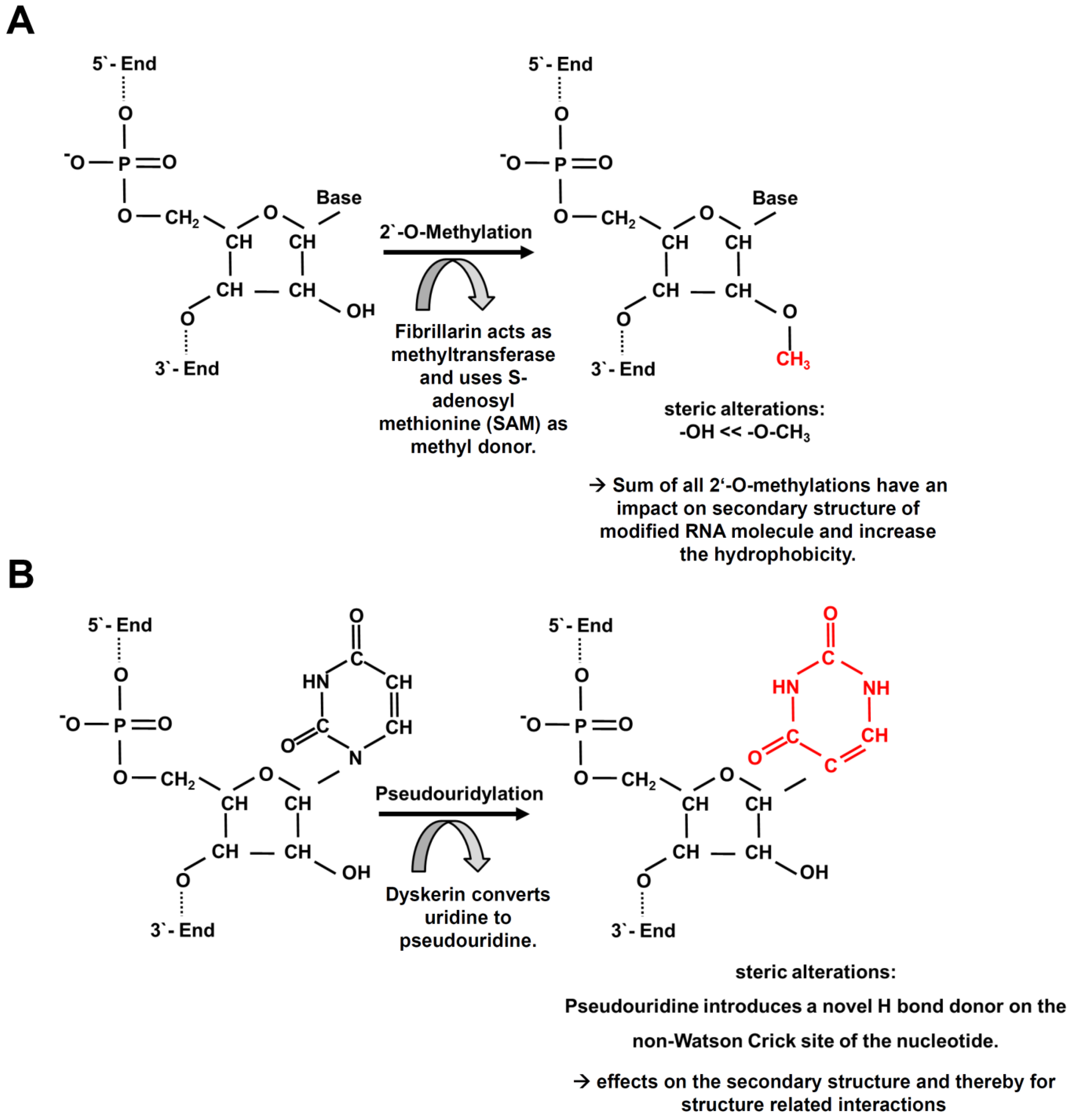
1. Introduction
2. Results
2.1. Expression Pattern and Localization of RNA-Modifying Proteins in the Skin
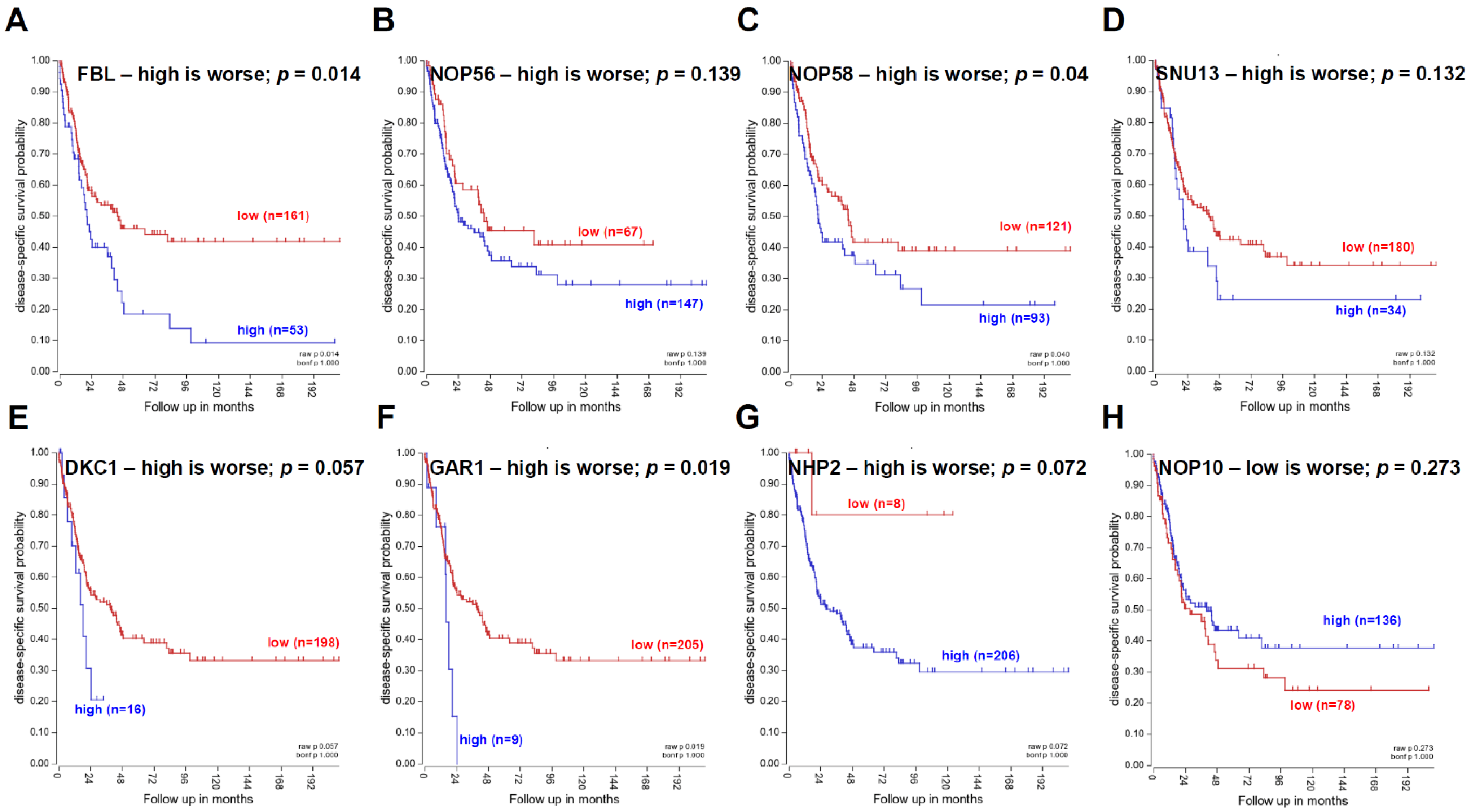
2.2. Correlation of RNA-Modifying Proteins with Tumor Cell Proliferation
2.3. Correlation of the Expression of RNA Modifying Factors with Immune Modulatory Genes
2.4. Correlation of miR Expression with RNA-Modifying Factors
3. Discussion
4. Materials and Methods
Immunohistochemistry
5. Conclusions
Author Contributions
Funding
Institutional Review Board Statement
Informed Consent Statement
Data Availability Statement
Conflicts of Interest
Abbreviations
References
- Haass, N.K.; Herlyn, M. Normal human melanocyte homeostasis as a paradigm for understanding melanoma. J. Investig. Dermatol. Symp. Proc. 2005, 10, 153–163. [Google Scholar] [CrossRef] [Green Version]
- Laikova, K.V.; Oberemok, V.V.; Krasnodubets, A.M.; Gal’chinsky, N.V.; Useinov, R.Z.; Novikov, I.A.; Temirova, Z.Z.; Gorlov, M.V.; Shved, N.A.; Kumeiko, V.V.; et al. Advances in the Understanding of Skin Cancer: Ultraviolet Radiation, Mutations, and Antisense Oligonucleotides as Anticancer Drugs. Molecules 2019, 24, 1516. [Google Scholar] [CrossRef] [Green Version]
- Bray, F.; Ren, J.S.; Masuyer, E.; Ferlay, J. Global estimates of cancer prevalence for 27 sites in the adult population in 2008. Int. J. Cancer 2013, 132, 1133–1145. [Google Scholar] [CrossRef] [PubMed]
- Sample, A.; He, Y.Y. Mechanisms and prevention of UV-induced melanoma. Photodermatol. Photoimmunol. Photomed. 2018, 34, 13–24. [Google Scholar] [CrossRef] [PubMed]
- Hawkes, J.E.; Truong, A.; Meyer, L.J. Genetic predisposition to melanoma. Semin. Oncol. 2016, 43, 591–597. [Google Scholar] [CrossRef]
- Debniak, T. Familial malignant melanoma—Overview. Hered. Cancer Clin. Pract. 2004, 2, 123–129. [Google Scholar] [CrossRef] [PubMed] [Green Version]
- Damsky, W.E.; Bosenberg, M. Melanocytic nevi and melanoma: Unraveling a complex relationship. Oncogene 2017, 36, 5771–5792. [Google Scholar] [CrossRef] [Green Version]
- Stepinski, D. The nucleolus, an ally, and an enemy of cancer cells. Histochem. Cell Biol. 2018, 150, 607–629. [Google Scholar] [CrossRef]
- Pierard, G.E. Cell proliferation in cutaneous malignant melanoma: Relationship with neoplastic progression. ISRN Dermatol. 2012, 2012, 828146. [Google Scholar] [CrossRef] [PubMed] [Green Version]
- Szavits-Nossan, J.; Ciandrini, L. Inferring efficiency of translation initiation and elongation from ribosome profiling. Nucleic Acids Res. 2020, 48, 9478–9490. [Google Scholar] [CrossRef]
- von der Haar, T. Mathematical and Computational Modelling of Ribosomal Movement and Protein Synthesis: An overview. Comput. Struct. Biotechnol. J. 2012, 1, e201204002. [Google Scholar] [CrossRef] [Green Version]
- Szavits-Nossan, J.; Ciandrini, L. Accurate measures of translational efficiency and traffic using ribosome profiling. bioRxiv 2019. [Google Scholar] [CrossRef] [Green Version]
- Pelletier, J.; Thomas, G.; Volarevic, S. Ribosome biogenesis in cancer: New players and therapeutic avenues. Nat. Rev. Cancer 2018, 18, 51–63. [Google Scholar] [CrossRef] [PubMed]
- Penzo, M.; Montanaro, L.; Trere, D.; Derenzini, M. The Ribosome Biogenesis-Cancer Connection. Cells 2019, 8, 55. [Google Scholar] [CrossRef] [PubMed] [Green Version]
- Brighenti, E.; Calabrese, C.; Liguori, G.; Giannone, F.A.; Trere, D.; Montanaro, L.; Derenzini, M. Interleukin 6 downregulates p53 expression and activity by stimulating ribosome biogenesis: A new pathway connecting inflammation to cancer. Oncogene 2014, 33, 4396–4406. [Google Scholar] [CrossRef] [Green Version]
- Maden, B.E. The numerous modified nucleotides in eukaryotic ribosomal RNA. Prog. Nucleic Acid Res. Mol. Biol. 1990, 39, 241–303. [Google Scholar] [CrossRef]
- Ofengand, J.; Bakin, A.; Wrzesinski, J.; Nurse, K.; Lane, B.G. The pseudouridine residues of ribosomal RNA. Biochem. Cell Biol. 1995, 73, 915–924. [Google Scholar] [CrossRef]
- Jady, B.E.; Kiss, T. A small nucleolar guide RNA functions both in 2′-O-ribose methylation and pseudouridylation of the U5 spliceosomal RNA. EMBO J. 2001, 20, 541–551. [Google Scholar] [CrossRef] [PubMed]
- Silipo, M.; Gautrey, H.; Tyson-Capper, A. Deregulation of splicing factors and breast cancer development. J. Mol. Cell Biol. 2015, 7, 388–401. [Google Scholar] [CrossRef] [Green Version]
- Somme, J.; Van Laer, B.; Roovers, M.; Steyaert, J.; Versees, W.; Droogmans, L. Characterization of two homologous 2′-O-methyltransferases showing different specificities for their tRNA substrates. RNA 2014, 20, 1257–1271. [Google Scholar] [CrossRef] [Green Version]
- Peng, Y.; Yu, G.; Tian, S.; Li, H. Co-expression and co-purification of archaeal and eukaryal box C/D RNPs. PLoS ONE 2014, 9, e103096. [Google Scholar] [CrossRef]
- Sproat, B.S.; Lamond, A.I.; Beijer, B.; Neuner, P.; Ryder, U. Highly efficient chemical synthesis of 2′-O-methyloligoribonucleotides and tetrabiotinylated derivatives; novel probes that are resistant to degradation by RNA or DNA specific nucleases. Nucleic Acids Res. 1989, 17, 3373–3386. [Google Scholar] [CrossRef] [PubMed] [Green Version]
- Inoue, H.; Hayase, Y.; Imura, A.; Iwai, S.; Miura, K.; Ohtsuka, E. Synthesis and hybridization studies on two complementary nona(2′-O-methyl)ribonucleotides. Nucleic Acids Res. 1987, 15, 6131–6148. [Google Scholar] [CrossRef]
- Lacoux, C.; Di Marino, D.; Boyl, P.P.; Zalfa, F.; Yan, B.; Ciotti, M.T.; Falconi, M.; Urlaub, H.; Achsel, T.; Mougin, A.; et al. BC1-FMRP interaction is modulated by 2′-O-methylation: RNA-binding activity of the tudor domain and translational regulation at synapses. Nucleic Acids Res. 2012, 40, 4086–4096. [Google Scholar] [CrossRef] [PubMed] [Green Version]
- Elliott, B.A.; Ho, H.T.; Ranganathan, S.V.; Vangaveti, S.; Ilkayeva, O.; Abou Assi, H.; Choi, A.K.; Agris, P.F.; Holley, C.L. Modification of messenger RNA by 2′-O-methylation regulates gene expression in vivo. Nat. Commun. 2019, 10, 3401. [Google Scholar] [CrossRef] [PubMed] [Green Version]
- Li, X.; Ma, S.; Yi, C. Pseudouridine: The fifth RNA nucleotide with renewed interests. Curr. Opin. Chem. Biol. 2016, 33, 108–116. [Google Scholar] [CrossRef] [PubMed]
- van der Feltz, C.; DeHaven, A.C.; Hoskins, A.A. Stress-induced Pseudouridylation Alters the Structural Equilibrium of Yeast U2 snRNA Stem II. J. Mol. Biol. 2018, 430, 524–536. [Google Scholar] [CrossRef]
- Penzo, M.; Guerrieri, A.N.; Zacchini, F.; Trere, D.; Montanaro, L. RNA Pseudouridylation in Physiology and Medicine: For Better and for Worse. Genes 2017, 8, 301. [Google Scholar] [CrossRef] [Green Version]
- Ge, J.; Yu, Y.T. RNA pseudouridylation: New insights into an old modification. Trends Biochem. Sci. 2013, 38, 210–218. [Google Scholar] [CrossRef] [Green Version]
- Zhao, Y.; Dunker, W.; Yu, Y.T.; Karijolich, J. The Role of Noncoding RNA Pseudouridylation in Nuclear Gene Expression Events. Front. Bioeng. Biotechnol. 2018, 6, 8. [Google Scholar] [CrossRef] [Green Version]
- Song, J.; Zhuang, Y.; Zhu, C.; Meng, H.; Lu, B.; Xie, B.; Peng, J.; Li, M.; Yi, C. Differential roles of human PUS10 in miRNA processing and tRNA pseudouridylation. Nat. Chem. Biol. 2020, 16, 160–169. [Google Scholar] [CrossRef] [PubMed]
- Chen, J.; Weiss, W.A. Alternative splicing in cancer: Implications for biology and therapy. Oncogene 2015, 34, 1–14. [Google Scholar] [CrossRef] [PubMed]
- Ma, F.C.; He, R.Q.; Lin, P.; Zhong, J.C.; Ma, J.; Yang, H.; Hu, X.H.; Chen, G. Profiling of prognostic alternative splicing in melanoma. Oncol. Lett. 2019, 18, 1081–1088. [Google Scholar] [CrossRef] [Green Version]
- Singh, B.; Eyras, E. The role of alternative splicing in cancer. Transcription 2017, 8, 91–98. [Google Scholar] [CrossRef] [Green Version]
- Guzinska-Ustymowicz, K.; Pryczynicz, A.; Kemona, A.; Czyzewska, J. Correlation between proliferation markers: PCNA, Ki-67, MCM-2 and antiapoptotic protein Bcl-2 in colorectal cancer. Anticancer Res. 2009, 29, 3049–3052. [Google Scholar]
- Ladstein, R.G.; Bachmann, I.M.; Straume, O.; Akslen, L.A. Ki-67 expression is superior to mitotic count and novel proliferation markers PHH3, MCM4 and mitosin as a prognostic factor in thick cutaneous melanoma. BMC Cancer 2010, 10, 140. [Google Scholar] [CrossRef] [PubMed] [Green Version]
- Ohsie, S.J.; Sarantopoulos, G.P.; Cochran, A.J.; Binder, S.W. Immunohistochemical characteristics of melanoma. J. Cutan. Pathol. 2008, 35, 433–444. [Google Scholar] [CrossRef]
- Reddy, V.B.; Gattuso, P.; Aranha, G.; Carson, H.J. Cell proliferation markers in predicting metastases in malignant melanoma. J. Cutan. Pathol. 1995, 22, 248–251. [Google Scholar] [CrossRef]
- Bresnick, A.R.; Weber, D.J.; Zimmer, D.B. S100 proteins in cancer. Nat. Rev. Cancer 2015, 15, 96–109. [Google Scholar] [CrossRef] [Green Version]
- Fander, J.; Kielstein, H.; Buttner, M.; Koelblinger, P.; Dummer, R.; Bauer, M.; Handke, D.; Wickenhauser, C.; Seliger, B.; Jasinski-Bergner, S. Characterizing CD44 regulatory microRNAs as putative therapeutic agents in human melanoma. Oncotarget 2019, 10, 6509–6525. [Google Scholar] [CrossRef] [Green Version]
- Ilmonen, S.; Hernberg, M.; Pyrhonen, S.; Tarkkanen, J.; Asko-Seljavaara, S. Ki-67, Bcl-2 and p53 expression in primary and metastatic melanoma. Melanoma Res. 2005, 15, 375–381. [Google Scholar] [CrossRef] [PubMed]
- Weinstein, D.; Leininger, J.; Hamby, C.; Safai, B. Diagnostic and prognostic biomarkers in melanoma. J. Clin. Aesthet. Dermatol. 2014, 7, 13–24. [Google Scholar] [PubMed]
- Kycler, W.; Grodecka-Gazdecka, S.; Breborowicz, J.; Filas, V.; Teresiak, M. Prognostic factors in melanoma. Rep. Pract. Oncol. Radiother. 2006, 11, 39–48. [Google Scholar] [CrossRef] [Green Version]
- Cirenajwis, H.; Ekedahl, H.; Lauss, M.; Harbst, K.; Carneiro, A.; Enoksson, J.; Rosengren, F.; Werner-Hartman, L.; Torngren, T.; Kvist, A.; et al. Molecular stratification of metastatic melanoma using gene expression profiling: Prediction of survival outcome and benefit from molecular targeted therapy. Oncotarget 2015, 6, 12297–12309. [Google Scholar] [CrossRef] [Green Version]
- Quandt, D.; Fiedler, E.; Boettcher, D.; Marsch, W.; Seliger, B. B7-h4 expression in human melanoma: Its association with patients’ survival and antitumor immune response. Clin. Cancer Res. 2011, 17, 3100–3111. [Google Scholar] [CrossRef] [PubMed] [Green Version]
- Starega-Roslan, J.; Krol, J.; Koscianska, E.; Kozlowski, P.; Szlachcic, W.J.; Sobczak, K.; Krzyzosiak, W.J. Structural basis of microRNA length variety. Nucleic Acids Res. 2011, 39, 257–268. [Google Scholar] [CrossRef] [Green Version]
- Friedrich, M.; Vaxevanis, C.K.; Biehl, K.; Mueller, A.; Seliger, B. Targeting the coding sequence: Opposing roles in regulating classical and non-classical MHC class I molecules by miR-16 and miR-744. J. Immunother. Cancer 2020, 8. [Google Scholar] [CrossRef] [PubMed]
- Jasinski-Bergner, S.; Mandelboim, O.; Seliger, B. The role of microRNAs in the control of innate immune response in cancer. J. Natl. Cancer Inst. 2014, 106. [Google Scholar] [CrossRef] [PubMed] [Green Version]
- Jasinski-Bergner, S.; Mandelboim, O.; Seliger, B. Molecular mechanisms of human herpes viruses inferring with host immune surveillance. J. Immunother. Cancer 2020, 8. [Google Scholar] [CrossRef]
- Fattore, L.; Costantini, S.; Malpicci, D.; Ruggiero, C.F.; Ascierto, P.A.; Croce, C.M.; Mancini, R.; Ciliberto, G. MicroRNAs in melanoma development and resistance to target therapy. Oncotarget 2017, 8, 22262–22278. [Google Scholar] [CrossRef] [Green Version]
- Kurimoto, R.; Chiba, T.; Ito, Y.; Matsushima, T.; Yano, Y.; Miyata, K.; Yashiro, Y.; Suzuki, T.; Tomita, K.; Asahara, H. The tRNA pseudouridine synthase TruB1 regulates the maturation of let-7 miRNA. EMBO J. 2020, 39, e104708. [Google Scholar] [CrossRef]
- Penzo, M.; Montanaro, L. Turning Uridines around: Role of rRNA Pseudouridylation in Ribosome Biogenesis and Ribosomal Function. Biomolecules 2018, 8, 38. [Google Scholar] [CrossRef] [PubMed] [Green Version]
- Hirano, M.; Das, S.; Guo, P.; Cooper, M.D. The evolution of adaptive immunity in vertebrates. Adv. Immunol. 2011, 109, 125–157. [Google Scholar] [CrossRef]
- Bukur, J.; Jasinski, S.; Seliger, B. The role of classical and non-classical HLA class I antigens in human tumors. Semin. Cancer Biol. 2012, 22, 350–358. [Google Scholar] [CrossRef] [PubMed]
- Motorin, Y.; Marchand, V. Detection and Analysis of RNA Ribose 2′-O-Methylations: Challenges and Solutions. Genes 2018, 9, 642. [Google Scholar] [CrossRef] [PubMed] [Green Version]
- Abe, M.; Naqvi, A.; Hendriks, G.J.; Feltzin, V.; Zhu, Y.; Grigoriev, A.; Bonini, N.M. Impact of age-associated increase in 2′-O-methylation of miRNAs on aging and neurodegeneration in Drosophila. Genes Dev. 2014, 28, 44–57. [Google Scholar] [CrossRef] [Green Version]
- Liang, H.; Jiao, Z.; Rong, W.; Qu, S.; Liao, Z.; Sun, X.; Wei, Y.; Zhao, Q.; Wang, J.; Liu, Y.; et al. 3′-Terminal 2′-O-methylation of lung cancer miR-21-5p enhances its stability and association with Argonaute 2. Nucleic Acids Res. 2020, 48, 7027–7040. [Google Scholar] [CrossRef]
- Ponten, F.; Jirstrom, K.; Uhlen, M. The Human Protein Atlas--a tool for pathology. J. Pathol. 2008, 216, 387–393. [Google Scholar] [CrossRef]
- Thul, P.J.; Akesson, L.; Wiking, M.; Mahdessian, D.; Geladaki, A.; Ait Blal, H.; Alm, T.; Asplund, A.; Bjork, L.; Breckels, L.M.; et al. A subcellular map of the human proteome. Science 2017, 356. [Google Scholar] [CrossRef]
- Uhlen, M.; Fagerberg, L.; Hallstrom, B.M.; Lindskog, C.; Oksvold, P.; Mardinoglu, A.; Sivertsson, A.; Kampf, C.; Sjostedt, E.; Asplund, A.; et al. Proteomics. Tissue-based map of the human proteome. Science 2015, 347, 1260419. [Google Scholar] [CrossRef] [PubMed]


| RNA Modification | Factor | Localization within Skin | Localization within MM |
|---|---|---|---|
| factors involved into 2′-O-methylation | FBL | nuclear, extra strong within the cells of the epidermis especially in the stratum germinativum | nuclear |
| NOP56 | nuclear | nuclear | |
| NOP58 | nuclear strongest expression within the cells of the epidermal layers, especially within the stratum germinativum | cytoplasmic and nuclear | |
| 15.5K (SNU13) | cytoplasmic and membranous restricted to the cells of the epidermis | nuclear | |
| factors involved into pseudouridylation | DKC1 | nuclear | nuclear |
| GAR1 | nuclear | nuclear | |
| NHP2 | nuclear | nuclear | |
| NHP10 | nuclear | nuclear |
| Correlated Expression | FBL | NOP56 | NOP58 | SNU13 | DKC1 | GAR1 | NHP2 | NOP10 |
|---|---|---|---|---|---|---|---|---|
| MKI67 | R = 0.081 | R = 0.216 | R = 0.235 | R = 0.050 | R = 0.185 | R = 0.154 | R = −0.133 | R = 0.011 |
| p = 0.241 | p = 1.47 × 10⁻³ | p = 5.22 × 10⁻⁴ | p = 0.468 | p = 6.50 × 10⁻³ | p = 0.024 | p = 0.053 | p = 0.874 | |
| PCNA | R = 0.152 | R = 0.581 | R = 0.448 | R = 0.106 | R = 0.463 | R = 0.238 | R = 0.372 | R = 0.258 |
| p = 0.026 | p = 9.59 × 10⁻²¹ | p = 5.58 × 10⁻¹² | p = 0.122 | p = 9.06 × 10⁻¹³ | p = 4.55 × 10⁻⁴ | p = 1.92 × 10⁻⁸ | p = 1.36 × 10⁻⁴ | |
| CCNA1 | R = −0.046 | R = 0.065 | R = 0.131 | R = −0.079 | R = 0.060 | R = −0.063 | R = 0.066 | R = 0.172 |
| p = 0.499 | p = 0.341 | p = 0.056 | p = 0.250 | p = 0.383 | p = 0.356 | p = 0.339 | p = 0.011 | |
| CCNB1 | R = 0.265 | R = 0.439 | R = 0.492 | R = 0.136 | R = 0.398 | R = 0.423 | R = 0.463 | R = 0.165 |
| p = 8.62 × 10⁻⁵ | p = 1.78 × 10⁻¹¹ | p = 1.88 × 10⁻¹⁴ | p = 0.047 | p = 1.50 × 10⁻⁹ | p = 1.10 × 10⁻¹⁰ | p = 9.44 × 10⁻¹³ | p = 0.015 | |
| MCM2 | R = 0.236 | R = 0.251 | R = 0.302 | R = 0.166 | R = 0.325 | R = 0.233 | R = 0.123 | R = 0.031 |
| p = 4.94 × 10⁻⁴ | p = 2.07 × 10⁻⁴ | p = 6.94 × 10⁻⁶ | p = 0.015 | p = 1.16 × 10⁻⁶ | p = 5.80 × 10⁻⁴ | p = 0.072 | p = 0.656 | |
| MCM4 | R = 0.334 | R = 0.546 | R = 0.466 | R = 0.262 | R = 0.554 | R = 0.482 | R = 0.377 | R = 0.057 |
| p = 5.60 × 10⁻⁷ | p = 4.78 × 10⁻¹⁸ | p = 6.15 × 10⁻¹³ | p = 1.04 × 10⁻⁴ | p = 1.34 × 10⁻¹⁸ | p = 7.93 × 10⁻¹⁴ | p = 1.29 × 10⁻⁸ | p = 0.403 | |
| CENPF | R = 0.363 | R = 0.428 | R = 0.495 | R = 0.162 | R = 0.509 | R = 0.515 | R = 0.198 | R = 0.040 |
| p = 4.69 × 10⁻⁸ | p = 6.01 × 10⁻¹¹ | p = 1.33 × 10⁻¹⁴ | p = 0.018 | p = 1.62 × 10⁻¹⁵ | p = 6.59 × 10⁻¹⁶ | p = 3.61 × 10⁻³ | p = 0.556 |
| Correlated Expression | FBL | NOP56 | NOP58 | SNU13 | DKC1 | GAR1 | NHP2 | NOP10 |
|---|---|---|---|---|---|---|---|---|
| MART1 | R = 0.009 | R = 0.304 | R = −0.024 | R = 0.134 | R = 0.265 | R = 0.203 | R = 0.259 | R = 0.115 |
| p = 0.893 | p = 6.12 × 10⁻⁶ | p = 0.728 | p = 0.050 | p = 8.59 × 10⁻⁵ | p = 2.92 × 10⁻³ | p = 1.25 × 10⁻⁴ | p = 0.094 | |
| S100B | R = −0.248 | R = −0.038 | R = −0.152 | R = −0.085 | R = −0.090 | R = −0.067 | R = 0.133 | R = 0.021 |
| p = 2.53 × 10⁻⁴ | p = 0.577 | p = 0.026 | p = 0.218 | p = 0.191 | p = 0.327 | p = 0.053 | p = 0.757 | |
| S100A4 | R = −0.325 | R = −0.414 | R = −0.223 | R = −0.240 | R = −0.342 | R = −0.274 | R = −0.258 | R = 0.042 |
| p = 1.20 × 10⁻⁶ | p = 2.79 × 10⁻¹⁰ | p = 1.01 × 10⁻³ | p = 3.96 × 10⁻⁴ | p = 2.98 × 10⁻⁷ | p = 4.75 × 10⁻⁵ | p = 1.33 × 10⁻⁴ | p = 0.539 | |
| S100A9 | R = −0.234 | R = −0.192 | R = −0.212 | R = −0.185 | R = −0.295 | R = −0.234 | R = −0.147 | R = 0.104 |
| p = 5.57 × 10⁻⁴ | p = 4.92 × 10⁻³ | p = 1.81 × 10⁻³ | p = 6.72 × 10⁻³ | p = 1.13 × 10⁻⁵ | p = 5.58 × 10⁻⁴ | p = 0.031 | p = 0.130 | |
| MITF | R = 0.076 | R = 0.388 | R = 0.050 | R = 0.124 | R = 0.345 | R = 0.256 | R = 0.304 | R = 0.124 |
| p = 0.271 | p = 4.05 × 10⁻⁹ | p = 0.468 | p = 0.070 | p = 2.24 × 10⁻⁷ | p = 1.51 × 10⁻⁴ | p = 5.90 × 10⁻⁶ | p = 0.070 | |
| MMP2 | R = −0.129 | R = −0.321 | R = −0.271 | R = −0.190 | R = −0.226 | R = −0.040 | R = −0.309 | R = −0.050 |
| p = 0.059 | p = 1.67 × 10⁻⁶ | p = 5.96 × 10⁻⁵ | p = 5.20 × 10⁻³ | p = 8.54 × 10⁻⁴ | p = 0.561 | p = 4.10 × 10⁻⁶ | p = 0.465 | |
| NM23 | R = −0.005 | R = 0.534 | R = 0.424 | R = 0.053 | R = 0.442 | R = 0.425 | R = 0.531 | R = 0.321 |
| p = 0.944 | p = 3.56 × 10⁻¹⁷ | p = 9.77 × 10⁻¹¹ | p = 0.445 | p = 1.21 × 10⁻¹¹ | p = 8.80 × 10⁻¹¹ | p = 5.60 × 10⁻¹⁷ | p = 1.66 × 10⁻⁶ | |
| CD44 | R = −0.091 | R = −0.026 | R = −0.174 | R = 0.034 | R = 0.101 | R = 0.012 | R = −0.040 | R = −0.040 |
| p = 0.184 | p = 0.705 | p = 0.011 | p = 0.618 | p = 0.139 | p = 0.863 | p = 0.557 | p = 0.556 | |
| PMEL | R = −0.033 | R = 0.281 | R = −0.061 | R = 0.029 | R = 0.171 | R = 0.126 | R = 0.160 | R = 0.179 |
| p = 0.630 | p = 2.96 × 10⁻⁵ | p = 0.371 | p = 0.670 | p = 0.012 | p = 0.066 | p = 0.019 | p = 8.58 × 10⁻³ | |
| BCL2 | R = 0.217 | R = −0.059 | R = −0.072 | R = 0.183 | R = 0.008 | R = 0.031 | R = −0.101 | R = −0.235 |
| p = 1.40 × 10⁻³ | p = 0.392 | p = 0.292 | p = 7.21 × 10⁻³ | p = 0.907 | p = 0.652 | p = 0.140 | p = 5.15 × 10⁻⁴ |
| Correlated Expression | FBL | NOP56 | NOP58 | SNU13 | DKC1 | GAR1 | NHP2 | NOP10 | |
|---|---|---|---|---|---|---|---|---|---|
| (NHP2L1) | |||||||||
| molecules contributing to recognition by immune effector cells | MICA | R = −0.341 | R = 0.100 | R = −0.152 | R = −0.128 | R = −0.112 | R = −0.189 | R = 0.121 | R = 0.245 |
| p = 3.10 × 10⁻⁷ | p = 0.146 | p = 0.026 | p = 0.061 | p = 0.103 | p = 5.59 × 10⁻³ | p = 0.079 | p = 2.88 × 10⁻⁴ | ||
| MICB | R = −0.241 | R = −0.119 | R = −0.069 | R = −0.014 | R = −0.149 | R = −0.304 | R = −0.106 | R = 0.175 | |
| p = 3.72 × 10⁻⁴ | p = 0.082 | p = 0.314 | p = 0.834 | p = 0.029 | p = 5.75 × 10⁻⁶ | p = 0.121 | p = 0.010 | ||
| ULBP1 | R = 0.109 | R = 0.020 | R = 0.109 | R = 0.011 | R = 0.067 | R = 0.056 | R = 0.077 | R = 0.029 | |
| p = 0.112 | p = 0.767 | p = 0.112 | p = 0.877 | p = 0.328 | p = 0.412 | p = 0.264 | p = 0.677 | ||
| ULBP2 | R = 0.047 | R = 0.078 | R = 0.107 | R = −0.020 | R = 0.096 | R = 0.120 | R = 0.093 | R = −0.023 | |
| p = 0.496 | p = 0.255 | p = 0.117 | p = 0.766 | p = 0.162 | p = 0.080 | p = 0.176 | p = 0.742 | ||
| ULBP3 | R = −0.091 | R = 0.028 | R = 0.005 | R = −0.008 | R = 0.036 | R = 0.058 | R = 0.060 | R = 0.098 | |
| p = 0.185 | p = 0.688 | p = 0.947 | p = 0.912 | p = 0.603 | p = 0.398 | p = 0.380 | p = 0.154 | ||
| ULBP4 | R = 0.048 | R = −0.104 | R = −0.171 | R = 0.202 | R = −0.015 | R = −0.008 | R = −0.024 | R = −0.107 | |
| p = 0.488 | p = 0.129 | p = 0.012 | p = 2.93 × 10⁻³ | p = 0.825 | p = 0.906 | p = 0.729 | p = 0.117 | ||
| ULBP5 | R = −0.011 | R = −0.007 | R = 0.018 | R = 0.022 | R = −0.001 | R = 0.069 | R = −0.022 | R = 0.024 | |
| p = 0.867 | p = 0.915 | p = 0.791 | p = 0.744 | p = 1.000 | p = 0.313 | p = 0.752 | p = 0.730 | ||
| ULBP6 | R = −0.005 | R = 0.020 | R = −0.132 | R = −0.029 | R = −0.093 | R = −0.138 | R = 0.025 | R = −0.004 | |
| p = 0.940 | p = 0.772 | p = 0.055 | p = 0.674 | p = 0.175 | p = 0.044 | p = 0.715 | p = 0.951 | ||
| HLA-A | R = −0.364 | R = −0.261 | R = −0.370 | R = −0.054 | R = −0.195 | R = −0.238 | R = −0.229 | R = 0.110 | |
| p = 4.16 × 10⁻⁸ | p = 1.13 × 10⁻⁴ | p = 2.47 × 10⁻⁸ | p = 0.431 | p = 4.12 × 10⁻³ | p = 4.55 × 10⁻⁴ | p = 7.55 × 10⁻⁴ | p = 0.109 | ||
| HLA-B | R = −0.369 | R = −0.353 | R = −0.342 | R = −0.024 | R = −0.305 | R = −0.365 | R = −0.218 | R = 0.114 | |
| p = 2.66 × 10⁻⁸ | p = 1.16 × 10⁻⁷ | p = 2.81 × 10⁻⁷ | p = 0.723 | p = 5.60 × 10⁻⁶ | p = 3.92 × 10⁻⁸ | p = 1.30 × 10⁻³ | p = 0.096 | ||
| HLA-C | R = −0.351 | R = −0.264 | R = −0.323 | R = −0.126 | R = −0.298 | R = −0.360 | R = −0.048 | R = 0.145 | |
| p = 1.29 × 10⁻⁷ | p = 9.52 × 10⁻⁵ | p = 1.35 × 10⁻⁶ | p = 0.066 | p = 9.08 × 10⁻⁶ | p = 5.96 × 10⁻⁸ | p = 0.481 | p = 0.034 | ||
| B2M | R = −0.407 | R = −0.281 | R = −0.224 | R = −0.150 | R = −0.198 | R = −0.318 | R = −0.178 | R = 0.314 | |
| p = 6.26 × 10⁻¹⁰ | p = 2.97 × 10⁻⁵ | p = 9.60 × 10⁻⁴ | p = 0.028 | p = 3.67 × 10⁻³ | p = 2.04 × 10⁻⁶ | p = 9.12 × 10⁻³ | p = 2.73 × 10⁻⁶ | ||
| TAP1 | R = −0.251 | R = −0.074 | R = −0.176 | R = 0.067 | R = −0.114 | R = −0.154 | R = 0.018 | R = 0.189 | |
| p = 2.05 × 10⁻⁴ | p = 0.278 | p = 9.93 × 10⁻³ | p = 0.331 | p = 0.097 | p = 0.024 | p = 0.788 | p = 5.58 × 10⁻³ | ||
| TAP2 | R = −0.102 | R = −0.042 | R = −0.064 | R = 0.144 | R = 0.027 | R = −0.031 | R = −0.081 | R = 0.034 | |
| p = 0.137 | p = 0.538 | p = 0.355 | p = 0.035 | p = 0.695 | p = 0.655 | p = 0.241 | p = 0.624 | ||
| TAPBP | R = −0.210 | R = −0.309 | R = −0.374 | R = 0.070 | R = −0.156 | R = −0.219 | R = −0.328 | R = −0.088 | |
| p = 1.99 × 10⁻³ | p = 3.94 × 10⁻⁶ | p = 1.70 × 10⁻⁸ | p = 0.311 | p = 0.022 | p = 1.25 × 10⁻³ | p = 9.18 × 10⁻⁷ | p = 0.19 | ||
| LMP2 | R = −0.342 | R = −0.214 | R = −0.209 | R = −0.038 | R = −0.262 | R = −0.338 | R = −0.014 | R = 0.190 | |
| p = 2.96 × 10⁻⁷ | p = 1.60 × 10⁻³ | p = 2.07 × 10⁻³ | p = 0.577 | p = 1.03 × 10⁻⁴ | p = 4.19 × 10⁻⁷ | p = 0.833 | p = 5.41 × 10⁻³ | ||
| LMP7 | R = −0.299 | R = −0.159 | R = −0.233 | R = 0.055 | R = −0.164 | R = −0.180 | R = 0.092 | R = 0.165 | |
| p = 8.42 × 10⁻⁶ | p = 0.020 | p = 5.83 × 10⁻⁴ | p = 0.420 | p = 0.016 | p = 8.24 × 10⁻³ | p = 0.179 | p = 0.015 | ||
| LMP10 | R = −0.092 | R = −0.148 | R = −0.205 | R = 0.008 | R = −0.180 | R = −0.245 | R = −0.057 | R = 0.138 | |
| p = 0.180 | p = 0.030 | p = 2.55 × 10⁻³ | p = 0.909 | p = 8.33 × 10⁻³ | p = 2.99 × 10⁻⁴ | p = 0.406 | p = 0.044 | ||
| ERAAP | R = −0.208 | R = −0.141 | R = −0.122 | R = −0.104 | R = −0.161 | R = −0.197 | R = −0.006 | R = 0.036 | |
| p = 2.24 × 10⁻³ | p = 0.040 | p = 0.075 | p = 0.131 | p = 0.019 | p = 3.73 × 10⁻³ | p = 0.932 | p = 0.602 | ||
| ERP57 | R = 0.004 | R = 0.077 | R = 0.014 | R = −0.031 | R = 0.081 | R = −0.017 | R = 0.012 | R = −0.073 | |
| p = 0.951 | p = 0.262 | p = 0.841 | p = 0.648 | p = 0.239 | p = 0.804 | p = 0.859 | p = 0.285 | ||
| CALR | R = −0.169 | R = 0.073 | R = −0.102 | R = −0.241 | R = −0.010 | R = −0.072 | R = −0.191 | R = 0.204 | |
| p = 0.013 | p = 0.288 | p = 0.138 | p = 3.64 × 10⁻⁴ | p = 0.888 | p = 0.296 | p = 5.11 × 10⁻³ | p = 2.74 × 10⁻³ | ||
| CANX | R = −0.303 | R = −0.430 | R = −0.332 | R = −0.147 | R = −0.300 | R = −0.370 | R = −0.320 | R = −0.022 | |
| p = 6.34 × 10⁻⁶ | p = 4.64 × 10⁻¹¹ | p = 6.62 × 10⁻⁷ | p = 0.031 | p = 8.14 × 10⁻⁶ | p = 2.48 × 10⁻⁸ | p = 1.79 × 10⁻⁶ | p = 0.744 | ||
| molecules contributing to immune evasion | PD-L1 (B7-H1) | R = −0.107 | R = −0.051 | R = −0.004 | R = −0.103 | R = 0.010 | R = −0.062 | R = 0.003 | R = 0.041 |
| p = 0.117 | p = 0.460 | p = 0.954 | p = 0.132 | p = 0.888 | p = 0.363 | p = 0.966 | p = 0.548 | ||
| PD-L2 (PDCD1LG2) | R = −0.151 | R = −0.150 | R = −0.092 | R = −0.096 | R = −0.114 | R = −0.215 | R = 0.014 | R = 0.068 | |
| p = 0.027 | p = 0.028 | p = 0.182 | p = 0.163 | p = 0.097 | p = 1.59 × 10⁻³ | p = 0.838 | p = 0.319 | ||
| HLA-G | R = −0.230 | R = −0.190 | R = −0.345 | R = −0.008 | R = −0.217 | R = −0.280 | R = −0.111 | R = 0.081 | |
| p = 6.90 × 10⁻⁴ | p = 5.32 × 10⁻³ | p = 2.20 × 10⁻⁷ | p = 0.905 | p = 1.39 × 10⁻³ | p = 3.19 × 10⁻⁵ | p = 0.104 | p = 0.236 | ||
| HLA-E | R = −0.321 | R = −0.290 | R = −0.418 | R = −0.045 | R = −0.296 | R = −0.380 | R = −0.236 | R = −0.013 | |
| p = 1.59 × 10⁻⁶ | p = 1.65 × 10⁻⁵ | p = 1.95 × 10⁻¹⁰ | p = 0.509 | p = 1.06 × 10⁻⁵ | p = 9.50 × 10⁻⁹ | p = 4.92 × 10⁻⁴ | p = 0.845 | ||
| HLA-F | R = −0.323 | R = −0.274 | R = −0.329 | R = −0.005 | R = −0.267 | R = −0.318 | R = −0.182 | R = 0.098 | |
| p = 1.41 × 10⁻⁶ | p = 4.71 × 10⁻⁵ | p = 8.52 × 10⁻⁷ | p = 0.944 | p = 7.79 × 10⁻⁵ | p = 1.98 × 10⁻⁶ | p = 7.47 × 10⁻³ | p = 0.154 | ||
| CD155 (PVR) | R = 0.029 | R = 0.436 | R = 0.098 | R = 0.011 | R = 0.227 | R = 0.263 | R = 0.125 | R = 0.148 | |
| p = 0.678 | p = 2.41 × 10⁻¹¹ | p = 0.151 | p = 0.874 | p = 8.28 × 10⁻⁴ | p = 1.00 × 10⁻⁴ | p = 0.067 | p = 0.030 | ||
| B7-H4 (VTCN1) | R = −0.003 | R = −0.059 | R = −0.016 | R = −0.048 | R = −0.031 | R = 0.034 | R = 0.062 | R = −0.086 | |
| p = 0.963 | p = 0.388 | p = 0.814 | p = 0.482 | p = 0.657 | p = 0.624 | p = 0.369 | p = 0.210 |
| Induced miRs in MM with Diagnostic/Prognostic Relevance | So Far in Literature Mentioned Enzymes with Putative Role for 2′-O-Methylation of miRs | So Far in Literature Mentioned Enzymes with Putative Role for Pseudouridylation of miRs | ||
|---|---|---|---|---|
| FBL | HENMT1 | DKC1 | TRUB1 | |
| miR-9-5p | R = 0.007 | R = 0.050 | R = −0.026 | R = 0.132 |
| p = 0.916 | p = 0.469 | p = 0.708 | p = 0.053 | |
| miR-10a | R = 0.020 | R = −0.036 | R = −0.004 | R = −0.047 |
| p = 0.767 | p = 0.603 | p = 0.957 | p = 0.492 | |
| miR-10b | R = −0.012 | R = 0.009 | R = −0.064 | R = −0.200 |
| p = 0.857 | p = 0.896 | p = 0.351 | p = 3.33 × 10⁻³ | |
| miR-17-5p | n.d. | n.d. | n.d. | n.d. |
| miR-18a | n.d. | n.d. | n.d. | n.d. |
| miR-21 | R = −0.168 | R = 0.017 | R = −0.304 | R = −0.139 |
| p = 0.014 | p = 0.800 | p = 5.88 × 10⁻⁶ | p = 0.042 | |
| miR-26b | R = 0.191 | R = −0.008 | R = 0.112 | R = −0.091 |
| p = 4.96 × 10⁻³ | p = 0.906 | p = 0.101 | p = 0.184 | |
| miR-92a | R = 0.106 | R = −0.055 | R = 0.155 | R = 0.059 |
| p = 0.122 | p = 0.424 | p = 0.023 | p = 0.390 | |
| miR-221 | R = −0.029 | R = 0.100 | R = 0.016 | R = 0.064 |
| p = 0.676 | p = 0.144 | p = 0.815 | p = 0.352 | |
| miR-222 | R = −0.015 | R = 0.083 | R = 0.083 | R = −0.073 |
| p = 0.825 | p = 0.227 | p = 0.224 | p = 0.285 | |
| miR-126 | R = 0.088 | R = −0.025 | R = −0.034 | R = −0.091 |
| p = 0.198 | p = 0.711 | p = 0.620 | p = 0.187 | |
| miR-145 | n.d. | n.d. | n.d. | n.d. |
| miR146a | R = −0.015 | R = −0.081 | R = 0.003 | R = 0.098 |
| p = 0.822 | p = 0.235 | p = 0.968 | p = 0.154 | |
| miR-182 | R = 0.008 | R = −0.068 | R = −0.045 | R = 0.071 |
| p = 0.905 | p = 0.322 | p = 0.510 | p = 0.299 | |
| miR-514 | R = −0.110 | R = 0.065 | R = 0.011 | R = 0.052 |
| p = 0.109 | p = 0.340 | p = 0.874 | p = 0.446 | |
| miR-520d | n.d. | n.d. | n.d. | n.d. |
| miR-527 | n.d. | n.d. | n.d. | n.d. |
Publisher’s Note: MDPI stays neutral with regard to jurisdictional claims in published maps and institutional affiliations. |
© 2021 by the authors. Licensee MDPI, Basel, Switzerland. This article is an open access article distributed under the terms and conditions of the Creative Commons Attribution (CC BY) license (http://creativecommons.org/licenses/by/4.0/).
Share and Cite
Jasinski-Bergner, S.; Blümke, J.; Wickenhauser, C.; Seliger, B. Relevance of 2′-O-Methylation and Pseudouridylation for the Malignant Melanoma. Cancers 2021, 13, 1167. https://doi.org/10.3390/cancers13051167
Jasinski-Bergner S, Blümke J, Wickenhauser C, Seliger B. Relevance of 2′-O-Methylation and Pseudouridylation for the Malignant Melanoma. Cancers. 2021; 13(5):1167. https://doi.org/10.3390/cancers13051167
Chicago/Turabian StyleJasinski-Bergner, Simon, Juliane Blümke, Claudia Wickenhauser, and Barbara Seliger. 2021. "Relevance of 2′-O-Methylation and Pseudouridylation for the Malignant Melanoma" Cancers 13, no. 5: 1167. https://doi.org/10.3390/cancers13051167