
Second Relapse of Pediatric Patients with Acute Myeloid Leukemia: A Report on Current Treatment Strategies and Outcome of the AML-BFM Study Group

 , ,
, ,  , , , , ,
Abstract
:Simple Summary
Abstract
1. Introduction
2. Materials and Methods
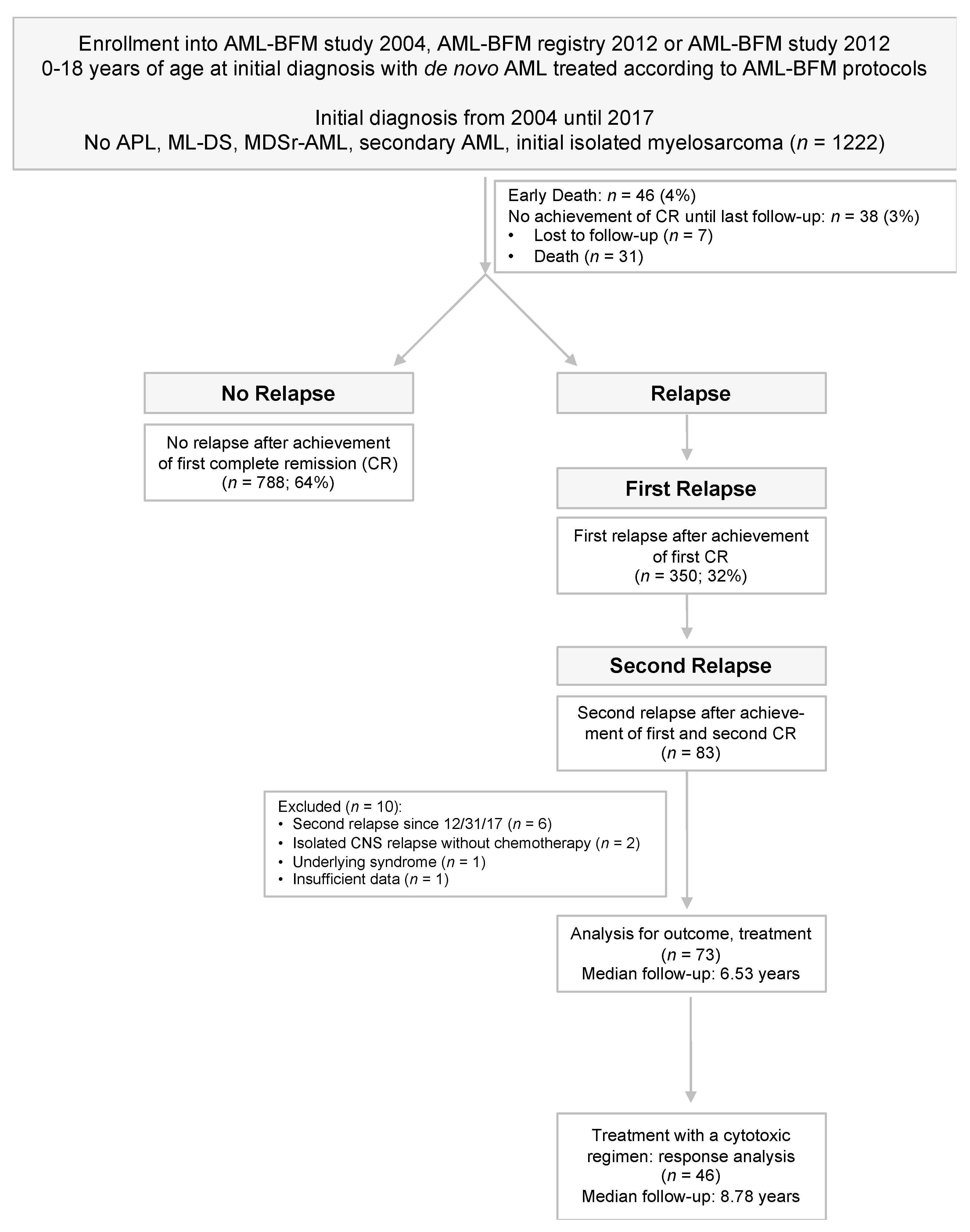
2.1. Patients
2.2. Previous Treatment Approaches
2.3. Definitions and Statistical Analysis
3. Results
3.1. Patient Characteristics
3.2. Treatment
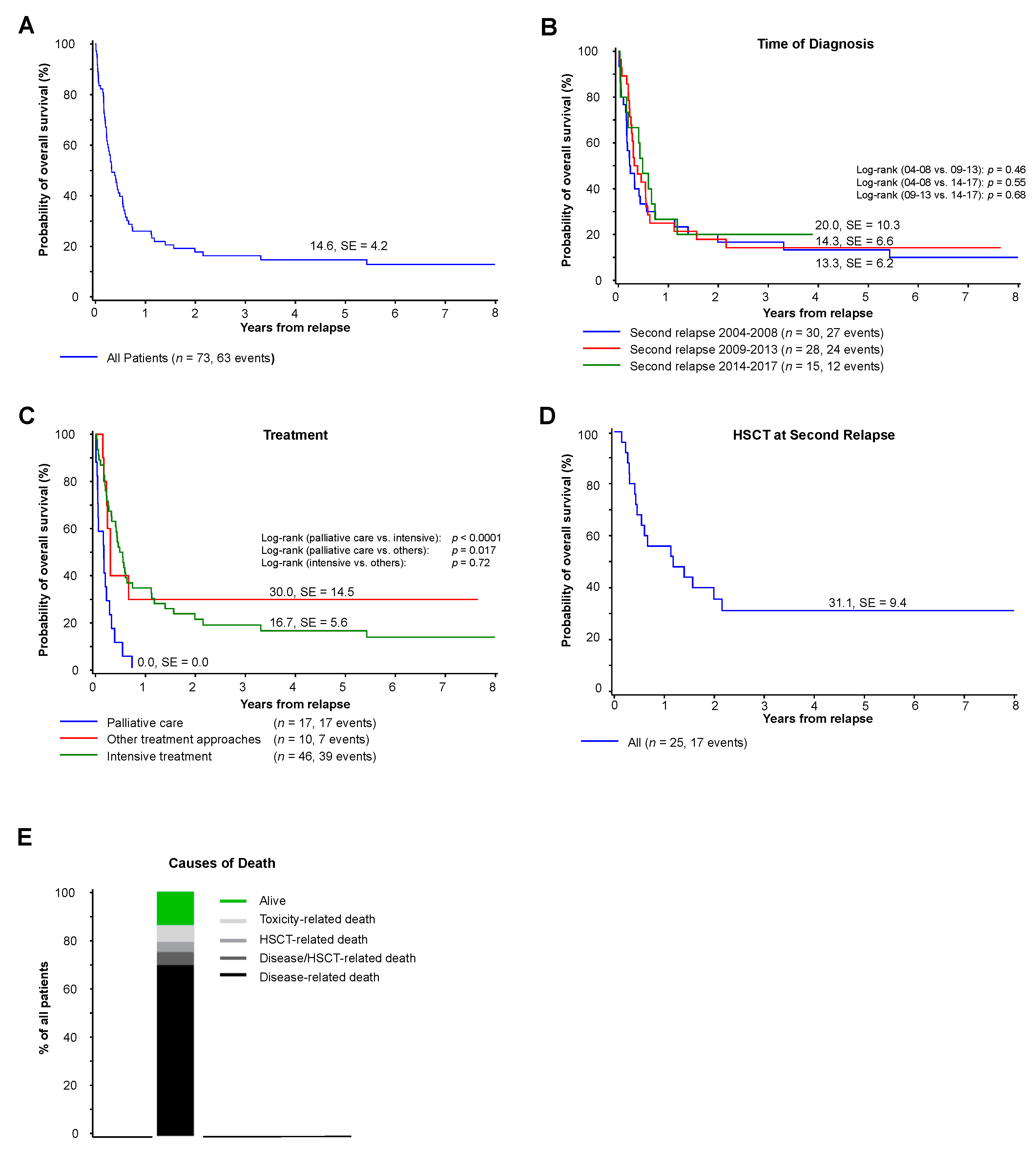
3.3. Survival
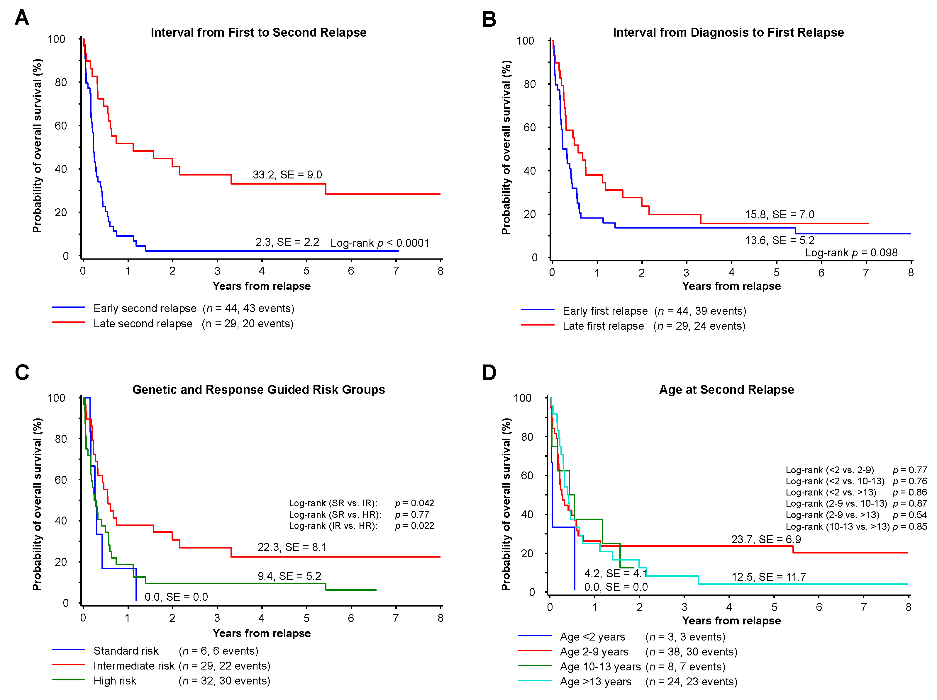
3.4. Prognostic Factors
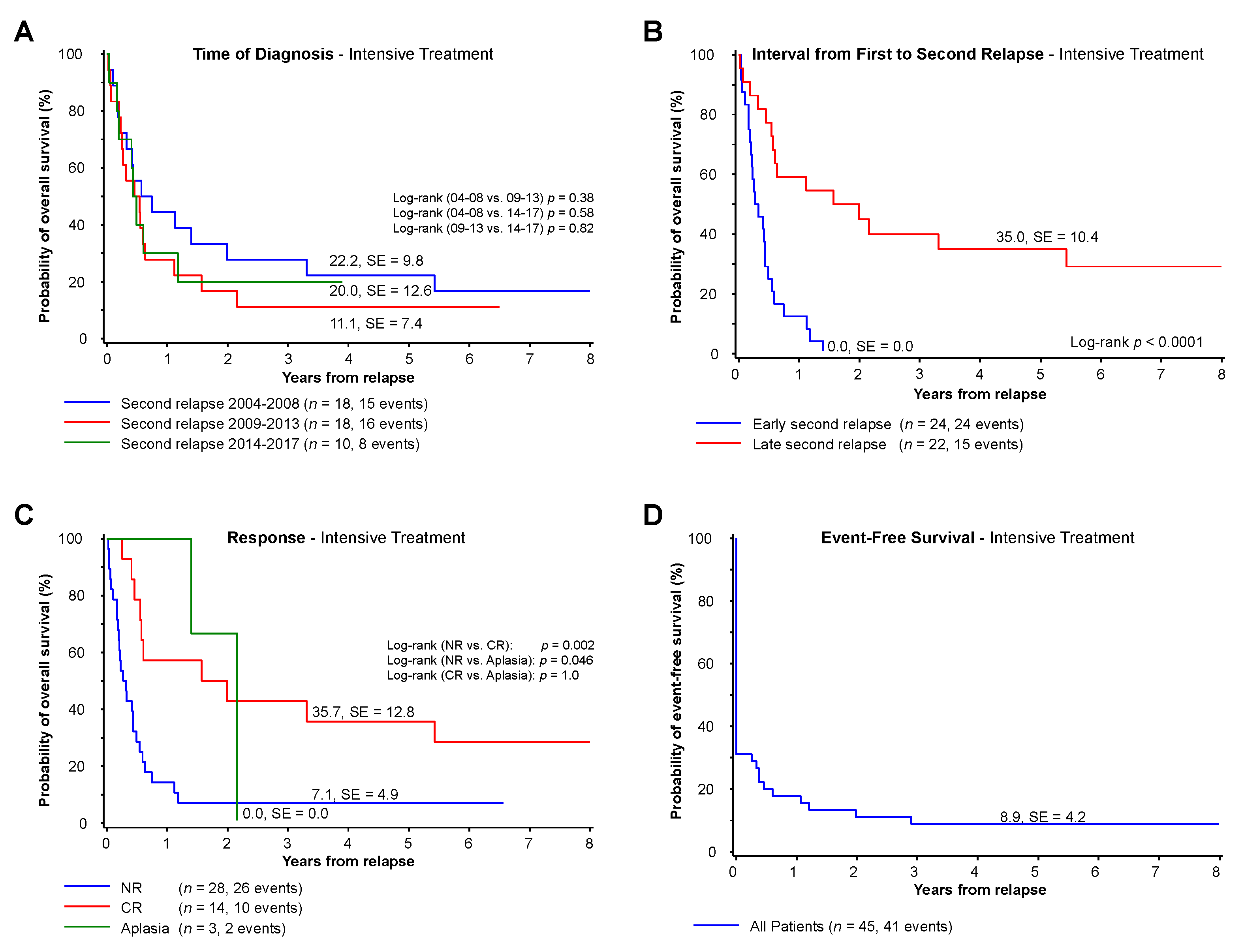
3.5. Patients Receiving an Intensive Treatment with the Intent to Induce Remission
4. Discussion
5. Conclusions
Supplementary Materials
Author Contributions
Funding
Institutional Review Board Statement
Informed Consent Statement
Data Availability Statement
Acknowledgments
Conflicts of Interest
References
- Abrahamsson, J.; Forestier, E.; Heldrup, J.; Jahnukainen, K.; Jonsson, O.G.; Lausen, B.; Palle, J.; Zeller, B.; Hasle, H. Response-guided induction therapy in pediatric acute myeloid leukemia with excellent remission rate. J. Clin. Oncol. 2011, 29, 310–315. [Google Scholar] [CrossRef] [PubMed] [Green Version]
- Gibson, B.E.; Wheatley, K.; Hann, I.M.; Stevens, R.F.; Webb, D.; Hills, R.K.; De Graaf, S.S.N.; Harrison, C.J. Treatment strategy and long-term results in paediatric patients treated in consecutive, U.K. AML trials. Leukemia 2005, 19, 2130–2138. [Google Scholar] [CrossRef] [PubMed] [Green Version]
- Pession, A.; Masetti, R.; Rizzari, C.; Putti, M.C.; Casale, F.; Fagioli, F.; Luciani, M.; Lo Nigro, L.; Menna, G.; Micalizzi, C.; et al. Results of the, A.I.EOP, A.M.L 2002/01 multicenter prospective trial for the treatment of children with acute myeloid leukemia. Blood 2013, 122, 170–178. [Google Scholar] [CrossRef] [Green Version]
- Creutzig, U.; Zimmermann, M.; Bourquin, J.P.; Dworzak, M.N.; Fleischhack, G.; Graf, N.; Klingebiel, T.; Kremens, B.; Lehrnbecher, T.; von Neuhoff, C.; et al. Randomized trial comparing liposomal daunorubicin with idarubicin as induction for pediatric acute myeloid leukemia: Results from Study AML-BFM 2004. Blood 2013, 122, 37–43. [Google Scholar] [CrossRef] [Green Version]
- Rubnitz, J.E.; Inaba, H.; Dahl, G.; Ribeiro, R.C.; Bowman, W.P.; Taub, J.; Pounds, S.; Razzouk, B.I.; Lacayo, N.J.; Cao, X.; et al. Minimal residual disease-directed therapy for childhood acute myeloid leukaemia: Results of the, A.M.L02 multicentre trial. Lancet Oncol. 2010, 11, 543–552. [Google Scholar] [CrossRef] [Green Version]
- Aplenc, R.; Meshinchi, S.; Sung, L.; Alonzo, T.; Choi, J.; Fisher, B.; Gerbing, R.; Hirsch, B.; Horton, T.; Kahwash, S.; et al. Bortezomib with standard chemotherapy for children with acute myeloid leukemia does not improve treatment outcomes: A report from the Children’s Oncology Group. Haematologica 2020, 105, 1879. [Google Scholar] [CrossRef] [PubMed]
- Rasche, M.; Zimmermann, M.; Borschel, L.; Bourquin, J.P.; Dworzak, M.; Klingebiel, T.; Lehrnbecher, T.; Creutzig, U.; Klusmann, J.H.; Reinhardt, D. Successes and challenges in the treatment of pediatric acute myeloid leukemia: A retrospective analysis of the AML-BFM trials from 1987 to 2012. Leukemia 2018, 32, 2167–2177. [Google Scholar] [CrossRef] [Green Version]
- Zwaan, C.M.; Kolb, E.A.; Reinhardt, D.; Abrahamsson, J.; Adachi, S.; Aplenc, R.; De Bont, E.S.; De Moerloose, B.; Dworzak, M.; Gibson, B.E.; et al. Collaborative Efforts Driving Progress in Pediatric Acute Myeloid Leukemia. J. Clin. Oncol. 2015, 33, 2949–2962. [Google Scholar] [CrossRef] [PubMed] [Green Version]
- Creutzig, U.; van den Heuvel-Eibrink, M.M.; Gibson, B.; Dworzak, M.N.; Adachi, S.; de Bont, E.; Harbott, J.; Hasle, H.; Johnston, D.; Kinoshita, A.; et al. Diagnosis and management of acute myeloid leukemia in children and adolescents: Recommendations from an international expert panel. Blood 2012, 120, 3187–3205. [Google Scholar] [CrossRef] [PubMed]
- Kaspers, G.J.; Zimmermann, M.; Reinhardt, D.; Gibson, B.E.; Tamminga, R.Y.; Aleinikova, O.; Armendariz, H.; Dworzak, M.; Ha, S.Y.; Hasle, H.; et al. Improved outcome in pediatric relapsed acute myeloid leukemia: Results of a randomized trial on liposomal daunorubicin by the International BFM Study Group. J. Clin. Oncol. 2013, 31, 599–607. [Google Scholar] [CrossRef]
- Sander, A.; Zimmermann, M.; Dworzak, M.; Fleischhack, G.; von Neuhoff, C.; Reinhardt, D.; Kaspers, G.J.L.; Creutzig, U. Consequent and intensified relapse therapy improved survival in pediatric, A.M.L: Results of relapse treatment in 379 patients of three consecutive, A.M.L-BFM trials. Leukemia 2010, 24, 1422–1428. [Google Scholar] [CrossRef] [Green Version]
- Mustafa, O.; Abdalla, K.; AlAzmi, A.A.; Elimam, N.; Abrar, M.B.; Jastaniah, W. FLAG/FLAG-IDA regimen for children with relapsed/refractory acute leukemia in the era of targeted novel therapies. J. Oncol Pharm Pract. 2019, 25, 1831–1838. [Google Scholar] [CrossRef] [PubMed]
- Rubnitz, J.E.; Razzouk, B.I.; Lensing, S.; Pounds, S.; Pui, C.H.; Ribeiro, R.C. Prognostic factors and outcome of recurrence in childhood acute myeloid leukemia. Cancer 2007, 109, 157–163. [Google Scholar] [CrossRef]
- Stahnke, K.; Boos, J.; Bender-Gotze, C.; Ritter, J.; Zimmermann, M.; Creutzig, U. Duration of first remission predicts remission rates and long-term survival in children with relapsed acute myelogenous leukemia. Leukemia 1998, 12, 1534–1538. [Google Scholar] [CrossRef] [PubMed] [Green Version]
- Webb, D.K.; Wheatley, K.; Harrison, G.; Stevens, R.F.; Hann, I.M. Outcome for children with relapsed acute myeloid leukaemia following initial therapy in the Medical Research Council (MRC) AML 10 trial. MRC Childhood Leukaemia Working Party. Leukemia 1999, 13, 25–31. [Google Scholar] [CrossRef] [PubMed] [Green Version]
- Nakayama, H.; Tabuchi, K.; Tawa, A.; Tsukimoto, I.; Tsuchida, M.; Morimoto, A.; Yabe, H.; Horibe, K.; Hanada, R.; Imaizumi, M.; et al. Outcome of children with relapsed acute myeloid leukemia following initial therapy under the, A.M.L99 protocol. Int. J. Hematol. 2014, 100, 171–179. [Google Scholar] [CrossRef] [PubMed]
- Karlsson, L.; Forestier, E.; Hasle, H.; Jahnukainen, K.; Jonsson, O.G.; Lausen, B.; Norén Nyström, U.; Palle, J.; Tierens, A.; Zeller, B.; et al. Outcome after intensive reinduction therapy and allogeneic stem cell transplant in paediatric relapsed acute myeloid leukaemia. Br. J. Haematol. 2017, 178, 592–602. [Google Scholar] [CrossRef] [PubMed] [Green Version]
- Hoffman, A.E.; Schoonmade, L.J.; Kaspers, G.J. Pediatric relapsed acute myeloid leukemia: A systematic review. Expert Rev. Anticancer Ther. 2020, 1–8. [Google Scholar] [CrossRef]
- Abrahamsson, J.; Clausen, N.; Gustafsson, G.; Hovi, L.; Jonmundsson, G.; Zeller, B.; Forestier, E.; Heldrup, J.; Hasle, H.; Nordic Society for Paediatric Haematology and Oncology (NOPHO). Improved outcome after relapse in children with acute myeloid leukaemia. Br. J. Haematol. 2007, 136, 229–236. [Google Scholar] [CrossRef] [PubMed]
- Zwaan, C.M.; Reinhardt, D.; Zimmerman, M.; Hasle, H.; Stary, J.; Stark, B.; Dworzak, M.; Creutzig, U.; Kaspers, G.J.L.; International BFM Study Group on Paediatric AML. Salvage treatment for children with refractory first or second relapse of acute myeloid leukaemia with gemtuzumab ozogamicin: Results of a phase, I.I. study. Br. J. Haematol. 2010, 148, 768–776. [Google Scholar] [CrossRef]
- Van Eijkelenburg, N.K.A.; Rasche, M.; Ghazaly, E.; Dworzak, M.N.; Klingebiel, T.; Rossig, C.; Leverger, G.; Stary, J.; De Bont, E.S.; Chitu, D.A.; et al. Clofarabine, high-dose cytarabine and liposomal daunorubicin in pediatric relapsed/refractory acute myeloid leukemia: A phase IB study. Haematologica 2018, 103, 1484–1492. [Google Scholar] [CrossRef] [Green Version]
- Aladjidi, N.; Auvrignon, A.; Leblanc, T.; Perel, Y.; Benard, A.; Bordigoni, P.; Gandemer, V.; Thuret, I.; Hugues Dalle, J.; Piguet, C.; et al. Outcome in children with relapsed acute myeloid leukemia after initial treatment with the French Leucemie Aique Myeloide Enfant (LAME) 89/91 protocol of the French Society of Pediatric Hematology and Immunology. J. Clin. Oncol. 2003, 21, 4377–4385. [Google Scholar] [CrossRef] [PubMed]
- Wells, R.J.; Adams, M.T.; Alonzo, T.A.; Arceci, R.J.; Buckley, J.; Buxton, A.B.; Dusenbery, K.; Gamis, A.; Masterson, M.; Vik, T.; et al. Mitoxantrone and cytarabine induction, high-dose cytarabine, and etoposide intensification for pediatric patients with relapsed or refractory acute myeloid leukemia: Children’s Cancer Group Study 2951. J. Clin. Oncol. 2003, 21, 2940–2947. [Google Scholar] [CrossRef]
- Creutzig, U.; Zimmermann, M.; Dworzak, M.N.; Gibson, B.; Tamminga, R.; Abrahamsson, J.; Ha, S.-Y.; Hasle, H.; Maschan, A.; Bertrand, Y.; et al. The prognostic significance of early treatment response in pediatric relapsed acute myeloid leukemia: Results of the international study Relapsed AML 2001/01. Haematologica 2014, 99, 1472–1478. [Google Scholar] [CrossRef] [PubMed]
- Goemans, B.F.; Tamminga, R.Y.; Corbijn, C.M.; Hahlen, K.; Kaspers, G.J. Outcome for children with relapsed acute myeloid leukemia in the Netherlands following initial treatment between 1980 and 1998: Survival after chemotherapy only? Haematologica 2008, 93, 1418–1420. [Google Scholar] [CrossRef]
- Niewerth, D.; Creutzig, U.; Bierings, M.B.; Kaspers, G.J. A review on allogeneic stem cell transplantation for newly diagnosed pediatric acute myeloid leukemia. Blood 2010, 116, 2205–2214. [Google Scholar] [CrossRef] [Green Version]
- Bachas, C.; Schuurhuis, G.J.; Reinhardt, D.; Creutzig, U.; Kwidama, Z.J.; Zwaan, C.M.; van den Heuvel-Eibrink, M.M.; De Bont, E.S.; Elitzur, S.; Rizzari, C.; et al. Clinical relevance of molecular aberrations in paediatric acute myeloid leukaemia at first relapse. Br. J. Haematol. 2014, 166, 902–910. [Google Scholar] [CrossRef] [Green Version]
- Sauer, M.G.; Lang, P.J.; Albert, M.H.; Bader, P.; Creutzig, U.; Eyrich, M.; Greil, J.; Gruhn, B.; Holter, W.; Klingebiel, T.; et al. Hematopoietic stem cell transplantation for children with acute myeloid leukemia-results of the AML, SCT-BFM 2007 trial. Leukemia 2020, 34, 613–624. [Google Scholar] [CrossRef] [PubMed]
- Karol, S.E.; Alexander, T.B.; Budhraja, A.; Pounds, S.B.; Canavera, K.; Wang, L.; Wolf, J.; Klco, J.M.; Mead, P.E.; Das Gupta, S.; et al. Venetoclax in combination with cytarabine with or without idarubicin in children with relapsed or refractory acute myeloid leukaemia: A phase 1, dose-escalation study. Lancet Oncol. 2020, 21, 551–560. [Google Scholar] [CrossRef]
- Bunin, N.J.; Davies, S.M.; Aplenc, R.; Camitta, B.M.; DeSantes, K.B.; Goyal, R.K.; Kapoor, N.; Kernan, N.A.; Rosenthal, J.; Smith, F.O.; et al. Unrelated donor bone marrow transplantation for children with acute myeloid leukemia beyond first remission or refractory to chemotherapy. J. Clin. Oncol. 2008, 26, 4326–4332. [Google Scholar] [CrossRef] [Green Version]
- Nishikawa, T.; Inagaki, J.; Nagatoshi, Y.; Fukano, R.; Nakashima, K.; Ito, N.; Sawa, D.; Kawano, Y.; Okamura, J. The second therapeutic trial for children with hematological malignancies who relapsed after their first allogeneic, S.C.T: Long-term outcomes. Pediatr. Transplant. 2012, 16, 722–728. [Google Scholar] [CrossRef] [PubMed]
- Stoiser, B.; Knobl, P.; Fonatsch, C.; Haas, O.A.; Mitterbauer, G.; Weltermann, A.; Geissler, K.; Valent, P.; Sperr, W.; Pabinger, I.; et al. Prognosis of patients with a second relapse of acute myeloid leukemia. Leukemia 2000, 14, 2059–2063. [Google Scholar] [CrossRef] [PubMed] [Green Version]


| Characteristics | Second Relapse | Second Relapse Intensive Treatment | |
|---|---|---|---|
| Number of patients | n (%) | 73 (100%) | 46 (100%) |
| Initial characteristics | |||
| Age | (years), median (range) | 7.4 (0.2–17.8) | 7.7 (0.2–17.8) |
| Gender | Male | 44 (60%) | 28 (61%) |
| Female | 29 (40%) | 18 (39%) | |
| FAB | M0 | 4 (5%) | 3 (7%) |
| M1/M2 | 27 (37%) | 17 (37%) | |
| M4/M5 | 33 (45%) | 21 (46%) | |
| M4eo | 1 (1%) | 1 (2%) | |
| M6 | 2 (3%) | 2 (4%) | |
| M7 | 5 (7%) | 2 (4%) | |
| Non-classified | 1 (1%) | --- | |
| Blood counts | WBC (×103/dL) median (range) | 13.7 (1.1–331.0) | 14.9 (1.1–331.0) |
| Risk group * | Standard | 6 (8%) | 3 (7%) |
| Intermediate | 29 (40%) | 18 (39%) | |
| High | 32 (44%) | 23 (50%) | |
| No data | 6 (8%) | 2 (4%) | |
| Initial response | CR | 68 (93%) | 43 (94%) |
| Previous treatment regimen | |||
| Initial treatment protocol | AML-BFM study 2004 | 62 (85%) | 38 (83%) |
| AML-BFM registry 2012 | 9 (12%) | 6 (13%) | |
| AML-BFM study 2012 | 2 (3%) | 2 (4%) | |
| Previous relapse treatment | DNX-FLA(G)+/−FLA(G) | 51 (70%) | 32 (70%) |
| DNX-FLA(G) + other intensive regimen ** | 14 (19%) | 9 (20%) | |
| Clofarabine-cont. regimen | 2 (3%) | 2 (4%) | |
| Others | 6 (8%) | 3 (7%) | |
| Previous HSCT | No HSCT | 9 (12%) | 7 (15%) |
| HSCT at initial disease only | 2 (3%) | 1 (2%) | |
| HSCT at first relapse | 56 (77%) | 35 (76%) | |
| HSCT at initial disease and relapse | 5 (7%) | 3 (7%) | |
| Unknown | 1 (1%) | -- | |
| Relapse characteristics | |||
| Age | At first relapse (years), median (range) | 8.4 (0.8–18.9) | 8.6 (0.8–18.8) |
| At second relapse (years), median (range) | 9.2 (1.6–20.2) | 9.5 (1.8–20.2) | |
| Time to subsequent relapse | Early first relapse | 44 (60%) | 26 (57%) |
| Late first relapse | 29 (40%) | 20 (44%) | |
| Early second relapse | 44 (60%) | 24 (52%) | |
| Late second relapse | 29 (40%) | 22 (48%) | |
| Second Relapse Treatment and Response of Pediatric AML | Patients (%) | |
| 73 (100%) | ||
| Chemotherapy * | Intensive treatment with the intent to induce remission | 46 (63%) |
| • Re-induction including (DNX)-FLA(G) +/−FLA (G) | 12 (16%) | |
| - Additional treatment including GO | 3 | |
| - Additional treatment including clofarabine | 3 | |
| • Re-Induction including clofarabine or GO, no (DNX)-FLA(G) | 20 (27%) | |
| • Individual approaches | 14 (19%) | |
| Others (including withdrawal immunosuppression, DLI or direct HSCT) | 10 (14%) | |
| Palliative care | 17 (23%) | |
| HSCT | No HSCT | 47 † (65%) |
| HSCT | 25 (35%) | |
| • First HSCT | 2 (8%) | |
| • Second HSCT | 21 (88%) | |
| • Third HSCT | 1 (4%) | |
| • Unknown | 1 | |
| Unknown | 1 | |
| Response after intensive treatment | Response evaluation available after up to two cycles of therapy | 45 (98%) |
| • CR | 14 (31%) | |
| • NEL without peripheral regeneration | 3 (7%) | |
| • NR | 28 (62%) | |
| Response evaluation available at the end of treatment including HSCT | 46 (100%) | |
| • CR | 19 (41%) | |
| • NEL without peripheral regeneration | 2 (4%) | |
| • NR | 25 (54%) | |
Publisher’s Note: MDPI stays neutral with regard to jurisdictional claims in published maps and institutional affiliations. |
© 2021 by the authors. Licensee MDPI, Basel, Switzerland. This article is an open access article distributed under the terms and conditions of the Creative Commons Attribution (CC BY) license (http://creativecommons.org/licenses/by/4.0/).
Share and Cite
Rasche, M.; Steidel, E.; Zimmermann, M.; Bourquin, J.-P.; Boztug, H.; Janotova, I.; Kolb, E.A.; Lehrnbecher, T.; von Neuhoff, N.; Niktoreh, N.; et al. Second Relapse of Pediatric Patients with Acute Myeloid Leukemia: A Report on Current Treatment Strategies and Outcome of the AML-BFM Study Group. Cancers 2021, 13, 789. https://doi.org/10.3390/cancers13040789
Rasche M, Steidel E, Zimmermann M, Bourquin J-P, Boztug H, Janotova I, Kolb EA, Lehrnbecher T, von Neuhoff N, Niktoreh N, et al. Second Relapse of Pediatric Patients with Acute Myeloid Leukemia: A Report on Current Treatment Strategies and Outcome of the AML-BFM Study Group. Cancers. 2021; 13(4):789. https://doi.org/10.3390/cancers13040789
Chicago/Turabian StyleRasche, Mareike, Emma Steidel, Martin Zimmermann, Jean-Pierre Bourquin, Heidrun Boztug, Iveta Janotova, E. Anders Kolb, Thomas Lehrnbecher, Nils von Neuhoff, Naghmeh Niktoreh, and et al. 2021. "Second Relapse of Pediatric Patients with Acute Myeloid Leukemia: A Report on Current Treatment Strategies and Outcome of the AML-BFM Study Group" Cancers 13, no. 4: 789. https://doi.org/10.3390/cancers13040789