Article

CSF1R Inhibition Combined with GM-CSF Reprograms Macrophages and Disrupts Protumoral Interplays with AML Cells

by
Tatiana Smirnova
*,†,
Caroline Spertini
and
Olivier Spertini
*
Service and Central Laboratory of Hematology, Centre Hospitalier Universitaire Vaudois, University of Lausanne, 1011 Lausanne, Switzerland
*
Authors to whom correspondence should be addressed.
These authors contributed equally to the study and are regarded as joint first authors on this manuscript.
Cancers 2021, 13(21), 5289; https://doi.org/10.3390/cancers13215289
Submission received: 3 September 2021 / Revised: 4 October 2021 / Accepted: 15 October 2021 / Published: 21 October 2021

Abstract

:

Simple Summary

Acute myeloid leukemia is a blood disease whose long-term treatment is not satisfactory due to frequent (>50%) relapse despite complete initial remission. By adopting protumoral properties, macrophages in the leukemia cell microenvironment may promote resistance to treatment leading to relapse. Our goal was to assess whether macrophages in contact with leukemia cells have an impact on blast survival and sensitivity to chemotherapy. We observed that leukemia cells can educate macrophages to support their survival and proliferation and reduce their sensitivity to therapy, as long as the CSF1 receptor remains active and they are in close contact. By inhibiting the activity of the CSF1 receptor, in the presence of GM-CSF, we could modify the macrophage phenotype and increase blast apoptosis and sensitivity to treatment. Our results indicate that leukemia therapies should not only target blasts but also their microenvironment and specifically macrophages and their receptor for CSF1.

Abstract

Relapse is a major issue in acute myeloid leukemia (AML) and while the contribution of gene mutations in developing drug resistance is well established, little is known on the role of macrophages (MΦs) in an AML cell microenvironment. We examined whether myeloblasts could educate MΦs to adopt a protumoral orientation supporting myeloblast survival and resistance to therapy. Flow cytometry analyses demonstrated that M2-like CD163+ MΦs are abundantly present, at diagnosis, in the bone marrow of AML patients. We showed that myeloblasts, or their conditioned medium, polarize monocytes to M2-like CD163+ MΦs, induce the secretion of many protumoral factors, and promote myeloblast survival and proliferation as long as close intercellular contacts are maintained. Importantly, pharmacologic inhibition of the CSF1 receptor (CSF1R), in the presence of GM-CSF, reprogrammed MΦ polarization to an M1-like orientation, induced the secretion of soluble factors with antitumoral activities, reduced protumoral agonists, and promoted the apoptosis of myeloblasts interacting with MΦs. Furthermore, myeloblasts, which became resistant to venetoclax or midostaurin during their interplay with protumoral CD163+ MΦs, regained sensitivity to these targeted therapies following CSF1R inhibition in the presence of GM-CSF. These data reveal a crucial role of CD163+ MΦ interactions with myeloblasts that promote myeloblast survival and identify CSF1R inhibition as a novel target for AML therapy.

Graphical Abstract

1. Introduction

AML is a very aggressive disease with poor long-term survival [1]. Seventy to ninety percent of adult patients under the age of 60 achieve complete remission; however, 50–70%, with intermediate or high genetic risk, relapse within 5 years. The European LeukemiaNet (ELN) 2017 recommendations for AML treatment are presently based on risk stratification by genetics and also consider the possibility to target the AML microenvironment as novel therapies [2].
Chemotherapy and hematopoietic stem cell transplantation most often allow the achievement of sustained complete remission. Moreover, targeted therapies improve the AML response to chemotherapy and favorably affect survival [3]. However, they remain restricted to a small number of AML categories. Over time and regardless of the treatment, the tumor microenvironment strongly contributes to promoting the growth and survival of drug-resistant subclones, leading to relapse [4,5]. Several clinical studies have demonstrated that heavy tumor infiltration by MΦs is of poor prognosis: tumor-associated macrophages (TAMs) promote tumor cell growth, resistance to treatment, and escape from immune surveillance [4,6].
TAMs are often called M2-like protumoral MΦs while the classical M1-like MΦs have inflammatory, antitumor properties [7,8]. However, MΦ plasticity is complex [9,10], and the M1/M2 nomenclature does not correspond to observations made in tumors, where mixed phenotype MΦs, expressing both M1 and M2 markers, are observed. In the tumor microenvironment, malignant cells activate and educate MΦs, which secrete cytokines and promote angiogenesis, matrix remodeling, metastasis, immunosuppression, cancer cell survival, and drug resistance [9,11,12,13].
Tumor escape from immune surveillance is further promoted by the expression of the “do not eat me” signal, CD47 [11,14] and the increased expression of programmed cell death protein-1 (PD1) by tumor-infiltrating T-cells and of its ligand PD-L1 (programmed death-ligand 1) by TAMs [15]. While the involvement of TAMs in the solid cancer microenvironment is established [16,17], little information is available on their role in human AML. In mice, heterogeneity in MΦ polarization was observed in an MLL-AF9 AML model [18,19]. Myeloblasts can recruit MΦs to support their proliferation in bone marrow (BM) and spleen [20].
The scavenger receptor CD163 is a marker of TAMs with protumoral characteristics [7,12,13]. Several studies reported that CD163 is a poor prognosis marker in solid tumors [13] and in AML [21]. CD163 expression appears to be required to allow mouse and human MΦs to adopt a protumoral phenotype and support malignant cell proliferation. Thus, in a mouse model of sarcoma, tumor growth was abrogated in CD163 deficient mice [22]. CD163-expressing TAMs may contribute to create an immunosuppressive microenvironment. In melanoma, the depletion of CD163+ MΦs promotes tumor infiltration by activated T lymphocytes and tumor regression [7].
Targeting TAM polarization is a novel important therapeutic approach in aggressive cancers [16,23]. The colony-stimulating factor-1 (CSF-1, also called M-CSF), which controls the survival, proliferation, differentiation, and polarization of MΦs by binding to its receptor, the CSF1R, is a promising therapeutic target in aggressive tumors and possibly in AML [24]. In multiple myeloma and mantle cell lymphoma, the inhibition of CSF1R can reprogram TAMs and reverse drug-resistance [25,26,27,28]. However, whether MΦs contribute to AML cell proliferation and/or survival in creating a protumorigenic niche associated with a protumoral MΦ orientation is not established.
Using human AML cell lines and primary myeloblasts, we examined the impact of AML cells on allogeneic and autologous MΦs. We showed that the co-culture of primary myeloblasts with healthy donor (HD) monocytes or with BM cells in their microenvironment induces a protumoral MΦ orientation (AML-MΦ), which supports blast cell proliferation and survival. Furthermore, using CSF1R inhibitors and granulocyte-macrophage colony-stimulating factor (GM-CSF), we succeeded in reprogramming protumoral AML-MΦs and in reversing drug-resistance induced by myeloblast cross-talk with MΦs.

2. Materials and Methods

2.1. Patient Samples

Heparinized blood and BM samples were obtained from 42 newly diagnosed AML patients at CHUV (Centre Hospitalier Universitaire Vaudois, Lausanne, Switzerland). Informed consent was obtained from all subjects involved in the study. After red cell lysis with ammonium chloride, the samples were processed for leukocyte immunostaining and analysis by flow cytometry (FC) [29]. Leukemia diagnosis was based on WHO 2016 classification [30] and ELN 2017 risk stratification by genetics [2] and are indicated in Table S1. The study was conducted in accordance with the Declaration of Helsinki and approved by the Commission Cantonale d’Ethique sur la recherche sur l’être humain CER-VD (protocol 2017-01509, 9 November 2017).

2.2. Immunophenotypic Analyzes by Flow Cytometry

Cell proliferation, apoptosis, and immunophenotypic analyzes were performed using a Beckman Coulter Gallios flow cytometer. Patient samples were analyzed with a 10-color/24-antibody panel [29] according to ELN recommendations [31]. MΦs were detached from culture dishes in phosphate-buffered saline (PBS) containing 10 mM ethylenediaminetetraacetic acid (EDTA), for 10 min at 37 °C, washed with Roswell Park Memorial Institute (RPMI) 1640 and characterized using CD45, CD14, CD163, CD80, CD206, and CSF1R; additional blast and lymphocyte markers were used for patient BM and peripheral blood (PB) sample and BM co-culture analyses (Table S2).

2.3. Healthy Donor-Derived Macrophages

The monocytic fraction was isolated by centrifuging HD PB anticoagulated with citrate phosphate dextrose-adenine 1 and diluted 1/2 with PBS for 10 min at 1000 g to prepare a buffy coat. The concentrated leukocytic fraction was then centrifuged at 400 g for 40 min on a Ficoll-Paque PLUS (GE Healthcare, Dietikon, Switzerland) gradient. Mononuclear cells were collected and plated in RPMI + 10% heat-inactivated fetal bovine serum (FBS) = plain medium (PM), see Figure S1 for the schematic protocol; monocyte-derived MΦs exposed to 10 ng/mL recombinant human M-CSF (ImmunoTools, Friesoythe, Germany) were called M-MΦs and those exposed to 1000 U/mL GM-CSF (Miltenyi Biotec, Solothurn, Switzerland): GM-MΦs [25,32].
After overnight culture, the wells were rinsed to deplete non-adherent cells; >95% of adherent cells were CD14+ monocytes. The medium was changed, with the added factors, every 2 days, for 7–9 days, when CD163 expression was checked by FC. Alternatively, to obtain HDAML-MΦs, HD monocytes were stimulated for 7–9 days with (1) primary myeloblast or cell line conditioned medium (CM) diluted 1:1 with PM; with (2) AML cell lines (1–5 × 10⁴/well) or primary blasts (0.5–1 × 10⁶/well), co-cultured in direct contact with HD monocytes; or (3) separated from MΦs by a 0.4 µm-pore membrane insert (Transwell (TW), Falcon, Dietikon, Switzerland).
For MΦ reprogramming experiments, HD MΦ monolayers initially activated with M-CSF (M-MΦ) or blast CM (HDAML-MΦ) were washed with PBS and cultured for 7–9 more days in PM containing GM-CSF and GW2580 (1 μM, Sigma-Aldrich, Buchs, Switzerland) [25] or PLX3397 (100 nM, MedChemExpress, Luzern, Switzerland), renewed every 2 days. The change in MΦ CD163 expression after reprogramming was assessed by FC, as described above.

2.4. Cell Lines and Cell Culture

HL-60 (AML with maturation), U937 (monoblastic leukemia), NB4 (acute promyelocytic leukemia), OCI-AML3 (NPM1 mutated) cell lines were cultured in plain medium (PM) [29] and MV-4-11 (monoblastic AML with FLT3-ITD mutation) in Iscove’s Modified Dulbecco Medium + 10% FBS.

2.5. PKH Labeling and Apoptosis Detection

Cells were labeled with 10 µM PKH26 (Sigma-Aldrich) according to the manufacturer’s protocol and immediately added to rinsed wells containing adherent MΦs for assays. A fraction of the labeled cells were fixed in 1% paraformaldehyde and used to calculate the normalized PKH mean fluorescence intensity (MFI), as PKH MFI at day 4 divided by PKH MFI at day 0. Blast cell apoptosis was assessed by FC, analyzing cells stained with annexin V (AnnV, eBioscience, Zug, Switzerland).

2.6. Cleaved Caspase-3 Detection

Cells were cultured in 100% PM or in 75% PM supplemented with 25% MΦ-CM. After 48 h, the cells were lysed, and the cell lysates were subjected to SDS-PAGE, transferred to nitrocellulose membranes, immunoblotted with anti-cleaved caspase-3 antibody (Cell Signaling Technology) followed by horseradish peroxidase-linked goat anti-rabbit immunoglobulin G (IgG) antibody (Cell Signaling Technology), and revealed with Luminata Forte chemiluminescence (Millipore, Schaffhausen, Switzerland) [29]. As a loading control, membranes were also immunoblotted with anti-α-tubulin antibody (Sigma-Aldrich) followed by horseradish peroxidase-conjugated sheep anti-mouse IgG antibody (Amersham, Dietikon, Switzerland) and revealed with Luminata Crescendo chemiluminescence (Millipore). Blocking antibodies were from InvivoGen (anti-IL-6) and R&D Systems (anti-tumor necrosis factor α (anti-TNF-α)).

2.7. Cytokine Arrays

The CM of the indicated leukemia cell lines, primary myeloblasts or MΦs were collected and centrifuged twice at 1500 rpm for 10 min, twice for 10 min at 2500 rpm, and filtered with 0.22 μm low protein binding syringe filter. CM was collected at day 7, from M- or GM-MΦs cultured in medium containing M- or GM-CSF, respectively. CM from reprogrammed MΦs (RGM/GW-MΦs) was obtained after one week of culture with M-CSF, followed by another week in medium supplemented with GM-CSF and GW2580. CM from M-, GM-, and RGM/GW-MΦs were obtained from at least three different HD and pooled for cytokine assays. The CM from primary AML blasts (mononuclear cell suspension containing >95% blast cells) co-cultured with MΦ were also analyzed and processed as above.
Cytokine array assays were conducted according to the manufacturer’s protocol (R&D Systems, Human XL Cytokine Array kit #ARY022B). X-ray films were digitalized with ImageScanner III (GE Healthcare) and dot pixel density was measured with ImageQuant software (Molecular Dynamics, Glattbrugg, Switzerland). The mean of duplicates was normalized to the mean pixel density of the six reference spots.

2.8. Myeloblast Resistance to Venetoclax and Midostaurin

HD MΦs were polarized for 7 days with M-CSF and then either reprogrammed for 7 more days in medium containing GW2580 or PLX3397 and GM-CSF or kept in M-CSF-containing medium. MΦ orientation and reprogramming was confirmed by FC. Medium was then removed, and MΦs were washed with PBS. Following this, 150 × 103 NB4 or MV-4-11 were added to MΦ monolayers in PM with inhibitors (venetoclax from LC Laboratories, or midostaurin from Sigma-Aldrich). After 48 h, cells were removed from the wells, stained for AnnV and/or 7-aminoactinomycin D (7-AAD), and analyzed by FC. Experiments were repeated using MΦs from at least three different HD.

2.9. Statistical Analysis

Statistical significance of differences between groups was examined with the Mann-Whitney (M-W) test between two nonparametric unpaired groups, the paired t-test between parametric matched samples, or the Wilcoxon matched-pairs signed rank (WMP) test between nonparametric matched samples. p values < 0.05 were considered as significant.

3. Results

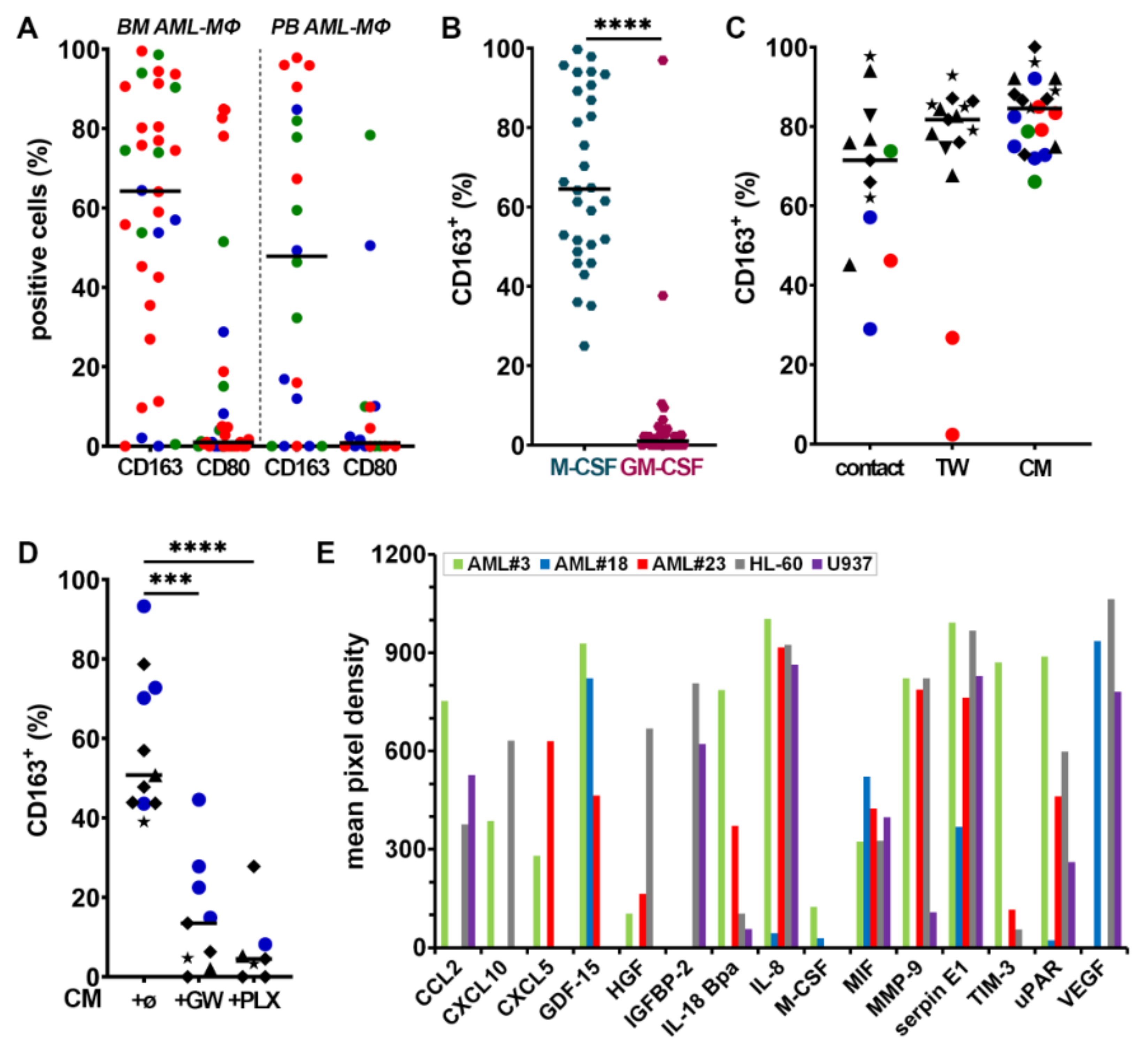
3.1. Presence of CD163-Expressing Macrophages in BM from AML Patients

BM and/or PB were obtained from 42 adult patients with newly diagnosed AML (Table S1). MΦs and myeloblasts were identified by multiparameter FC with a panel of antibodies (Table S2, see Methods for details) [29,31]. BM and PB analyses showed that independently of ELN risk categories [2], the majority of MΦs in AML patients expressed CD163 (Figure 1A), a marker frequently associated with a protumoral phenotype [18,19,20,21]. By contrast, CD80, a marker typically associated with the inflammatory phenotype and whose expression and functions remain poorly characterized in AML, was expressed at much lower frequency [25,27].

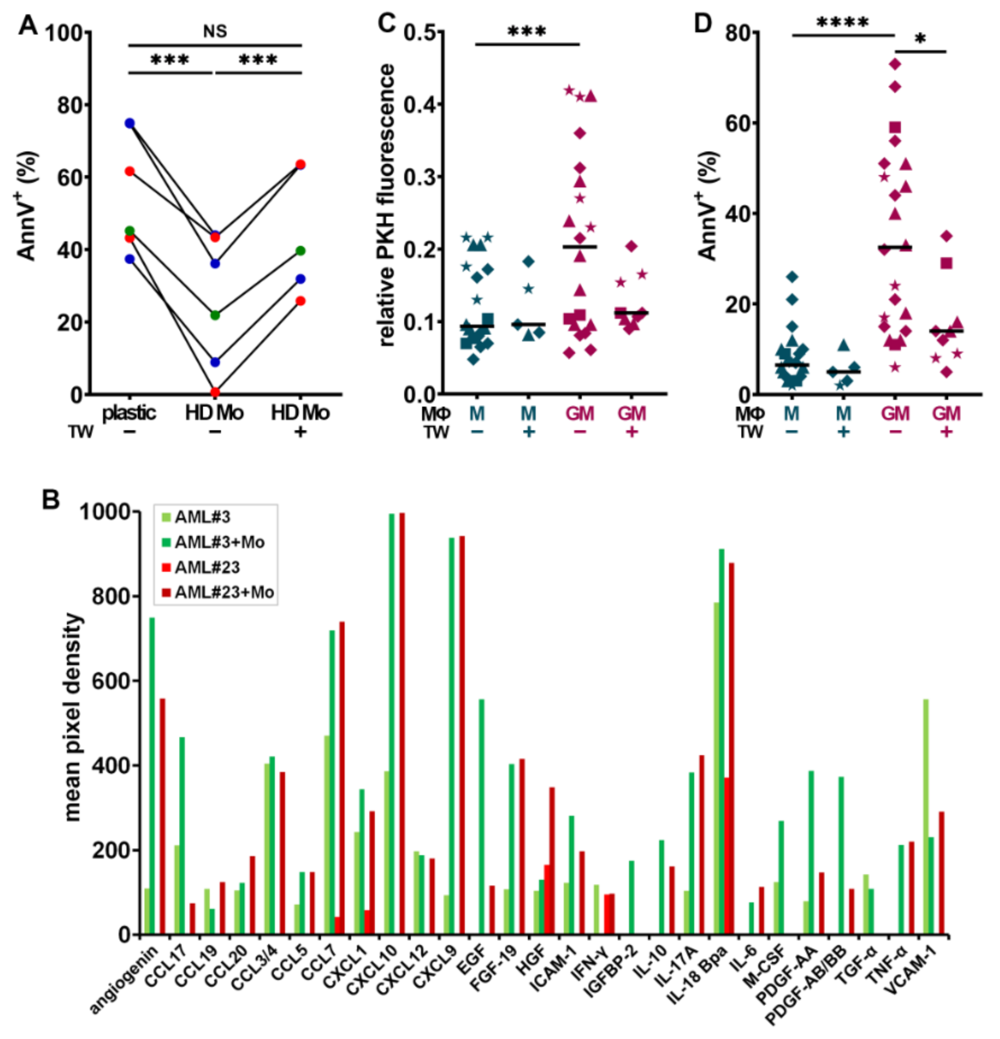
3.2. Myeloblasts Polarize HD Monocytes to Macrophages

We first monitored the differentiation of HD Mo into MΦs by adding either M- or GM-CSF in PM for one week. Monocytes cultivated in M-CSF differentiated into M2-like CD163high MΦs, whereas those cultured in GM-CSF differentiated into M1-like CD163low MΦs (Figure 1B). To determine whether myeloblasts can polarize protumoral MΦs and induce CD163 expression at their surface, we performed co-cultures of HD Mo with human AML cell lines or primary myeloblasts obtained from AML patients. After 7 days, HD Mo differentiated into MΦs (HDAML-MΦs; Figure 1C, contact), whose CD163 expression was similar to that of HD MΦs cultured in medium supplemented with M-CSF.
To determine if direct contact is required for myeloblasts to polarize monocytes into MΦs, they were separated with a 0.4 μm-pore membrane-insert (TW). CD163 expression on HDAML-MΦs was consistently observed even in the absence of direct contact (Figure 1C). Similar results were obtained by stimulating monocytes with CM from AML cell lines or primary myeloblasts. As control, the co-culture of normal hematopoietic stem progenitor cells (HSPC) with HD Mo, under the same conditions as AML cells, did not induce CD163 expression at the surface of monocytes (Figure S2A). In addition, culture in PM or with other inflammatory factors (lipopolysaccharide, TNF-α, IL-1β) did not induce CD163 expression; moreover, boiled U937 CM also failed to induce CD163 expression (Figure S3). These results suggest that soluble factors released by myeloblasts contribute to inducing MΦ polarization.
Several other polarization markers were analyzed: the expression level of CD80 was expectedly higher in GM- than in M-MΦs (Figure S4A) but exhibited some variability among HDAML-MΦs (Figure S4B). M- and GM-MΦs exhibited variable expression of CD206 (Figure S4C), a marker typically associated with protumoral MΦ activation in different tumor types including AML [33]. CD206 was also expressed by most HDAML-MΦs (Figure S4D), both after contact with blasts or with their CM only.
As M-CSF plays a crucial role in polarizing M-MΦs, we assessed whether inhibiting its receptor could prevent the up-regulation of CD163 expression induced by myeloblasts CM. Indeed, GW2580 and PLX3397, when added into CM, significantly prevented CD163 upregulation (Figure 1D), confirming a major role for CSF1R signaling in inducing CD163 expression.
To understand what factors in AML may be important in contributing to alternative MΦ polarization and the induction of CD163 expression, we performed semi-quantitative cytokine array analyses using myeloblast CM from three AML patients with favorable, intermediate, or high genetic risk (patients #3, #18, and #23, respectively) and from two AML cell lines (HL-60 and U937; Figure 1E). The assay can detect 105 factors, indicated in Table S3, which shows the pixel density of dots revealed on membranes illustrated in Figure S5A), some of which may contribute to promote cancer growth, cell migration, angiogenesis, and metastasis, such as CXCL5, the urokinase-type plasminogen activator receptor (uPAR), the matrix metalloproteinase 9 (MMP-9), serpin E1 [34], and the growth differentiation factor-15 (GDF-15, a novel targetable immune checkpoint [35]).
VEGF (vascular endothelial growth factor), and other angiopoietic factors, with CXCL5 [36], M-CSF [37], and CD163-expressing MΦs promote angiogenesis [38]. The hepatocyte growth factor (HGF) is a key player in AML cell proliferation [24], drug-resistance, and poor survival [39,40]. The insulin-like growth factor (IGF) and its binding protein insulin-like growth factor binding protein 2 (IGFBP-2), secreted by HL-60 and U937 cells, have important functions in promoting blast proliferation/survival and resistance to chemotherapy [41,42,43]. Cytokines, such as IL-18 and the IL-18-binding protein, gained increasing attention for their roles in MΦ activation syndrome [44]. Furthermore, M-CSF, macrophage migration inhibitory factor (MIF), IL-8, and CCL2 may promote a protumoral MΦ polarization [25,45] contributing to forming a protective niche for leukemia stem cells [17].
CXCL10 could contribute to either a protumoral or an antitumoral activation, depending on the context [46]. The relative levels of cytokine secretion strongly differ between patients and/or leukemia cell lines, which is consistent with the heterogeneity of AML genetics, phenotypes, and biology and may contribute to the variability in the expression of markers associated to HDAML-MΦ polarization, such as CD163, CD80, and CD206.

3.3. HD Macrophages Promote Primary Myeloblast Survival

Since myeloblasts can polarize monocyte-derived MΦs (Figure 1C), we examined whether co-culturing primary human myeloblasts with HD monocytes (HD Mo) would affect blast cell survival and proliferation. Myeloblasts obtained from nine newly-diagnosed patients were cultured alone or with freshly isolated HD Mo, in direct contact or separated by a TW, for 7 days. Myeloblasts labeled with PKH26 did not reveal significant changes in proliferation when they interacted with HD Mo (T. S., personal observation, 2020). However, the co-culture of six patient cells in direct contact with HD Mo significantly improved their survival at day 7, compared to culture in plastic (Figure 2A).
This survival advantage was dependent on direct contact with the MΦs, as it was abrogated when blasts and MΦs were separated by TW inserts. These observations suggest that the survival of certain sub-types of AML could be dependent on direct interactions with MΦs. In contrast, the co-culture of normal HSPC, under the same conditions, with HD Mo did not affect their survival (Figure S2B).
The cross-talk between myeloblasts, MΦs, and other components of the BM microenvironment induces the secretion of multiple proinflammatory and protumoral growth factors, which promote the remodeling of the leukemia stem cell niche and may stimulate leukemia cell proliferation and/or survival [47]. The identification of the involved soluble molecules (SM) and/or their receptors is important as they may be therapeutic targets. SM resulting from the interplay between HD Mo co-cultured with primary myeloblasts from patients #3 and #23 were identified after 7 days using a cytokine array (Figure S5B, Table S3).
The results were compared to those of myeloblasts cultured without monocytes (Figure S5A) and both conditions were illustrated in Figure 2B. Common and distinct characteristics are shared by AML#3 with favorable vs. #23 with adverse genetic risk. Several survival and proliferation factors were clearly upregulated when myeloblasts were in contact with HD Mo as opposed to cultured on plastic, such as IGFBP-2, IL-10, and epidermal growth factor (EGF) [48] (Figure 2B).
HGF was secreted at higher levels in co-culture with AML#23. M-CSF was upregulated in co-culture with AML#3, involved in paracrine cross-talks previously observed to drive solid tumor invasion [32,49]. Adhesion receptors VCAM-1 (vascular cell adhesion protein 1) and ICAM-1 (intercellular adhesion molecule-1) were both increased in the co-culture medium from patient#23. Several proangiogenic factors were strongly upregulated by co-culture, including angiogenin, PDGF-AA (platelet-derived growth factor), -AB/BB, and fibroblast growth factor 19 (FGF-19). Moreover, CXCL10, CXCL12, and CCL7, which may promote tumor progression, were also highly induced [32,50,51]. IL-17A, known to promote multiple myeloma cell proliferation, was highly secreted by primary myeloblasts co-cultured with MΦs [52].

3.4. CD163+ MΦs Activated with M-CSF Support Myeloblast Survival and Proliferation

As MΦs can contribute to creating a protumoral blast niche in BM microenvironment, we tested more specifically the effect of MΦ orientation on blast survival and proliferation. Myeloblasts were co-cultured for 4 days with HD MΦs, which had been oriented during one week in medium containing GM-CSF (GM-MΦs) or M-CSF (M-MΦs). Co-cultures were performed in PM, either in direct contact or separated with a TW.
The analysis of PKH26 dilution at day 4 showed that myeloblasts co-cultured with the CD163 GM-MΦs exhibited a significantly lower proliferative activity, correlated to higher PKH26 fluorescence, compared to those cultured on CD163+ M-MΦs (Figure 2C). Moreover, a significantly higher proportion of AnnV+ myeloblasts was detected, when they were cultured in contact with GM- than with M-MΦs (Figure 2D). Interestingly, in the absence of direct contact between myeloblasts and GM-MΦs, blasts in TW inserts largely escaped from apoptosis. By contrast, the proportion of AnnV+ blasts co-cultured with M-MΦs did not significantly differ in the presence or absence of contact.

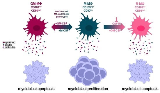
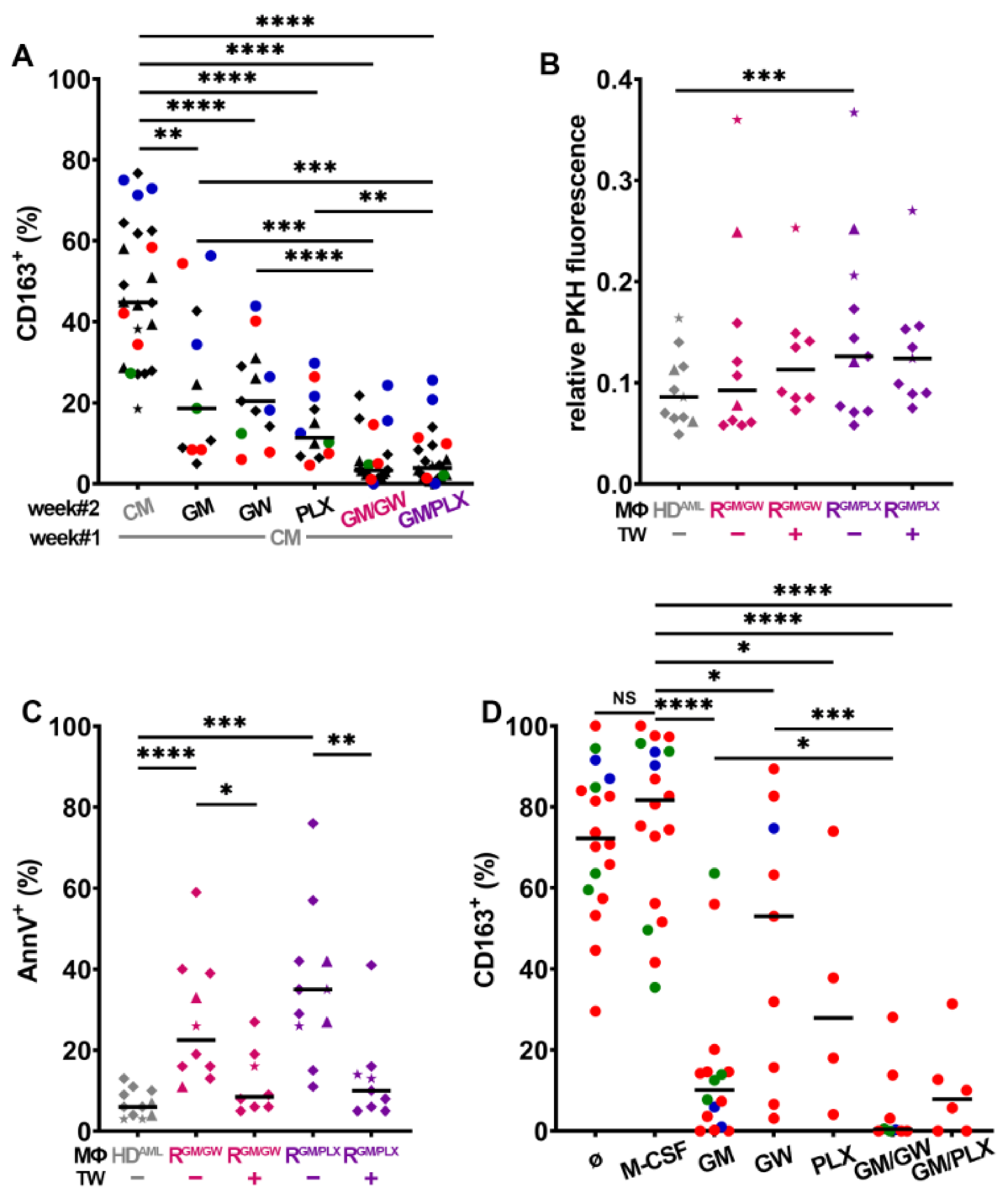
3.5. CSF1R Inhibition and Exposure to GM-CSF Reprograms CD163+ M- and HDAML-MΦs

As the upregulation of CD163 expression on HDAML-MΦs can be prevented by the inhibition of CSF1R activity (Figure 1D), we assessed whether selective CSF1R tyrosine kinase inhibitors GW2580 [25] or PLX3397 [53] could modify MΦ polarization. As illustrated in Figure 3A, high CD163 expression on M-MΦs was maintained after 7 additional days of culture in M-CSF (week#2, Figure 3A). CSF1R inhibition or addition of GM-CSF during week#2 lowered CD163 expression, with a stronger effect when GM-CSF was used in combination with either CSF1R inhibitor. In parallel, all treatments also significantly increased CD80 surface expression (Figure S6A).
The effect of M-MΦ reprogramming on myeloblast proliferation was assessed by comparing the proliferation of HL-60, MV-4-11, NB4, U937, and OCI-AML3 myeloblasts co-cultured for 4 days with M- vs. R-MΦs. Compared to myeloblasts in direct contact with CD163+ M-MΦs, the co-culture with R-MΦs (reprogrammed with GM-CSF combined with either GW2580 = RGM/GW, or PLX3397 = RGM/PLX), significantly slowed down their proliferation and promoted myeloblast apoptosis (Figure 3B,C). Interestingly, a significant decrease in myeloblast apoptosis was observed when they were separated from R-MΦs by TW inserts. We next assessed the survival of primary myeloblasts of all three risk categories cultured on R-MΦs (Figure 3D): their proliferation was not significantly affected by R-MΦs (T. S., personal observation, 2020), but their survival was strongly and significantly reduced compared to survival on M-MΦs.
We then investigated the impact of HDAML-MΦ reprogramming: after 7 days of reorientation, single treatments significantly lowered CD163 expression, whereas the combination of GW2580 or PLX3397 with GM-CSF almost abolished it (Figure 4A); in parallel, reorientation increased CD80 expression (Figure S6B). The co-culture of HL-60, NB4, and U937 cells with HDAML R-MΦs, lowered their proliferative activity (Figure 4B) and significantly induced their apoptosis (Figure 4C). As observed with R-MΦs (Figure 3C), the separation of myeloblasts from HDAML R-MΦs by a TW, protected them from apoptosis (Figure 4C). These findings reveal a major role for HDAML-MΦ polarization, and most likely for adhesive interactions, in controlling myeloblast survival.
The impact of MΦ reprogramming using CSF1R inhibitors combined to GM-CSF was then assessed on autologous primary BM MΦs from AML patients. BM cells were plated at diagnosis, and MΦs were co-cultured with myeloblasts, leukocytes, and stroma for 7 days under the conditions indicated in Figure 4D. Autologous BM AML-MΦs maintained their CD163 positivity in PM, as in medium supplemented with M-CSF.
CSF1R inhibition with GW2580 was least efficient, while GM-CSF supplementation reduced CD163 expression, but not as strongly as when combined with GW2580. The combination of PLX3397 with GM-CSF also efficiently inhibited CD163 surface expression. In parallel, we monitored CD80 expression, which significantly increased with GM-CSF either alone or combined with GW2580 or PLX3397 (Figure S6C). Finally, we measured primary patient blast apoptosis in four different BM patient co-cultures in medium with M-CSF vs. reprogramming conditions; twice as many apoptotic blast cells were found in both reprogramming media compared to what was observed in M-CSF (Figure S6D).
Innate and adaptive immune checkpoint deregulation have been shown to play important roles in myeloid malignancies. We, therefore, analyzed the expression of CD47, which couples to SIRPα on MΦs [14,54], and of PD-L1 [15]. Interestingly, PD-L1 expression was unchanged or even increased on CD163low HD or autologous primary MΦs exposed to CSF1R inhibitors, compared to CD163high cells cultured with M-CSF (Figure S7A,B). Further FC analyses showed that the reprogramming of autologous MΦs co-cultured with primary myeloblasts did not change CD47 on myeloblasts nor SIRPα expression on MΦs (Figure S7C,D).
Taken together, our results indicate that CSF1R plays a critical role in the polarization of HD M-MΦs and of allogeneic HDAML- or autologous AML-MΦs toward a protumoral M2-like phenotype and that inhibiting the CSF1R activity, in the presence of GM-CSF, efficiently reverses MΦ orientation. This is evidenced by a down-regulation of CD163 and an increase of CD80 expression, which has a direct impact on myeloblast proliferation and survival.

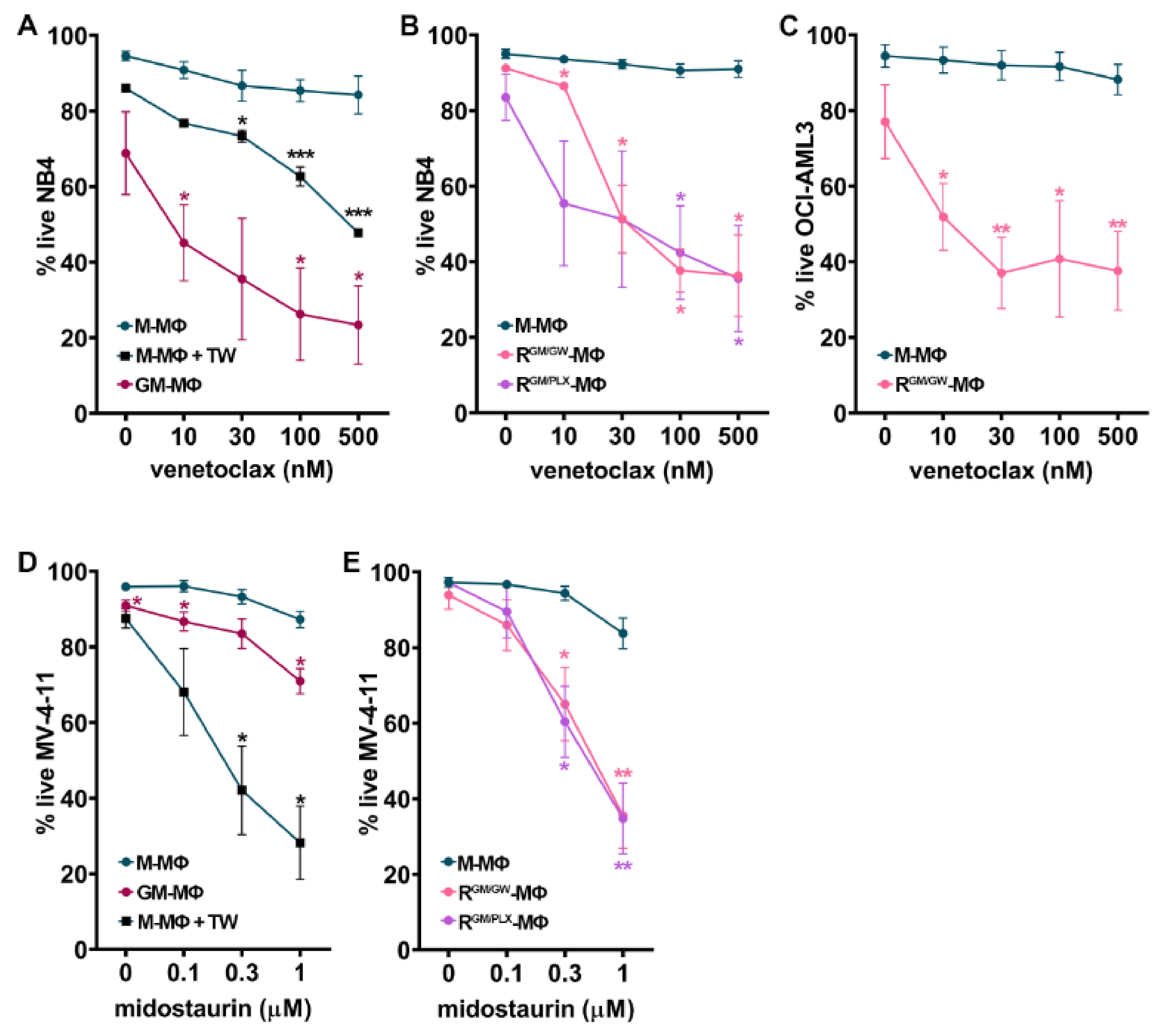
3.6. Macrophage Reprogramming Reverses Drug Resistance

We next assessed whether MΦ orientation affects the sensitivity of myeloblasts to targeted therapy. We first examined whether MΦ polarization has an effect on myeloblast sensitivity to venetoclax, a BCL-2 inhibitor. Myeloblasts were cultured in direct contact with HD M- vs. GM-MΦs (week#1) or M- vs. R-MΦs (week#2) in presence of vehicle or escalating doses of venetoclax. After 48 h, the percentage of live cells (AnnV and 7-AAD) was measured by FC.
We observed that the co-culture of NB4 cells with M-MΦs induced blast resistance to venetoclax, as compared to blasts cultured alone (T. S., personal observation, 2020). Direct contact contributed to the resistance of myeloblasts to venetoclax, as NB4 cells became significantly more sensitive to it, when they were separated from M-MΦs by TW inserts (Figure 5A). By contrast, venetoclax strongly induced NB4 cell apoptosis, even at low concentrations, when blasts were cultured on GM-MΦs (Figure 5A). Importantly, this was also the case when NB4 were cultured on R-MΦs (reprogrammed with either GW2580 or PLX3397, and GM-CSF, RGM/GW-, or RGM/PLX-MΦs; Figure 5B). MΦ reprogramming with GW2580 and GM-CSF also significantly sensitized OCI-AML-3 myeloblasts to venetoclax (Figure 5C).
FLT3-mutated AML have a poor prognosis [55]. To determine the impact of MΦ orientation and reprogramming on myeloblasts sensitivity to midostaurin (a FLT3 inhibitor used in AML patients [56]), we co-cultured the MLL-AF4 and FLT3-ITD mutated MV-4-11 AML cells with M- vs. GM- (week#1) or M- vs. R-MΦs (week#2). MV-4-11 cells co-cultured in direct contact with M-MΦs were resistant to midostaurin (Figure 5D). In contrast, when myeloblasts were separated from M-MΦs by a TW insert, midostaurin efficiently induced MV-4-11 apoptosis, suggesting an important role for cell adhesion in promoting resistance to midostaurin (Figure 5D). As observed with venetoclax, the co-culture of MV-4-11 myeloblasts on GM- or R-MΦs strongly increased their sensitivity to midostaurin (Figure 5D,E). Taken together, our results suggest a critical role of MΦ orientation in promoting drug-resistance to midostaurin and venetoclax.

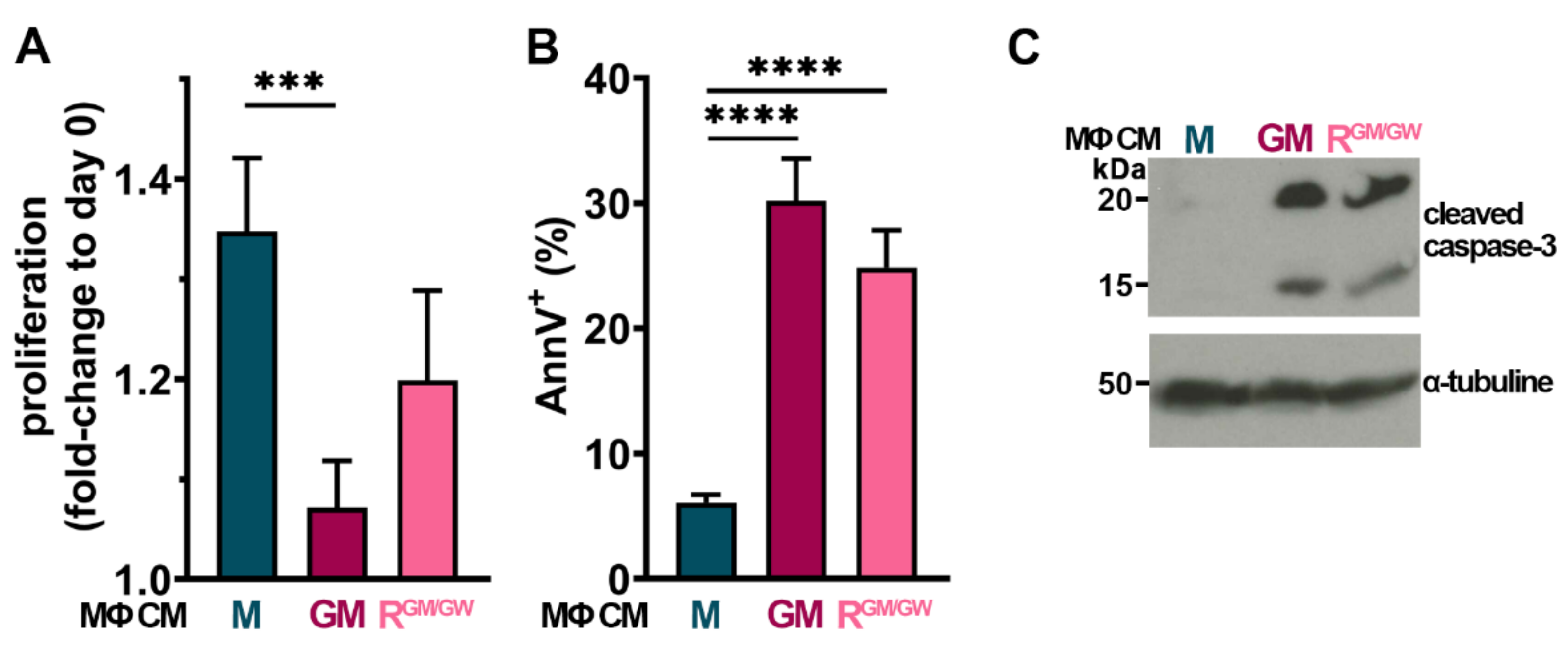
3.7. Factors Secreted by GM- and R-MΦs Play a Role in Myeloblast Apoptosis

In order to assess whether factors secreted by MΦs could also have an impact on myeloblasts, HL-60, NB4, OCI-AML3, and U937 cells were cultured with MΦ CM. Of the four tested cell lines, only U937 displayed sensitivity to MΦ CM: after 24 h, GM-MΦ CM significantly impaired their proliferation, while RGM/GW-MΦ CM strongly decreased it (Figure 6A). Moreover, both GM- and RGM/GW-MΦ CM induced >25% apoptosis in U937 cells (Figure 6B).
Effects on proliferation and apoptosis were already detectable at 6 h (C. S., personal observation, 2020). Moreover, after 48 h of incubation with MΦ CM, cleaved caspase-3 was detected by western blot (Figure 6C; uncropped version Figure S8); of note, caspase-3 cleavage was already detectable at 24 h, but less markedly. M-MΦ CM had no impact on apoptosis induction and caspase-3 cleavage. These data indicate at least a partial contribution to apoptosis of both inflammatory/cytotoxic molecules contained in polarized GM- and RGM/GW-MΦ CM.

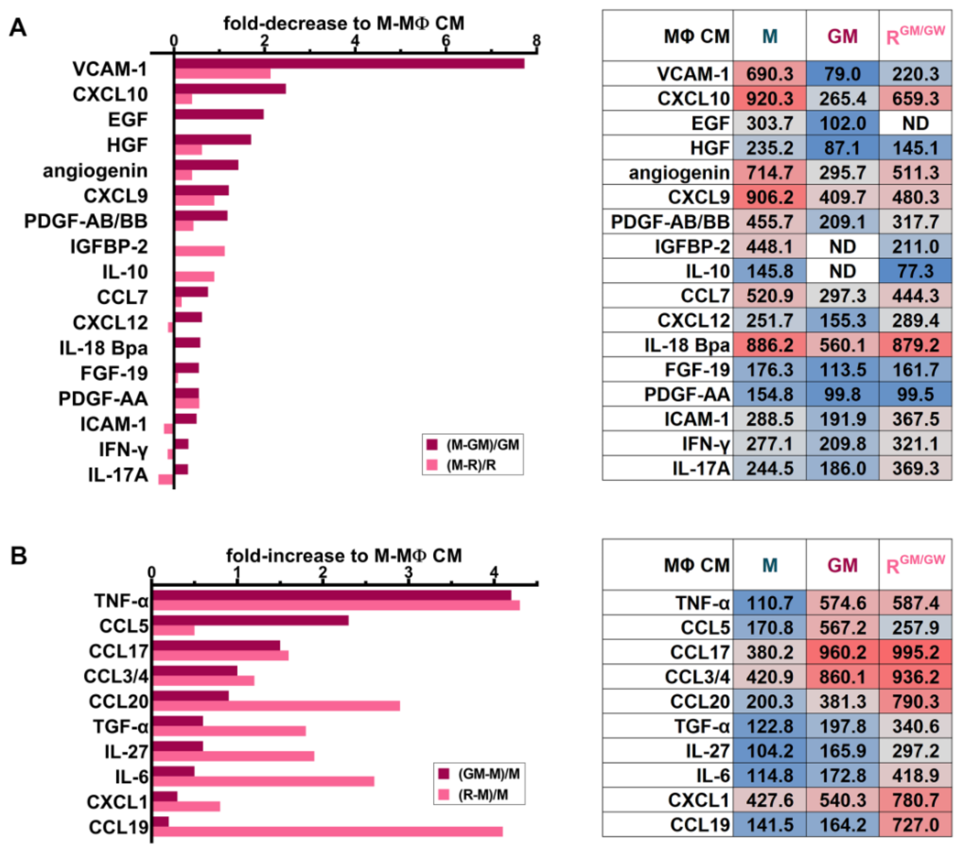
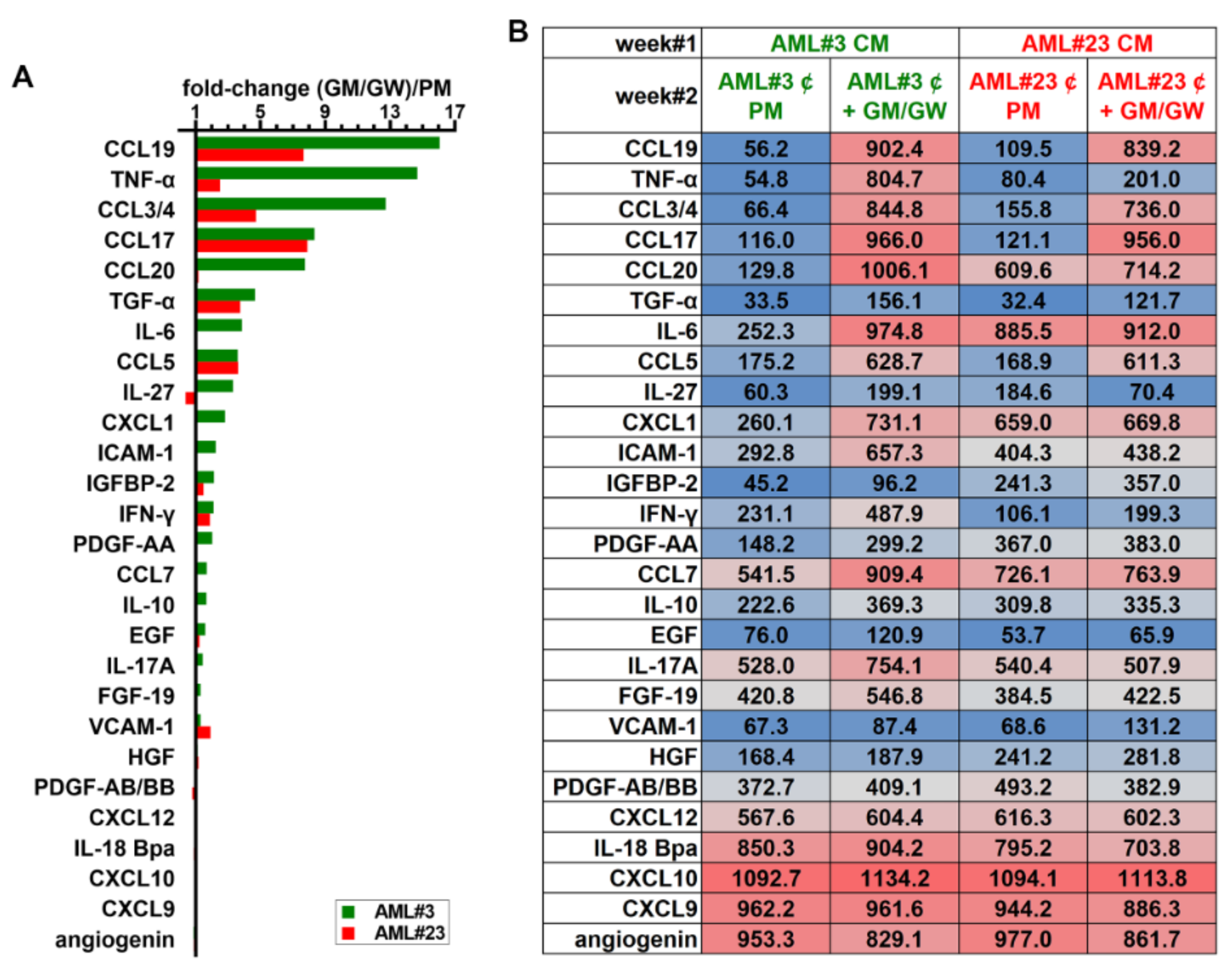
3.8. Identification of Soluble Molecules Secreted by MΦs

To facilitate the identification of SM predominantly secreted by M-, GM-, and RGM/GW-MΦs, we illustrated several relevant cytokines as their fold-decrease (Figure 7A) or -increase (Figure 7B) in GM- or R-MΦ CM relative to their quantification in M-MΦ CM, while the short tables indicate the SM relative levels (data from all analyzed SM are shown in Table S3 and revealed membranes are shown in Figure S5C). M-MΦs secrete prosurvival factors, such as HGF, and EGF, and high levels of VCAM-1, which has been demonstrated to play an essential role in the BM stromal compartment in mediating leukemia cell chemoresistance [57].
Other M-MΦ SM detected in the array may play a role, such as CXCL12 or IL-8 (Figure 7A and Table S3), as they contribute to support myeloblast survival and proliferation [58,59] and to orient MΦs toward immunosuppressive and proangiogenic functions [60]. Furthermore, M-MΦ reprogramming with GM-CSF and GW2580 reduced the secretion of many factors responsible for survival, angiogenesis, and cross-talk with stromal cells, for example HGF, angiogenin, IGFBP-2, IL-10, and PDGF-AB/BB. The tumor-promoting chemokine CCL7 [61] is also more abundantly secreted by M-MΦs than GM- or R-MΦs. CXCL9 and CXCL10, which may exhibit pro- or anti-tumoral activities, depending on the splice variant of their receptor CXCR3 [62], were reduced in CM of both GM- and R-MΦs [63].
The inflammatory cytokine TNF-α is highly secreted in GM- and RGM/GW-MΦ CM (Figure 7B). Other factors, with context-dependent antitumoral or immune cell chemotactic activity, such as CCL5 [64], CCL17 [65], CCL3/4, CCL20 [66], CXCL1, and CCL19 [67], were also predominantly detected in CM of GM- and RGM/GW-MΦs. Compared to MΦs stimulated with M-CSF, GM- and R-MΦs secreted increased levels of IL-6 and IL-27, which may also contribute to inhibiting myeloblast proliferation and promoting blast apoptosis [39,68].
We next determined the effect of MΦ reprogramming on soluble factor secretion resulting from the interplay between HD MΦs co-cultured with primary myeloblasts from patient #3 and #23. HD Mo were first stimulated for one week with CM from patient #3 or #23. On day 7, after confirming the induction of CD163 expression in HDAML-MΦs, primary blasts of patients #3 or #23 were added to the respective MΦ monolayers for one more week (week#2), in PM vs. PM supplemented with GM-CSF and GW2580 (Figure S5D). The level of each SM was illustrated as the ratio of its level in PM supplemented with GM-CSF and GW2580 divided by that secreted by co-cultures in PM (key cytokines in Figure 8).
Consistent with analyses of GM- and R-MΦ CM shown in Figure 7, the relative levels of TNF-α increased in both co-cultures of AML #3 and #23 with HD MΦs in the presence of GM-CSF and GW2580. The secretion of interferon γ, a key inflammatory molecule, also increased in both co-cultures under reprogramming conditions, as were CCL5, CCL17, CCL3/4, CCL19 and CCL20. IL-6 was highly secreted in CM of AML #3 co-cultured with HDAML-MΦs in reprogramming conditions. IL-27 exhibited an opposite trend between patients. Interestingly, a number of other SM known for their protumoral activity, such as IL-10, EGF, HGF, IGFBP-2, PDGF-AB/BB, CXCL12, FGF-19, IL-18 Bpa, and VCAM-1, slightly increased or did not exhibit any significant relative change in their levels in this experiment.

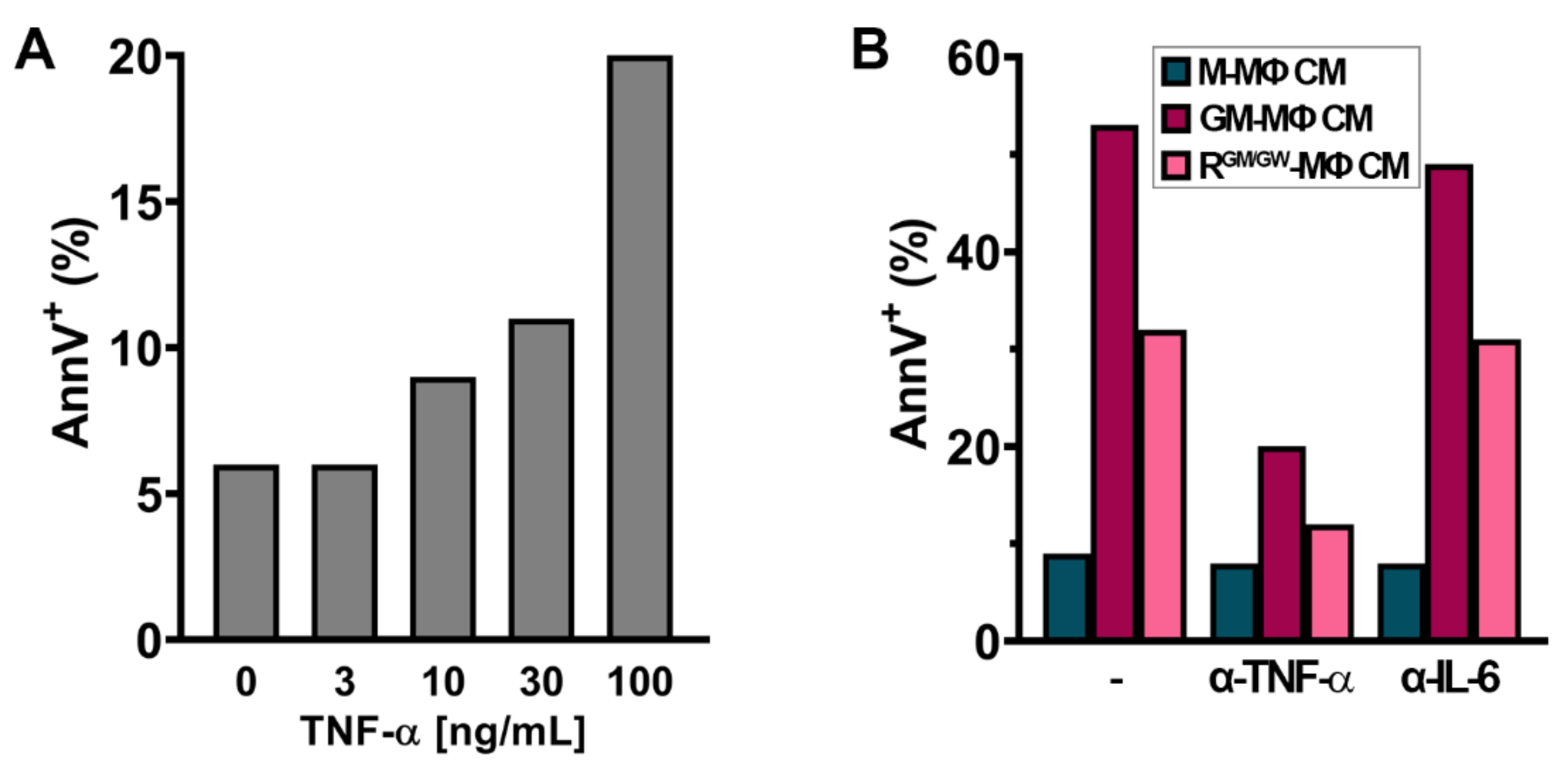
3.9. TNF-α Induces Apoptosis in U937 Cells

TNF-α is highly increased in GM- and R-MΦ CM (Figure 7B); while it could have a major function in modulating the surrounding microenvironment cross-talks, and inducing the expression and secretion of other inflammatory factors, it was shown to be cytotoxic to U937 cells [69]. We, therefore, tested the effect of escalating doses of recombinant TNF-α on U937 cells. After 24 h, we confirmed that TNF-α triggered apoptosis of U937 cells in a dose-dependent manner (Figure 9A). We next investigated whether adding a neutralizing antibody against TNF-α in MΦ CM would affect the induction of apoptosis. As illustrated on Figure 9B, adding a monoclonal antibody against TNF-α in the CM strongly decreased apoptosis, whereas adding an antibody against IL-6, an inflammatory factor which is also increased in both MΦ CM, had no impact.

4. Discussion

AML form a heterogeneous group of diseases, frequently of poor prognosis, with a high risk of relapse after completion of therapy. In patients with solid tumors and leukemia, the presence of TAMs has been reported as a bad prognosis factor, which may be related to the change in MΦ polarization inside tissues infiltrated by malignant cells, promoting tumor growth and survival [23]. As little information is available on AML-associated MΦs, we examined their impact on myeloblast survival, proliferation, and resistance to venetoclax or midostaurin.
In addition, we examined the possibility of changing the MΦ orientation by inhibiting the CSF1R. Our results show that (1) BM AML MΦs frequently express the protumoral orientation marker CD163 at diagnosis; (2) myeloblasts can educate HD MΦs to express CD163 in a CSF1R-dependent manner and promote their growth and survival; (3) numerous SM are secreted that support myeloblasts proliferation, survival, migration, and angiogenesis; (4) resistance to venetoclax and midostaurin is promoted by myeloblast interactions with MΦs; and (5) MΦ orientation can be reprogrammed by inhibition of CSF1R combined with GM-CSF exposure, leading to the reversal of resistance to targeted therapies and myeloblast apoptosis.
MΦs are highly plastic cells that can change their phenotype and function in response to microenvironmental stimuli, which modulate their polarization [70]. In vitro, they adopt either an antimicrobial and antitumoral activity when they are exposed to cytokines secreted by Th1 lymphocytes (such as TNF-α or interferon γ) or an anti-inflammatory phenotype promoting angiogenesis and tumor growth when they are exposed to Th2 lymphocytic cytokines (such as IL-4, -5, and -13) [71]. In tumors, depending on local cytokines secreted by malignant cells and tissue environment, TAM orientation can be shared between M1 and M2 status, with multiple phenotypes between these two extremes [15].
In the majority of AML patients, we observed that the BM and PB MΦs significantly express the M2-like marker CD163 at diagnosis (Figure 1A). Interestingly, similar observations were reported in B- and T-cell acute lymphoblastic leukemia, suggesting that leukemia cells and their microenvironment may dysregulate MΦ function [72]. In a minority of AML patients, we also observed MΦs expressing CD80, suggesting that a continuum of phenotypes between M1- and M2-like extremes are present at diagnosis, whose function may change in response to local signals.
The co-culture of AML cell lines or primary myeloblasts with HD Mo highly induced CD163 expression on HDAML-MΦs (Figure 1C), mimicking the effect of M-CSF (Figure 1B). This induction did not require direct contact between myeloblasts and MΦs as it was also observed with exposure to AML CM, suggesting that it may depend on soluble factors, mainly cytokines [73]. However, cytokines known to induce the expression of CD163, such as IL-4, -13, and -10 [74], were not detected in AML CM (Table S3). Alternatively, we hypothesized that it may depend on CSF1/CSF1R pathway activation, as in solid cancers [15] and lymphoma [27,28], which was confirmed by the abrogation of CD163 expression induced by AML CM supplemented with GW2580 or PLX3397 (Figure 1D).
Previous observations reported a sensitivity of primary AML cells to CSF1R inhibition, in favorable-risk patients, through the secretion of cytokines, in particular HGF, by CSF1R-expressing supportive cells [24]. Inhibiting CSF1R in order to reprogram protumoral MΦ was crucial in our experiment, but MΦs were simultaneously cultured with GM-CSF to induce an M1-like polarization. GM-CSF, as a key driver of inflammatory reaction, contributes to the antitumoral effect of R-MΦs. It may do so by enhancing the MΦ phagocytic activity, the production of reactive oxygen species, and the secretion of proinflammatory cytokines that affect T-lymphocyte response against malignant cells [75,76].
M-MΦs (Figure 3A), HDAML-MΦs (Figure 4A), and primary autologous MΦs in their BM microenvironment (Figure 4D) were efficiently reprogrammed by CSF1R inhibition leading to a drastic decrease in CD163 expression; addition of GM-CSF potentiated this down-regulation. In co-culture assays, R-MΦs efficiently induced primary myeloblasts and AML cell line apoptosis, but as with GM-MΦs, myeloblasts were partially rescued from apoptosis in the absence of direct contact, suggesting a role for cell adhesion molecules. However, SM also participated in the proapoptotic effect of R-MΦs. CM from GM- or R-MΦs induced caspase-3 cleavage and the apoptosis of >25% of U937 monoblasts.
Interestingly, mAb blocking studies indicated that TNF-α may contribute to inducing U937 apoptosis. High TNF-α levels were detected in CM of GM- and R-MΦs (Figure 7B) and recombinant TNF-α induced U937 cell apoptosis. In myeloblasts, TNF-α could activate the apoptosis cascades through binding to its receptor TNFR1, FADD (Fas-associated protein with death domain) recruitment, and caspase-8 and -3 activation or by activation of the mitochondrial pathway leading to caspase-9 and -3 activation [77].
In a mouse model of chronic lymphocytic leukemia, targeting MΦs using an anti-CSF1R mAb efficiently inhibited disease progression; interestingly, the leukemic cell death was dependent on TNF-α signaling and tumor microenvironment reprogramming toward an antitumoral phenotype [78]. In vivo, TNF-α could have not only an effect on myeloblasts but also a major impact on MΦ orientation by counterbalancing the emergence of M2-like AML-MΦs by inhibiting IL-13, and possibly IL-4, secreted by eosinophils in tumors [79].
Moreover, TNF-α can induce the secretion of other proinflammatory effectors, like GM-CSF, which activates interferon regulatory factor 4 (IRF4) and induces the biosynthesis of CCL17 in MΦs, a cytokine loop with proinflammatory properties [80] promoting MΦ M1-like orientation and myeloblasts apoptosis. However, the CM of GM- or R-MΦs had no proapoptotic effect on three other AML cell lines; this may be due to the ability of TNF-α to simultaneously activate pathways leading to cell apoptosis or survival and proliferation [77].
Other mechanisms may contribute in vivo to the proapoptotic effect of R-MΦs. Figure 7 shows that the cytokine profile of R-MΦs differs from that of GM-MΦs by the secretion of higher levels of chemokines, such as CCL20, CCL19, IL-6, and IL-27. The crucial role played by CCL19 in the immune response is illustrated by its ability with IL-7 to promote tumor infiltration by T cells and dendritic cells and to improve the therapeutic effect of CAR-T cells against solid tumors [81] or multiple myeloma [82].
The proinflammatory molecule IL-6 may also contribute to immune response by promoting T-cell-mediated immune defense [83] and was even reported to stimulate myeloblast differentiation by MΦs [84]. Finally, blocking CSF1R in vivo inhibits monocyte recruitment into the tumor environment, leading to MΦ depletion. MΦ reprogramming induced by CSF1R inhibition may also strengthen immune defense by stimulating T-cell recruitment in a malignant cell environment [85].
Drug resistance is a major problem in relapsed/refractory AML. The combination of the BCL-2 inhibitor venetoclax with hypomethylating agents or low dose cytarabine demonstrated high initial efficiency in patients with de novo AML, who were ineligible for intensive chemotherapy [86]. However, the response to treatment is most often lost after several months, with poor survival of relapsing patients [87].
Several mechanisms of resistance to venetoclax have been reported [88] involving the up-regulation of antiapoptotic molecules Mcl-1 and Bcl-xL, mutations in genes controlling different kinase pathways, transcription factors, epigenetic modifiers, and tumor suppressors. Similarly, drug resistance affects the sensitivity of FLT3-mutated AML to midostaurin. Like for venetoclax, the mechanisms of resistance may be complex and involve the intrinsic properties of AML cells as well as of the microenvironment [89].
The ability of reprogrammed macrophages to increase the sensitivity of myeloblasts to midostaurin or venetoclax suggests a role for macrophage polarization in promoting resistance to these targeted therapies. Interestingly, close contacts between macrophages and myeloblasts were required to promote resistance to midostaurin or venetoclax, which may suggest the involvement of adhesion receptors. The presence of high soluble VCAM-1 levels in M-MΦ CM and its strong decrease in the CM of GM- and R-MΦs may suggest the involvement of VCAM-1/CD49d, which were previously reported to trigger NF-κB signaling and promote chemoresistance [57].
Aiming to modulate the MΦ phenotype in AML is a promising therapeutic approach. Currently, clinical development of CSF1R inhibitors, including PLX3397, for AML treatment is at the early stages [90]. The efficacy of PLX3397, a FLT3 and CSF1R inhibitor, was demonstrated in relapsed/refractory FLT3-ITD-mutated AML with a safety profile similar to that of other FLT3 inhibitors [91]. Its impact on CSF1R inhibition is unclear in relapsing FLT3-ITD AML, and its clinical activity is predominantly related to its ability to inhibit FLT3 [91]. CSF1R inhibition alone may not be sufficient to overcome resistance mechanisms in vivo [92,93], but it may represent a general approach to target the microenvironment of AML cells and may be promising when used in combination with inhibitors of the immune checkpoints, angiogenesis, or with the adoptive T-cell transfer, which are undergoing clinical investigations [94].
To reprogram human MΦs in AML, combining CSF1R inhibition with GM-CSF might be more efficient for inducing an antitumoral response. It may however not be sufficient to restore MΦ phagocytic activity and antigen-presenting capacity in vivo. Indeed, PD-L1 expression is unchanged or higher on CD163 MΦs exposed to CSF1R inhibitors, than on CD163+ cells. PD1/PD-L1 inhibition may improve the antitumoral activity of R-MΦs and conversely, CSF1R inhibition may improve the effect of anti-PD1/PD-L1 monoclonal antibody in relapsed/refractory AML.
In addition to the T-cell immune checkpoints, CD47 is the dominant MΦ checkpoint, which is overexpressed in myeloid malignancies and inhibits phagocytosis through “do not eat me” signals upon its binding to SIRPα on MΦs [54]. As observed for PD1/PD-L1, the reprogramming of AML-MΦs co-cultured with primary myeloblasts did not change CD47 expression on myeloblasts nor SIRPα expression on macrophages. As the combination of the anti-CD47mAb, magrolimab, with 5-azacitidine strongly improves the antitumoral activity of MΦs [54], considering a combination of CD47 blockade and CSF1R inhibitors, with 5-azacitidine, might be a way to improve the in vivo antitumoral activity of reprogrammed MΦs.

5. Conclusions

Relapse and resistance to treatment remain major issues in the treatment of AML. We showed here that protumoral CD163+ MΦs predominate, at diagnosis, in the BM of AML patients and that the induction of CD163 expression on MΦs by myeloblasts was dependent on CSF1R. We observed that numerous SM were secreted by MΦs and myeloblasts, which promoted an M2-like MΦ orientation, leading to increased blast survival and drug resistance in the presence of close intercellular contacts. Finally, inhibiting CSF1R, in the presence of GM-CSF, reprogramed MΦ orientation, reversed the resistance to targeted therapies, and promoted myeloblast apoptosis. CSF1R inhibition may serve as a novel way to target the microenvironment of myeloblasts and improve the efficiency of AML therapy.

Supplementary Materials

The following are available online at https://www.mdpi.com/article/10.3390/cancers13215289/s1, Figure S1: Schematic protocol of MΦ differentiation process, Figure S2: HSPC do not upregulate CD163 expression on MΦs, Figure S3: Inflammatory or inactivated factors do not upregulate CD163 expression on HD MΦs, Figure S4: CD80 and CD206 are frequently expressed by HD and HDAML-MΦs, Figure S5: Autoradiographies of cytokine array membranes used for densitometry, Figure S6: CD80 expression is increased in R-MΦs, Figure S7: PD-L1 is increased on R-MΦs, while SIRPα is unchanged, Figure S8: Uncropped western blot of Figure 6C, Table S1: FAB, WHO 2016 and ELN 2017 genetic risk classifications from included patients, Table S2: Antibodies for FC, Table S3: Relative quantification of all cytokines from membranes depicted on Figure S5.

Author Contributions

Conceptualization, T.S. and O.S.; methodology, C.S., T.S. and O.S.; investigation, T.S. and C.S.; project administration, T.S.; Validation, C.S., T.S. and O.S.; resources, T.S. and O.S.; data curation, C.S. and T.S.; writing—review and editing, C.S., T.S. and O.S.; funding acquisition, T.S. All authors have read and agreed to the published version of the manuscript.

Funding

This research was supported by a grant from Fond’Action contre le cancer and the Foundation Dr. Henri Dubois-Ferrière Dinu Lipatti.

Institutional Review Board Statement

The study was conducted according to the guidelines of the Declaration of Helsinki, and approved by the Ethics Committee of CHUV (protocol 2017-01509, 9 November 2017).

Informed Consent Statement

Informed consent was obtained from all subjects involved in the study.

Data Availability Statement

Data is contained within the article or Supplementary Materials.

Acknowledgments

We thank the patients and the clinical diagnostic laboratory of hematology for providing samples after diagnostic immunophenotyping.

Conflicts of Interest

The authors declare no conflict of interest.

References

  1. Burnett, A.K.; Hills, R.K.; Russell, N. Twenty five years of UK trials in acute myeloid leukaemia: What have we learned? Br. J. Haematol. 2020, 188, 86–100. [Google Scholar] [CrossRef] [PubMed]
  2. Dohner, H.; Estey, E.; Grimwade, D.; Amadori, S.; Appelbaum, F.R.; Buchner, T.; Dombret, H.; Ebert, B.L.; Fenaux, P.; Larson, R.A.; et al. Diagnosis and management of AML in adults: 2017 ELN recommendations from an international expert panel. Blood 2017, 129, 424–447. [Google Scholar] [CrossRef] [PubMed] [Green Version]
  3. Morita, K.; Wang, F.; Jahn, K.; Hu, T.; Tanaka, T.; Sasaki, Y.; Kuipers, J.; Loghavi, S.; Wang, S.A.; Yan, Y.; et al. Clonal evolution of acute myeloid leukemia revealed by high-throughput single-cell genomics. Nat. Commun. 2020, 11, 5327. [Google Scholar] [CrossRef] [PubMed]
  4. Son, B.; Lee, S.; Youn, H.; Kim, E.; Kim, W.; Youn, B. The role of tumor microenvironment in therapeutic resistance. Oncotarget 2017, 8, 3933–3945. [Google Scholar] [CrossRef] [Green Version]
  5. Gurnari, C.; Pagliuca, S.; Visconte, V. Deciphering the Therapeutic Resistance in Acute Myeloid Leukemia. Int. J. Mol. Sci. 2020, 21, 8505. [Google Scholar] [CrossRef]
  6. Ruffell, B.; Coussens, L.M. Macrophages and therapeutic resistance in cancer. Cancer Cell 2015, 27, 462–472. [Google Scholar] [CrossRef] [Green Version]
  7. Etzerodt, A.; Tsalkitzi, K.; Maniecki, M.; Damsky, W.; Delfini, M.; Baudoin, E.; Moulin, M.; Bosenberg, M.; Graversen, J.H.; Auphan-Anezin, N.; et al. Specific targeting of CD163(+) TAMs mobilizes inflammatory monocytes and promotes T cell-mediated tumor regression. J. Exp. Med. 2019, 216, 2394–2411. [Google Scholar] [CrossRef]
  8. Mills, C.D. M1 and M2 Macrophages: Oracles of Health and Disease. Crit. Rev. Immunol. 2012, 32, 463–488. [Google Scholar] [CrossRef] [Green Version]
  9. Qian, B.Z.; Pollard, J.W. Macrophage diversity enhances tumor progression and metastasis. Cell 2010, 141, 39–51. [Google Scholar] [CrossRef] [Green Version]
  10. Murray, P.J. Macrophage Polarization. Annu. Rev. Physiol. 2017, 79, 541–566. [Google Scholar] [CrossRef]
  11. Cassetta, L.; Fragkogianni, S.; Sims, A.H.; Swierczak, A.; Forrester, L.M.; Zhang, H.; Soong, D.Y.H.; Cotechini, T.; Anur, P.; Lin, E.Y.; et al. Human Tumor-Associated Macrophage and Monocyte Transcriptional Landscapes Reveal Cancer-Specific Reprogramming, Biomarkers, and Therapeutic Targets. Cancer Cell 2019, 35, 588–602.e10. [Google Scholar] [CrossRef] [Green Version]
  12. Neubert, N.J.; Schmittnaegel, M.; Bordry, N.; Nassiri, S.; Wald, N.; Martignier, C.; Tille, L.; Homicsko, K.; Damsky, W.; Maby-El Hajjami, H.; et al. T cell-induced CSF1 promotes melanoma resistance to PD1 blockade. Sci. Transl. Med. 2018, 10, eaan3311. [Google Scholar] [CrossRef] [Green Version]
  13. Komohara, Y.; Jinushi, M.; Takeya, M. Clinical significance of macrophage heterogeneity in human malignant tumors. Cancer Sci. 2014, 105, 1–8. [Google Scholar] [CrossRef] [Green Version]
  14. Takizawa, H.; Manz, M.G. Macrophage tolerance: CD47-SIRP-α-mediated signals matter. Nat. Immunol. 2007, 8, 1287–1289. [Google Scholar] [CrossRef]
  15. Noy, R.; Pollard, J.W. Tumor-associated macrophages: From mechanisms to therapy. Immunity 2014, 41, 49–61. [Google Scholar] [CrossRef] [Green Version]
  16. Cassetta, L.; Kitamura, T. Macrophage targeting: Opening new possibilities for cancer immunotherapy. Immunology 2018, 155, 285–293. [Google Scholar] [CrossRef]
  17. Mantovani, A.; Marchesi, F.; Malesci, A.; Laghi, L.; Allavena, P. Tumour-associated macrophages as treatment targets in oncology. Nat. Rev. Clin. Oncol. 2017, 14, 399–416. [Google Scholar] [CrossRef]
  18. Yang, X.; Feng, W.; Wang, R.; Yang, F.; Wang, L.; Chen, S.; Ru, Y.; Cheng, T.; Zheng, G. Repolarizing heterogeneous leukemia-associated macrophages with more M1 characteristics eliminates their pro-leukemic effects. Oncoimmunology 2018, 7, e1412910. [Google Scholar] [CrossRef]
  19. Chen, C.; Wang, R.; Feng, W.; Yang, F.; Wang, L.; Yang, X.; Ren, L.; Zheng, G. Peritoneal resident macrophages in mice with MLL-AF9-induced acute myeloid leukemia show an M2-like phenotype. Ann. Transl. Med. 2021, 9, 266. [Google Scholar] [CrossRef]
  20. Al-Matary, Y.S.; Botezatu, L.; Opalka, B.; Hones, J.M.; Lams, R.F.; Thivakaran, A.; Schutte, J.; Koster, R.; Lennartz, K.; Schroeder, T.; et al. Acute myeloid leukemia cells polarize macrophages towards a leukemia supporting state in a Growth factor independence 1 dependent manner. Haematologica 2016, 101, 1216–1227. [Google Scholar] [CrossRef] [Green Version]
  21. Yan, H.; Qu, J.; Cao, W.; Liu, Y.; Zheng, G.; Zhang, E.; Cai, Z. Identification of prognostic genes in the acute myeloid leukemia immune microenvironment based on TCGA data analysis. Cancer Immunol. Immunother. 2019, 68, 1971–1978. [Google Scholar] [CrossRef]
  22. Shiraishi, D.; Fujiwara, Y.; Horlad, H.; Saito, Y.; Iriki, T.; Tsuboki, J.; Cheng, P.; Nakagata, N.; Mizuta, H.; Bekki, H.; et al. CD163 Is Required for Protumoral Activation of Macrophages in Human and Murine Sarcoma. Cancer Res. 2018, 78, 3255–3266. [Google Scholar] [CrossRef] [Green Version]
  23. Lopez-Yrigoyen, M.; Cassetta, L.; Pollard, J.W. Macrophage targeting in cancer. Ann. N. Y. Acad. Sci. 2020, 1499, 18–41. [Google Scholar] [CrossRef]
  24. Edwards, D.K.t.; Watanabe-Smith, K.; Rofelty, A.; Damnernsawad, A.; Laderas, T.; Lamble, A.; Lind, E.F.; Kaempf, A.; Mori, M.; Rosenberg, M.; et al. CSF1R inhibitors exhibit antitumor activity in acute myeloid leukemia by blocking paracrine signals from support cells. Blood 2019, 133, 588–599. [Google Scholar] [CrossRef]
  25. Gutierrez-Gonzalez, A.; Martinez-Moreno, M.; Samaniego, R.; Arellano-Sanchez, N.; Salinas-Munoz, L.; Relloso, M.; Valeri, A.; Martinez-Lopez, J.; Corbi, A.L.; Hidalgo, A.; et al. Evaluation of the potential therapeutic benefits of macrophage reprogramming in multiple myeloma. Blood 2016, 128, 2241–2252. [Google Scholar] [CrossRef] [Green Version]
  26. Wang, Q.; Liu, F.; Liu, L. Prognostic significance of PD-L1 in solid tumor: An updated meta-analysis. Medicine 2017, 96, e6369. [Google Scholar] [CrossRef]
  27. Papin, A.; Tessoulin, B.; Bellanger, C.; Moreau, A.; Le Bris, Y.; Maisonneuve, H.; Moreau, P.; Touzeau, C.; Amiot, M.; Pellat-Deceunynck, C.; et al. CSF1R and BTK inhibitions as novel strategies to disrupt the dialog between mantle cell lymphoma and macrophages. Leukemia 2019, 33, 2442–2453. [Google Scholar] [CrossRef]
  28. Valero, J.G.; Matas-Cespedes, A.; Arenas, F.; Rodriguez, V.; Carreras, J.; Serrat, N.; Guerrero-Hernandez, M.; Yahiaoui, A.; Balague, O.; Martin, S.; et al. The receptor of the colony-stimulating factor-1 (CSF-1R) is a novel prognostic factor and therapeutic target in follicular lymphoma. Leukemia 2021, 35, 2635–2649. [Google Scholar] [CrossRef]
  29. Spertini, C.; Baïsse, B.; Bellone, M.; Gikic, M.; Smirnova, T.; Spertini, O. Acute Myeloid and Lymphoblastic Leukemia Cell Interactions with Endothelial Selectins: Critical Role of PSGL-1, CD44 and CD43. Cancers 2019, 11, 1253. [Google Scholar] [CrossRef] [Green Version]
  30. Swerdlow, S.; Campo, E.; Harris, N.; Jaffe, E.; Pileri, S.; Stein, H.; Thiele, J. WHO Classification of Tumours of Haematopoietic and Lymphoid Tissues; IARC Press: Lyon, France, 2016. [Google Scholar]
  31. Bene, M.C.; Nebe, T.; Bettelheim, P.; Buldini, B.; Bumbea, H.; Kern, W.; Lacombe, F.; Lemez, P.; Marinov, I.; Matutes, E.; et al. Immunophenotyping of acute leukemia and lymphoproliferative disorders: A consensus proposal of the European LeukemiaNet Work Package 10. Leukemia 2011, 25, 567–574. [Google Scholar] [CrossRef] [Green Version]
  32. Hernandez, L.; Smirnova, T.; Kedrin, D.; Wyckoff, J.; Zhu, L.; Stanley, E.R.; Cox, D.; Muller, W.J.; Pollard, J.W.; Van Rooijen, N.; et al. The EGF/CSF-1 paracrine invasion loop can be triggered by heregulin beta1 and CXCL12. Cancer Res. 2009, 69, 3221–3227. [Google Scholar] [CrossRef] [PubMed] [Green Version]
  33. Xu, Z.J.; Gu, Y.; Wang, C.Z.; Jin, Y.; Wen, X.M.; Ma, J.C.; Tang, L.J.; Mao, Z.W.; Qian, J.; Lin, J. The M2 macrophage marker CD206: A novel prognostic indicator for acute myeloid leukemia. Oncoimmunology 2020, 9, 1683347. [Google Scholar] [CrossRef] [PubMed] [Green Version]
  34. Fayard, B.; Bianchi, F.; Dey, J.; Moreno, E.; Djaffer, S.; Hynes, N.E.; Monard, D. The serine protease inhibitor protease nexin-1 controls mammary cancer metastasis through LRP-1-mediated MMP-9 expression. Cancer Res. 2009, 69, 5690–5698. [Google Scholar] [CrossRef] [PubMed] [Green Version]
  35. Wischhusen, J.; Melero, I.; Fridman, W.H. Growth/Differentiation Factor-15 (GDF-15): From Biomarker to Novel Targetable Immune Checkpoint. Front. Immunol. 2020, 11, 951. [Google Scholar] [CrossRef] [PubMed]
  36. Zhang, W.; Wang, H.; Sun, M.; Deng, X.; Wu, X.; Ma, Y.; Li, M.; Shuoa, S.M.; You, Q.; Miao, L. CXCL5/CXCR2 axis in tumor microenvironment as potential diagnostic biomarker and therapeutic target. Cancer Commun. 2020, 40, 69–80. [Google Scholar] [CrossRef] [Green Version]
  37. Kubota, Y.; Takubo, K.; Shimizu, T.; Ohno, H.; Kishi, K.; Shibuya, M.; Saya, H.; Suda, T. M-CSF inhibition selectively targets pathological angiogenesis and lymphangiogenesis. J. Exp. Med. 2009, 206, 1089–1102. [Google Scholar] [CrossRef]
  38. Kobori, T.; Hamasaki, S.; Kitaura, A.; Yamazaki, Y.; Nishinaka, T.; Niwa, A.; Nakao, S.; Wake, H.; Mori, S.; Yoshino, T.; et al. Interleukin-18 Amplifies Macrophage Polarization and Morphological Alteration, Leading to Excessive Angiogenesis. Front. Immunol. 2018, 9, 334. [Google Scholar] [CrossRef]
  39. Récher, C. Clinical Implications of Inflammation in Acute Myeloid Leukemia. Front. Oncol. 2021, 11, 623952. [Google Scholar] [CrossRef]
  40. Sanchez-Correa, B.; Bergua, J.M.; Campos, C.; Gayoso, I.; Arcos, M.J.; Banas, H.; Morgado, S.; Casado, J.G.; Solana, R.; Tarazona, R. Cytokine profiles in acute myeloid leukemia patients at diagnosis: Survival is inversely correlated with IL-6 and directly correlated with IL-10 levels. Cytokine 2013, 61, 885–891. [Google Scholar] [CrossRef]
  41. Li, T.; Forbes, M.E.; Fuller, G.N.; Li, J.; Yang, X.; Zhang, W. IGFBP2: Integrative hub of developmental and oncogenic signaling network. Oncogene 2020, 39, 2243–2257. [Google Scholar] [CrossRef]
  42. Kühnl, A.; Kaiser, M.; Neumann, M.; Fransecky, L.; Heesch, S.; Radmacher, M.; Marcucci, G.; Bloomfield, C.D.; Hofmann, W.K.; Thiel, E.; et al. High expression of IGFBP2 is associated with chemoresistance in adult acute myeloid leukemia. Leuk. Res. 2011, 35, 1585–1590. [Google Scholar] [CrossRef] [Green Version]
  43. Doepfner, K.T.; Spertini, O.; Arcaro, A. Autocrine insulin-like growth factor-I signaling promotes growth and survival of human acute myeloid leukemia cells via the phosphoinositide 3-kinase/Akt pathway. Leukemia 2007, 21, 1921–1930. [Google Scholar] [CrossRef]
  44. Girard-Guyonvarc’h, C.; Palomo, J.; Martin, P.; Rodriguez, E.; Troccaz, S.; Palmer, G.; Gabay, C. Unopposed IL-18 signaling leads to severe TLR9-induced macrophage activation syndrome in mice. Blood 2018, 131, 1430–1441. [Google Scholar] [CrossRef]
  45. Smirnova, T.; Bonapace, L.; MacDonald, G.; Kondo, S.; Wyckoff, J.; Ebersbach, H.; Fayard, B.; Doelemeyer, A.; Coissieux, M.M.; Heideman, M.R.; et al. Serpin E2 promotes breast cancer metastasis by remodeling the tumor matrix and polarizing tumor associated macrophages. Oncotarget 2016, 7, 82289–82304. [Google Scholar] [CrossRef] [Green Version]
  46. Vilgelm, A.E.; Richmond, A. Chemokines Modulate Immune Surveillance in Tumorigenesis, Metastasis, and Response to Immunotherapy. Front. Immunol. 2019, 10, 333. [Google Scholar] [CrossRef] [Green Version]
  47. Schepers, K.; Campbell, T.B.; Passegue, E. Normal and leukemic stem cell niches: Insights and therapeutic opportunities. Cell Stem Cell 2015, 16, 254–267. [Google Scholar] [CrossRef] [Green Version]
  48. Binder, S.; Luciano, M.; Horejs-Hoeck, J. The cytokine network in acute myeloid leukemia (AML): A focus on pro- and anti-inflammatory mediators. Cytokine Growth Factor Rev. 2018, 43, 8–15. [Google Scholar] [CrossRef]
  49. Wyckoff, J.; Wang, W.; Lin, E.Y.; Wang, Y.; Pixley, F.; Stanley, E.R.; Graf, T.; Pollard, J.W.; Segall, J.; Condeelis, J. A paracrine loop between tumor cells and macrophages is required for tumor cell migration in mammary tumors. Cancer Res. 2004, 64, 7022–7029. [Google Scholar] [CrossRef] [Green Version]
  50. Boimel, P.J.; Smirnova, T.; Zhou, Z.N.; Wyckoff, J.; Park, H.; Coniglio, S.J.; Qian, B.Z.; Stanley, E.R.; Cox, D.; Pollard, J.W.; et al. Contribution of CXCL12 secretion to invasion of breast cancer cells. Breast Cancer Res. 2012, 14, R23. [Google Scholar] [CrossRef] [Green Version]
  51. Liu, Y.; Cai, Y.; Liu, L.; Wu, Y.; Xiong, X. Crucial biological functions of CCL7 in cancer. PeerJ 2018, 6, e4928. [Google Scholar] [CrossRef]
  52. Prabhala, R.H.; Pelluru, D.; Fulciniti, M.; Prabhala, H.K.; Nanjappa, P.; Song, W.; Pai, C.; Amin, S.; Tai, Y.T.; Richardson, P.G.; et al. Elevated IL-17 produced by TH17 cells promotes myeloma cell growth and inhibits immune function in multiple myeloma. Blood 2010, 115, 5385–5392. [Google Scholar] [CrossRef]
  53. Quail, D.F.; Joyce, J.A. Molecular Pathways: Deciphering Mechanisms of Resistance to Macrophage-Targeted Therapies. Clin. Cancer Res. 2017, 23, 876–884. [Google Scholar] [CrossRef] [Green Version]
  54. Chao, M.P.; Takimoto, C.H.; Feng, D.D.; McKenna, K.; Gip, P.; Liu, J.; Volkmer, J.P.; Weissman, I.L.; Majeti, R. Therapeutic Targeting of the Macrophage Immune Checkpoint CD47 in Myeloid Malignancies. Front. Oncol. 2019, 9, 1380. [Google Scholar] [CrossRef]
  55. Ley, T.J.; Miller, C.; Ding, L.; Raphael, B.J.; Mungall, A.J.; Robertson, A.; Hoadley, K.; Triche, T.J., Jr.; Laird, P.W.; Baty, J.D.; et al. Genomic and epigenomic landscapes of adult de novo acute myeloid leukemia. N. Engl. J. Med. 2013, 368, 2059–2074. [Google Scholar] [CrossRef] [Green Version]
  56. Daver, N.; Schlenk, R.F.; Russell, N.H.; Levis, M.J. Targeting FLT3 mutations in AML: Review of current knowledge and evidence. Leukemia 2019, 33, 299–312. [Google Scholar] [CrossRef] [Green Version]
  57. Jacamo, R.; Chen, Y.; Wang, Z.; Ma, W.; Zhang, M.; Spaeth, E.L.; Wang, Y.; Battula, V.L.; Mak, P.Y.; Schallmoser, K.; et al. Reciprocal leukemia-stroma VCAM-1/VLA-4-dependent activation of NF-κB mediates chemoresistance. Blood 2014, 123, 2691–2702. [Google Scholar] [CrossRef]
  58. Abdul-Aziz, A.M.; Shafat, M.S.; Mehta, T.K.; Di Palma, F.; Lawes, M.J.; Rushworth, S.A.; Bowles, K.M. MIF-Induced Stromal PKCβ/IL8 Is Essential in Human Acute Myeloid Leukemia. Cancer Res. 2017, 77, 303–311. [Google Scholar] [CrossRef] [Green Version]
  59. Cho, B.S.; Zeng, Z.; Mu, H.; Wang, Z.; Konoplev, S.; McQueen, T.; Protopopova, M.; Cortes, J.; Marszalek, J.R.; Peng, S.B.; et al. Antileukemia activity of the novel peptidic CXCR4 antagonist LY2510924 as monotherapy and in combination with chemotherapy. Blood 2015, 126, 222–232. [Google Scholar] [CrossRef]
  60. Sanchez-Martin, L.; Estecha, A.; Samaniego, R.; Sanchez-Ramon, S.; Vega, M.A.; Sanchez-Mateos, P. The chemokine CXCL12 regulates monocyte-macrophage differentiation and RUNX3 expression. Blood 2011, 117, 88–97. [Google Scholar] [CrossRef] [Green Version]
  61. Lee, Y.S.; Cho, Y.B. CCL7 Signaling in the Tumor Microenvironment. Adv. Exp. Med. Biol. 2020, 1231, 33–43. [Google Scholar] [CrossRef] [PubMed]
  62. Urra, S.; Fischer, M.C.; Martinez, J.R.; Veliz, L.; Orellana, P.; Solar, A.; Bohmwald, K.; Kalergis, A.; Riedel, C.; Corvalan, A.H.; et al. Differential expression profile of CXCR3 splicing variants is associated with thyroid neoplasia. Potential role in papillary thyroid carcinoma oncogenesis? Oncotarget 2018, 9, 2445–2467. [Google Scholar] [CrossRef] [PubMed] [Green Version]
  63. Ding, Q.; Lu, P.; Xia, Y.; Ding, S.; Fan, Y.; Li, X.; Han, P.; Liu, J.; Tian, D.; Liu, M. CXCL9: Evidence and contradictions for its role in tumor progression. Cancer Med. 2016, 5, 3246–3259. [Google Scholar] [CrossRef] [PubMed]
  64. Marques, R.E.; Guabiraba, R.; Russo, R.C.; Teixeira, M.M. Targeting CCL5 in inflammation. Expert Opin. Ther. Targets 2013, 17, 1439–1460. [Google Scholar] [CrossRef] [PubMed]
  65. Achuthan, A.; Cook, A.D.; Lee, M.C.; Saleh, R.; Khiew, H.W.; Chang, M.W.; Louis, C.; Fleetwood, A.J.; Lacey, D.C.; Christensen, A.D.; et al. Granulocyte macrophage colony-stimulating factor induces CCL17 production via IRF4 to mediate inflammation. J. Clin. Investig. 2016, 126, 3453–3466. [Google Scholar] [CrossRef] [Green Version]
  66. Ruytinx, P.; Proost, P.; Van Damme, J.; Struyf, S. Chemokine-Induced Macrophage Polarization in Inflammatory Conditions. Front. Immunol. 2018, 9, 1930. [Google Scholar] [CrossRef] [Green Version]
  67. Zlotnik, A.; Yoshie, O. Chemokines: A new classification system and their role in immunity. Immunity 2000, 12, 121–127. [Google Scholar] [CrossRef] [Green Version]
  68. Chiba, Y.; Mizoguchi, I.; Furusawa, J.; Hasegawa, H.; Ohashi, M.; Xu, M.; Owaki, T.; Yoshimoto, T. Interleukin-27 Exerts Its Antitumor Effects by Promoting Differentiation of Hematopoietic Stem Cells to M1 Macrophages. Cancer Res. 2018, 78, 182–194. [Google Scholar] [CrossRef] [Green Version]
  69. Larrick, J.W.; Wright, S.C. Cytotoxic mechanism of tumor necrosis factor-alpha. FASEB J. 1990, 4, 3215–3223. [Google Scholar] [CrossRef]
  70. Sica, A.; Mantovani, A. Macrophage plasticity and polarization: In vivo veritas. J. Clin. Investig. 2012, 122, 787–795. [Google Scholar] [CrossRef]
  71. Shapouri-Moghaddam, A.; Mohammadian, S.; Vazini, H.; Taghadosi, M.; Esmaeili, S.A.; Mardani, F.; Seifi, B.; Mohammadi, A.; Afshari, J.T.; Sahebkar, A. Macrophage plasticity, polarization, and function in health and disease. J. Cell. Physiol. 2018, 233, 6425–6440. [Google Scholar] [CrossRef]
  72. Dander, E.; Fallati, A.; Gulic, T.; Pagni, F.; Gaspari, S.; Silvestri, D.; Cricri, G.; Bedini, G.; Portale, F.; Buracchi, C.; et al. Monocyte-macrophage polarization and recruitment pathways in the tumour microenvironment of B-cell acute lymphoblastic leukaemia. Br. J. Haematol. 2021, 193, 1157–1171. [Google Scholar] [CrossRef]
  73. Skytthe, M.K.; Graversen, J.H.; Moestrup, S.K. Targeting of CD163(+) Macrophages in Inflammatory and Malignant Diseases. Int. J. Mol. Sci. 2020, 21, 5497. [Google Scholar] [CrossRef]
  74. Etzerodt, A.; Moestrup, S.K. CD163 and inflammation: Biological, diagnostic, and therapeutic aspects. Antioxid. Redox Signal. 2013, 18, 2352–2363. [Google Scholar] [CrossRef] [Green Version]
  75. Lee, K.M.C.; Achuthan, A.A.; Hamilton, J.A. GM-CSF: A Promising Target in Inflammation and Autoimmunity. Immunotargets Ther. 2020, 9, 225–240. [Google Scholar] [CrossRef]
  76. Kim, I.K.; Koh, C.H.; Jeon, I.; Shin, K.S.; Kang, T.S.; Bae, E.A.; Seo, H.; Ko, H.J.; Kim, B.S.; Chung, Y.; et al. GM-CSF Promotes Antitumor Immunity by Inducing Th9 Cell Responses. Cancer Immunol. Res. 2019, 7, 498–509. [Google Scholar] [CrossRef]
  77. Aggarwal, B.B.; Gupta, S.C.; Kim, J.H. Historical perspectives on tumor necrosis factor and its superfamily: 25 years later, a golden journey. Blood 2012, 119, 651–665. [Google Scholar] [CrossRef] [Green Version]
  78. Galletti, G.; Scielzo, C.; Barbaglio, F.; Rodriguez, T.V.; Riba, M.; Lazarevic, D.; Cittaro, D.; Simonetti, G.; Ranghetti, P.; Scarfo, L.; et al. Targeting Macrophages Sensitizes Chronic Lymphocytic Leukemia to Apoptosis and Inhibits Disease Progression. Cell Rep. 2016, 14, 1748–1760. [Google Scholar] [CrossRef] [Green Version]
  79. Kratochvill, F.; Neale, G.; Haverkamp, J.M.; Van de Velde, L.A.; Smith, A.M.; Kawauchi, D.; McEvoy, J.; Roussel, M.F.; Dyer, M.A.; Qualls, J.E.; et al. TNF Counterbalances the Emergence of M2 Tumor Macrophages. Cell Rep. 2015, 12, 1902–1914. [Google Scholar] [CrossRef] [Green Version]
  80. Cook, A.D.; Lee, M.C.; Saleh, R.; Khiew, H.W.; Christensen, A.D.; Achuthan, A.; Fleetwood, A.J.; Lacey, D.C.; Smith, J.E.; Forster, I.; et al. TNF and granulocyte macrophage-colony stimulating factor interdependence mediates inflammation via CCL17. JCI Insight 2018, 3, e99249. [Google Scholar] [CrossRef] [Green Version]
  81. Adachi, K.; Kano, Y.; Nagai, T.; Okuyama, N.; Sakoda, Y.; Tamada, K. IL-7 and CCL19 expression in CAR-T cells improves immune cell infiltration and CAR-T cell survival in the tumor. Nat. Biotechnol. 2018, 36, 346–351. [Google Scholar] [CrossRef]
  82. Duan, D.; Wang, K.; Wei, C.; Feng, D.; Liu, Y.; He, Q.; Xu, X.; Wang, C.; Zhao, S.; Lv, L.; et al. The BCMA-Targeted Fourth-Generation CAR-T Cells Secreting IL-7 and CCL19 for Therapy of Refractory/Recurrent Multiple Myeloma. Front. Immunol. 2021, 12, 609421. [Google Scholar] [CrossRef] [PubMed]
  83. Li, B.; Jones, L.L.; Geiger, T.L. IL-6 Promotes T Cell Proliferation and Expansion under Inflammatory Conditions in Association with Low-Level RORγt Expression. J. Immunol. 2018, 201, 2934–2946. [Google Scholar] [CrossRef] [PubMed]
  84. Kawashima, T.; Murata, K.; Akira, S.; Tonozuka, Y.; Minoshima, Y.; Feng, S.; Kumagai, H.; Tsuruga, H.; Ikeda, Y.; Asano, S.; et al. STAT5 induces macrophage differentiation of M1 leukemia cells through activation of IL-6 production mediated by NF-kappaB p65. J. Immunol. 2001, 167, 3652–3660. [Google Scholar] [CrossRef] [PubMed] [Green Version]
  85. Fujiwara, T.; Yakoub, M.A.; Chandler, A.; Christ, A.B.; Yang, G.; Ouerfelli, O.; Rajasekhar, V.K.; Yoshida, A.; Kondo, H.; Hata, T.; et al. CSF1/CSF1R Signaling Inhibitor Pexidartinib (PLX3397) Reprograms Tumor-Associated Macrophages and Stimulates T-cell Infiltration in the Sarcoma Microenvironment. Mol. Cancer Ther. 2021, 20, 1388–1399. [Google Scholar] [CrossRef] [PubMed]
  86. DiNardo, C.D.; Pratz, K.; Pullarkat, V.; Jonas, B.A.; Arellano, M.; Becker, P.S.; Frankfurt, O.; Konopleva, M.; Wei, A.H.; Kantarjian, H.M.; et al. Venetoclax combined with decitabine or azacitidine in treatment-naive, elderly patients with acute myeloid leukemia. Blood 2019, 133, 7–17. [Google Scholar] [CrossRef] [Green Version]
  87. Maiti, A.; Rausch, C.R.; Cortes, J.E.; Pemmaraju, N.; Daver, N.G.; Ravandi, F.; Garcia-Manero, G.; Borthakur, G.; Naqvi, K.; Ohanian, M.; et al. Outcomes of relapsed or refractory acute myeloid leukemia after frontline hypomethylating agent and venetoclax regimens. Haematologica 2021, 106, 894–898. [Google Scholar] [CrossRef]
  88. Saliba, A.N.; John, A.J.; Kaufmann, S.H. Resistance to venetoclax and hypomethylating agents in acute myeloid leukemia. Cancer Drug Resist. 2021, 4, 125–142. [Google Scholar] [CrossRef]
  89. Yang, X.; Sexauer, A.; Levis, M. Bone marrow stroma-mediated resistance to FLT3 inhibitors in FLT3-ITD AML is mediated by persistent activation of extracellular regulated kinase. Br. J. Haematol. 2014, 164, 61–72. [Google Scholar] [CrossRef]
  90. Sletta, K.Y.; Castells, O.; Gjertsen, B.T. Colony Stimulating Factor 1 Receptor in Acute Myeloid Leukemia. Front. Oncol. 2021, 11, 654817. [Google Scholar] [CrossRef]
  91. Smith, C.C.; Levis, M.J.; Frankfurt, O.; Pagel, J.M.; Roboz, G.J.; Stone, R.M.; Wang, E.S.; Severson, P.L.; West, B.L.; Le, M.H.; et al. A phase 1/2 study of the oral FLT3 inhibitor pexidartinib in relapsed/refractory FLT3-ITD-mutant acute myeloid leukemia. Blood Adv. 2020, 4, 1711–1721. [Google Scholar] [CrossRef]
  92. Quail, D.F.; Bowman, R.L.; Akkari, L.; Quick, M.L.; Schuhmacher, A.J.; Huse, J.T.; Holland, E.C.; Sutton, J.C.; Joyce, J.A. The tumor microenvironment underlies acquired resistance to CSF-1R inhibition in gliomas. Science 2016, 352, aad3018. [Google Scholar] [CrossRef] [Green Version]
  93. Cai, H.; Zhang, Y.; Wang, J.; Gu, J. Defects in Macrophage Reprogramming in Cancer Therapy: The Negative Impact of PD-L1/PD-1. Front. Immunol. 2021, 12, 690869. [Google Scholar] [CrossRef]
  94. Ries, C.H.; Hoves, S.; Cannarile, M.A.; Ruttinger, D. CSF-1/CSF-1R targeting agents in clinical development for cancer therapy. Curr. Opin. Pharmacol. 2015, 23, 45–51. [Google Scholar] [CrossRef]
Figure 1. CD163 expression on MΦs is modulated by stimuli and depends on CSF1R activity (A) Expression frequency of CD163 and CD80 in BM and/or PB of 42 AML patient MΦs (within CD14+/CD45+ gate) at diagnosis. Each patient is color-coded according to ELN genetic risk (green = favorable, blue = intermediate, and red = high risk). The median is indicated by a black horizontal line. (B) Expression frequency of CD163 in HD MΦs after one week of culture in PM supplemented with M- or GM-CSF, n = 30. (C) CD163 expression allogeneic HDAML-MΦs obtained from monocytes cultured with AML cell lines (★ = HL-60, ▲ = NB4, ♦ = U937, and ▼ = OCI-AML3) or primary patient blasts (dots color-coded according to genetic risk) either in direct contact or separated with a TW. AML conditioned medium (CM) was also tested. The median is indicated by a black horizontal line, n = 13–21. (D) HD Mo were cultured for 7 days with primary patient blasts (blue) or leukemia cell lines (symbols) CM, supplemented with GW2580 (GW) or PLX3397 (PLX) before analyzing CD163 expression by FC. CM from: ★ = HL-60, ▲ = NB4, and ♦ = U937, blue dot = patient#18; n = 3–6 HD. (E) Relative quantification of selected cytokines secreted by patient blasts with three different genetic risks and two leukemia cell lines. *** p < 0.001 and **** p < 0.0001 (M-W test).
Cancers 13 05289 g001
Figure 2. CD163+ MΦs support myeloblast survival and proliferation. (A) Frequency of AnnV+ AML blasts after 7 days of culture alone (plastic), or with monocytes either in direct contact (HD Mo) or separated by a TW. n = 6 patients (#9, 11, 17, 18, 26, and 42), color-coded according to ELN genetic risk. *** p < 0.001 (paired t-test). (B) Selected cytokines that were increased in CM when primary myeloblasts (favorable, green vs. high risk, red) were cultivated for 7 days with HD Mo vs. alone are illustrated. (C,D) PKH-labeled leukemia cell lines were cultured for 4 days on a monolayer of M- or GM-MΦs and analyzed for proliferation (C) and survival (D). * p < 0.05 (M-W test), *** p < 0.001, and **** p < 0.0001 (WMP test). (★ = HL-60, ■ = MV-4-11, ▲ = NB4, and ♦ = U937).
Cancers 13 05289 g002
Figure 3. R-MΦs induce myeloblast apoptosis. (A) Expression frequency of CD163+ MΦs whose culture medium has been supplemented with M-CSF for 2 weeks or switched to reorienting medium with GM-CSF (GM) and/or GW2580 (GW) and/or PLX3397 (PLX) for one week, n = 11–26. (B,C) PKH-labeled leukemia cell lines were cultured for 4 days on monolayers of M- or RGM/GW- or RGM/PLX-MΦs +/− TW and analyzed for proliferation (B) and survival (C), n = 14–30. ★ = HL-60, ■ = MV-4-11, ▲ = NB4, ♦ = U937, and ▼ = OCI-AML3. (D) Primary patient myeloblasts color-coded according to ELN genetic risk were co-cultured for 4 days on monolayers of M- or RGM/GW- or RGM/PLX-MΦs and analyzed for survival; AnnV positivity is expressed as fold-change compared to culture on M-MΦs. n = 11–12. * p < 0.05, ** p < 0.01, *** p < 0.001, and **** p < 0.0001 (using M-W test (AC) or WMP test (D)).
Cancers 13 05289 g003
Figure 4. Reprogrammed AML macrophages induce myeloblast apoptosis. (A) After one week of stimulation in CM from U937 (♦), HL-60 (★), NB4 (▲), or primary patient blasts (colored dots), HDAML-MΦs were either left in the same CM or reoriented in PM with indicated supplements for one additional week and CD163 expression was analyzed by FC. n = 11–24. (B) Myeloblast proliferation and (C) apoptosis induced after 4 days of co-culture on HDAML-MΦs. ★ = HL-60, ▲ = NB4, and ♦ = U937. (D) Primary BM patient samples color-coded according to genetic risk were cultured for 7 days in PM + indicated supplements and analyzed by FC. (GM/GW = GM-CSF + GW2580, GM/PLX = GM-CSF + PLX3397). * p < 0.05, ** p < 0.01, *** p < 0.001, and **** p < 0.0001 (using M-W test except between paired groups that were compared with WMP test).
Cancers 13 05289 g004
Figure 5. MΦ reprogramming reverses myeloblast resistance to targeted therapy. AML cell lines were cultured with indicated MΦ monolayers for 48 h, and the frequency of live (AnnV/7-AAD-negative) cells was measured by FC in the presence of escalating doses of venetoclax (AC), or midostaurin (DE). (A) Frequency of live NB4 cells after culture with GM- or M-MΦs either in direct contact or separated by a TW. (B) Frequency of live NB4 cells cultured with MΦ whose culture medium has been supplemented with M-CSF for 2 weeks (M-MΦs) or switched to reorienting medium supplemented with GM-CSF (GM) and GW2580 (GW) or PLX3397 (PLX) (RGM/GW- or RGM/PLX-MΦs) during week#2. (C) Frequency of live OCI-AML3 as above (B) cultured with M- or RGM/GW-MΦs. (D) Frequency of live (7AAD-negative) MV-4-11 cells after culture with GM- or M-MΦs either in direct contact or separated by a TW. (E) The % of live MV-4-11 cells cultured with MΦs whose culture medium has been supplemented with M-CSF for 2 weeks (M-MΦs) or switched to reorienting medium (RGM/GW- or RGM/PLX-MΦs). Experiments were performed using monocytes isolated from 3–13 different HD. Data points represent the mean ± SEM; * p < 0.05, ** p < 0.01, and *** p < 0.001 to M-MΦs (M-W test).
Cancers 13 05289 g005
Figure 6. GM- and R- MΦ CM impair U937 proliferation and induce their apoptosis. (A,B) U937 cells were cultured in PM supplemented with 25% MΦ CM and counted after 24 h, n = 5–8 (A) or analyzed by FC with AnnV staining, n = 7–13 (B). The mean + SEM is plotted for each condition. *** p < 0.001, and **** p < 0.0001 (M-W test). (C) Lysates from 106 U937 cells cultivated for 48 h with indicated MΦ CM were immunoblotted with anti-cleaved caspase-3 antibody (upper panel) or anti-α-tubulin (lower panel) antibody as control.
Cancers 13 05289 g006
Figure 7. Cytokine secretion profiles differ with MΦ polarization status. HD monocytes were stimulated for 7 days with M- or GM-CSF and CM was collected and analyzed. Alternatively, CM of MΦs whose culture medium has been supplemented with M-CSF for 1 week and switched to reorienting medium supplemented with GM-CSF (GM) and GW2580 (GW) for one more week was also collected. (A) A selection of cytokines is illustrated, which are decreased in GM- (dark red) and R-MΦ (pink) CM compared to M-MΦ CM, according to the formula in graph legend. (B) Selected increased cytokines in GM- (dark red) or R-MΦ (pink) CM compared to M-MΦ CM are illustrated and calculated according to the formula in the graph legend. For both A and B, the relative optical density of the selected cytokines are indicated in the tables and colored from blue (low) to grey (medium) to red (high) according to their mean pixel intensity.
Cancers 13 05289 g007
Figure 8. The secretion of proinflammatory cytokines is increased in co-cultures of primary AML blasts with reprogrammed HDAML-MΦs. HD Mo were stimulated for 7 days with CM from primary blasts from patients #3 or #23 and primary blasts from the same patients were then added to the respective HDAML-MΦs with or without GM-CSF + GW2580 for 7 more days (week#2). At the end of week#2, CM from the four distinct co-cultures were collected and analyzed in cytokine arrays. (A) Fold change in relative secretion of selected cytokines secreted by blasts from patient #3 (favorable risk, green bars) and #23 (high risk, red bars) co-cultured with reprogrammed HDAML MΦs in PM containing GM-CSF and GW2580, divided by their respective relative levels in control co-cultures of AML blasts with HDAML MΦs in PM. (B) Relative optical density of the selected cytokines illustrated in the graph colored from blue (low) to grey (medium) to red (high) according to their mean pixel density.
Cancers 13 05289 g008
Figure 9. TNF-α in MΦ CM can trigger apoptosis in U937 cells. (A) U937 cells were cultured for 24 h with increasing concentrations of TNF-α and apoptosis was monitored by FC. (B) U937 cells were cultured for 24 h with 25% indicated MΦ CM and anti-TNF-α (30 µg/mL) or anti-IL-6 (300 ng/mL) antibodies. AnnV positivity was analyzed by FC.
Cancers 13 05289 g009
Publisher’s Note: MDPI stays neutral with regard to jurisdictional claims in published maps and institutional affiliations.

