Article

Colorectal Cancer Screening Outcomes of 2412 Prostate Cancer Patients Considered for Carbon Ion Radiotherapy

1
Department of Radiation Oncology, Gunma University Graduate School of Medicine, 3-39-22 Showamachi, Maebashi 371-8511, Japan
2
Gunma University Heavy Ion Medical Center, 3-39-22 Showa-machi, Maebashi 371-8511, Japan
3
Department of Radiology, University of Toyama, 2630 Sugitani, Toyama 930-0194, Japan
4
Department of Radiation Oncology, Kanagawa Cancer Center, 2-3-2 Nakao, Asahi-ku, Yokohama 241-8515, Japan
*
Author to whom correspondence should be addressed.
Cancers 2021, 13(17), 4481; https://doi.org/10.3390/cancers13174481
Submission received: 10 August 2021 / Revised: 3 September 2021 / Accepted: 3 September 2021 / Published: 6 September 2021
(This article belongs to the Special Issue Prostate Cancer Radiotherapy: Recent Advances and Challenges)

Abstract

:

Simple Summary

Colorectal cancer (CRC) screening is effective for cancer detection in average-risk adults. For prostate cancer (PCa) patients considered for carbon ion radiotherapy (CIRT), pre-treatment CRC screening is performed empirically to avoid post-treatment colonoscopic manipulation. However, the outcomes of screening remain unclear. To address this, we analyzed the outcomes of 2412 PCa patients at average risk for CRC who underwent routine pre-CIRT CRC screening and found that the estimated CRC prevalence was greater than that reported by 17 previous large-scale screening studies analyzing average-risk adults. These data indicate the possibility that the prevalence of CRC in PCa patients is greater than that in general average-risk adults, warranting further research.

Abstract

Colorectal cancer (CRC) screening is effective for detecting cancer in average-risk adults. For prostate cancer (PCa) patients considered for carbon ion radiotherapy (CIRT), pre-treatment CRC screening is performed empirically to avoid post-treatment colonoscopic manipulation. However, the outcomes of screening this population remain unclear. Here, we compared the outcomes of routine pre-CIRT CRC screening of 2412 PCa patients at average risk for CRC with data from two published datasets: the Japan National Cancer Registry (JNCR) and a series of 17 large-scale screening studies analyzing average-risk adults. The estimated prevalence rate was calculated using the pooled sensitivity elucidated by a previous meta-analysis. Consequently, 28 patients (1.16%) were diagnosed with CRC. CRC morbidity was significantly associated with high pre-treatment levels of prostate-specific antigen (p = 0.023). The screening positivity rate in this study cohort exceeded the annual incidence reported in the JNCR for most age brackets. Furthermore, the estimated prevalence rate in this study cohort (1.46%) exceeded that reported in all 17 large-scale studies, making the result an outlier (p = 0.005). These data indicate the possibility that the prevalence of CRC in PCa patients is greater than that in general average-risk adults, warranting further research in a prospective setting.

1. Introduction

Colorectal cancer (CRC) and prostate cancer (PCa) are the third and fifth leading causes of death, respectively, among all cancers in men worldwide [1]. For localized PCa, radiation therapy is the standard treatment [2]; however, carbon ion radiotherapy (CIRT) shows promise as a definitive treatment that achieves a 5-year cause-specific survival of 99% [3]. Radiation-induced proctitis is an adverse effect that develops in patients with PCa treated with CIRT [4], and subsequent colonoscopic manipulation of the irradiated site can exacerbate this rectal condition [5]. However, CRC screening using the fecal immunochemical test (FIT), followed by colonoscopy for FIT-positive cases, is recommended for average-risk adults [6,7]. Therefore, it is preferrable that patients with PCa who are considered for CIRT receive CRC screening prior to treatment. From this perspective, CRC screening is routine practice for PCa patients on their first referral to our CIRT center. However, the incidence and clinical course of CRC, and its associated clinical factors, in this population remain unclear. To address this, we analyzed outcomes after routine pre-treatment CRC screening performed at our center.

2. Materials and Methods

2.1. Study Design

Patients with pathologically-confirmed PCa who were at average risk for CRC [7] and referred to Gunma University Heavy Ion Medical Center (GHMC, Maebashi, Japan) in 2012–2020 were enrolled retrospectively. Medical charts were reviewed retrospectively and the outcomes of CRC screening were recorded. The association between CRC morbidity and clinical risk factors for PCa (i.e., initial prostate-specific antigen (PSA) value, T stage (based on the TNM classification of the International Union Against Cancer 2009), Gleason score, and tumor risk group) was analyzed [8,9].
The outcomes of CRC screening in this study cohort were compared with those in two datasets. One was the Japan National Cancer Registry 2015 (JNCR) [10]. The strength of this dataset is that it reports the real-world incidence of CRC and enables a nation-, gender-, and age-matched comparison with the present study cohort. The other is a series of 17 large-scale studies presented in the consensus statement for CRC screening published by the U.S. Multi-Society Task Force (USMSTF) (Table 1) [6,11,12,13,14,15,16,17,18,19,20,21,22,23,24,25,26,27]. These studies report the outcomes of CRC screening in an average-risk population comprising more than 1000 participants (median, 4202; range, 1204–27,860). The strength of this dataset is the robustness of the reported prevalence rate, i.e., in all studies, the prevalence of CRC was confirmed either by colonoscopy or by 2-year follow up.
The estimated prevalence of CRC in the present study cohort was calculated by dividing the raw screening positivity by 0.79; this number was based on the results of a meta-analysis of 19 studies of average-risk adults that reported the pooled sensitivity of CRC screening in average-risk adults as 0.79 (95% confidence interval, 0.69–0.86) [28].

2.2. Colorectal Cancer Screening

All participants received CRC screening at the first referral to GHMC. This screening comprised a two-sample FIT followed by colonoscopy for cases with at least one FIT-positive result [6,7]. If a patient had received screening within 1 year of the first referral to GHMC, the results were used without performing a new test; therefore, CRC screening in a subset of participants was carried out at institutes other than Gunma University. The definition of advanced adenoma, as well as the indications for resection of polyps, was not standardized among institutes. For a maximally precise interpretation of the report contents from this perspective, the colonoscopy findings were recorded using the following classification: CRC (i.e., invasive carcinoma), resected adenoma, unresected polyp, and other benign findings.

2.3. Statistical Analysis Subsection
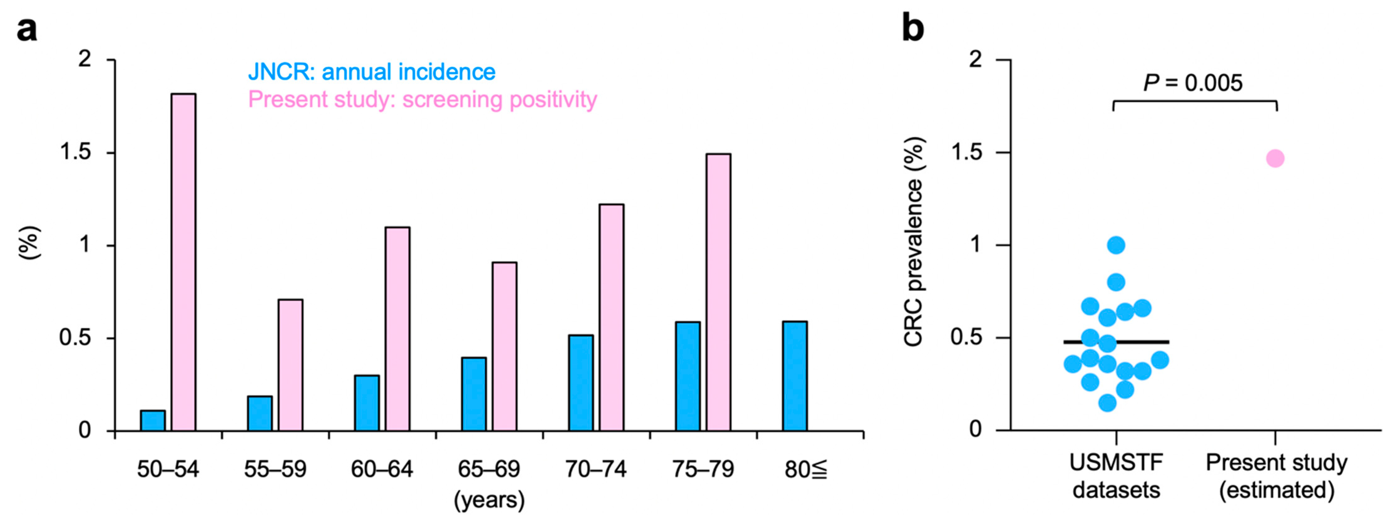
Outliers were tested using the Smirnov–Grubbs test after confirming the normality of the dataset using the Shapiro–Wilk test. The association between CRC morbidity and clinical risk factors for PCa was examined using Fisher’s exact test. The level of statistical significance was set at p < 0.05 after Bonferroni correction. Statistical analysis was performed using R (R Foundation for Statistical Computing, Vienna, Austria) on the EZR platform [29].

3. Results

Of the 2555 consecutive PCa patients enrolled in the study, 2537 were at average risk for CRC; 125 patients were excluded due to incomplete medical records (including the absence of screening information). Therefore, 2412 patients were analyzed (Figure 1). The average age was 69 years (range, 50–94 years). Initial PSA values, T stage, Gleason score, and PCa risk group are summarized in Table 2. Of all patients, 604 patients (25.0%) were positive for FIT; of these, 586 (97.0%) underwent colonoscopy. Figure 2 shows the colonoscopy findings of FIT-positive patients. CRC was detected in 28 (1.16%) patients. Meanwhile, non-malignant findings associated with lower gastrointestinal hemorrhage were identified in 421 (17.4%) patients.
All 28 patients diagnosed with CRC prioritized treatment for CRC before treatment for PCa; 24 patients (85.7%) completed CIRT successfully after treatment for CRC. Of the remaining four patients, two received chemotherapy; therefore, CIRT for PCa was not indicated. Of the remaining two patients, one had pelvic lymph node involvement and was not considered eligible for CIRT, and the other received surgery for CRC and at the time of writing is considering CIRT for PCa when his health recovers. Of note, none of the 24 patients treated with CIRT for PCa after treatment for CRC developed symptomatic proctitis post-CIRT.
The association between CRC morbidity and major clinical risk factors for PCa was analyzed. A high initial PSA value showed a significant association with CRC morbidity (p = 0.023), whereas T stage, Gleason score, or tumor risk group did not (Table 2). Age was not associated with CRC morbidity (69.6 ± 6.4 vs. 69.2 ± 6.8 for CRC patients and non-CRC patients, respectively; p = 0.62).
The outcomes of CRC screening in this study cohort were compared with the nation-matched JNCR dataset (see Section 2.1 for details). In the JNCR dataset, there was a trend toward a higher incidence of CRC among the elderly (Figure 3a). The same trend in the screening positivity rate was observed in this study cohort, except for age brackets 50–54 years and ≥80 years; it is likely that these exceptions are due to the small number of analyses. The screening positivity rate in this study cohort exceeded the annual incidence of CRC for all age brackets observed in the JNCR dataset; the exception was the ≥80 years population (Figure 3a). These data indicate that the performance of CRC screening in this study cohort is robust.
The estimated prevalence rate of CRC in this study cohort was 1.46%, based on the pooled sensitivity for CRC screening among asymptomatic average-risk adults (i.e., 0.79; see Section 2.1 for details). Interestingly, the estimated prevalence rate of CRC in this study cohort was greater than that in any of the 17 large-scale cohorts of average-risk adults, making the current cohort an outlier (p = 0.005, see Section 2.1 for details of the control cohorts) (Figure 3b). These data indicate that the prevalence of CRC in PCa patients considered for CIRT is greater than that in average-risk adults.

4. Discussion

Here, we found that the prevalence of CRC in PCa patients referred to GHMC for the purpose of CIRT was greater than that in the general population. To the best of our knowledge, this is the first study to report a greater prevalence rate of CRC in PCa patients compared with a risk-matched general population. The comparison with the USMSTF-reported studies was performed under risk-matched settings. Meanwhile, the comparison with the JNCR dataset was performed under nation-, gender-, and age-matched settings. These data indicate the robustness of the main finding of this study.
A possible explanation for the high prevalence of CRC in our study is that CRC and PCa share the same potential risk factors. Risk factors can be classified as endogenous or exogenous, although some factors are not exclusively one or the other (e.g., race, aging, and oxidative stress). Current alcohol intake was associated with an increased risk of CRC if one consumed an average of one or more alcoholic drinks per day [30], and the risk increased with alcohol intake for PCa as well [31]. It has been suggested that a diet high in meat-derived fats may also increase the risk of advanced colorectal neoplasia [30]. Fat intake, especially polyunsaturated fats, has also been reported to be strongly positively correlated with incidence and mortality in PCa [31], and this mechanism may be due to fat-induced changes in hormone profiles [32], the effects of fat metabolites as protein and DNA reactive intermediates [33], or elevated oxidative stress due to fat [34]. It has been reported that dietary fiber intake from cereals was associated with a lower relative risk of advanced neoplasia of the colon [30]. Cereal consumption has also been shown to be inversely associated with mortality in PCa [31]. It is suggested that plant ligands and isoflavonoids in grains and cereals are converted by intestinal bacteria into compounds with weak estrogenic and antioxidant activity, which have anticancer effects [35]. With regard to vitamin D intake, it has also been suggested to be inversely related to the risk of CRC [30]. In PCa, 1α-25-dihydroxyvitamin D (1,25-D), the hormonal form of vitamin D, has been reported to inhibit PCa cell invasion in vitro [36] and to exhibit antiproliferative and differentiation-promoting effects in the Dunning rat PCa model [37], suggesting that vitamin D deficiency has been a potential risk factor for PCa [31]. It is not likely that a potential age bias in this study cohort affected the greater CRC morbidity, considering that the trend of CRC morbidity by age group in this study cohort was consistent with that in the JNCR. It may be possible that this study cohort suffered from selection bias in that it included more health-conscious patients that favored CIRT, which is a promising, albeit scarce medical resource; even if that was the case, the bias should not have caused an increase (rather, it would have caused a decrease) in the prevalence of CRC associated with more frequent routine cancer screening of such patients.
Another possibility is the presence of unknown host genetic variants that contribute to predisposition to the two cancers; indeed, a subset of genes is responsible for cancer predisposing syndromes (e.g., the BRCA genes responsible for Hereditary Breast and Ovarian Cancers) [38]. In fact, tyrosine kinase receptors, which was isolated as an oncogene in CRC, is also expressed in PCa [39]. Tyrosine kinase receptors are thought to transmit proximal signals for neurotrophin-mediated growth in cancer, and the tyrosine kinase inhibitor K 252a inhibited the growth of cancer cell lines in vitro [40], further supporting the role of tyrosine kinase receptors in neurotrophin-mediated growth of PCa. Further research is needed to explore factors predictive of PCa sub-populations at high risk of CRC.
In this study, a high initial PSA value was associated with high CRC-related morbidity. Additionally, most immunohistochemical studies have concluded that abnormalities in the tumor suppressor gene TP53, which functions as a regulator of the cell cycle and is associated with many human malignancies, including colorectal cancer, are usually present in localized prostate cancers with high Gleason scores (>7) [41,42,43]. From this perspective, simultaneous screening for such common cancers is of high importance [44,45]. More importantly, there are no established treatment strategies for synchronous CRC and PCa. Corbin et al. performed a retrospective review of medical records at Duke University Medical Center and the Durham Veterans Affairs Medical Center between 1988 and 2017, and identified 54 patients with synchronous rectosigmoid cancer and PCa [46]. Seretis et al. performed a literature review of synchronous rectal cancer and PCa, and summarized 23 cases [47]. These studies report various (i.e., unstandardized) treatments for each patient. In this study, we report a series of 24 patients with synchronous CRC (including 14 patients of rectosigmoid cancer) and PCa who were treated successfully with surgical resection for the former, followed by CIRT for the latter. Furthermore, CIRT resulted in no symptomatic proctitis.
The screening positivity for CRC in our study cohort was markedly higher than the annual incidence of CRC in the JNCR dataset (1.16% vs. 0.37%, respectively) [10]. It is easy to imagine that in a real-world setting a portion of cases diagnosed with CRC would have been dropped from registration in the JNCR; therefore, it would be crude to compare the screening outcomes of this study cohort with the incidence reported in JNCR. Nevertheless, the incidence of CRC in the JNCR dataset is broadly consistent with the prevalence of CRC observed in nation-matched large-scale screening studies (i.e., median, 0.36%; range, 0.32–0.61%) [11,15,17,21,25], providing a certain level of justification for the current comparison. Radiotherapy for localized PCa may be postponed by performing androgen deprivation therapy [48]; therefore, detection of CRC prior to radiotherapy for PCa is of benefit to patients. In addition, in this study, we found benign lesions associated with lower gastrointestinal hemorrhage in 17.4% of participants; such information will be useful during post-CIRT follow up to avoid post-treatment colonoscopic manipulation that can exacerbate radiation-induced proctitis. Taken together, the results of the present study suggest that pre-treatment CRC screening is beneficial for PCa patients considered for CIRT. This benefit may be interpreted broadly with respect to other external beam radiotherapies based on photons or protons because, similar to CIRT, these modalities can cause radiation-induced proctitis in patients with localized PCa.
The study has several limitations. First, we were unable to standardize the details of CRC screening in terms of the brand and cut-off value of FIT, or the method and reporting of colonoscopy; this is because screening was performed at multiple institutes. Additionally, the clinical and epidemiological information is limited to PCa progression and to standard risk groups for CRC.

5. Conclusions

To elucidate the outcomes of CRC screening in PCa patients considered for CIRT, we performed a large-scale retrospective analysis of 2412 participants at average risk for CRC. These data indicate the possibility that the prevalence of CRC in PCa patients is greater than that in general average-risk adults, warranting further research in a prospective setting.

Author Contributions

Conceptualization, N.K. (Nao Kobayashi) and T.O. (Takahiro Oike); formal analysis, N.K. (Nao Kobayashi), N.K. (Nobuteru Kubo), T.O. (Takahiro Oike), Y.M., T.M., H.S., A.A., H.K. (Hiroyuki Katoh) and H.K. (Hidemasa Kawamura); data curation, N.K. (Nao Kobayashi), N.K. (Nobuteru Kubo) and T.O. (Takahiro Oike); writing—original draft preparation, N.K. (Nao Kobayashi); writing—review and editing, T.O. (Takahiro Oike) and T.O. (Tatsuya Ohno); visualization, N.K. (Nao Kobayashi) and T.O. (Takahiro Oike); supervision, T.O. (Takahiro Oike) and T.O. (Tatsuya Ohno); project administration, T.O. (Takahiro Oike); funding acquisition, T.O. (Tatsuya Ohno). All authors read and agreed to the published version of the manuscript.

Funding

This study was supported by the Gunma University Heavy Ion Medical Center.

Institutional Review Board Statement

The study was conducted in accordance with the Declaration of Helsinki and was approved by the Institutional Ethical Review Committee of the Gunma University Hospital (approval number, HS2019-039; date of approval, 29 June 2020).

Informed Consent Statement

Patient consent was waived by the Institutional Ethical Review Committee of the Gunma University Hospital due to the retrospective observational nature of the study.

Data Availability Statement

The data are not publicly available due to the study protocol approved by the Institutional Ethical Review Committee of the Gunma University Hospital.

Acknowledgments

We thank Takashi Aikawa of Gunma University for helpful advice. We also thank Yuka Hirota of Gunma University for technical assistance.

Conflicts of Interest

The authors declare no conflict of interest. The funders played no role in the design of the study; in the collection, analyses, or interpretation of data; in the writing of the manuscript; or in the decision to publish the results.

References

  1. Sung, H.; Ferlay, J.; Siegel, R.L.; Laversanne, M.; Soerjomataram, I.; Jemal, A.; Bray, F. Global Cancer Statistics 2020: GLOBOCAN Estimates of Incidence and Mortality Worldwide for 36 Cancers in 185 Countries. CA Cancer J. Clin. 2021, 71, 209–249. [Google Scholar] [CrossRef]
  2. National Comprehensive Cancer Network. Prostate Cancer (Version 2.2021). Available online: https://www.nccn.org/professionals/physician_gls/pdf/prostate.pdf (accessed on 15 June 2021).
  3. Nomiya, T.; Tsuji, H.; Kawamura, H.; Ohno, T.; Toyama, S.; Shioyama, Y.; Nakayama, Y.; Nemoto, K.; Tsujii, H.; Kamada, T. A multi-institutional analysis of prospective studies of carbon ion radiotherapy for prostate cancer: A report from the Japan Carbon ion Radiation Oncology Study Group (J-CROS). Radiother. Oncol. 2016, 121, 288–293. [Google Scholar] [CrossRef] [Green Version]
  4. Ishikawa, H.; Tsuji, H.; Kamada, T.; Akakura, K.; Suzuki, H.; Shimazaki, J.; Tsujii, H. Carbon-ion radiation therapy for prostate cancer. Int. J. Urol. 2012, 19, 296–305. [Google Scholar] [CrossRef] [Green Version]
  5. Marguet, C.; Raj, G.V.; Brashears, J.H.; Anscher, M.S.; Ludwig, K.; Mouraviev, V.; Robertson, C.N.; Polascik, T.J. Rectourethral fistula after combination radiotherapy for prostate cancer. Urology 2007, 69, 898–901. [Google Scholar] [CrossRef]
  6. Robertson, D.J.; Lee, J.K.; Boland, C.R.; Dominitz, J.A.; Giardiello, F.M.; Johnson, D.A.; Kaltenbach, T.; Lieberman, D.; Levin, T.R.; Rex, D.K. Recommendations on Fecal Immunochemical Testing to Screen for Colorectal Neoplasia: A Consensus Statement by the US Multi-Society Task Force on Colorectal Cancer. Gastroenterology 2017, 152, 1217–1237. [Google Scholar] [CrossRef] [PubMed] [Green Version]
  7. Provenzale, D.; Gupta, S.; Ahnen, D.J.; Markowitz, A.J.; Chung, D.C.; Mayer, R.J.; Regenbogen, S.E.; Blanco, A.M.; Bray, T.; Cooper, G.; et al. NCCN Guidelines Insights: Colorectal Cancer Screening, Version 1.2018. J. Natl. Compr. Cancer Netw. 2018, 16, 939–949. [Google Scholar] [CrossRef]
  8. Kawamura, H.; Kubo, N.; Sato, H.; Mizukami, T.; Katoh, H.; Ishikawa, H.; Ohno, T.; Matsui, H.; Ito, K.; Suzuki, K.; et al. Moderately hypofractionated carbon ion radiotherapy for prostate cancer; a prospective observational study “GUNMA0702”. BMC Cancer 2020, 20, 75. [Google Scholar] [CrossRef] [PubMed] [Green Version]
  9. Ishikawa, H.; Katoh, H.; Kaminuma, T.; Kawamura, H.; Ito, K.; Matsui, H.; Hirato, J.; Shimizu, N.; Takezawa, Y.; Tsuji, H.; et al. Carbon-ion Radiotherapy for Prostate Cancer: Analysis of Morbidities and Change in Health-related Quality of Life. Anticancer Res. 2015, 35, 5559–5566. [Google Scholar] [PubMed]
  10. Cancer Registry and Statistics. Cancer Information Service, National Cancer Center, Japan (Monitoring of Cancer Incidence in Japan). Available online: https://ganjoho.jp/reg_stat/statistics/dl/index.html#incidence (accessed on 30 May 2021).
  11. Morikawa, T.; Kato, J.; Yamaji, Y.; Wada, R.; Mitsushima, T.; Shiratori, Y. A comparison of the immunochemical fecal occult blood test and total colonoscopy in the asymptomatic population. Gastroenterology 2005, 129, 422–428. [Google Scholar] [CrossRef] [PubMed]
  12. Imperiale, T.F.; Ransohoff, D.F.; Itzkowitz, S.H.; Levin, T.R.; Lavin, P.; Lidgard, G.P.; Ahlquist, D.A.; Berger, B.M. Multitarget stool DNA testing for colorectal-cancer screening. N. Engl. J. Med. 2014, 370, 1287–1297. [Google Scholar] [CrossRef] [PubMed] [Green Version]
  13. Chiu, H.M.; Lee, Y.C.; Tu, C.H.; Chen, C.C.; Tseng, P.H.; Liang, J.T.; Shun, C.T.; Lin, J.T.; Wu, M.S. Association between early stage colon neoplasms and false-negative results from the fecal immunochemical test. Clin. Gastroenterol. Hepatol. 2013, 11, 832–838. [Google Scholar] [CrossRef]
  14. Cheng, T.I.; Wong, J.M.; Hong, C.F.; Cheng, S.H.; Cheng, T.J.; Shieh, M.J.; Lin, Y.M.; Tso, C.Y.; Huang, A.T. Colorectal cancer screening in asymptomaic adults: Comparison of colonoscopy, sigmoidoscopy and fecal occult blood tests. J. Formos. Med. Assoc. 2002, 101, 685–690. [Google Scholar] [PubMed]
  15. Nakama, H.; Yamamoto, M.; Kamijo, N.; Li, T.; Wei, N.; Fattah, A.S.; Zhang, B. Colonoscopic evaluation of immunochemical fecal occult blood test for detection of colorectal neoplasia. Hepatogastroenterology 1999, 46, 228–231. [Google Scholar]
  16. Sohn, D.K.; Jeong, S.Y.; Choi, H.S.; Lim, S.B.; Huh, J.M.; Kim, D.H.; Kim, D.Y.; Kim, Y.H.; Chang, H.J.; Jung, K.H.; et al. Single immunochemical fecal occult blood test for detection of colorectal neoplasia. Cancer Res. Treat. 2005, 37, 20–23. [Google Scholar] [CrossRef] [PubMed] [Green Version]
  17. Nakazato, M.; Yamano, H.O.; Matsushita, H.O.; Sato, K.; Fujita, K.; Yamanaka, Y.; Imai, Y. Immunologic fecal occult blood test for colorectal cancer screening. Jpn. Med. Assoc. J. 2006, 49, 203. [Google Scholar]
  18. Chiang, T.H.; Lee, Y.C.; Tu, C.H.; Chiu, H.M.; Wu, M.S. Performance of the immunochemical fecal occult blood test in predicting lesions in the lower gastrointestinal tract. CMAJ 2011, 183, 1474–1481. [Google Scholar] [CrossRef] [PubMed] [Green Version]
  19. Brenner, H.; Tao, S. Superior diagnostic performance of faecal immunochemical tests for haemoglobin in a head-to-head comparison with guaiac based faecal occult blood test among 2235 participants of screening colonoscopy. Eur. J. Cancer 2013, 49, 3049–3054. [Google Scholar] [CrossRef]
  20. De Wijkerslooth, T.R.; Stoop, E.M.; Bossuyt, P.M.; Meijer, G.A.; van Ballegooijen, M.; Van Roon, A.H.; Stegeman, I.; Kraaijenhagen, R.A.; Fockens, P.; van Leerdam, M.E.; et al. Immunochemical fecal occult blood testing is equally sensitive for proximal and distal advanced neoplasia. Am. J. Gastroenterol. 2012, 107, 1570–1578. [Google Scholar] [CrossRef] [PubMed]
  21. Itoh, M.; Takahashi, K.; Nishida, H.; Sakagami, K.; Okubo, T. Estimation of the optimal cut off point in a new immunological faecal occult blood test in a corporate colorectal cancer screening programme. J. Med. Screen. 1996, 3, 66–71. [Google Scholar] [CrossRef] [Green Version]
  22. Allison, J.E.; Tekawa, I.S.; Ransom, L.J.; Adrain, A.L. A comparison of fecal occult-blood tests for colorectal-cancer screening. N. Engl. J. Med. 1996, 334, 155–160. [Google Scholar] [CrossRef]
  23. Launoy, G.D.; Bertrand, H.J.; Berchi, C.; Talbourdet, V.Y.; Guizard, A.V.; Bouvier, V.M.; Caces, E.R. Evaluation of an immunochemical fecal occult blood test with automated reading in screening for colorectal cancer in a general average-risk population. Int. J. Cancer 2005, 115, 493–496. [Google Scholar] [CrossRef]
  24. Allison, J.E.; Sakoda, L.C.; Levin, T.R.; Tucker, J.P.; Tekawa, I.S.; Cuff, T.; Pauly, M.P.; Shlager, L.; Palitz, A.M.; Zhao, W.K.; et al. Screening for colorectal neoplasms with new fecal occult blood tests: Update on performance characteristics. J. Natl. Cancer Inst. 2007, 99, 1462–1470. [Google Scholar] [CrossRef] [Green Version]
  25. Nakama, H.; Kamijo, N.; Abdul Fattah, A.S.; Zhang, B. Validity of immunological faecal occult blood screening for colorectal cancer: A follow up study. J. Med. Screen. 1996, 3, 63–65. [Google Scholar] [CrossRef] [PubMed] [Green Version]
  26. Parra-Blanco, A.; Gimeno-García, A.Z.; Quintero, E.; Nicolás, D.; Moreno, S.G.; Jiménez, A.; Hernández-Guerra, M.; Carrillo-Palau, M.; Eishi, Y.; López-Bastida, J. Diagnostic accuracy of immunochemical versus guaiac faecal occult blood tests for colorectal cancer screening. J. Gastroenterol. 2010, 45, 703–712. [Google Scholar] [CrossRef] [PubMed]
  27. Levi, Z.; Birkenfeld, S.; Vilkin, A.; Bar-Chana, M.; Lifshitz, I.; Chared, M.; Maoz, E.; Niv, Y. A higher detection rate for colorectal cancer and advanced adenomatous polyp for screening with immunochemical fecal occult blood test than guaiac fecal occult blood test, despite lower compliance rate. A prospective, controlled, feasibility study. Int. J. Cancer 2011, 128, 2415–2424. [Google Scholar] [CrossRef] [PubMed]
  28. Lee, J.K.; Liles, E.G.; Bent, S.; Levin, T.R.; Corley, D.A. Accuracy of fecal immunochemical tests for colorectal cancer: Systematic review and meta-analysis. Ann. Intern. Med. 2014, 160, 171. [Google Scholar] [CrossRef] [PubMed]
  29. Kanda, Y. Investigation of the freely available easy-to-use software ’EZR’ for medical statistics. Bone Marrow Transplant. 2013, 48, 452–458. [Google Scholar] [CrossRef] [PubMed] [Green Version]
  30. Lieberman, D.A.; Prindiville, S.; Weiss, D.G.; Willett, W. Risk factors for advanced colonic neoplasia and hyperplastic polyps in asymptomatic individuals. JAMA 2003, 290, 2959–2967. [Google Scholar] [CrossRef] [Green Version]
  31. Bostwick, D.G.; Burke, H.B.; Djakiew, D.; Euling, S.; Ho, S.M.; Landolph, J.; Morrison, H.; Sonawane, B.; Shifflett, T.; Waters, D.J.; et al. Human prostate cancer risk factors. Cancer 2004, 101, 2371–2490. [Google Scholar] [CrossRef]
  32. Bishop, G.A.; McMillan, M.S.; Haughton, G.; Frelinger, J.A. Signaling to a B-cell clone by Ek, but not Ak, does not reflect alteration of Ak genes. Immunogenetics 1988, 28, 184–192. [Google Scholar] [CrossRef]
  33. Hietanen, E.; Bartsch, H.; Béréziat, J.C.; Camus, A.M.; McClinton, S.; Eremin, O.; Davidson, L.; Boyle, P. Diet and oxidative stress in breast, colon and prostate cancer patients: A case-control study. Eur. J. Clin. Nutr. 1994, 48, 575–586. [Google Scholar]
  34. Ho, P.J.; Baxter, R.C. Insulin-like growth factor-binding protein-2 in patients with prostate carcinoma and benign prostatic hyperplasia. Clin. Endocrinol. 1997, 46, 333–342. [Google Scholar] [CrossRef]
  35. Hebert, J.R.; Hurley, T.G.; Olendzki, B.C.; Teas, J.; Ma, Y.; Hampl, J.S. Nutritional and socioeconomic factors in relation to prostate cancer mortality: A cross-national study. J. Natl. Cancer Inst. 1998, 90, 1637–1647. [Google Scholar] [CrossRef] [Green Version]
  36. Schwartz, G.G.; Whitlatch, L.W.; Chen, T.C.; Lokeshwar, B.L.; Holick, M.F. Human prostate cells synthesize 1,25-dihydroxyvitamin D3 from 25-hydroxyvitamin D3. Cancer Epidemiol. Biomark. Prev. 1998, 7, 391–395. [Google Scholar]
  37. Getzenberg, R.H.; Light, B.W.; Lapco, P.E.; Konety, B.R.; Nangia, A.K.; Acierno, J.S.; Dhir, R.; Shurin, Z.; Day, R.S.; Trump, D.L.; et al. Vitamin D inhibition of prostate adenocarcinoma growth and metastasis in the Dunning rat prostate model system. Urology 1997, 50, 999–1006. [Google Scholar] [CrossRef]
  38. Yoshida, R. Hereditary breast and ovarian cancer (HBOC): Review of its molecular characteristics, screening, treatment, and prognosis. Breast Cancer 2020, 1–14. [Google Scholar] [CrossRef] [PubMed]
  39. Pflug, B.R.; Dionne, C.; Kaplan, D.R.; Lynch, J.; Djakiew, D. Expression of a Trk high affinity nerve growth factor receptor in the human prostate. Endocrinology 1995, 136, 262–268. [Google Scholar] [CrossRef]
  40. Delsite, R.; Djakiew, D. Anti-proliferative effect of the kinase inhibitor K252a on human prostatic carcinoma cell lines. J. Androl. 1996, 17, 481–490. [Google Scholar]
  41. Navone, N.M.; Troncoso, P.; Pisters, L.L.; Goodrow, T.L.; Palmer, J.L.; Nichols, W.W.; von Eschenbach, A.C.; Conti, C.J. p53 protein accumulation and gene mutation in the progression of human prostate carcinoma. J. Natl. Cancer Inst. 1993, 85, 1657–1669. [Google Scholar] [CrossRef] [PubMed]
  42. Hall, M.C.; Navone, N.M.; Troncoso, P.; Pollack, A.; Zagars, G.K.; von Eschenbach, A.C.; Conti, C.J.; Chung, L.W. Frequency and characterization of p53 mutations in clinically localized prostate cancer. Urology 1995, 45, 470–475. [Google Scholar] [CrossRef]
  43. Heidenberg, H.B.; Sesterhenn, I.A.; Gaddipati, J.P.; Weghorst, C.M.; Buzard, G.S.; Moul, J.W.; Srivastava, S. Alteration of the tumor suppressor gene p53 in a high fraction of hormone refractory prostate cancer. J. Urol. 1995, 154, 414–421. [Google Scholar] [CrossRef]
  44. Gohagan, J.K.; Prorok, P.C.; Hayes, R.B.; Kramer, B.S.; Prostate, Lung, Colorectal and Ovarian Cancer Screening Trial Project Team. The Prostate, Lung, Colorectal and Ovarian (PLCO) Cancer Screening Trial of the National Cancer Institute: History, organization, and status. Control. Clin. Trials 2000, 21, 251S–272S. [Google Scholar] [CrossRef]
  45. Ilunga-Tshiswaka, D.; Donley, T.; Okafor, A.; Memiah, P.; Mbizo, J. Prostate and Colorectal Cancer Screening Uptake among US and Foreign-Born Males: Evidence from the 2015 NHIS Survey. J. Community Health 2017, 42, 612–623. [Google Scholar] [CrossRef]
  46. Jacobs, C.D.; Trotter, J.; Palta, M.; Moravan, M.J.; Wu, Y.; Willett, C.G.; Lee, W.R.; Czito, B.G. Multi-Institutional Analysis of Synchronous Prostate and Rectosigmoid Cancers. Front. Oncol. 2020, 10, 345. [Google Scholar] [CrossRef]
  47. Seretis, C.; Seretis, F.; Liakos, N. Multidisciplinary approach to synchronous prostate and rectal cancer: Current experience and future challenges. J. Clin. Med. Res. 2014, 6, 157–161. [Google Scholar] [CrossRef] [PubMed] [Green Version]
  48. Dee, E.C.; Mahal, B.A.; Arega, M.A.; D’Amico, A.V.; Mouw, K.W.; Nguyen, P.L.; Muralidhar, V. Relative timing of radiotherapy and androgen deprivation for prostate cancer and implications for treatment during the COVID-19 pandemic. JAMA Oncol. 2020, 6, 1630–1632. [Google Scholar] [CrossRef] [PubMed]
Figure 1. Flow diagram showing patient enrollment. GHMC, Gunma University Heavy Ion Medical Center; IBD, inflammatory bowel disease; CRC, colorectal cancer.
Cancers 13 04481 g001
Figure 2. Colonoscopy findings among fecal immunochemical test-positive patients. See Section 2.2 for the definition of adenoma and polyp.
Cancers 13 04481 g002
Figure 3. Comparison of colorectal cancer (CRC) screening outcomes between this study cohort and (a) the Japan National Cancer Registry (JNCR) or (b) a series of large-scale CRC screening studies reported in the U.S. Multi-Society Task Force (USMSTF) consensus statement. The details of the JNCR and USMSTF datasets are summarized in Section 2.1. p-values were assessed using the Smirnov–Grubbs test.
Cancers 13 04481 g003
Table 1. Prevalence of CRC in average-risk adults reported in large-scale studies.
AuthorsYearCohortCRCRef.
nn%Confirmed by
Morikawa et al.200521,805790.36Colonoscopy[11]
Imperiale et al.20149899650.66Colonoscopy[12]
Chiu et al.20138822130.15Colonoscopy[13]
Cheng et al.20027411160.22Colonoscopy[14]
Nakama et al.19994611180.39Colonoscopy[15]
Sohn et al.20053794120.32Colonoscopy[16]
Nakazato et al.20063090190.61Colonoscopy[17]
Chiang et al.20112796281.00Colonoscopy[18]
Brenner et al.20132235150.67Colonoscopy[19]
Wijkerslooth et al.2012125680.64Colonoscopy[20]
Itoh et al.199627,860890.322-year f/u[21]
Allison et al.19967493350.472-year f/u[22]
Launoy et al.20057421280.382-year f/u[23]
Allison et al.20075356140.262-year f/u[24]
Nakama et al.19963365120.362-year f/u[25]
Parra-Blanco et al.20101756140.802-year f/u[26]
Levi et al.2011120460.502-year f/u[27]
CRC, colorectal cancer; Ref, reference; f/u, follow up.
Table 2. Association between CRC morbidity and clinical risk factors for PCa.
Prostate Cancer Risk FactorsNon-CRC PatientsCRC Patientsp-Value
n%n%
Initial PSA (ng/mL) 0.023
<10151063.41242.9
10–19.959024.8725.0
20≤28211.8932.1
T stage 0.20
129612.4621.3
2124852.5932.2
3 + 483535.11346.4
Gleason score >0.99
61918.0314.3
7135256.81257.1
846819.6628.6
9 + 1037115.670.0
Tumor risk group >0.99
Low30.100.0
Intermediate113448.51140.7
High120251.41659.3
CRC, colorectal cancer; PCa, prostate cancer; PSA, prostate-specific antigen. p-values, assessed by Fisher’s exact test, are shown after Bonferroni correction.
Publisher’s Note: MDPI stays neutral with regard to jurisdictional claims in published maps and institutional affiliations.

