
How Has Prostate Cancer Radiotherapy Changed in Italy between 2004 and 2011? An Analysis of the National Patterns-Of-Practice (POP) Database by the Uro-Oncology Study Group of the Italian Society of Radiotherapy and Clinical Oncology (AIRO)

 , , , , , and add
Show full author list
, , , , , and add
Show full author list
Abstract
:Simple Summary
Abstract
1. Introduction
2. Materials and Methods
Statistical Analysis
3. Results
3.1. Patients’ Clinical Features
3.2. Diagnostic Workup
3.3. Treatment Features
3.4. Acute Toxicity
3.5. Late Toxicity
3.6. Overall Survival (OS) and Biochemical Relapse-Free Survival (BRFS)
4. Discussion
5. Conclusions
Author Contributions
Funding
Institutional Review Board Statement
Informed Consent Statement
Data Availability Statement
Acknowledgments
Conflicts of Interest
References
- Center, M.M.; Jemal, A.; Lortet-Tieulent, J.; Ward, E.; Ferlay, J.; Brawley, O.; Bray, F. International variation in prostate cancer incidence and mortality rates. Eur. Urol. 2012, 61, 1079–1092. [Google Scholar] [CrossRef] [PubMed]
- I Numeri del Cancro in Italia 2019|Associazione Italiana Registri Tumori. Available online: https://www.registri-tumori.it/cms/pubblicazioni/i-numeri-del-cancro-italia-2019 (accessed on 12 September 2019).
- de Crevoisier, R.; Bayar, M.A.; Pommier, P.; Muracciole, X.; Pene, F.; Dudouet, P.; Latorzeff, I.; Beckendorf, V.; Bachaud, J.M.; Laplanche, A.; et al. Daily Versus Weekly Prostate Cancer Image Guided Radiation Therapy: Phase 3 Multicenter Randomized Trial. Int. J. Radiat. Oncol. Biol. Phys. 2018, 102, 1420–1429. [Google Scholar] [CrossRef]
- Murray, J.; Griffin, C.; Gulliford, S.; Syndikus, I.; Staffurth, J.; Panades, M.; Scrase, C.; Parker, C.; Khoo, V.; Dean, J.; et al. A randomised assessment of image guided radiotherapy within a phase 3 trial of conventional or hypofractionated high dose intensity modulated radiotherapy for prostate cancer. Radiother. Oncol. J. Eur. Soc. Ther. Radiol. Oncol. 2020, 142, 62–71. [Google Scholar] [CrossRef] [PubMed]
- Magrini, S.M.; Bertoni, F.; Vavassori, V.; Villa, S.; Cagna, E.; Maranzano, E.; Pertici, M.; Pradella, R.; Spediacci, M.A.; Chiavacci, A.; et al. Practice patterns for prostate cancer in nine central and northern Italy radiation oncology centers: A survey including 1759 patients treated during two decades (1980–1998). Int. J. Radiat. Oncol. Biol. Phys. 2002, 52, 1310–1319. [Google Scholar] [CrossRef]
- Pegurri, L.; Buglione, M.; Girelli, G.; Guarnieri, A.; Meattini, I.; Ricardi, U.; Mangoni, M.; Gabriele, P.; Bellavita, R.; Krengli, M.; et al. Changes in patterns of practice for prostate cancer radiotherapy in Italy 1995-2003. A survey of the Prostate Cancer Study Group of the Italian Radiation Oncology Society. Tumori 2014, 100, 31–37. [Google Scholar] [CrossRef]
- Cox, J.D.; Stetz, J.; Pajak, T.F. Toxicity criteria of the Radiation Therapy Oncology Group (RTOG) and the European organization for research and treatment of cancer (EORTC). Int. J. Radiat. Oncol. Biol. Phys. 1995, 31, 1341–1346. [Google Scholar] [CrossRef]
- American Society for Therapeutic Radiology and Oncology Consensus Panel. Consensus statement: Guidelines for PSA following radiation therapy. Int. J. Radiat. Oncol. Biol. Phys. 1997, 37, 1035–1041. [Google Scholar] [CrossRef]
- Egevad, L.; Delahunt, B.; Srigley, J.R.; Samaratunga, H. International Society of Urological Pathology (ISUP) grading of prostate cancer—An ISUP consensus on contemporary grading. APMIS Acta Pathol. Microbiol. Immunol. Scand. 2016, 124, 433–435. [Google Scholar] [CrossRef]
- Brierley, J.D.; Gospodarowicz, M.K.; Wittekind, C. TNM Classification of Malignant Tumours, 8th ed.; John Wiley & Sons, Inc.: Chichester, UK; Hoboken, NJ, USA, 2017; ISBN 978-1-119-26357-9. [Google Scholar]
- Fowler, J.F.; Ritter, M.A.; Chappell, R.J.; Brenner, D.J. What hypofractionated protocols should be tested for prostate cancer? Int. J. Radiat. Oncol. Biol. Phys. 2003, 56, 1093–1104. [Google Scholar] [CrossRef]
- Ritter, M. Rationale, conduct, and outcome using hypofractionated radiotherapy in prostate cancer. Semin Radiat. Oncol. 2008, 18, 249–256. [Google Scholar] [CrossRef] [PubMed] [Green Version]
- National Comprehensive Cancer Network Prostate Cancer (Version 4.2019). Available online: https://www.nccn.org/professionals/physician_gls/pdf/prostate.pdf (accessed on 19 August 2019).
- European Association of Urology. European Association of Urology Guidelines. 2020 Edition; European Association of Urology Guidelines Office: Arnhem, The Netherlands, 2020; ISBN 978-94-92671-07-3. [Google Scholar]
- Santoni, R.; Arcangeli, S.; Ingrosso, G.; Antognoni, P.; Mazzeo, E.; D’Angelillo, R.M.; Buglione, M.; Borghesi, S.; Petitti, T.; Vinciguerra, A.; et al. Linee guida Carcinoma della Prostata—AIRO, 2016. Tumori 2016, 102, S1–S79. [Google Scholar] [CrossRef]
- Linee Guida Carcinoma Della Prostata. Available online: https://www.aiom.it/linee-guida-aiom-carcinoma-della-prostata-2019/ (accessed on 25 October 2019).
- Buglione, M.; Noale, M.; Bruni, A.; Antonelli, A.; Bertoni, F.; Corvo, R.; Ricardi, U.; Borghetti, P.; Maddalo, M.; Simeone, C.; et al. Treatment paths for localised prostate cancer in Italy: The results of a multidisciplinary, observational, prospective study (Pros-IT CNR). PLoS ONE 2019, 14, e0224151. [Google Scholar] [CrossRef] [PubMed]
- Peeters, S.T.; Heemsbergen, W.D.; Koper, P.C.; van Putten, W.L.; Slot, A.; Dielwart, M.F.; Bonfrer, J.M.; Incrocci, L.; Lebesque, J.V. Dose-response in radiotherapy for localized prostate cancer: Results of the Dutch multicenter randomized phase III trial comparing 68 Gy of radiotherapy with 78 Gy. J. Clin. Oncol. Official J. Am. Soc. Clin. Oncol. 2006, 24, 1990–1996. [Google Scholar] [CrossRef] [PubMed]
- Pollack, A.; Zagars, G.K.; Starkschall, G.; Antolak, J.A.; Lee, J.J.; Huang, E.; von Eschenbach, A.C.; Kuban, D.A.; Rosen, I. Prostate cancer radiation dose response: Results of the M. D. Anderson phase III randomized trial. Int. J. Radiat. Oncol. Biol. Phys. 2002, 53, 1097–1105. [Google Scholar] [CrossRef]
- Zietman, A.L.; DeSilvio, M.L.; Slater, J.D.; Rossi, C.J., Jr.; Miller, D.W.; Adams, J.A.; Shipley, W.U. Comparison of conventional-dose vs high-dose conformal radiation therapy in clinically localized adenocarcinoma of the prostate: A randomized controlled trial. Jama 2005, 294, 1233–1239. [Google Scholar] [CrossRef]
- Kuban, D.A.; Tucker, S.L.; Dong, L.; Starkschall, G.; Huang, E.H.; Cheung, M.R.; Lee, A.K.; Pollack, A. Long-term results of the M. D. Anderson randomized dose-escalation trial for prostate cancer. Int. J. Radiat. Oncol. Biol. Phys. 2008, 70, 67–74. [Google Scholar] [CrossRef] [PubMed]
- Dearnaley, D.P.; Sydes, M.R.; Graham, J.D.; Aird, E.G.; Bottomley, D.; Cowan, R.A.; Huddart, R.A.; Jose, C.C.; Matthews, J.H.; Millar, J.; et al. Escalated-dose versus standard-dose conformal radiotherapy in prostate cancer: First results from the MRC RT01 randomised controlled trial. Lancet Oncol. 2007, 8, 475–487. [Google Scholar] [CrossRef]
- Epstein, J.I.; Allsbrook, W.C.; Amin, M.B.; Egevad, L.L.; ISUP Grading Committee. The 2005 International Society of Urological Pathology (ISUP) Consensus Conference on Gleason Grading of Prostatic Carcinoma. Am. J. Surg. Pathol. 2005, 29, 1228–1242. [Google Scholar] [CrossRef] [Green Version]
- Epstein, J.I.; Egevad, L.; Amin, M.B.; Delahunt, B.; Srigley, J.R.; Humphrey, P.A.; Grading Committee. The 2014 International Society of Urological Pathology (ISUP) Consensus Conference on Gleason Grading of Prostatic Carcinoma: Definition of Grading Patterns and Proposal for a New Grading System. Am. J. Surg. Pathol. 2016, 40, 244–252. [Google Scholar] [CrossRef]
- Arcangeli, G.; Saracino, B.; Arcangeli, S.; Gomellini, S.; Petrongari, M.G.; Sanguineti, G.; Strigari, L. Moderate Hypofractionation in High-Risk, Organ-Confined Prostate Cancer: Final Results of a Phase III Randomized Trial. J. Clin. Oncol. Official J. Am. Soc. Clin. Oncol. 2017, 35, 1891–1897. [Google Scholar] [CrossRef]
- Pollack, A.; Walker, G.; Horwitz, E.M.; Price, R.; Feigenberg, S.; Konski, A.A.; Stoyanova, R.; Movsas, B.; Greenberg, R.E.; Uzzo, R.G.; et al. Randomized trial of hypofractionated external-beam radiotherapy for prostate cancer. J. Clin. Oncol. Official J. Am. Soc. Clin. Oncol. 2013, 31, 3860–3868. [Google Scholar] [CrossRef] [PubMed]
- Incrocci, L.; Wortel, R.C.; Alemayehu, W.G.; Aluwini, S.; Schimmel, E.; Krol, S.; van der Toorn, P.P.; Jager, H.; Heemsbergen, W.; Heijmen, B.; et al. Hypofractionated versus conventionally fractionated radiotherapy for patients with localised prostate cancer (HYPRO): Final efficacy results from a randomised, multicentre, open-label, phase 3 trial. Lancet Oncol. 2016, 17, 1061–1069. [Google Scholar] [CrossRef]
- Dearnaley, D.; Syndikus, I.; Mossop, H.; Khoo, V.; Birtle, A.; Bloomfield, D.; Graham, J.; Kirkbride, P.; Logue, J.; Malik, Z.; et al. Conventional versus hypofractionated high-dose intensity-modulated radiotherapy for prostate cancer: 5-year outcomes of the randomised, non-inferiority, phase 3 CHHiP trial. Lancet Oncol. 2016, 17, 1047–1060. [Google Scholar] [CrossRef] [Green Version]
- Murthy, V.; Maitre, P.; Kannan, S.; Panigrahi, G.; Krishnatry, R.; Bakshi, G.; Prakash, G.; Pal, M.; Menon, S.; Phurailatpam, R.; et al. Prostate-Only Versus Whole-Pelvic Radiation Therapy in High-Risk and Very High-Risk Prostate Cancer (POP-RT): Outcomes From Phase III Randomized Controlled Trial. J. Clin. Oncol. 2021, JCO2003282. [Google Scholar] [CrossRef]
- Ohri, N.; Dicker, A.P.; Showalter, T.N. Late toxicity rates following definitive radiotherapy for prostate cancer. Can. J. Urol. 2012, 19, 6373–6380. [Google Scholar] [PubMed]
- Dearnaley, D.P.; Jovic, G.; Syndikus, I.; Khoo, V.; Cowan, R.A.; Graham, J.D.; Aird, E.G.; Bottomley, D.; Huddart, R.A.; Jose, C.C.; et al. Escalated-dose versus control-dose conformal radiotherapy for prostate cancer: Long-term results from the MRC RT01 randomised controlled trial. Lancet Oncol. 2014, 15, 464–473. [Google Scholar] [CrossRef] [Green Version]
- Xu, N.; Rossi, P.J.; Jani, A.B. Toxicity analysis of dose escalation from 75.6 gy to 81.0 gy in prostate cancer. Am. J. Clin. Oncol. 2011, 34, 11–15. [Google Scholar] [CrossRef]
- Goldner, G.; Bombosch, V.; Geinitz, H.; Becker, G.; Wachter, S.; Glocker, S.; Zimmermann, F.; Wachter-Gerstner, N.; Schrott, A.; Bamberg, M.; et al. Moderate risk-adapted dose escalation with three-dimensional conformal radiotherapy of localized prostate cancer from 70 to 74 Gy. First report on 5-year morbidity and biochemical control from a prospective Austrian-German multicenter phase II trial. Strahlenther. Onkol. Organ. Dtsch. Rontgenges. 2009, 185, 94–100. [Google Scholar] [CrossRef]
- Zelefsky, M.J.; Levin, E.J.; Hunt, M.; Yamada, Y.; Shippy, A.M.; Jackson, A.; Amols, H.I. Incidence of late rectal and urinary toxicities after three-dimensional conformal radiotherapy and intensity-modulated radiotherapy for localized prostate cancer. Int. J. Radiat. Oncol. Biol. Phys. 2008, 70, 1124–1129. [Google Scholar] [CrossRef] [PubMed]
- Jani, A.B.; Su, A.; Correa, D.; Gratzle, J. Comparison of late gastrointestinal and genitourinary toxicity of prostate cancer patients undergoing intensity-modulated versus conventional radiotherapy using localized fields. Prostate Cancer Prostatic Dis. 2007, 10, 82–86. [Google Scholar] [CrossRef] [Green Version]
- Jacobs, B.L.; Zhang, Y.; Skolarus, T.A.; Wei, J.T.; Montie, J.E.; Miller, D.C.; Hollenbeck, B.K. Comparative effectiveness of external-beam radiation approaches for prostate cancer. Eur. Urol. 2014, 65, 162–168. [Google Scholar] [CrossRef] [PubMed] [Green Version]
- Barelkowski, T.; Wust, P.; Kaul, D.; Zschaeck, S.; Wlodarczyk, W.; Budach, V.; Ghadjar, P.; Beck, M. Image-guided dose-escalated radiation therapy for localized prostate cancer with helical tomotherapy. Strahlenther. Onkol. Organ. Dtsch. Rontgenges. 2020, 196, 229–242. [Google Scholar] [CrossRef] [PubMed]
- Detti, B.; Baki, M.; Becherini, C.; Saieva, C.; Scartoni, D.; Giacomelli, I.; Trombetta, L.; Muntoni, C.; Olmetto, E.; Francolini, G.; et al. High-dose intensity-modulated radiation therapy as primary treatment of prostate cancer: Genitourinary/gastrointestinal toxicity and outcomes, a single-institution experience. Radiol. Med. 2019, 124, 422–431. [Google Scholar] [CrossRef] [PubMed]


| Clinical Feature | POPI 1995–1998 1005 Patients (No., %) | POPII 1999–2003 3001 Patients (No., %) | Current Series (POPIII) 2004–2011 2525 Patients (No., %) |
|---|---|---|---|
| Median age (range, yr) | 70 (range, 28–89) | 71 (range, 46–86) | 72 (range, 46–88) |
| T stage | |||
| 1 | 99 (10) | 597 (20) | 866 (34) |
| 2 | 577 (57) | 1732 (58) | 1177 (47) |
| 3 | 284 (28) | 621 (21) | 450 (18) |
| 4 | 17 (2) | 29 (1) | 11 (<1) |
| Unknown | 28 (3) | 5 (>1) | 21 (<1) |
| N stage | |||
| N+ | 32 (3) | 75 (3) | 42 (2) |
| N0 | 504 (50) | 2502 (83) | 1761 (70) |
| Nx | 469 (47) | 424 (14) | 722 (28) |
| Grade group (ISUP) | |||
| Group 1 (Gleason score ≤ 6) | Not evaluable | 1380 (46) | 1152 (46) |
| Group 2 (Gleason score 3 + 4 = 7) | Not evaluable | 460 (16) | 548 (22) |
| Group 3 (Gleason score 4 + 3 = 7) | Not evaluable | 279 (9) | 254 (10) |
| Group 4 (Gleason score 8) | Not evaluable | 314 (10) | 356 (14) |
| Group 5 (Gleason scores 9–10) | Not evaluable | 153 (5) | 188 (7) |
| Unknown | Not evaluable | 409 (14) | 27 (1) |
| Patients with Gleason score 2–5 | |||
| Not evaluable | 814 (27) | 163 (6) | |
| Gleason sum | |||
| 2–6 | 606 (61) | 1676 (56) | 1152 (46) |
| 7 | 142 (14) | 777 (26) | 805 (32) |
| 8–10 | 114 (11) | 506 (17) | 543 (22) |
| Unknown | 143 (14) | 42 (1) | 25 (1) |
| Baseline PSA (ng/mL) | |||
| <1 | 0 (0) | 0 (0) | 24 (<1) |
| 1–10 | 345 (34) | 1292 (43) | 1481 (59) |
| 10.1–20 | 277 (28) | 886 (30) | 617 (24) |
| >20 | 326 (32) | 810 (27) | 395 (15) |
| Unknown | 57 (6) | 13 (<1) | 8 (<1) |
| Median baseline PSA (ng/mL) | 13 | 12 | 8.2 |
| D’Amico category risk | |||
| Low risk | 34 (3) | 516 (17) | 739 (29) |
| Intermediate risk | 342 (33) | 1057 (35) | 756 (30) |
| High Risk | 432 (43) | 1356 (46) | 1016 (40) |
| Not assessable | 197 (21) | 72 (2) | 14 (<1) |
| Diagnostic Procedures | POPI 1995–1998 1005 Patients (No., %) | POPII 1999–2003 3001 Patients (No., %) | Current Series (POPIII) 2004–2011 2525 Patients (No., %) | Chi-Square Test 1999–2003 Serie vs. 2004–2011 Serie |
|---|---|---|---|---|
| Abdominal/pelvic computed tomography | ||||
| No | 395 (39) | 798 (27) | 1186 (47) | p < 0.001 |
| Yes | 610 (61) | 2203 (73) | 1339 (53) | |
| Abdominal/pelvic magnetic resonance | ||||
| No | 867 (86) | 2808 (94) | 2335 (92) | p = 0.171 |
| Yes | 138 (14) | 193 (6) | 190 (8) | |
| Transrectal ultrasound | ||||
| No | 254 (25) | 385 (13) | 809 (32) | p < 0.001 |
| Yes | 751 (75) | 2616 (87) | 1716 (68) | |
| Magnetic resonance with endorectal coil | ||||
| No | 911 (91) | 2714 (90) | 2251 (89) | p = 0.114 |
| Yes | 94 (9) | 287 (10) | 274 (11) | |
| Bone scan | ||||
| No | 86 (9) | 471 (16) | 1181 (47) | p < 0.001 |
| Yes | 919 (91) | 2530 (84) | 1344 (53) | |
| Staging lymphadenectomy | ||||
| No | 928 (92) | 2945 (98) | Not evaluated | \\ |
| Yes | 77 (8) | 56 (2) | Not evaluated | |
| Choline PET | ||||
| No | Not evaluated | Not evaluated | 2476 (98) | \\ |
| Yes | Not evaluated | Not evaluated | 49 (2) | |
| TURP | ||||
| No | Not evaluated | 2741 (91) | 2299 (91) | \\ |
| Yes | Not evaluated | 259 (9) | 215 (8) | |
| Unknown | Not evaluated | 1 (<1) | 11 (<1) |
| Therapeutic Feature | POPI 1995–1998 1005 Patients (No., %) | POPII 1999–2003 3001 Patients (No., %) | Current Series (POPIII) 2004–2011 2525 Patients (No., %) |
|---|---|---|---|
| Dose to prostate | |||
| <70 Gy s.f | 315 (31) | 218 (7) | 20 (<1) |
| 70–75 Gy s.f. | 689 (69) | 2531 (84) | 1257 (50) |
| ≥76 Gy s.f. | 1 (<1) | 252 (9) | 1136 (45) |
| ≤70 hypo 2.5–3.1 Gy Dmean to prostate EqD2 (α/β = 1.5): 78 Gy * | Not performed | Not performed | 47 (2) |
| >70 hypo 2.2–2.35 Gy Dmean to prostate EqD2 (α/β = 1.5): 79 Gy * | Not performed | Not performed | 20 (<1) |
| >70 hypo 2.7 Gy Dmean to prostate EqD2 (α/β = 1.5): 84.2 Gy * | Not performed | Not performed | 40 (2) |
| Unknown | 5 (<1) | ||
| Dmean to prostate (Gy) ** | |||
| Conventional fractionation | 69 | 71.6 | 74.1 |
| EqD2 (α/β = 1.5) | 74.5 | ||
| Treated volume | |||
| No pelvic irradiation | 784 (78) | 2497 (83) | 2416 (95) |
| Pelvic irradiation | 221 (22) | 504 (17) | 107 (4) |
| Unknown | 2 (<1) | ||
| Beam energy, MV | |||
| ≤10 | 62 (6) | 174 (6) | 1479 (59) |
| 10–18 | 421 (42) | 2026 (68) | 613 (24) |
| ≥18 | 522 (52) | 252 (8) | 283 (11) |
| Unknown | - | 549 (18) | 150 (6) |
| Conformal techniques | |||
| 2D-RT | 593 (59) | 725 (24) | - |
| Static 3D-CRT | 412 (41) | 2174 (72) | 1953 (77) |
| Dynamic 3D-CRT | Not performed | 95 (3) | 4 (<1) |
| IMRT | Not performed | 7 (<1) | 568 (22) |
| IGRT | |||
| No | All | All | 2188 (87) |
| Yes | None | None | 337 (13) |
| CTV-PTV margin (mm) | |||
| <8 | 625 (25) | ||
| ≥8–10 | 1747 (70) | ||
| Unknown | All | All | 153 (5) |
| Concomitant hormone therapy | |||
| No | 201 (20) | 644 (22) | 581 (23) |
| Yes | 804 (80) | 2226 (74) | 1911 (76) |
| Unknown | - | 131 (4) | 33 (1) |
| Treatment Features and Toxicity | Period | p | |
|---|---|---|---|
| 2004–2007 (1246 Patients, 49%) | 2008–2011 (1279 Patients, 51%) | ||
| Pts treated with IMRT (%) | 7% | 38% | p < 0.001 |
| Pts treated with IGRT (%) | 7% | 19% | p < 0.001 |
| Pts treated with EqD2 ≥ 78 Gy (%) | 22% | 28% | p < 0.001 |
| Mean dose (EqD2) (±SD) Gy | 74 Gy (± 3.6) | 75 Gy (± 3.5) | p < 0.001 |
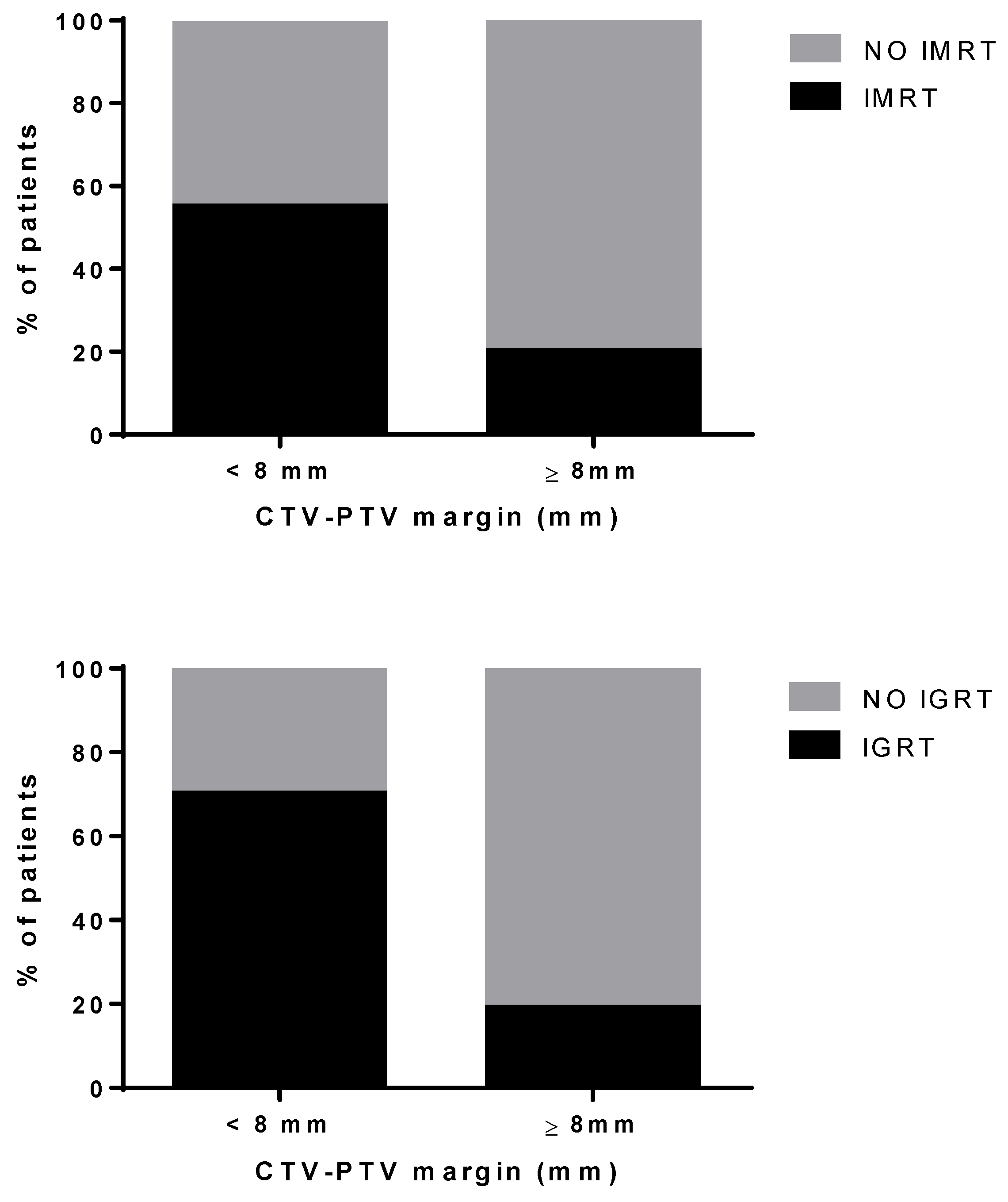
| CTV-PTV margin < 8 mm (%) (*) | 15% | 38% | p < 0.001 |
| Hypofractionation (%) | 1% | 7% | p < 0.001 |
| Acute GI (≥G2) toxicity (%) | 15% | 8% | p < 0.001 |
| Acute GU (≥G2) toxicity (%) | 16% | 14% | p = NS |
| Late GI (≥G2) toxicity (%) | 13% | 13% | p = NS |
| Late GU (≥G2) toxicity (%) | 14% | 8% | p < 0.001 |
Publisher’s Note: MDPI stays neutral with regard to jurisdictional claims in published maps and institutional affiliations. |
© 2021 by the authors. Licensee MDPI, Basel, Switzerland. This article is an open access article distributed under the terms and conditions of the Creative Commons Attribution (CC BY) license (https://creativecommons.org/licenses/by/4.0/).
Share and Cite
Mazzeo, E.; Triggiani, L.; Frassinelli, L.; Guarneri, A.; Bartoncini, S.; Antognoni, P.; Gottardo, S.; Greco, D.; Borghesi, S.; Nanni, S.; et al. How Has Prostate Cancer Radiotherapy Changed in Italy between 2004 and 2011? An Analysis of the National Patterns-Of-Practice (POP) Database by the Uro-Oncology Study Group of the Italian Society of Radiotherapy and Clinical Oncology (AIRO). Cancers 2021, 13, 2702. https://doi.org/10.3390/cancers13112702
Mazzeo E, Triggiani L, Frassinelli L, Guarneri A, Bartoncini S, Antognoni P, Gottardo S, Greco D, Borghesi S, Nanni S, et al. How Has Prostate Cancer Radiotherapy Changed in Italy between 2004 and 2011? An Analysis of the National Patterns-Of-Practice (POP) Database by the Uro-Oncology Study Group of the Italian Society of Radiotherapy and Clinical Oncology (AIRO). Cancers. 2021; 13(11):2702. https://doi.org/10.3390/cancers13112702
Chicago/Turabian StyleMazzeo, Ercole, Luca Triggiani, Luca Frassinelli, Alessia Guarneri, Sara Bartoncini, Paolo Antognoni, Stefania Gottardo, Diana Greco, Simona Borghesi, Sara Nanni, and et al. 2021. "How Has Prostate Cancer Radiotherapy Changed in Italy between 2004 and 2011? An Analysis of the National Patterns-Of-Practice (POP) Database by the Uro-Oncology Study Group of the Italian Society of Radiotherapy and Clinical Oncology (AIRO)" Cancers 13, no. 11: 2702. https://doi.org/10.3390/cancers13112702