Recent Advances in Nanotherapeutics for Multiple Myeloma

 , ,
, ,
Abstract
:Simple Summary
Abstract
1. Introduction
2. Nanotherapeutics for MM Therapy
2.1. Liposomes
2.2. Micelles
2.3. Polymeric Nanoparticles
2.4. Inorganic Nanoparticles
2.5. Carbon-Based Nanoparticles
3. Conclusions
Funding
Acknowledgments
Conflicts of Interest
References
- Rajkumar, S.V. Multiple myeloma: 2020 Update on diagnosis, risk-stratification and management. Am. J. Hematol. 2020, 95, 548–567. [Google Scholar] [CrossRef] [PubMed] [Green Version]
- International Agency for Research on Cancer; World Health Organization. The Global Cancer Observatory. 2019. Available online: https://gco.iarc.fr/today/data/factsheets/cancers/35-Multiple-myeloma-fact-sheet.pdf (accessed on 25 June 2020).
- National Cancer Institute, Cancer Stat Facts: Myeloma. 2020. Available online: https://seer.cancer.gov/statfacts/html/mulmy.html (accessed on 25 June 2020).
- Costello, C.; Davies, F.E.; Cook, G.; Vela-Ojeda, J.; Omel, J.; Rifkin, R.M.; Berdeja, J.; Puig, N.; Usmani, S.Z.; Weisel, K.; et al. INSIGHT MM: A large, global, prospective, non-interventional, real-world study of patients with multiple myeloma. Future Oncol. 2019, 15, 1411–1428. [Google Scholar] [CrossRef] [Green Version]
- Fairfield, H.; Falank, C.; Avery, L.; Reagan, M.R. Multiple myeloma in the marrow: Pathogenesis and treatments. Ann. N. Y. Acad. Sci. 2016, 1364, 32–51. [Google Scholar] [CrossRef] [Green Version]
- Garderet, L.; D’Souza, A.; Jacobs, P.; Van Biezen, A.; Schönland, S.O.; Kroeger, N.; Morris, C.; Hari, P. Response Assessment in Myeloma: Practical Manual on Consistent Reporting in an Era of Dramatic Therapeutic Advances. Biol. Blood Marrow Transplant. 2017, 23, 1193–1202. [Google Scholar] [CrossRef] [Green Version]
- Furukawa, Y.; Jiro Kikuchi, J. Molecular Pathogenesis of Multiple Myeloma. Int. J. Clin. Oncol. 2015, 20, 413–422. [Google Scholar] [CrossRef]
- Scalzulli, E.; Grammatico, S.; Vozella, F.; Petrucci, M.T. Proteasome inhibitors for the treatment of multiple myeloma. Expert Opin. Pharmacother. 2018, 19, 375–386. [Google Scholar] [CrossRef]
- Gandolfi, S.; Laubach, J.P.; Hideshima, T.; Chauhan, D.; Anderson, K.C.; Richardson, P.G. The proteasome and proteasome inhibitors in multiple myeloma. Cancer Metastasis Rev. 2017, 36, 561–584. [Google Scholar] [CrossRef]
- Pozzi, S.; Raje, N. The Role of Bisphosphonates in Multiple Myeloma: Mechanisms, Side Effects, and the Future. Oncologist 2011, 16, 651–662. [Google Scholar] [CrossRef] [Green Version]
- Burwick, N.; Sharma, S. Glucocorticoids in multiple myeloma: Past, present, and future. Ann. Hematol. 2018, 98, 19–28. [Google Scholar] [CrossRef]
- Holstein, S.A.; McCarthy, P.L. Immunomodulatory Drugs in Multiple Myeloma: Mechanisms of Action and Clinical Experience. Drugs 2017, 77, 505–520. [Google Scholar] [CrossRef]
- Ramdial, J.; Ma, J.; Milton, D.R.; Delgado, R.; Bashir, Q.; Srour, S.A.; Saini, N.; Nieto, Y.; Hosing, C.M.; Popat, U.R.; et al. Peripheral Blood Stem Cell Mobilization for Autologous Hematopoietic Stem Cell Transplantation in Multiple Myeloma: Growth Factors Only vs. Growth Factors + Chemotherapy. Biol. Blood Marrow Transplant. 2020, 26, S286–S287. [Google Scholar] [CrossRef]
- Branagan, A.; Lei, M.; Lou, U.; Raje, N. Current Treatment Strategies for Multiple Myeloma. JCO Oncol. Pract. 2020, 16, 5–14. [Google Scholar] [CrossRef] [PubMed]
- Mair, M.; Straka, C.; Buratti, T.; Tauber, M.; Mitterer, M.; Fong, D. Clinical outcomes and prognostic factors in patients with multiple myeloma in South Tyrol: A retrospective single-center analysis. Ann. Hematol. 2020, 99, 1031–1040. [Google Scholar] [CrossRef] [PubMed]
- Guedes, R.A.; Aniceto, N.; Andrade, M.A.P.; Salvador, J.A.R.; Guedes, R.C.; Guedes, R.C. Chemical Patterns of Proteasome Inhibitors: Lessons Learned from Two Decades of Drug Design. Int. J. Mol. Sci. 2019, 20, 5326. [Google Scholar] [CrossRef] [PubMed] [Green Version]
- De La Puente, P.; Azab, A.K. Nanoparticle delivery systems, general approaches, and their implementation in multiple myeloma. Eur. J. Haematol. 2017, 98, 529–541. [Google Scholar] [CrossRef] [PubMed]
- Zheleznyak, A.; Shokeen, M.; Achilefu, S. Nanotherapeutics for multiple myeloma. Wiley Interdiscip. Rev. Nanomed. Nanobiotechnol. 2018, 10, e1526. [Google Scholar] [CrossRef]
- Ribatti, D.; Vacca, A. New Insights in Anti-Angiogenesis in Multiple Myeloma. Int. J. Mol. Sci. 2018, 19, 2031. [Google Scholar] [CrossRef] [Green Version]
- Detappe, A.; Bustoros, M.; Mouhieddine, T.H.; Ghoroghchian, P.P. Advancements in Nanomedicine for Multiple Myeloma. Trends Mol. Med. 2018, 24, 560–574. [Google Scholar] [CrossRef]
- Visani, G.; Loscocco, F.; Isidori, A. Nanomedicine strategies for hematological malignancies: What is next? Nanomedicine 2014, 9, 2415–2428. [Google Scholar] [CrossRef]
- Ashley, J.D.; Quinlan, C.J.; Schroeder, V.A.; Suckow, M.A.; Pizzuti, V.J.; Kiziltepe, T.; Bilgicer, B. Dual Carfilzomib and Doxorubicin–Loaded Liposomal Nanoparticles for Synergistic Efficacy in Multiple Myeloma. Mol. Cancer Ther. 2016, 15, 1452–1459. [Google Scholar] [CrossRef] [Green Version]
- Maes, A.; Menu, E.; De Veirman, K.; Maes, K.; Erkerken, K.V.; De Bruyne, E. The therapeutic potential of cell cycle targeting in multiple myeloma. Oncotarget 2017, 8, 90501–90520. [Google Scholar] [CrossRef] [PubMed] [Green Version]
- Nakayama, M.; Akimoto, J.; Okano, T. Polymeric micelles with stimuli-triggering systems for advanced cancer drug targeting. J. Drug Target. 2014, 22, 584–599. [Google Scholar] [CrossRef] [PubMed]
- Jhaveri, A.; Deshpande, P.; Torchilin, V.P. Stimuli-sensitive nanopreparations for combination cancer therapy. J. Control. Release 2014, 190, 352–370. [Google Scholar] [CrossRef] [PubMed]
- Cheng, F.-Y.; Su, C.-H.; Wu, P.-C.; Yeh, C.-S. Multifunctional polymeric nanoparticles for combined chemotherapeutic and near-infrared photothermal cancer therapy in vitro and in vivo. Chem. Commun. 2010, 46, 3167–3169. [Google Scholar] [CrossRef]
- Husseini, G.A.; Pitt, W.G. Micelles and nanoparticles for ultrasonic drug and gene delivery. Adv. Drug Deliv. Rev. 2008, 60, 1137–1152. [Google Scholar] [CrossRef] [Green Version]
- Zhong, H.; Chan, G.; Hu, Y.; Hu, H.; Ouyang, D. A Comprehensive Map of FDA-Approved Pharmaceutical Products. Pharmaceutics 2018, 10, 263. [Google Scholar] [CrossRef] [Green Version]
- Damiano, J.S.; Cress, A.E.; Hazlehurst, L.A.; Shtil, A.A.; Dalton, W.S. Cell adhesion mediated drug resistance (CAM-DR): Role of integrins and resistance to apoptosis in human myeloma cell lines. Blood 1999, 93, 1658–1667. [Google Scholar] [CrossRef] [Green Version]
- Bae, J.; Parayath, N.; Ma, W.; Amiji, M.; Munshi, N.; Anderson, K.C. Correction: BCMA peptide-engineered nanoparticles enhance induction and function of antigen-specific CD8+ cytotoxic T lymphocytes against multiple myeloma: Clinical applications. Leukemia 2020, 34, 1971. [Google Scholar] [CrossRef] [Green Version]
- Deshantri, A.K.; Fens, M.H.; Ruiter, R.W.; Metselaar, J.M.; Storm, G.; Van Bloois, L.; Varela-Moreira, A.; Mandhane, S.N.; Mutis, T.; Martens, A.C.; et al. Liposomal dexamethasone inhibits tumor growth in an advanced human-mouse hybrid model of multiple myeloma. J. Control. Release 2019, 296, 232–240. [Google Scholar] [CrossRef]
- Zhang, C.; Wang, X.; Cheng, R.; Zhong, Z. A6 Peptide-Tagged Core-Disulfide-Cross-Linked Micelles for Targeted Delivery of Proteasome Inhibitor Carfilzomib to Multiple Myeloma In Vivo. Biomacromolecules 2020, 21, 2049–2059. [Google Scholar] [CrossRef]
- Varela-Moreira, A.; Van Straten, D.; Van Leur, H.F.; Ruiter, R.W.; Deshantri, A.K.; Hennink, W.E.; Fens, M.H.; Groen, R.W.; Schiffelers, R.M. Polymeric micelles loaded with carfilzomib increase tolerability in a humanized bone marrow-like scaffold mouse model. Int. J. Pharm. X 2020, 2, 100049. [Google Scholar] [CrossRef] [PubMed]
- Gu, Z.; Wang, X.; Cheng, R.; Cheng, L.; Zhong, Z. Hyaluronic acid shell and disulfide-crosslinked core micelles for in vivo targeted delivery of bortezomib for the treatment of multiple myeloma. Acta Biomater. 2018, 80, 288–295. [Google Scholar] [CrossRef] [PubMed]
- Kotagiri, N.; Cooper, M.L.; Rettig, M.; Egbulefu, C.; Prior, J.; Cui, G.; Karmakar, P.; Zhou, M.; Yang, X.; Sudlow, G.; et al. Radionuclides transform chemotherapeutics into phototherapeutics for precise treatment of disseminated cancer. Nat. Commun. 2018, 9, 1–12. [Google Scholar] [CrossRef]
- Cosco, D.; Cilurzo, F.; Maiuolo, J.; Federico, C.; Di Martino, M.T.; Cristiano, M.C.; Tassone, P.; Fresta, M.; Paolino, D. Delivery of miR-34a by chitosan/PLGA nanoplexes for the anticancer treatment of multiple myeloma. Sci. Rep. 2015, 5, 17579. [Google Scholar] [CrossRef] [PubMed] [Green Version]
- Zhong, Y.; Meng, F.; Deng, C.; Mao, X.; Zhong, Z. Targeted inhibition of human hematological cancers in vivo by doxorubicin encapsulated in smart lipoic acid-crosslinked hyaluronic acid nanoparticles. Drug Deliv. 2017, 24, 1482–1490. [Google Scholar] [CrossRef] [Green Version]
- Karri, V.V.S.R.; Dhandapani, N.V.; Mannemala, S.S.; Radhakrishna, K.; Mulukutla, S.; Sudunagunta, D. Ameliorating the antitumor activity of lenalidomide using PLGA nanoparticles for the treatment of multiple myeloma. Braz. J. Pharm. Sci. 2017, 53, e15185. [Google Scholar] [CrossRef] [Green Version]
- De La Puente, P.; Luderer, M.J.; Federico, C.; Jin, A.; Gilson, R.; Egbulefu, C.; Alhallak, K.; Shah, S.; Muz, B.; Sun, J.; et al. Enhancing proteasome-inhibitory activity and specificity of bortezomib by CD38 targeted nanoparticles in multiple myeloma. J. Control. Release 2017, 270, 158–176. [Google Scholar] [CrossRef]
- Lee, A.L.Z.; Voo, Z.X.; Chin, W.; Ono, R.J.; Yang, C.; Gao, S.; Hedrick, J.L.; Yang, Y.Y. Injectable Coacervate Hydrogel for Delivery of Anticancer Drug-Loaded Nanoparticles in vivo. ACS Appl. Mater. Interfaces 2018, 10, 13274–13282. [Google Scholar] [CrossRef]
- Huang, Y.-H.; Vakili, M.R.; Molavi, O.; Morrissey, Y.; Wu, C.; Paiva, I.; Soleimani, A.H.; Sanaee, F.; Lavasanifar, A.; Lai, R. Decoration of Anti-CD38 on Nanoparticles Carrying a STAT3 Inhibitor Can Improve the Therapeutic Efficacy Against Myeloma. Cancers 2019, 11, 248. [Google Scholar] [CrossRef] [Green Version]
- Guo, S.; Xiao, P.; Li, B.; Wang, W.; Wang, S.; Lv, T.; Xu, X.; Chen, C.; Huang, L.; Li, Z.; et al. Co-immunizing with PD-L1 induces CD8+ DCs-mediated anti-tumor immunity in multiple myeloma. Int. Immunopharmacol. 2020, 84, 106516. [Google Scholar] [CrossRef]
- Al-Sadoon, M.K.; Rabah, D.M.; Badr, G. Enhanced anticancer efficacy of snake venom combined with silica nanoparticles in a murine model of human multiple myeloma: Molecular targets for cell cycle arrest and apoptosis induction. Cell. Immunol. 2013, 284, 129–138. [Google Scholar] [CrossRef]
- Sze, J.H.; Raninga, P.V.; Nakamura, K.; Casey, M.; Khanna, K.K.; Berners-Price, S.J.; Di Trapani, G.; Tonissen, K.F. Anticancer activity of a Gold(I) phosphine thioredoxin reductase inhibitor in multiple myeloma. Redox Biol. 2020, 28, 101310. [Google Scholar] [CrossRef] [PubMed]
- Yang, C.; He, X.; Song, L.; Zhan, X.; Zhang, Y.; Hu, K.; Gu, N. γ-Fe2O3 Nanoparticles Increase Therapeutic Efficacy of Combination with Paclitaxel and Anti-ABCG2 Monoclonal Antibody on Multiple Myeloma Cancer Stem Cells in Mouse Model. J. Biomed. Nanotechnol. 2014, 10, 336–344. [Google Scholar] [CrossRef]
- Chen, B.; Wang, F.; Zhang, W.; Wang, X.; Senthilkumar, R.; Qiao, L. Inducing cell cycle arrest and apoptosis by dimercaptosuccinic acid modified Fe3O4 magnetic nanoparticles combined with nontoxic concentration of bortezomib and gambogic acid in RPMI-8226 cells. Int. J. Nanomed. 2015, 10, 3275–3289. [Google Scholar] [CrossRef] [PubMed] [Green Version]
- Cholujova, D.; Bujnakova, Z.; Dutkova, E.; Hideshima, T.; Groen, R.W.; Mitsiades, K.S.; Richardson, P.G.; Dorfman, D.M.; Balaz, P.; Anderson, K.C.; et al. Realgar nanoparticles versus ATO arsenic compounds induce in vitro and in vivo activity against multiple myeloma. Br. J. Haematol. 2017, 179, 756–771. [Google Scholar] [CrossRef] [PubMed] [Green Version]
- Li, Z.; Guo, D.; Yin, X.; Ding, S.; Shen, M.; Zhang, R.; Wang, Y.; Xu, R. Zinc oxide nanoparticles induce human multiple myeloma cell death via reactive oxygen species and Cyt-C/Apaf-1/Caspase-9/Caspase-3 signaling pathway in vitro. Biomed. Pharmacother. 2020, 122, 109712. [Google Scholar] [CrossRef]
- Tang, R.; Zheleznyak, A.; Mixdorf, M.; Ghai, A.; Prior, J.; Black, K.C.L.; Shokeen, M.; Reed, N.; Biswas, P.; Achilefu, S. Osteotropic Radiolabeled Nanophotosensitizer for Imaging and Treating Multiple Myeloma. ACS Nano 2020, 14, 4255–4264. [Google Scholar] [CrossRef] [PubMed]
- Zhao, X.; Zhao, C.; Wang, Y.; Li, Y.; Du, L.; Cui, Z.; Wu, S. Cytotoxicity of graphene oxide and graphene oxide loaded with doxorubicin on human multiple myeloma cells. Int. J. Nanomed. 2014, 9, 1413–1421. [Google Scholar] [CrossRef] [Green Version]
- Lin, J.; Hu, Y.; Zhao, J.-J. Repression of Multiple Myeloma Cell Growth In Vivo by Single-wall Carbon Nanotube (SWCNT)-delivered MALAT1 Antisense Oligos. J. Vis. Exp. 2018, 142, e58598. [Google Scholar] [CrossRef] [PubMed]
- Chen, D.; Chen, B.; Yao, F. Doxorubicin-Loaded PEG-CdTe Quantum Dots as a Smart Drug Delivery System for Extramedullary Multiple Myeloma Treatment. Nanoscale Res. Lett. 2018, 13, 373. [Google Scholar] [CrossRef]
- Zylberberg, C.; Matosevic, S. Pharmaceutical liposomal drug delivery: A review of new delivery systems and a look at the regulatory landscape. Drug Deliv. 2016, 23, 3319–3329. [Google Scholar] [CrossRef] [PubMed] [Green Version]
- Neri, P.; Ren, L.; Azab, A.K.; Brentnall, M.; Gratton, K.; Klimowicz, A.C.; Lin, C.; Duggan, P.; Tassone, P.; Mansoor, A.; et al. Integrin β7-mediated regulation of multiple myeloma cell adhesion, migration, and invasion. Blood 2011, 117, 6202–6213. [Google Scholar] [CrossRef] [PubMed] [Green Version]
- Ruoslahti, E. Integrins. J. Clin. Investig. 1991, 87, 1–5. [Google Scholar] [CrossRef] [PubMed]
- Stefanick, J.F.; Omstead, D.T.; Kiziltepe, T.; Bilgicer, B. Dual-receptor targeted strategy in nanoparticle design achieves tumor cell selectivity through cooperativity. Nanoscale 2019, 11, 4414–4427. [Google Scholar] [CrossRef] [PubMed]
- Coquery, C.M.; Erickson, L.D. Regulatory Roles of the Tumor Necrosis Factor Receptor BCMA. Crit. Rev. Immunol. 2012, 32, 287–305. [Google Scholar] [CrossRef] [Green Version]
- Sawant, R.R.; Torchilin, V.P. Multifunctionality of lipid-core micelles for drug delivery and tumour targeting. Mol. Membr. Biol. 2010, 27, 232–246. [Google Scholar] [CrossRef]
- Cho, H.K.; Cheong, I.W.; Lee, J.M.; Kim, J.H. Polymeric nanoparticles, micelles and polymersomes from amphiphilic block copolymer. Korean J. Chem. Eng. 2010, 27, 731–740. [Google Scholar] [CrossRef]
- Raliya, R.; Chadha, T.S.; Haddad, K.; Biswas, P. Perspective on Nanoparticle Technology for Biomedical Use. Curr. Pharm. Des. 2016, 22, 2481–2490. [Google Scholar] [CrossRef] [Green Version]
- Yan, Y.; Fu, J.; Liu, X.; Wang, T.; Lu, X. Acid-responsive intracellular doxorubicin release from click chemistry functionalized mesoporous silica nanoparticles. RSC Adv. 2015, 5, 30640–30646. [Google Scholar] [CrossRef]
- Trewyn, B.G.; Slowing, I.I.; Giri, S.; Chen, H.-T.; Lin, V.S.-Y. Synthesis and Functionalization of a Mesoporous Silica Nanoparticle Based on the Sol–Gel Process and Applications in Controlled Release. Acc. Chem. Res. 2007, 40, 846–853. [Google Scholar] [CrossRef] [Green Version]
- Tagaya, M.; Abe, S.; Motozuka, S.; Shiba, K.; Takemura, T.; Hayashi, I.; Sakaguchi, Y. Surface-engineered mesoporous silica particles with luminescent, cytocompatible and targeting properties for cancer cell imaging. RSC Adv. 2017, 7, 13643–13652. [Google Scholar] [CrossRef] [Green Version]
- Sayed, D.; Al-Sadoon, M.K.; Badr, G. Silica Nanoparticles Sensitize Human Multiple Myeloma Cells to Snake (Walterinnesia aegyptia) Venom-Induced Apoptosis and Growth Arrest. Oxidative Med. Cell. Longev. 2012, 2012, 1–10. [Google Scholar] [CrossRef] [PubMed] [Green Version]
- Long, S.; Qin, Q.; Wang, Y.; Yang, Y.; Wang, Y.; Deng, A.; Qiao, L.; Liu, B. Nanoporous silica coupled MALDI-TOF MS detection of Bence-Jones proteins in human urine for diagnosis of multiple myeloma. Talanta 2019, 200, 288–292. [Google Scholar] [CrossRef] [PubMed]
- Hayashi, K.; Nakamura, M.; Miki, H.; Ozaki, S.; Abe, M.; Matsumoto, T.; Kori, T.; Ishimura, K. Photostable Iodinated Silica/Porphyrin Hybrid Nanoparticles with Heavy-Atom Effect for Wide-Field Photodynamic/Photothermal Therapy Using Single Light Source. Adv. Funct. Mater. 2014, 24, 503–513. [Google Scholar] [CrossRef]
- Bhattacharya, R.; Patra, C.R.; Verma, R.; Kumar, S.; Greipp, P.R.; Mukherjee, P. Gold Nanoparticles Inhibit the Proliferation of Multiple Myeloma Cells. Adv. Mater. 2007, 19, 711–716. [Google Scholar] [CrossRef]
- Patra, H.K.; Dasgupta, A.K.; Sarkar, S.; Biswas, I.; Chattopadhyay, A. Dual role of nanoparticles as drug carrier and drug. Cancer Nanotechnol. 2011, 2, 37–47. [Google Scholar] [CrossRef] [PubMed] [Green Version]
- Dutta, R.; Liba, O.; SoRelle, E.D.; Winetraub, Y.; Ramani, V.C.; Jeffrey, S.S.; Sledge, G.W.; De La Zerda, A. Real-Time Detection of Circulating Tumor Cells in Living Animals Using Functionalized Large Gold Nanorods. Nano Lett. 2019, 19, 2334–2342. [Google Scholar] [CrossRef]
- Ansari, S.A.M.K.; Ficiarà, E.; Ruffinatti, F.A.; Stura, I.; Argenziano, M.; Abollino, O.; Cavalli, R.; Guiot, C.; D’Agata, F. Magnetic Iron Oxide Nanoparticles: Synthesis, Characterization and Functionalization for Biomedical Applications in the Central Nervous System. Materials 2019, 12, 465. [Google Scholar] [CrossRef] [Green Version]
- Shi, M.; Cheng, L.; Zhang, Z.; Liu, Z.; Mao, X. Ferroferric oxide nanoparticles induce prosurvival autophagy in human blood cells by modulating the Beclin 1/Bcl-2/VPS34 complex. Int. J. Nanomed. 2015, 10, 207–216. [Google Scholar]
- Sharma, M.; Khan, H.; Thall, P.F.; Orlowski, R.Z.; Bassett, R.; Shah, N.; Bashir, Q.; Parmar, S.; Wang, M.L.; Shah, J.J.; et al. A randomized phase 2 trial of a preparative regimen of bortezomib, high-dose melphalan, arsenic trioxide, and ascorbic acid. Cancer 2012, 118, 2507–2515. [Google Scholar] [CrossRef] [Green Version]
- Mishra, P.K.; Mishra, H.; Ekielski, A.; Talegaonkar, S.; Vaidya, B. Zinc oxide nanoparticles: A promising nanomaterial for biomedical applications. Drug Discov. Today 2017, 22, 1825–1834. [Google Scholar] [CrossRef] [PubMed]
- Zhu, J.; Xu, M.; Gao, M.; Zhang, Z.; Xu, Y.; Xia, T.; Liu, S. Graphene Oxide Induced Perturbation to Plasma Membrane and Cytoskeletal Meshwork Sensitize Cancer Cells to Chemotherapeutic Agents. ACS Nano 2017, 11, 2637–2651. [Google Scholar] [CrossRef]
- Guan, M.; Li, J.; Jia, Q.; Ge, J.; Chen, D.; Zhou, Y.; Wang, P.; Zou, T.; Zhen, M.; Wang, C.; et al. A Versatile and Clearable Nanocarbon Theranostic Based on Carbon Dots and Gadolinium Metallofullerene Nanocrystals. Adv. Healthc. Mater. 2016, 5, 2283–2294. [Google Scholar] [CrossRef]
- Misra, C.; Thotakura, N.; Kumar, R.; Singh, B.; Sharma, G.; Katare, O.P.; Raza, K. Improved cellular uptake, enhanced efficacy and promising pharmacokinetic profile of docetaxel employing glycine-tethered C60-fullerenes. Mater. Sci. Eng. C 2017, 76, 501–508. [Google Scholar] [CrossRef] [PubMed]
- Iannazzo, D.; Ziccarelli, I.; Pistone, A. Graphene quantum dots: Multifunctional nanoplatforms for anticancer therapy. J. Mater. Chem. B 2017, 5, 6471–6489. [Google Scholar] [CrossRef]
- Wang, Y.; Wu, S.; Zhao, X.; Su, Z.; Du, L.; Sui, A. In vitro toxicity evaluation of graphene oxide on human RPMI 8226 cells. Bio Med. Mater. Eng. 2014, 24, 2007–2013. [Google Scholar] [CrossRef] [PubMed] [Green Version]


| Nanotechnology Platform/Anticancer Agent | Targeting Ligand | Trigger System | Imaging Studies | Biological Test | Ref. |
|---|---|---|---|---|---|
| PEG-liposomes/DOX/CFZ | - | - | - | MM.1S and NCI-H929 cells, SCID mice | [22] |
| PEG-liposomes | VLA-4 and LPAM-1 | - | - | NCI-H929, MM.1S, U266, IM9, RPMI-8226 cells | [29] |
| BCMA-liposomes | BCMA-specific peptide | - | - | CD138+ cells | [30] |
| PEG-liposomes/DEX | - | - | in vivo | MM.1S cells, RAG2−/−γc−/−mice | [31] |
| PEG-P(DTC-co-CL) micelles/CFZ | A6 peptide (KPSSPPEE) | - | in vivo and ex vivo | LP-1 cells, LP-1 MM-bearing nude mice | [32] |
| PEG-b-p(HPMA-Bz) micelles/CFZ | - | - | in vivo | MM1.S, L363 and UM-9 cells, Female RAG2−/−γc−/−mice | [33] |
| HA-CCMs micelles/BTZ | HA | - | in vivo | LP-1 cells | [34] |
| TC micelles/FDG | VLA-4- | in vivo | MM1.S-Luc cells, C57BL/6J mice | [35] | |
| chitosan-PLGA/miR-34a | - | - | - | SKMM1 and 8226 cells, SKMM1 xenograft models in SCID mice | [36] |
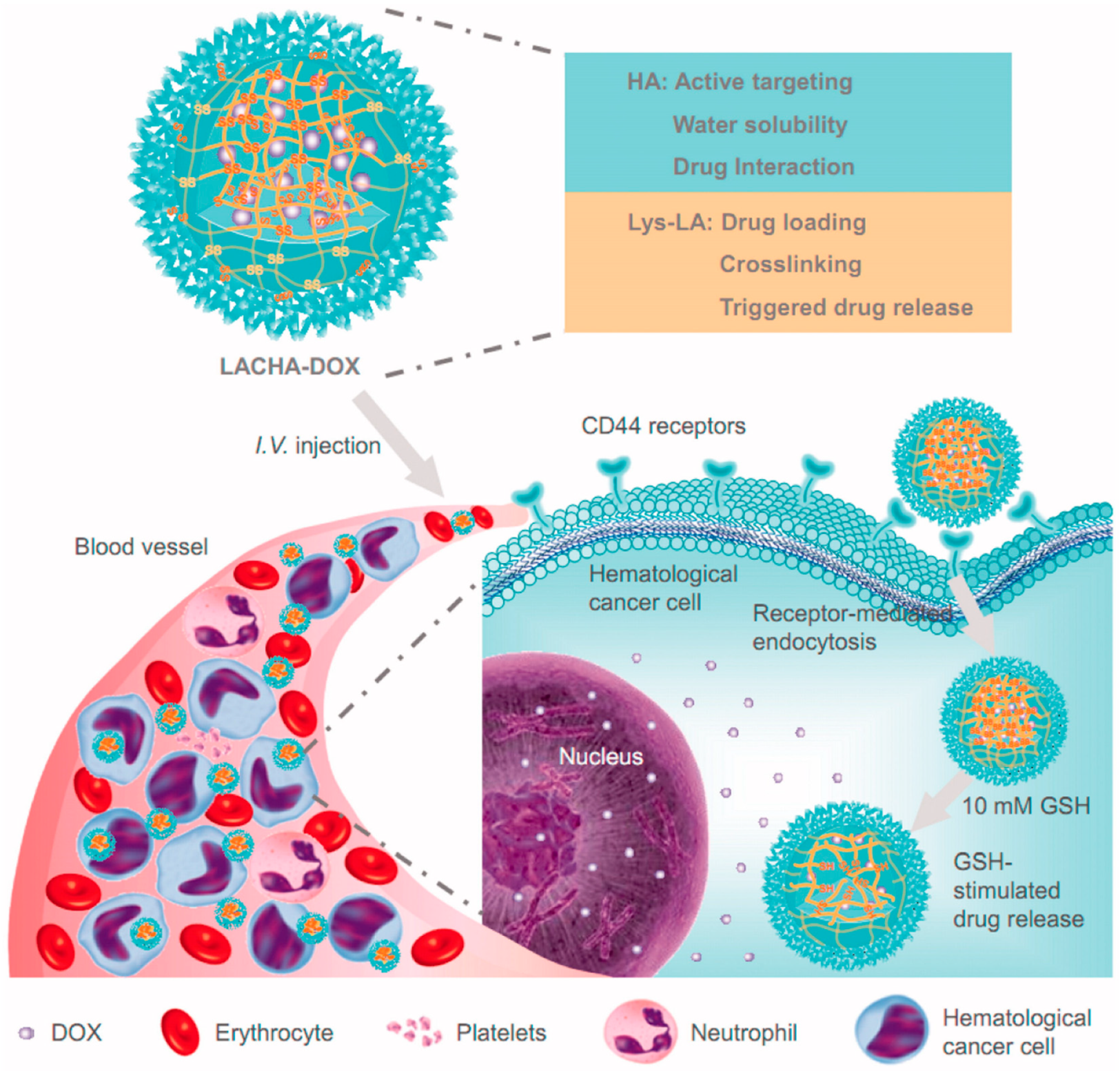
| LACHA/DOX | HA | - | in vivo and ex vivo | LP-1 and AML-2 cells, LP-1 and AML-2 tumor xenografts | [37] |
| PLGA/LND | - | - | U266 cells, Wistar rats | [38] | |
| Chitosan/BTZ | biotin-anti-CD38 | - | in vivo | MM.1S, H929, RPMI8226 and U266 cells, SCID mice | [39] |
| catechol-polycarbonate-PBA/BTZ | - | pH | - | MM.1S and HDF cells, MM.1S xenograft mouse | [40] |
| PEO-b-PBCL/S3I-1757 | anti-CD38 | - | in vivo and ex vivo | U266 and RPMI8226 cells, SCID xenograft mouse | [41] |
| PLGA | BCMA-specific peptide | - | - | CD138+ cells | [30] |
| PLGA-PEI -pPD-L1/pDDK-1 | pDDK-1 | - | - | splenocytes, female BALB/c mice | [42] |
| silica/porphyrin | FA | LED | in vivo | RPMI 8226 cells, female CB17/Icr-Prkdc scid mice | [43] |
| [Au(d2pype)2]Cl | - | - | in vivo | JJN3, RPMI8226 and U266 cells, NOD/SCID mice | [44] |
| γ-Fe2O3/paclitaxel | Anti-ABCG2 | - | - | RPMI 8226 cells, male NOD/SCID mice | [45] |
| Fe3O4/BTZ/GA | - | - | - | RPMI-8226 cells, male BALB/c nude mice | [46] |
| As4S4/melphalan or lenalidomide | - | - | - | RPMI 8226-S, RPMI-Dox40, RPMI-LR5, RPMI-MR20, MM.1S, MM.1R, OPM-1, OPM-2, KMS-11, KMS-18, OCIMY5, U266, NCI-H929, HS-5 cells, huBMsc mouse models | [47] |
| ZnO | - | - | - | RPMI8226 cells | [48] |
| 89Zr radiolabeled TiO2 | transferrin | - | in vivo | MM1.S, SCID mices | [49] |
| GO/DOX | - | - | - | RPMI8226 cells | [50] |
| SWCNTs | anti-MALAT1 gapmer DNA | - | in vivo | H929, MM.1 S cells, SCID-beige mouse | [51] |
| PEG-CdTe/DOX | –pH | - | PRMI8226 cells | [52] |
Publisher’s Note: MDPI stays neutral with regard to jurisdictional claims in published maps and institutional affiliations. |
© 2020 by the authors. Licensee MDPI, Basel, Switzerland. This article is an open access article distributed under the terms and conditions of the Creative Commons Attribution (CC BY) license (http://creativecommons.org/licenses/by/4.0/).
Share and Cite
Iannazzo, D.; Ettari, R.; Giofrè, S.; Eid, A.H.; Bitto, A. Recent Advances in Nanotherapeutics for Multiple Myeloma. Cancers 2020, 12, 3144. https://doi.org/10.3390/cancers12113144
Iannazzo D, Ettari R, Giofrè S, Eid AH, Bitto A. Recent Advances in Nanotherapeutics for Multiple Myeloma. Cancers. 2020; 12(11):3144. https://doi.org/10.3390/cancers12113144
Chicago/Turabian StyleIannazzo, Daniela, Roberta Ettari, Salvatore Giofrè, Ali H. Eid, and Alessandra Bitto. 2020. "Recent Advances in Nanotherapeutics for Multiple Myeloma" Cancers 12, no. 11: 3144. https://doi.org/10.3390/cancers12113144