Review

Targeting Ca2+ Signaling in the Initiation, Promotion and Progression of Hepatocellular Carcinoma

1
Department of Medical Biochemistry, College of Medicine and Public Health, Flinders University, Adelaide 5001, South Australia, Australia
2
School of Medicine, The University of Adelaide, Adelaide 5005, South Australia, Australia
3
South Australian Health and Medical Research Institute, Adelaide 5005, South Australia, Australia
*
Author to whom correspondence should be addressed.
Cancers 2020, 12(10), 2755; https://doi.org/10.3390/cancers12102755
Submission received: 6 August 2020 / Revised: 20 September 2020 / Accepted: 21 September 2020 / Published: 24 September 2020
(This article belongs to the Special Issue Targeting Calcium Signaling in Cancer Cells)

Abstract

:

Simple Summary

Liver cancer (hepatocellular carcinoma) is a significant health burden worldwide. It is often not detected until at an advanced stage when there are few treatment options available. Changes in calcium concentrations within liver cancer cells are essential for regulating their growth, death, and migration (metastasis). Our aim was to review published papers which have identified proteins involved in calcium signaling as potential drug targets for the treatment of liver cancer. About twenty calcium signaling proteins were identified, including those involved in regulating calcium concentrations in the cytoplasm, endoplasmic reticulum and mitochondria. A few of these have turned out to be sites of action of natural products previously known to inhibit liver cancer. More systematic studies are now needed to determine which calcium signaling proteins might be used clinically for treatment of liver cancer, especially advanced stage cancers and those resistant to inhibition by current drugs.

Abstract

Hepatocellular carcinoma (HCC) is a considerable health burden worldwide and a major contributor to cancer-related deaths. HCC is often not noticed until at an advanced stage where treatment options are limited and current systemic drugs can usually only prolong survival for a short time. Understanding the biology and pathology of HCC is a challenge, due to the cellular and anatomic complexities of the liver. While not yet fully understood, liver cancer stem cells play a central role in the initiation and progression of HCC and in resistance to drugs. There are approximately twenty Ca2+-signaling proteins identified as potential targets for therapeutic treatment at different stages of HCC. These potential targets include inhibition of the self-renewal properties of liver cancer stem cells; HCC initiation and promotion by hepatitis B and C and non-alcoholic fatty liver disease (principally involving reduction of reactive oxygen species); and cell proliferation, tumor growth, migration and metastasis. A few of these Ca2+-signaling pathways have been identified as targets for natural products previously known to reduce HCC. Promising Ca2+-signaling targets include voltage-operated Ca2+ channel proteins (liver cancer stem cells), inositol trisphosphate receptors, store-operated Ca2+ entry, TRP channels, sarco/endoplasmic reticulum (Ca2++Mg2+) ATP-ase and Ca2+/calmodulin-dependent protein kinases. However, none of these Ca2+-signaling targets has been seriously studied any further than laboratory research experiments. The future application of more systematic studies, including genomics, gene expression (RNA-seq), and improved knowledge of the fundamental biology and pathology of HCC will likely reveal new Ca2+-signaling protein targets and consolidate priorities for those already identified.

Graphical Abstract

1. Introduction

Hepatocellular carcinoma (HCC)—which arises from chronic liver disease—is a significant contributor to cancer-related deaths and often has poor clinical outcomes. Due to the prevalence of obesity, non-alcoholic fatty liver disease (NAFLD), and metabolic syndrome the incidence of HCC is increasing [1,2,3]. The liver is a complex organ, composed of several different cell types, of which hepatocytes form the majority [4]. Hepatocytes are responsible for the central metabolism of the body including carbohydrate, lipid, protein and amino acid metabolism. Hepatocytes also mediate the uptake, synthesis and export of bile acids and the metabolism and excretion of drugs and other xenobiotic molecules [4]. Changes in intracellular free Ca2+ concentrations play a central role in the hormonal regulation of liver metabolism and of the pathways which regulate hepatocyte proliferation and apoptosis [5]. Factors that create an environment in the liver which initiate and promote HCC include hepatitis B (HBV), hepatitis C (HCV) and liver steatosis [1,2,3]. Liver cancer stem cells are thought to play an important role in the initiation and progression of HCC [6,7,8,9,10].
Numerous mutations in genes encoding Ca2+-signaling proteins have been identified in DNA extracted from HCC liver tissue, and a number of Ca2+-signaling proteins are over or under expressed in HCC [11,12]. These and many other studies conducted over the past 20 years have identified Ca2+-signaling proteins which appear to play important roles on the initiation and progression of HCC and which could be potential therapeutic targets. These potential targets are summarized in Table 1. The table was arranged so as to group the potential Ca2+-signaling protein targets in the different stages of HCC: liver cancer stem cells, initiation and promotion by HBV and HCV infection and non-alcoholic fatty liver disease, and in established HCC tumors, including migration and metastasis. The aim of this review is to identify what is known about these potential Ca2+-signaling proteins, to evaluate the evidence for this identification and explain the rationale for targeting these steps in the treatment of HCC.
Liver anatomy and biology, and the pathways of initiation and progression of HCC are complex. Therefore, the first part of this review sets out the biologic and pathologic background which forms the context for the identification and evaluation of potential Ca2+-signaling targets in HCC. Of particular importance are the roles of liver cancer stem cells in HCC initiation, progression and resistance to treatments. Also, the potential of drug-emitting bead transcatheter arterial chemoembolization to deliver a drug to the site of a tumor. Many studies have followed a general pattern. This has often involved initially identifying a given Ca2+-signaling protein or pathway of potential interest. Then experiments have been conducted to measure the expression of the Ca2+-signaling protein(s) in human HCC and non-tumor liver tissue and in HCC cell lines and to artificially alter expression of the protein in HCC cell lines and examine the effects on cell proliferation, apoptosis, and/or metastasis. Finally, downstream pathways potentially involved are often investigated. In other studies, bioinformatics analysis or other ideas have led to the identification of a protein which has turned out to be a Ca2+-signaling protein.

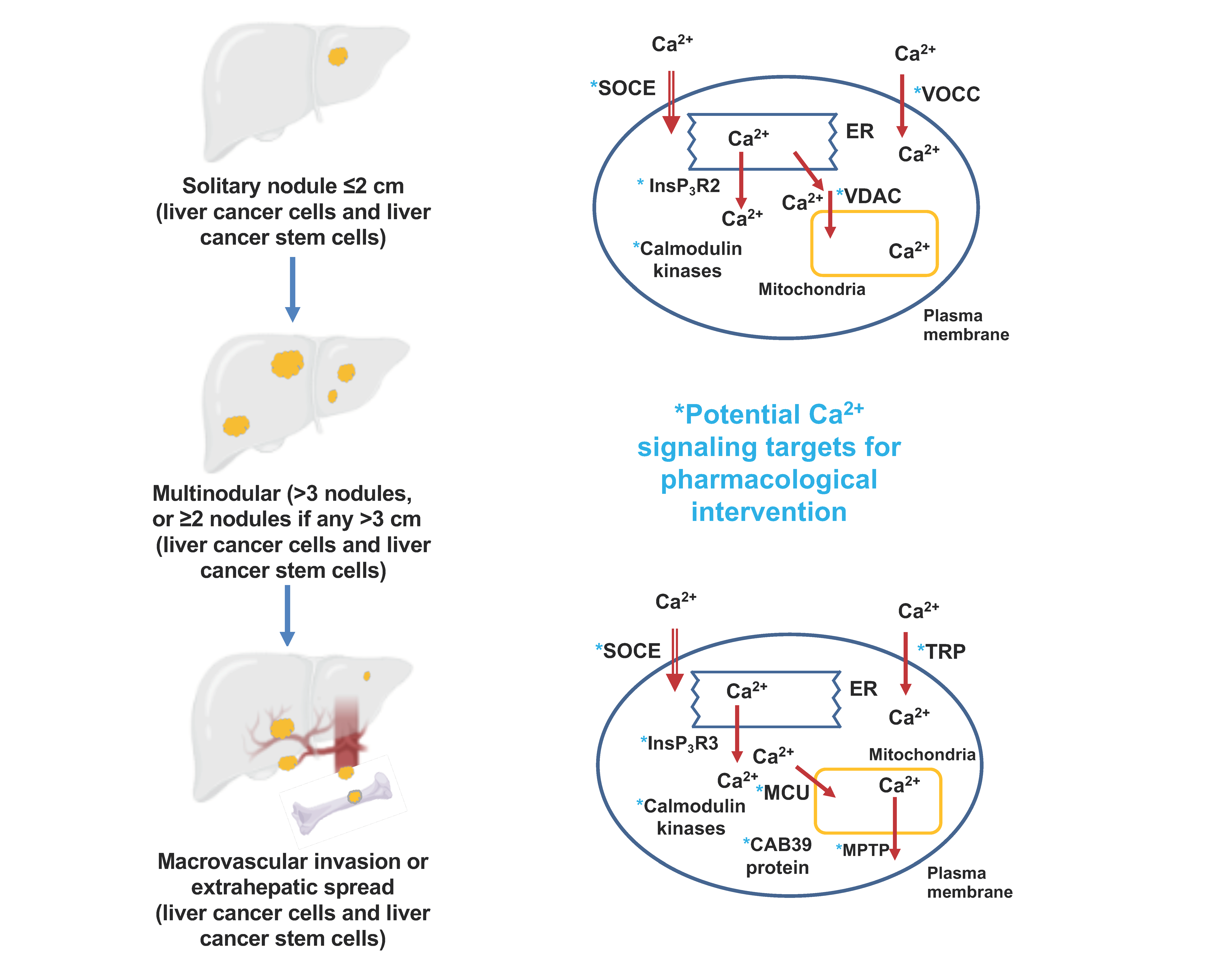
2. Overview of Liver Anatomy, Cell Types, Physiology and Metabolic Pathways

The metabolic roles of the liver include the metabolism of carbohydrates, lipids, proteins and amino acids and the synthesis and transport of the components of bile fluid and the metabolism and excretion via bile or blood of drugs and xenobiotic compounds [4,5,40]. These processes are principally conducted by hepatocytes (parenchymal cells). Several other cell types are also present in the liver. These include cholangiocytes, (which are also parenchymal cells) and fibroblasts, stellate cells, Kupffer cells and sinusoidal endothelial cells (which are all nonparenchymal cells) [4,5,40]. In addition, liver tissue harbors liver precursor/stem cells, as discussed in more detail below. Having an appreciation that all these different cell types are likely to be present in any given sample of liver tissue is an important aspect of interpreting both Ca2+-signaling pathways and changes in signaling proteins in HCC tissue.
Hepatocytes constitute 60–80% of the total liver mass. They are normally quite stable with an average lifetime (turnover) of many days. However, hepatocytes have a large capacity to proliferate. This allows the liver to regenerate following liver surgery [4,5,40]. About 30% of hepatocytes are polyploid, which means they contain at least two sets of homologous chromosomes. This is due to both an increase in the amount of DNA in a given nucleus and/or to an increase in the number of nuclei per cell [41,42].
The smallest defined subanatomical unit of the liver is the hexagonal lobule. In the human liver this has a diameter of about 1 mm. A portal triad is located at each of the six corners of the hexagon. This consists of a portal artery, portal vein and bile duct, arranged as shown schematically in Figure 1 [4]. This figure also shows the direction of blood and bile flow and the relative anatomic locations of hepatocytes, stellate, sinusoidal endothelial and Kupffer cells. To achieve their functions, hepatocytes are spatially polarized with sinusoidal, canalicular and basolateral membranes (Figure 1). Cholangiocytes line the bile duct and are responsible for the movement of bile fluid out of the liver to the common bile duct. Kupffer cells are resident macrophages and are responsible for protecting the liver from inflammation. Functions of stellate cells include mediating the development of liver fibrosis following liver inflammation and/or injury.

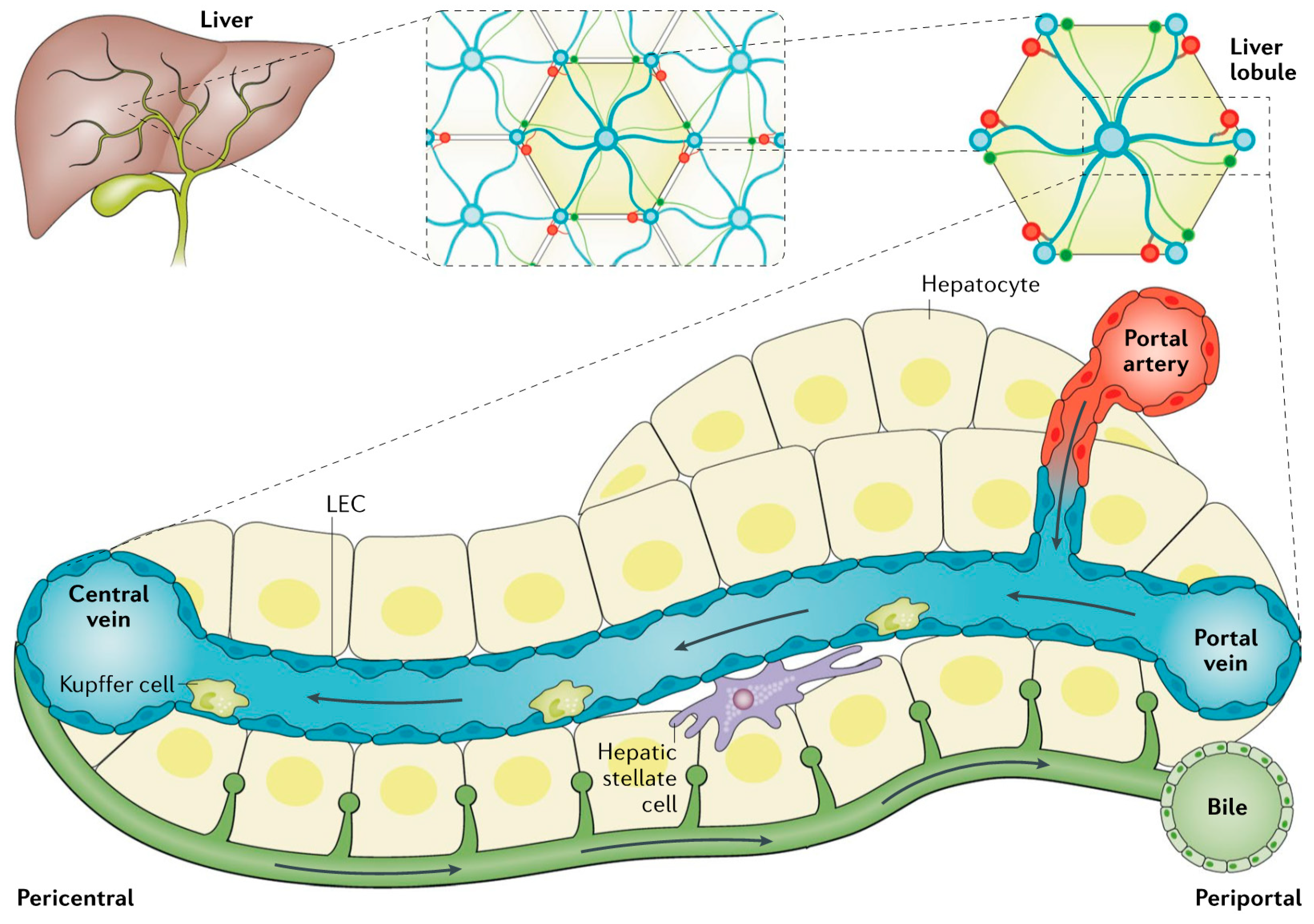
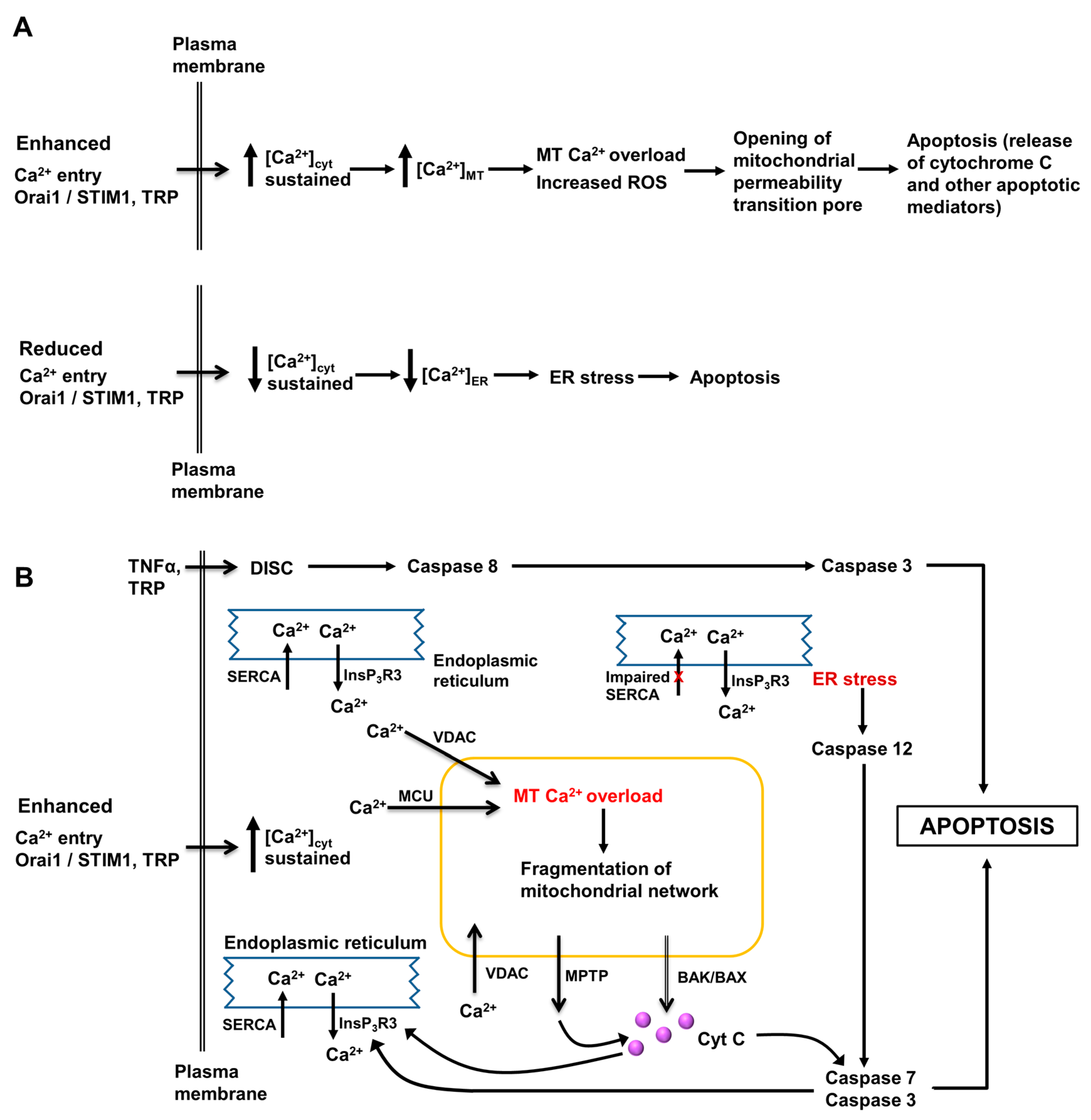
3. Role of Intracellular Ca2+ in Regulation of Hepatocyte Metabolism, Proliferation, Injury and Death

Pathways of intracellular Ca2+-signaling in hepatocytes have been well characterized over the past 30 to 40 years (reviewed in [5]). Hepatocytes were, in part, a model cell type for understanding Ca2+-signaling in non-excitable cells. These signaling pathways included the role of Ca2+ in the hormonal regulation of glycolysis and one of the first observations of Ca2+ oscillations in animal cells [43,44]. Likewise, intracellular Ca2+-signaling in cholangiocytes was well characterized (reviewed in [45,46]). However, much less is known about intracellular Ca2+-signaling in Kupffer, stellate and sinusoidal endothelial cells. Yet, as indicated above, these cell types play important roles in liver function. They are involved in the initiation and progression of HCC and are present in samples of HCC liver and non-tumor liver.
It is worthwhile considering why targeting Ca2+-signaling proteins, rather than targeting protein kinases (for example), may be a useful strategy in the prevention and treatment of HCC. Intracellular Ca2+ is a widespread and reversible signaling system in animal cells. At an early stage in the evolution of living cells, formation of the cell membrane was also associated with the creation of a very large Ca2+ gradient (about 20,000) across the cell membrane. This evolved cell membrane arrangement protected the cytoplasm and organelles within the cell from the high Ca2+ concentrations in the external environment [47,48]. Enclosure and definition of the intracellular space were coupled with the evolution of many proteins possessing the ability to bind Ca2+ at Ca2+-specific binding sites. This allowed Ca2+-signaling to impinge on most biochemical pathways [47,48]. On one hand, this makes Ca2+-signaling an attractive pharmaceutical target, as the inhibition or activation of a given process can have multiple downstream consequences which may potentially be beneficial. On the other hand, the broad nature of the Ca2+-signaling system means that interaction at one site has the potential to affect numerous downstream events and hence may not be very specific. Choosing Ca2+-signaling targets for the prevention and treatment of HCC may thus involve a delicate balance between these two extremes.
In adult hepatocytes, changes in the free Ca2+ concentration in the cytoplasmic space ([Ca2+]cyt), mitochondrial matrix ([Ca2+]MT), lumen of the endoplasmic reticulum ([Ca2+]ER), nucleus, and lysosomes play central roles in the hormonal regulation of glycogen metabolism, glycolysis, lipid synthesis and degradation, and protein and amino acid metabolism (reviewed in [5]). Intracellular Ca2+-signaling is also essential for regulating the transport and movement of the components of bile fluid. Of particular importance in the development, progression and therapeutic treatment of HCC are the roles of intracellular Ca2+ in regulating the proliferation, apoptosis and migration of hepatocytes and HCC cells. Some key pathways are shown in Figure 2 and Figure 3. Together with other signaling pathways, changes in intracellular Ca2+ can switch cell signaling from proliferation to apoptosis and cell death. Moreover, interventions causing a large influx of Ca2+ across the plasma membrane, such as may arise from the pathologic or pharmacological activation of some TRP channels, also induces cell death [31,32,49].
The “Ca2+-signaling tool-kit” in the adult hepatocyte is similar to that in many other non-excitable mammalian cells (reviewed in [5]). However, there are some differences in the expression of channels and transporters between adult hepatocytes and those in progenitor/stem cells, liver cancer stem cells and HCC cells. Moreover, in cholangiocytes, Kupffer cells, stellate and sinusoidal epithelial cells different aspects of Ca2+-signaling pathways are emphasized. As discussed below, one of these differences is the expression of voltage-operated Ca2+ channels which do not seem to be expressed in adult hepatocytes but may be expressed in other cells present in HCC tumors.
Most studies of Ca2+-signaling in hepatocytes have been conducted using isolated rat or mouse hepatocytes, liver cell lines established from rat or mouse liver tumors and in liver cell lines established from human liver tumors. Ca2+-signaling proteins and pathways in normal human hepatocytes are less well characterized. However, most of the studies reported in this review involve human liver tissue and liver cell lines derived from human liver. While the Ca2+-signaling pathways in normal human adult hepatocytes are likely to be broadly similar to those in normal rat and mouse adult hepatocytes, there are some differences.
The three sub-types of InsP3 receptors (InsP3R) are expressed in adult rat and mouse hepatocytes, although the level of expression of type 3 InsP3R (InsP3R3) is low. The roles of type 2 InsP3 receptors (InsP3R2) in generating oscillations in [Ca2+]cyt and in the regulation of bile secretion and Ca2+-signaling in the hepatocyte nucleus have been well characterized [45,50]. Type 1 InsP3 receptors (InsP3R1) are involved in the regulation of metabolism and may be specifically associated with the activation of Ca2+ entry (reviewed in [5,45]). It is unlikely that there are functional ryanodine receptors in adult rat and mouse hepatocytes, but this point is not fully resolved (reviewed in [5]).
In rat and mouse hepatocytes, the main Ca2+ entry pathway is via store-operated Ca2+ entry, mediated by Orai and STIM polypeptides [5]. These permit sustained hormone-induced oscillations in [Ca2+]cyt and maintain adequate Ca2+ in the ER lumen during hormonal Ca2+-signaling. Thus, Ca2+ entry through store-operated Ca2+ channels replenishes intracellular Ca2+ which is extruded from the cytoplasmic space via the plasma membrane (Ca2++Mg2+)ATP-ase following an increase in [Ca2+]cyt. In hepatocytes isolated from human liver, TRPC6 polypeptides may also make a contribution to store-operated Ca2+ entry [28]. Moreover, several other TRP non-selective cation channels are also expressed in hepatocytes. These likely serve to deliver Ca2+ and Na+ to the cytoplasmic space to serve specific regulatory functions [5].
Store-operated Ca2+ entry to hepatocytes and liver cell lines is frequently measured using an intracellular Ca2+ sensor (e.g., Fura-2) and fluorescence imaging employing a “Ca2+ add back” protocol. This involves measurement of the increase in [Ca2+]cyt following the addition of extracellular Ca2+ to the culture medium of cells previously incubated in the absence of extracellular Ca2+ plus an agent such as thapsigargin or a generator of InsP3 to release Ca2+ from the ER. While this is a useful and valuable technique, measurement of Ca2+ entry by patch clamp recording is needed to confirm the presence of functional store-operated Ca2+ channels. These are normally defined as possessing the properties of Ca2+-release-activated Ca2+ channels (CRAC) channels, originally identified in lymphocytes and mast cells [51].
To our knowledge, there is no evidence for the expression of functional voltage-operated Ca2+ channels in hepatocytes isolated from normal adult rat, mouse or human liver [5]. However, as discussed below, voltage-operated Ca2+ channels are likely expressed in liver cancer stem cells, in HCC cells and in immortalized HCC cell lines (reviewed in [5]). Moreover, stellate and Kupffer cells also express voltage-operated Ca2+ channels (reviewed in [5]).

4. The Pathology of Hepatocellular Carcinoma

In the context of all cancers, HCC poses a significant health burden and is a major cause of cancer-related deaths in both men and women [3,52,53,54,55]. With the current increased prevalence of obesity, non-alcoholic fatty liver disease and metabolic syndrome, the incidences of HCC and HCC-related deaths are increasing [56]. HCC is one of two main primary liver cancers (i.e., cancers that originate from mutations in hepatocytes or other cell types in the liver) and accounts for about 80% of all liver cancers [3,10]. The other main primary liver cancer is intrahepatic cholangiocarcinoma, which accounts for about 1% of all liver cancers. Other primary liver cancers are mixed hepatocellular cholangiocarcinoma, fibrolamellar hepatocellular carcinoma and pediatric hepatoblastoma. Metastatic cancers (i.e., non-primary liver cancers), the most important of which is colon cancer, can also be established in the liver [57]. Although these non-primary liver cancers are also important, in this review we only consider HCC.
HBV and HCV were previously the major risk factors for HCC [53,54]. However, hepatitis viral infections can now be well treated and controlled, so that non-alcoholic fatty liver disease is considered to be the leading risk factor for HCC. Other risk factors include aflatoxins and type 2 diabetes [53,58].
Stages in the development of HCC from initiation to metastasis to other regions of the liver and eventually to other tissues are summarized schematically in Figure 4 [3,10,59]. The development of HCC tumors is usually preceded by liver fibrosis and cirrhosis, although some HCCs develop without cirrhosis [1,3,10,60]. An example of an HCC tumor (being removed surgically by liver resection) and an example of the histology of liver tissue across a tumor, the tumor margin and para tumor tissue are shown in Figure 5. The transformation of HCC cells to acquire migratory and metastatic properties involves a change from an epithelial to a mesenchymal phenotype. In this transition, epithelial cells lose spatial cell polarity and cell–cell adhesiveness and gain migratory and invasive properties [3].
In the early stages, HCC does not normally give rise to many physical symptoms and signs. Early stage HCC can usually only be detected using ultrasound, imaging and measurement of blood alpha-fetoprotein concentrations. In the detection and monitoring of later stages of HCC, imaging and blood alpha-fetoprotein play major roles [10,53].
The mechanisms involved in the initiation and progression of HCC are complex and are only partly understood. Epigenetic as well as genetic changes are involved. Mutated genes which feature in many HCCs include those encoding proteins which regulate the Wnt/β-catenin pathway, the p53 cell cycle pathway, telomere maintenance and chromatin structure and function [10,11,60,62,63]. As discussed below, stem cells are thought to play an important role in the initiation and progression of HCC [6,7,8,9,10,60]. Development and progression of HCC is promoted by inflammation, such as that initiated by HBV and HCV and steatosis (non-alcoholic fatty liver disease) [53,64].

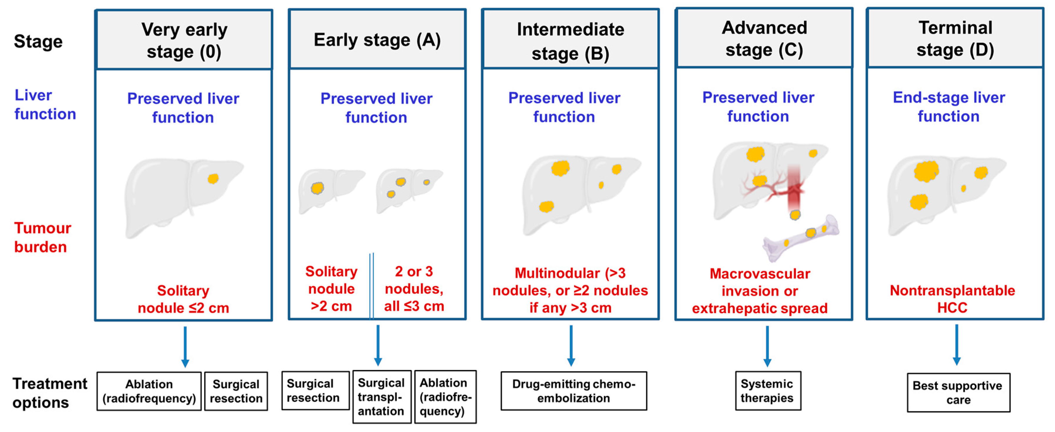
5. Current Treatments for Hepatocellular Carcinoma

Current treatment options for HCC at the different stages are summarized in Figure 4. Well established HCC is difficult to treat, resulting in uncertain and often poor outcomes [3,65,66]. If HCC is detected in the very early stages with only one, or a few, tumor nodules of small size, the tumor(s) can be removed surgically by liver resection or liver transplantation (surgical liver resection shown in Figure 5A). Examples of systemic agents used to treat later stage HCC include sorafenib and lenvatinib (multikinase inhibitors), PD-L1 (programmed death-ligand 1) receptor blockers, statins and metformin [3]. Unfortunately, for many treatments the risk of cancer recurrence is high.
Of particular interest in considering the potential administration of therapeutic agents targeted to Ca2+-signaling pathways in HCC is drug-emitting bead transcatheter arterial chemoembolization. This is employed to deliver therapeutic agents to the site of tumors in the treatment of HCC patients with intermediate stage HCC which cannot be treated surgically [67,68,69]. Examples of chemotherapeutic agents delivered by drug-emitting bead transcatheter arterial chemoembolization include doxorubicin, cisplatin, oxaliplatin and arsenic trioxide. Thus, drug-emitting bead transcatheter arterial chemoembolization offers a drug delivery mechanism which should enhance the ability to target HCC tumors, and hence reduce effects on non-tumor liver tissue and systemic side effects.
Drug-emitting bead transcatheter arterial chemoembolization is a radiological procedure. In principle, this involves the intra-arterial injection to the site of a tumor of a viscous emulsion composed of a drug-emitting bead (e.g., CalliSpheres microspheres) mixed with iodized oil to deliver one or more chemotherapeutic agents to the site of the tumor. This is followed by embolization of the blood vessel with gelatine sponge particles which confines the chemotherapeutic agent to the vicinity of the tumor and creates an ischemic environment which assists in killing HCC cells [67,68,69].

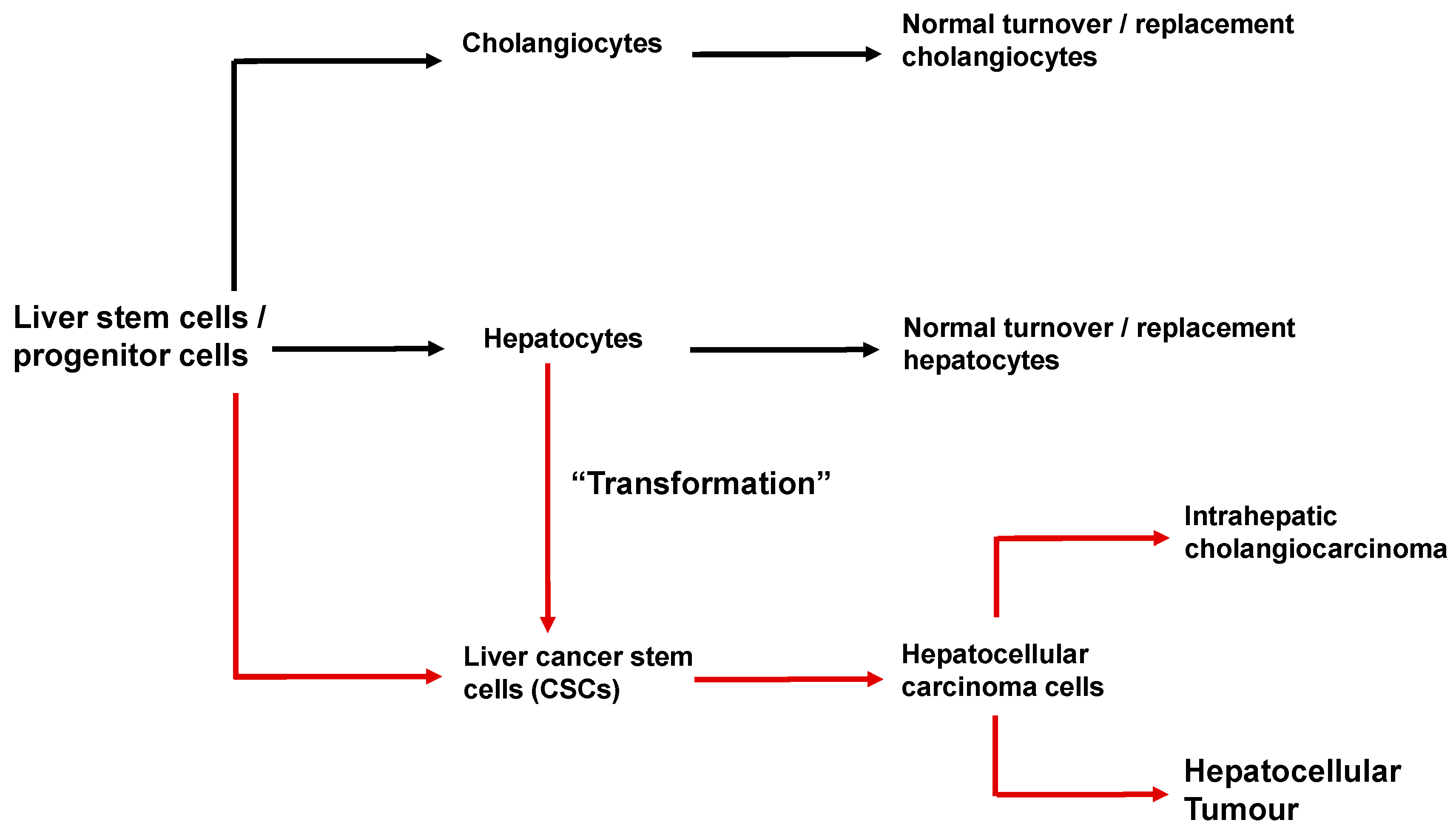
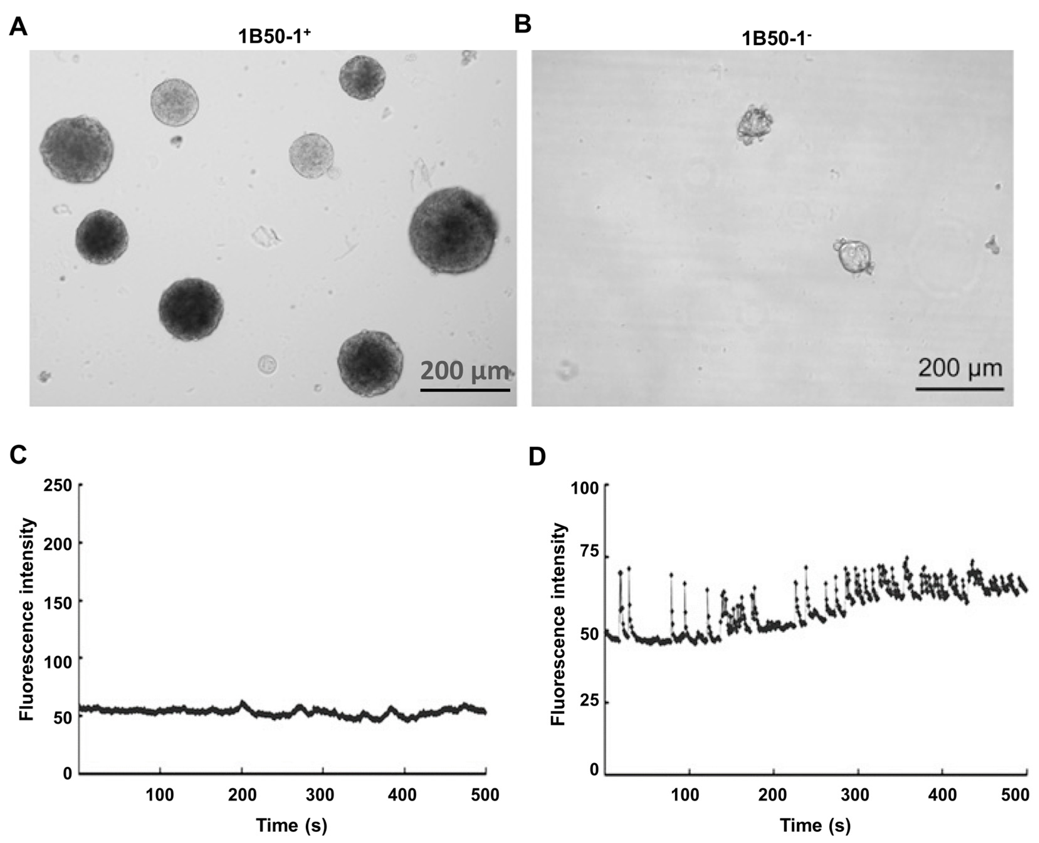
6. Cancer Stem Cells and the Initiation and Progression of Hepatocellular Carcinoma

As described above, the liver of a normal adult human harbors liver stem cells (also called progenitor cells) [6,7,8,9,10]. These liver stem cells are identified by a specific set of surface marker proteins. Stem cells/progenitor cells can differentiate to adult hepatocytes and cholangiocytes, which is the normal process for the turnover of these cells (shown schematically in Figure 6). Upon differentiation, liver stem cells/progenitor cells can also provide new hepatocytes for liver regeneration following some forms of liver damage, for example liver surgery. Mitosis of adult hepatocytes also contributes to replacement and expansion of the adult hepatocyte population [7,9,10].
Analysis of cells present in HCC liver tissue has identified a small subset of liver cancer cells called liver cancer stem cells. The properties of these cells are similar to those of the pools of cancer stem cells identified in many other cancers. Liver cancer stem cells are identified experimentally by a specific set of surface marker proteins, for example EpCAM, CD133, CD44, CD90, OV-6, ALDH, K19, which also distinguish them from “normal” liver stem cells/progenitor cells [7,8,9,10]. Liver cancer stem cells are characterized by their abilities to self-renew, differentiate, rapidly proliferate, and their multilineage potential. Liver cancer stem cells are thought to be involved in the initiation of HCC, recurrence of HCC following treatment, HCC metastasis and in the development of the resistance of HCC to therapies. There is now much evidence to suggest that the destruction of liver cancer stem cells is a desirable target in the treatment of HCC [7,8,9,10].
Liver cancer stem cells are thought to arise from liver stem cells/progenitor cells and also from adult hepatocytes (shown schematically in Figure 6) [7,8,9,10]. There is some evidence that liver cancer stem cells are physically located in “niches” in the liver. Under conditions which foster the initiation and promotion of HCC (chronic inflammation, HBV, HCV, alcohol, non-alcoholic fatty liver disease), liver cancer stem cells can undergo further genetic and epigenetic changes leading to HCC cells and HCC or intrahepatic cholangiocarcinoma.
The relationships between liver stem/progenitor cells, adult hepatocytes, cholangiocytes, liver cancer stem cells and HCC cells are complex, difficult to fully deduce from current observations and hence presently only partly understood. Therefore, both the discussion above and the scheme shown in Figure 6, should be viewed as a guide only.

7. Methodology and Terminology for Hepatocellular Carcinoma Liver Samples and Hepatocellular Carcinoma Cell Lines

Research directed towards understanding the role of intracellular Ca2+-signaling in the development and progression of HCC has historically employed hepatocytes and liver cell lines. These were derived principally from rat and mouse liver tumors. Over the past ten years or so, most studies have employed cell lines derived from human liver and human HCC tissue, mouse chemical or xenograft models of HCC tumors and the analysis of tissue samples taken from the livers of HCC patients. Examples of cell lines derived from human HCC tumors and from normal human liver and mouse models of HCC employed in the studies addressed in this review are listed in Table 2.
The process of obtaining human liver HCC tissue usually involves taking a tissue sample from a liver resection (such as that shown in Figure 5A), then the selection tissue samples taken from the actual tumor and from “non-tumor” (para-tumor) tissue obtained from outside the margins of the tumor (Figure 5B). The ideal situation is where both HCC tumor tissue and non-tumor (para-tumor) tissue is taken from the same liver. In the discussions below, the terms HCC liver tissue and non-tumor tissue will be employed to describe samples taken from the livers of HCC patients, without reference to exactly how the “control” non-tumor tissue was obtained. As implied in the discussion of liver cancer stem cells above, the para-tumor liver tissue may or may not be the same as “normal” liver tissue. Moreover, while hepatocytes or transformed hepatocytes (HCC cells), will be the main cell type present, these tissues will also contain all other cell types present in liver.
A number of HCC cell lines derived from human HCC or “normal” liver are listed in Table 2. These have varying degrees of dedifferentiation from normal adult hepatocytes. Some studies have also employed “normal” liver cell lines such as L01 and L02 liver cell lines [72]. These are derived from adult or fetal human liver. However, they are “immortalized” cells, so caution is needed in treating these as truly “normal” hepatocytes. In addition, constant vigilance is required to be assured that the cell line employed in a given study is true to type and is not contaminated by other cells [73].

8. Mutations and Altered Expression of Ca2+-Signaling Proteins in Hepatocellular Carcinoma

In considering Ca2+ signaling proteins as therapeutic targets in the prevention and treatment of HCC, it is useful to summarize the mutations and altered expression of Ca2+ signaling proteins so far detected in HCC in the human liver. Our current knowledge of these mutations and alterations in the expression of Ca2+ signaling proteins is summarized in Table 3 and Table 4. The data were obtained from an analysis of DNA and mRNA and/or protein expression present in HCC and “normal” liver tissue obtained from HCC patients. These show that mutations and altered gene expression have been detected in a wide range of Ca2+ channels, transporters and Ca2+-binding proteins. Moreover, comparison of the genes listed in Table 3 and Table 4 with the Ca2+-signaling proteins listed in Table 1 as potential targets for treatment of HCC indicates that many potential Ca2+-signaling targets are also mutated or under- or overexpressed in HCC.
As discussed above, the liver is heterogeneous with respect to cell types. Therefore, it is expected that a given HCC liver sample will contain a mixture of normal hepatocytes, HCC cells, liver progenitor/stem cells, liver cancer stem cells, stellate cells, Kupffer cells, sinusoidal endothelial cells, cholangiocytes and likely some other cell types. Thus, the observed mutations could be in the DNA of any one or more of these cell types. Nevertheless, the predominant cell types in HCC tissue removed for analysis are likely to be hepatocytes and transformed hepatocytes (HCC cells). Consequently, it is likely that the observed mutations and alterations in gene expression relate to DNA derived from HCC cells. The application of single-cell transcriptome analysis will help to resolve the liver cell types responsible for altered gene expression in HCC [76].

9. Voltage-Operated Ca2+ Channels, InsP3 Receptors and TRPV2 Channels in Liver Cancer Stem Cells

The results of several recent studies point to the importance of intracellular Ca2+-signaling, voltage-operated Ca2+ channels and InsP3Rs in maintaining the characteristics of liver cancer stem cells and the ability of these cells to self-renew (Table 1). In a search for marker proteins which could be used to identify liver cancer stem cells, Zhao and colleagues generated a monoclonal antibody, IB50-1, which turned out to bind to isoform 5 of α2δ1, a subunit of several voltage-operated Ca2+ channels [13]. Analysis of HCC cells isolated from frozen tissue sections obtained from HCC liver resections showed a subgroup of IB50-1 positive cells which were enriched in HCC tissue compared with non-tumor tissue. The IB50-1 antibody thus allowed the authors to identify tumor initiating cells with the properties of liver cancer stem cells. From these cell suspensions they created a stable cell line, Hep-12, of liver cancer stem cells which all expressed the α2δ1 protein and a stable cell line, Hep-11, which did not express the α2δ1. Hep-12 cells exhibited enhanced self-renewal properties, including spheroid formation (an indicator of self-renewal capacity) and initiated tumors in NOD/SCID HCC mice, whereas Hep-11 cells did not (Figure 7A,B).
Studies of Ca2+-signaling using fluo-4 showed that basal [Ca2+]cyt in Hep-12 cells was higher than that in Hep-11 cells [13]. Moreover, Hep-12 cells exhibited spontaneous oscillations in [Ca2+]cyt which were not observed in Hep-11 cells (Figure 7C,D). Pretreatment of Hep-12 cells with antibody IB50-1 reduced basal [Ca2+]cyt and reduced the amplitude of oscillations in [Ca2+]cyt, while overexpression of α2δ1 in Hep-11 cells increased basal [Ca2+]cyt. Some evidence for the expression of functional L- and N-type voltage-operated Ca2+ channels in Hep-12 cells was obtained using specific voltage-operated Ca2+-channel blockers and RT–PCR. In Hep-12 cells, inhibition of α2δ1 by antibody IB50-1 or knockdown of α2δ1 using shRNA induced apoptosis by a mechanism involving decreased phosphorylation of ERK1/2, decreased Bcl2, increased Bax and Bad and activation of caspases 3,8 and 9 (pathway scheme shown in Figure 3). It was concluded from these studies that α2δ1 and hence L- and N- type voltage-operated Ca2+ channels, expressed in tumor initiating cells (liver cancer stem cells), promote/maintain the self-renewal characteristics of these cells. Targeted inhibition of α2δ1 should induce apoptosis in liver cancer stem cells and hence reduce the development of HCC. This could be especially useful in preventing the recurrence of HCC tumors, following liver surgery or other initial treatment [13].
Further investigations using Hep-12 (subset of HCC cells enriched in liver cancer stem cells) and Hep-11 (HCC cells) provided additional evidence that oscillations in [Ca2+]cyt play a role in maintaining the ability of liver cancer stem cells to self-renew and form tumors and identified InsP3R2 as a contributor to these [Ca2+]cyt oscillations [16]. Agents expected to enhance the self-renewal of liver cancer stem cells, EGF/FGFβ/B27 and Il-6, induced oscillations in [Ca2+]cyt in Hep12 cells, but not in Hep-11 cells, suggesting that oscillations in [Ca2+]cyt are an important characteristic of liver cancer stem cells. The authors also employed the genetically encoded GCaMP-ER2 Ca2+ sensor targeted to the ER to investigate oscillations in [Ca2+]ER and the role of the ER and InsP3Rs in the generation of [Ca2+]cyt oscillations. Using RNA-seq, 10 genes encoding Ca2+-signaling proteins which were expressed at higher levels in Hep-12 cells compared with Hep-11 cells were identified [16]. These included voltage-operated Ca2+ channel subunits and InsP3Rs. Using sh-RNA to knockdown the expression of several of these genes, InsP3R2 was identified as important for the generation of [Ca2+]cyt oscillations in Hep-12 cells. Knockdown of InsP3R2 in Hep-12 cells reduced their capacity to form spheroids and reduced their capacity to form tumors in NOD/SCID mice.
The results of all these studies on liver cancer stem cells, identified by expression of the α2δ1 antigen, provide evidence that voltage-operated Ca2+ channels in the plasma membrane and IP3R2 in the ER are important in maintaining the stem cell characteristics of these cells. Inhibition of voltage-operated Ca2+ channels and/or InsP3R2 may provide a strategy to inhibit the pathologic roles of these cells. Further studies are warranted to gain a deeper understanding of the mechanisms involved, especially the roles of plasma membrane Ca2+ entry channels. For example, how expression of L- and N- type voltage-operated Ca2+ channels relates to the observation that liver cancer stem cells are depolarized relative to normal stem cells [84].
The α2δ1 protein could also be a potential biomarker for HCC. Badr and colleagues used ELISA to measure the α2δ1 peptide in the serum of patients with chronic HCV infection diagnosed with HCC and in the serum of healthy controls [85]. Serum concentrations of α2δ1 were compared with those of alpha-fetoprotein and annexin A2. A good correlation was observed between serum levels of α2δ1 and alpha-fetoprotein (but not with annexin-1, another putative marker protein) and the stage of HCC.
T-type voltage-operated Ca2+ channels may also be involved in regulating the proliferation and differentiation of liver cancer stem cells. Radiofrequency electromagnetic fields have been shown to induce tumor shrinkage in several cancers, including HCC (treatment options, Figure 4) [14]. In a study of the mechanisms involved, employing mouse HCC xenograft tumors and Huh-7 liver cells, Jimenez and colleagues found that the application of an amplitude modulated radiofrequency electromagnetic field (optimized for treatment of HCC tumors), increases Ca2+ entry to HCC cells via T-type voltage-operated Ca2+ channels, resulting in an increase in [Ca2+]cyt and cell death.
It has also been suggested that TRPV2 is a possible target for the therapeutic destruction of liver cancer stem cells [15,86]. Measurement of the expression of TRPV2 together with liver cancer stem cell marker proteins in several liver cell lines and in HCC liver samples has provided some evidence that increased expression of TRPV2 in HCC cells reduces their ability to act as liver cancer stem cells [86].
These studies with T-type voltage-operated Ca2+ channels and TRPV2 in liver cancer stem cells suggest that Ca2+-signaling pathways in these cells are worthy of further study. Such study should expand knowledge of their role in the initiation and progression of HCC and further evaluate the possibility that they may be future therapeutic targets.

10. Mitochondrial Ca2+ and Store-Operated Ca2+ Entry in HBV and HCV Infection

Infection of the liver by HBV (DNA virus) or HCV (RNA virus) alters intracellular Ca2+-signaling to create an environment that permits replication of the virus. This environment, especially virus-initiated increase in ROS, also favors the initiation and promotion of HCC [18,19,87]. Ca2+-induced production of ROS by mitochondria is an important source of this virally induced increase in ROS. Viral infection was shown to increase the transfer of Ca2+ from the ER to the mitochondria through mitochondrial membrane associated junctions (MAMs) involving the InsP3R, voltage-dependent anion channel (VDAC) and Ero1a proteins [17,18]. In the case of HCV this Ca2+ movement seems to be initiated by HCV-induced ER stress leading to the unfolded protein response [18]. In the case of HBV, Ca2+ movement from ER to mitochondria is initiated by the viral protein HBx, which binds to the voltage-dependent anion channel 3 (VDAC3) [18]. The resulting increase in [Ca2+]MT decreases the mitochondrial membrane potential, inhibits the electron transport chain and oxidative phosphorylation and increases the production of ROS and reactive nitrogen species by components of the electron transport chain [17,18]. Pharmacological inhibition of mitochondrial Ca2+ uptake has been proposed as a strategy to inhibit the downstream consequences of HBV and HCV infection, including the initiation of HCC [17].
HBV-initiated synthesis of the HBV Bax1 protein and the activation of Ca2+ uptake to mitochondria by Bax1 have also been implicated in the activation of store-operated Ca2+ entry in hepatocytes infected with HBV [19]. Expression of Bax1 increased store-operated Ca2+ entry in isolated rat hepatocytes in culture transfected with DNA encoding Bax1. ATP was used to release Ca2+ from the ER via production of InsP3 and a “Ca2+ add back” protocol and Fura-4 F fluorescence Ca2+ imaging were employed to measure store-operated Ca2+ entry. No change in the expression of Orai, STIM or TRP proteins (components of the store-operated Ca2+ entry pathways) was detected by RNA-seq. The authors suggested that Ca2+ uptake by mitochondria enhanced by HBV Bax1 reduces Ca2+-mediated feedback inhibition of store-operated Ca2+ entry thus increasing Ca2+ entry [19].

11. Store-Operated Ca2+ Entry, SERCA2b and Ca2+/Calmodulin-Dependent Protein Kinases in Initiation and Progression of Hepatocellular Carcinoma in Non-Alcoholic Fatty Liver Disease

As for hepatitis viral infection, a chronic increase in ROS promotes the initiation and progression of non-alcoholic fatty liver disease to non-alcoholic steatohepatitis and then to HCC [88,89]. Increased production of ROS is caused by an accumulation of excess lipid in cytoplasmic lipid droplets in hepatocytes [88,89]. Several Ca2+ channels, transporters and binding proteins are thought to be important in the initiation and progression of HCC (Table 5). These Ca2+-signaling proteins and pathways regulate the accumulation and removal of lipid from lipid droplets and lipid-induced production of ROS. They offer potential targets for therapeutic strategies directed towards normalizing intracellular Ca2+-signaling in hepatocytes, reducing cytoplasmic lipid and ROS, and hence preventing the initiation and promotion of HCC. The nature and rationale for identifying these targets has been reviewed elsewhere [5].

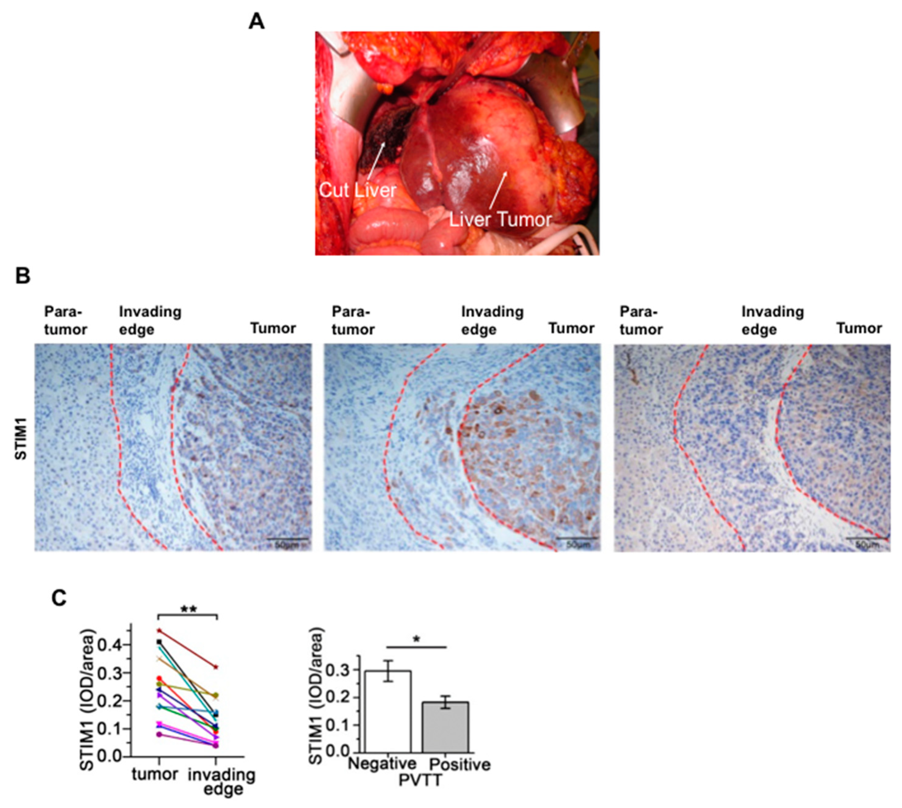
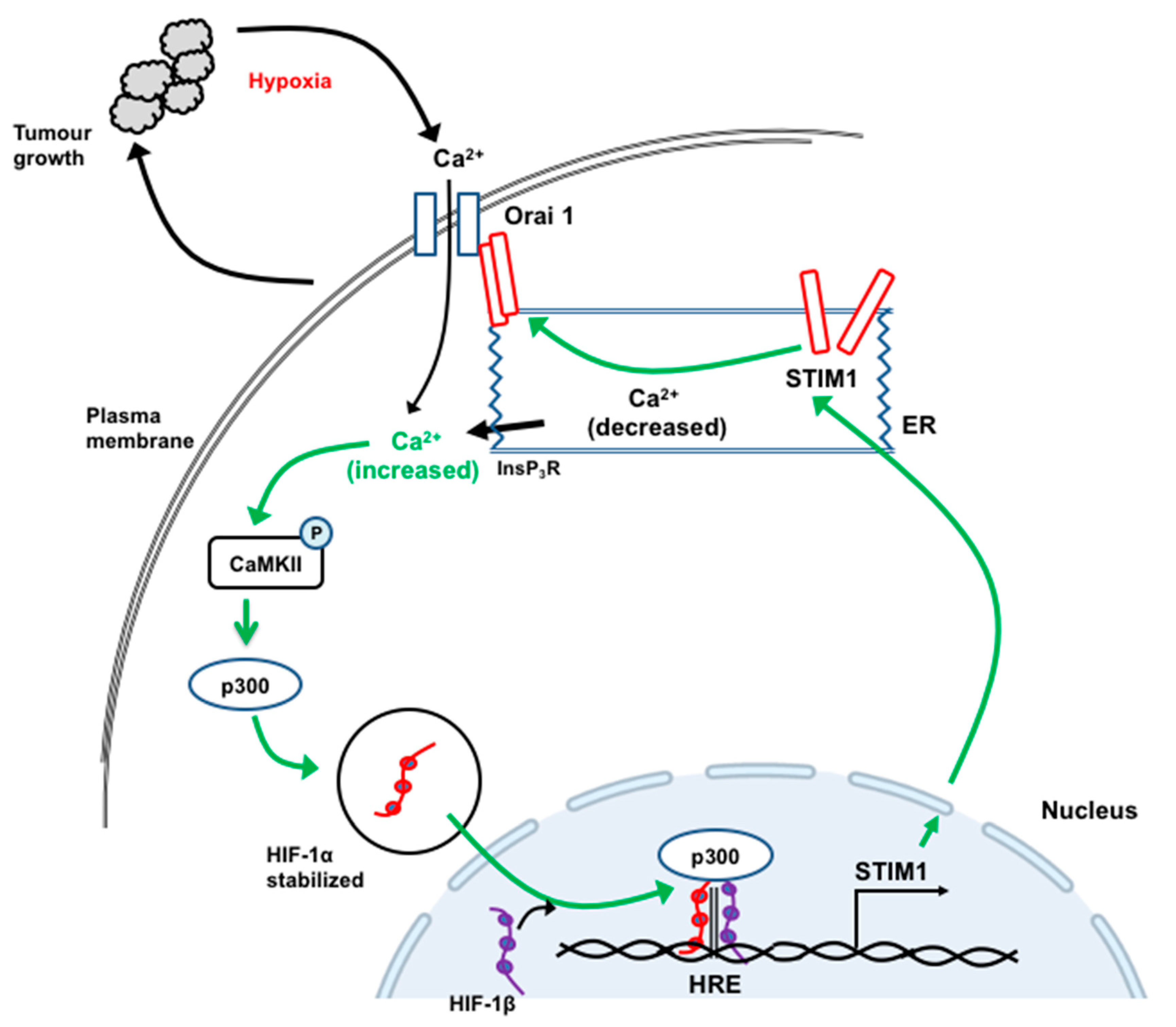
12. STIM1 and Orai1 in the Progression and Metastasis of Hepatocellular Carcinoma

STIM1, Orai1 and store-operated Ca2+ entry were shown to be important in HCC proliferation and metastasis and to be involved in mediating the ability of anticancer drugs to kill HCC cells (Table 1). However, the roles played by STIM1 and Orai1 appear complex and are presently only partly understood.
In an early study Yang and colleagues showed that the expression of STIM1 is higher in HCC tumor tissue than in paired non-tumor liver tissue [80]. They measured STIM1 expression in several HCC cell lines and found highest expression in the HCC-LM3 cell line, which also exhibited the highest migration ability. Inhibition of store-operated Ca2+ entry with SKF-96365 in HCC-LM3 cells or knockdown of STIM1 with siRNA decreased migration and invasion, assayed in vitro. It was concluded that inhibition of store-operated Ca2+ entry is a potential strategy to inhibit HCC migration and invasion.
Several experimental approaches have shown that overexpression of STIM1, leading to enhanced Ca2+ entry via store-operated Ca2+ channels, promotes the proliferation and metastasis of HCC cells [26]. Knockdown of STIM1 in human SMMC7721 HCC cells, using shRNA, was found to decrease cell proliferation and colony formation, arrest the cell cycle at G0/G1 and decrease DNA synthesis, while overexpression of STIM1 led to enhanced cell proliferation [26].
In studies employing human HepG2 HCC cells and a xenograft nude mouse model of HCC, Li and colleagues showed that hypoxia induced the expression of STIM1, increased [Ca2+]cyt and increased store-operated Ca2+ entry [95]. These changes were associated with increased expression of transcription factor HIF-1. Increased expression of STIM1 was also observed in HCC tumors in mice and in samples of human HCC tissue compared with non-tumor tissue [95]. Knockdown or overexpression of STIM1 were associated with reduced and increased tumor growth, respectively. The mechanisms involved were further investigated using YC-1, an inhibitor of HIF-1. It was concluded that the growth of HCC tumors leads to a decrease in O2 available to HCC cells, activation of HIF-1 and increased transcription of STIM1, mediated by the binding of HIF-1 to the HRE motif in the STIM1 promoter. This, in turn, increases store-operated Ca2+ entry and [Ca2+]cyt and activates p300 and CamKII, leading to the stabilization of HIF-1. This then re-enforces the increased transcription of STIM1 (shown schematically in Figure 8). While the central role of increased [Ca2+]cyt and STIM1 expression seem well established, further experiments may be needed to elucidate: the mechanisms involved in the first step (initial activation of HIF1 by hypoxia); whether factors additional to increased STIM1 protein are required to activate store-operated Ca2+ entry; and the downstream mechanisms by which increased STIM1 expression and increased [Ca2+]cyt enhance tumor cell growth.
However, as just discussed, overexpression of STIM1 contributes to enhanced HCC cell proliferation and tumor growth, whereas decreased expression of STIM1 may be associated with HCC metastasis [61]. Immunohistochemistry showed decreased expression of STIM1 at the edge of HCC tumors (Figure 5B,C). Using four different human HCC cell lines, grown under conditions which promoted either proliferation or metastasis, Zhao and colleagues found that STIM1 expression was lower in the cells exhibiting metastatic properties [61]. Further studies of the pathways involved led to the conclusion that during the proliferation and growth of HCC cells, STIM1 is expressed at a high level, leading to enhanced store-operated Ca2+ entry and activation of the CamKII/Akt/Gsk3β pathway which, in part, maintains expression of transcription factor Snail1 at a low level. In metastatic HCC cells, expression of Snail1 is increased, leading to a decrease in STIM1 expression (mediated by binding of Snail1 to the Ebox of the STIM1 promoter). This is also associated with decreased glycolysis and increased fatty acid oxidation [61].
Studies with TRPC1 have provided indirect evidence for a role for store-operated Ca2+ entry in the mechanisms by which cell proliferation is enhanced in HCC cells [96,97]. From the results of studies employing the Huh-7 HCC cell line and shRNA to suppress TRPC1 expression, the authors suggested possible roles for both store-operated Ca2+ entry and TRPC1 in the regulation of cell proliferation in HCC cells.
Orai1 may be involved in the progression of HCC and in the mechanisms by which the anticancer drug 5-fluorouracil kills HCC cells [27]. Expression of Orai1 was found to be increased in HCC tissue compared with that in non-tumor liver tissue. Experiments employing HepG2 cells provided evidence that 5-flurouracil induces autophagic cell death by inhibiting the PI3K/Akt/mTOR pathway. 5-Fluorouricil also decreased store-operated Ca2+ entry, reduced the expression of Orai1, but did not alter STIM1 or TRPC1 expression. Knockdown of Orai1 or pharmacological inhibition of store-operated Ca2+ entry, inhibited the PI3K/Akt/mTOR pathway and potentiated autophagic cell death. Increased expression of Orai1 lessened the ability of 5-fluorouracil to induce autophagic cell death. It was concluded that inhibition of Orai1, coincident with treatment with 5-fluorouracil, may enhance the sensitivity of HCC cells to the drug [27].
All these studies identify store-operated Ca2+ entry to HCC cells as an important signal for proliferation and metastasis and a potential target for therapeutic intervention—especially in the reduction of tumor growth. However, further studies are needed in order to gain a more complete understanding of the role of store-operated Ca2+ entry at different stages of HCC tumor development.

13. TRPC6, TRPV4 and TRPV1 in the Progression, Metastasis and Apoptosis of Hepatocellular Carcinoma

Three TRP non-selective cation channels have so far been identified as potential therapeutic targets for HCC. These are TRPC6 and TRPV4, implicated in mediating progression and metastasis, and TRPV1 which offers an avenue to kill HCC cells via Ca2+-signaling (Table 1). Some time ago, El Boustany and colleagues investigated the role of TRPC6-mediated Ca2+ entry in HCC cell proliferation in a study which employed Huh-7 HCC cells as well as hepatocytes isolated from liver tissue obtained from HCC patients [28]. Overexpression of TRPC6 was found to enhance cell proliferation and store-operated Ca2+ entry (measured using Fura-2 and a “Ca2+ add back” protocol) while TRPC6 knockdown inhibited cell proliferation and reduced store-operated Ca2+ entry. Knockdown of STIM and Orai also reduced store-operated Ca2+ entry. The expression of TRPC6 in hepatocytes isolated from HCC liver tissue was found to be somewhat greater than that in hepatocytes isolated from normal liver tissue. The authors concluded that these results provide evidence that Ca2+ entry to HCC cells via TRPC6 plays a role in promoting the proliferation of HCC cells and oncogenesis [28].
In experiments employing HepG2 and Huh-7 HCC cells, Xu and colleagues found that TRPC6 and the plasma membrane (Na+-Ca2+) exchange protein (NCX1) are required components of the pathway by which the cytokine TGFβ enhances HCC tumorigenesis and progression [98]. TGFβ was found to induce the phosphorylation of Smad proteins, which regulate gene transcription [98]. The authors found that TGFβ induces an increase in [Ca2+]cyt in HCC cells. The use of inhibitors of TRPC6 channels and of the (Na+-Ca2+) exchanger or shRNA to knockdown the expression of these proteins, showed that they are required for TGFβ -induced migration, invasion and metastasis in HCC cells. Co-immunoprecipitation experiments provided evidence that TGFβ increased the association of TRPC6 protein with the (Na+-Ca2+) exchange protein. In human HCC tissues, the expression of both TRPC6 and the (Na+-Ca2+) exchanger was higher in human HCC tissue compared to that in non-tumor tissue. Moreover, higher levels of expression of these proteins correlated with a higher tumor grade [98].
TRPC6 has been identified as a target of the 21 amino acid terebrid snail venom peptide (Tv1), which was found to inhibit cell proliferation and migration in BNL1MEA.7R.1 (1MEA) cells, a mouse liver carcinoma cell line and to reduce tumor size in tumor bearing mice [29]. Expression of TRPC6 (and TRPV1) was also found to be higher in mouse 1MEA HCC cells than that in BNL.CL.2 cells, a mouse liver epithelial cell line. Fluorescence imaging in 1MEA cells showed that Tv1 colocalizes with TRPC6. Further studies of the pathways involved, as well as molecular modeling of the interaction of Tv1 with TRPC6, led to the conclusion that in HCC cells, Tv1 binds to TRPC6 leading to decreases in: [Ca2+]cyt; NFAT activation; cyclooxygenase-2 (COX-2) expression; and PGE2 concentration, all of which contribute to inhibition of HCC cell proliferation and migration [29].
Elevated expression of TRPV4 has also been observed in human HCC tissue compared to that in non-tumor liver tissue [30]. Moreover, pharmacological inhibition of TRPV4 with HC-067,047 in a HCC cell line decreased cell proliferation, increased apoptosis and inhibited the epithelial-mesenchymal transition [30]. In a NOD-SCID mouse xenograft HCC model, intraperitoneal injection of HC-067,047 also inhibited tumor growth and induced apoptosis. Inhibition of TRPV4 has also been shown to reduce liver fibrosis, a risk factor for HCC [99]. Using a mouse model of CCl4-induced liver fibrosis, Fu and colleagues employed the TRPV4 agonist GSK1016790A and inhibitor HC-067,047 in order to activate and inhibit, respectively, TRPV4. Their results provide evidence that activation of TRPV4 contributes to the development of liver fibrosis. Taken together, the studies above suggest that targeting TRPC6 or possibly TRPV4 with a pharmacological inhibitor could be used to inhibit of HCC progression.
Pharmacological activation of TRPV1 provides a potential avenue for killing HCC cells, as suggested for some other cancers [31,32,100]. In many cell types, the activation of TRPV1 by capsaicin or other agonists enhances a large Ca2+ entry which can induce apoptosis [31,32,100]. Capsaicin offers the advantage of being a natural product and like other potential agents, could be physically targeted to a liver tumor using drug-emitting bead transcatheter arterial chemoembolization (discussed above). Numerous studies, principally all employing HepG2 cells, have shown that capsaicin induces apoptosis in HCC cells [31,101,102]. The mechanisms involved include an increase in [Ca2+]cyt and ROS and activation of the STAT3 pathway [31,101,102]. Capsaicin has also been shown to act synergistically with the multikinase inhibitor sorafenib and with the application of a static magnetic field, in reducing the proliferation of HCC cells [31,100]. Capsaicin enhanced HCC cell death induced by sorafenib or magnetic field and in the case of the application of capsaicin in combination with a magnetic field, cell death was reduced by the TRPV1 inhibitor SB-705,498 [31].
Most of the studies above focused on the ability of capsaicin to kill HCC cells, without necessarily linking the actions of capsaicin to the expression of TRPV1 in these cells. In an early study, Miao and colleagues reported that the TRPV1R protein is expressed in human HCC tissue and provided some evidence that higher expression of TRPV1R is associated with better HCC prognosis [103]. More recently, the role of TRPV1 in HCC has been studied using TRPV1 KO mice, diethylnitrosamine and xenograft mouse models of HCC and HCC cell lines [32]. TRPV1 was identified in HepG2 and Huh-7 HCC cells and in human liver HCC tissue. Knockout of TRPV1 promoted tumorigenesis and noticeably altered liver histology, while administration of capsaicin to mice inhibited tumor growth. The authors suggested that TRPV1 is a potential therapeutic target for HCC.
From these studies it can be concluded that Ca2+ entry through TRPC6 and TRPV4 contributes to the growth and progression of HCC, while activation of TRPV1 may be targeted to kill HCC cells. However, substantial further research is needed to clarify the roles of each of these channels, especially the potential of TRPV1 to mediate HCC apoptosis.

14. Type 3 InsP3 Receptors in the Progression of Hepatocellular Carcinoma

Two recent studies have shown that Ca2+ release from the ER through InsP3R3 mediates the progression of both HCC and cholangiocarcinoma [33,46]. In hepatocytes isolated from normal human liver, expression of InsP3R3 was found to be very low or not detectable. Immunohistochemistry showed increased expression of InsP3R3 in human HCC tissue compared with non-tumor liver tissue and increased expression in HCC cell lines [33]. High expression in HCC tissue was associated with poorer patient outcomes. CRISPR/Cas9 was used to delete InsP3R3 from HepG2 cells, and these were then used to grow tumors in a nude mouse xenograft model. Tumors derived from InsP3R3 KO HepG2 cells grew less than those derived from WT HepG2 cells. The InsP3R3 gene was found to be strongly methylated in normal human liver tissue but was demethylated at many sites in HCC tissue. Mice treated with a demethylating agent exhibited increased expression of InsP3R3 in specific regions of the liver and this was associated with increased InsP3-mediated Ca2+-signaling and enhanced cell proliferation and liver regeneration following hepatectomy [33].
Expression of InsP3R3 was also found to be elevated in both hilar and intrahepatic samples from human cholangiocarcinoma liver and in cholangiocarcinoma cell lines compared to normal cholangiocytes [46]. Deletion of InsP3R3 from cholangiocarcinoma cell lines decreased proliferation and migration. In MzChAl cholangiocarcinoma cells, InsP3R3 was found to be localized in regions of the ER close to the mitochondria, as well as in apical regions, where it is principally located in normal cholangiocytes. Increased expression of InsP3R3 induced an increase in [Ca2+]MT while deletion of InsP3R3 impaired the increase in [Ca2+]MT and caused cell death. The results of these studies with both HCC and cholangiocarcinoma suggest that targeting InsP3R3 with a pharmacological inhibitor would inhibit progression of HCC [33,46].

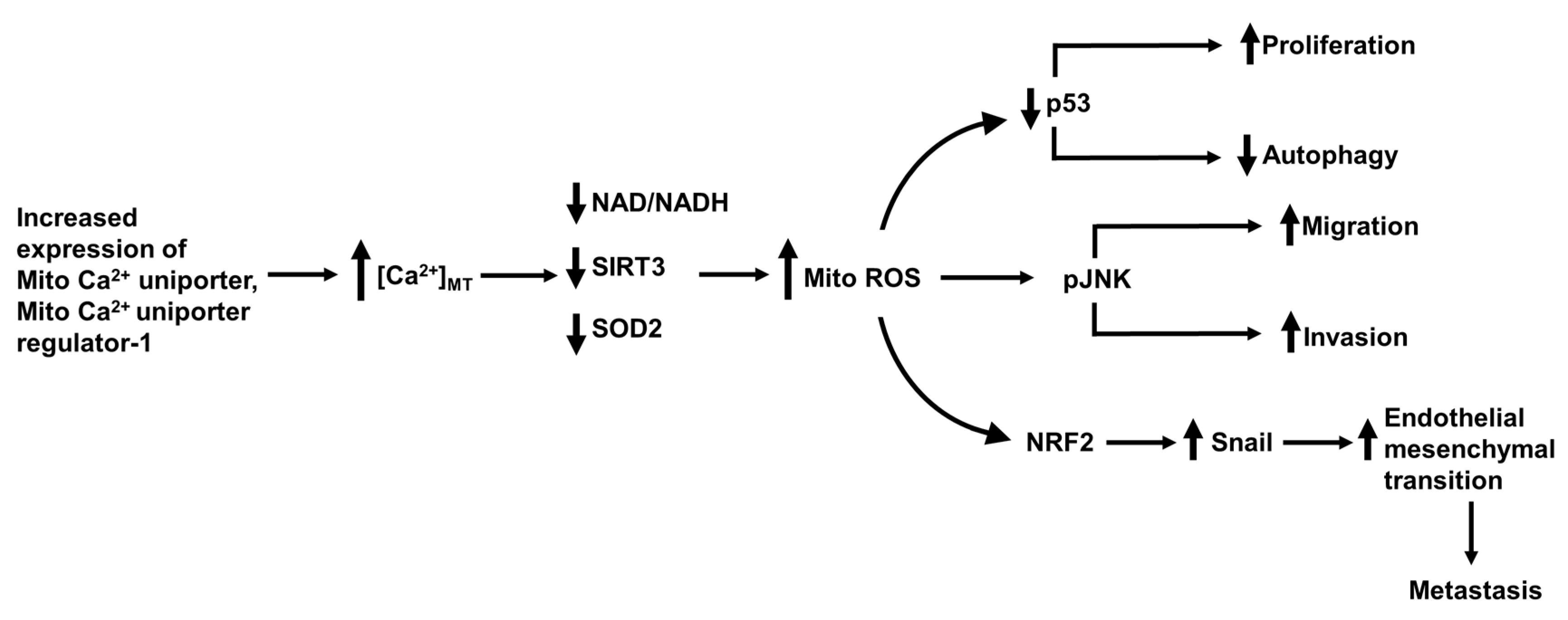
15. The Mitochondrial Ca2+ Uniporter, Permeability Transition Pore and Mitofusin-2 in Hepatocellular Carcinoma Metastasis

In liver cells, as in most other cell types, [Ca2+]MT plays important roles in the regulation of the citric acid cycle and ATP production and in the apoptotic pathway. Under normal conditions, Ca2+ is transported from the cytoplasmic space into the mitochondrial matrix via the mitochondrial Ca2+ uniporter. Ca2+ can also be transferred directly from the ER to the mitochondrial matrix via the mitochondrial-associated membranes (MAMs) and the voltage-dependent anion channel (VDAC). Ca2+ is normally transported out of mitochondria by the (Ca2+ - H+) exchanger [104]. When [Ca2+]cyt and/or [Ca2+]ER is chronically elevated the mitochondrial matrix may become overloaded with Ca2+. This then triggers the release of Ca2+ via the mitochondrial permeability transition pore (pathway shown schematically in Figure 3B) [34,81,104]. Uptake of Ca2+ through the mitochondrial Ca2+ uniporter is regulated by a number of proteins, including mitochondrial Ca2+ uniporter regulator 1 (MCUR1) and mitochondrial Ca2+ uptake 1 and 2 (MICU1 and 2) [34,81,104]. Several studies have shown that both the mitochondrial uniporter and mitochondrial permeability transition pore play important roles in HCC progression and metastasis [34,35,36,81,105].
In human HCC tissue, the mitochondrial Ca2+ uniporter protein was found to be expressed at higher levels than in paired non-tumor tissue. Higher expression correlated with a higher risk of recurrence and death in HCC [34]. Studies conducted using several HCC cell lines in which expression of the mitochondrial Ca2+ uniporter was experimentally either reduced or increased showed that high levels of mitochondrial Ca2+ uniporter enhance the uptake of Ca2+ to mitochondria and increase [Ca2+]MT, which in turn increases ROS and promotes HCC metastasis. Buffering of [Ca2+]MT, using parvalbumin genetically targeted to the mitochondrial matrix (PV-Mito), suppressed the production of ROS and development of metastatic properties. Further analysis of the pathways involved led to the conclusion that increased expression of mitochondrial Ca2+ uniporter and enhanced uptake of Ca2+ to the mitochondrial matrix cause an increase in ROS via changes in NAD/NADH, SIRT3 and superoxidase dismutase 2 (SOD2), as shown schematically in Figure 9 [34].
Two studies provide evidence that increased expression of mitochondrial Ca2+ uniporter regulator 1 enhances HCC cell survival via the p53 pathway and also enhances metastasis via the Nrf2 pathway. Expression of mitochondrial Ca2+ uniporter regulator 1, was found to be high in many HCC liver samples compared with that on non-tumor liver tissue [81]. Using selected HCC cell lines in which mitochondrial Ca2+ uniporter regulator 1 was expressed at either high or low levels, Ren and colleagues found that elevated expression of mitochondrial Ca2+ uniporter regulator 1 is associated with elevated [Ca2+]MT, increased mitochondrial ROS, decreased apoptosis and increased cell proliferation [81]. In a mouse xenograft HCC model, tumors arising from the implantation of HCC cells expressing high levels of mitochondrial Ca2+ uniporter regulator 1 exhibited enhanced growth compared with that of tumors derived for HCC cells exhibiting low mitochondrial Ca2+ uniporter regulator 1 expression [81]. Further investigation of the pathways involved provided evidence that increased expression of mitochondrial Ca2+ uniporter regulator 1 enhanced the proliferation of HCC cells by increasing [Ca2+]MT and generation of mitochondrial ROS, leading to enhanced degradation of p53, decreased autophagy and increased cell cycle activity and cell proliferation (shown schematically Figure 9) [81].
Higher levels of mitochondrial Ca2+ uniporter regulator 1 have also been shown to promote the epithelial mesenchymal transition in HCC cells [105]. Studies employing HCC cell lines showed that knockdown of mitochondrial Ca2+ uniporter regulator 1 and the associated decreased mitochondrial and cellular production of ROS, led to decreased invasion and migration, increased expression of epithelial cell markers (ZO-1 and E- cadherin), decreased expression of mesenchymal markers (N- cadherin and vimentin) and decreased expression of the transcription factor snail, a regulator of the epithelial mesenchymal transition [105]. Overexpression of mitochondrial Ca2+ uniporter regulator 1 caused the opposite changes. Measurement of the expression of mitochondrial Ca2+ uniporter regulator 1 in human HCC tissue samples indicated that higher levels of mitochondrial Ca2+ uniporter regulator 1 were associated with more advanced HCC. From these studies it was concluded that increased expression of mitochondrial Ca2+ uniporter regulator 1 increases [Ca2+]MT, ROS production, activates Nrf2 and the Notch pathway which mediates the epithelial mesenchymal transition (Figure 9) [105]. Taken together, these studies suggest that targeting the mitochondrial uniporter protein or one of its regulator proteins with a pharmacological inhibitor should inhibit the progression of HCC.
Enhanced opening of the mitochondrial permeability transition pore leading to increased apoptosis has been implicated in the mechanism by which the natural product erinacine, obtained from the mushroom Hericium erinaceus, inhibits the growth of HCC cells [35]. Expression of components of the PI3kinase/Akt/GSK-3β pathway was found to be increased in human HCC tissues compared to that in non-tumor liver tissue. Experiments with HepG2 HCC cells using the PI3kinase inhibitor LY294002 showed that inhibition of PI3kinase decreased the mitochondrial membrane potential and increased [Ca2+]MT (measured using Rhodamine 2). In HepG2 cells, erinacine decreased cell proliferation, colony formation, migration and invasion. These changes were associated with decreases in mitochondrial membrane potential, expression of components of the PI3kinase/Akt/GSK-3β pathway, cyclin D, vimentin, β-catenin and Bcl2 and increased expression of E- cadherin. Further, erinacine increased apoptosis, as indicted by increased Bax and caspase-9. Erinacine also enhanced the action of LY294002 on these pathways. In a mouse xenograft liver tumor model, erinacine reduced tumor size [35]. These results provide some insight into the ability of erinacine to inhibit the growth and progression of HCC and implicate [Ca2+]cyt, [Ca2+]MT, the mitochondrial permeability pore transition and the components of the PI3kinase/Akt/GSK-3β pathway in the mechanism of erinacine action. However, further experiments are needed to define the exact roles of [Ca2+]cyt, [Ca2+]MT and the mitochondrial permeability pore transition.
Activation of mitofusin-2 has been identified as a target for reduction of HCC through enhancing the transfer of Ca2+ from the ER to mitochondria, thereby inducing apoptosis [36]. Mitofusin-2 is located in the mitochondrial outer membrane and is involved in mitochondrial fusion [36]. Increased expression of mitofusion-2 in HCC cells induces apoptosis [36]. Wang and colleagues found that expression of mitofusin-2 in HCC tissue is lower than that in non-tumor liver tissue. Lower mitofusin-2 expression was found to be associated with poorer outcomes. Overexpression of mitofusin-2 in HepG2 HCC cells induced apoptosis and this was blocked by the combination of Ru360, an inhibitor of mitochondrial Ca2+ uptake and heparin, an inhibitor of InsP3-induced ER Ca2+ release. Mitofusin-2 overexpression also reduced the mitochondrial membrane potential, reduced [Ca2+]ER and increased [Ca2+]MT and cellular ROS. Decreased expression of mitochondrial Ca2+ uptake proteins 1 and 2 was also observed in HepG2 cells overexpressing mitofusin-2. The authors concluded that increasing the expression of mitofusin-2 in HCC cells induces apoptosis by enhancing Ca2+ entry to mitochondria from the ER [36]. Thus, these studies suggest that targeting mitofusin-2 with a pharmacological activator would inhibit progression of HCC.

16. Tuftelin1, Ca2+ Calmodulin Kinases and Ca2+ Binding Protein 39 in the Promotion and Metastasis of Hepatocellular Carcinoma

The tuftelin1 (TUFT1) protein is involved in the development and mineralization of tooth tissue and its expression is increased in hypoxia [106]. Noting that the tumor microenvironment is often hypoxic, as discussed above, Dou and colleagues investigated the expression and role of tuftelin1 in the growth and metastasis of HCC tumors [106]. Expression of tuftelin1 was found to be increased in HCC liver tissue compared with that in non-tumor tissue. Moreover, increased expression of tuftelin1 correlated with poor overall survival. The pathways involving tuftelin1 in HCC cells were investigated using five different HCC cell lines. Knockdown of tuftelin1 blocked the ability of hypoxia to promote HCC progression, as assessed in HCC cells by monitoring cell growth, metastatic properties and the epithelium mesenchymal transition. Ectopic (increased) expression of tuftelin1 increased [Ca2+]cyt (assessed as an increase in the percentage of cells positive for elevated [Ca2+]cyt) and increased the phosphorylation of Akt, but not that of ERK or JNK. Other experiments identified miR-671-5p as an important regulator of tuftelin1 expression. On the basis of these and other observations, Dou and colleagues proposed the following pathway for the involvement of tuftelin1 and Ca2+ in hypoxia-induced HCC progression. Hypoxia, via miR671-5p, activates transcription factor HIF-1α which induces increased expression of tuftelin1. This in turn increases [Ca2+]cyt, activates the Ca2+/PI3kinase/Akt pathway leading to an enhancement of the growth, metastasis and epithelial mesenchymal transition of HCC cells [106].
Experiments directed towards identifying the mechanism by which another micro RNA, miR-129-5p, inhibits HCC growth have identified Ca2+/calmodulin-dependent protein kinase IV (CamKIV) as a potential therapeutic target [38]. Expression of miR-129-5p in HCC liver tissue was found to be lower than that in paired non-tumor tissue. In two HCC cell lines, overexpression of miR-129-5p was associated with inhibition of cell proliferation, migration and invasion and the promotion of apoptosis. Using bioinformatics and luciferase reporter assays, the relevant target gene for miR-129-5p was identified as CaMKIV. This protein kinase was found to be expressed at a low level in HCC tissue compared to adjacent non-tumor tissue and was also expressed at a low level in two HCC cell lines, HepG2 and BEL-740 L. Ectopic (increased) expression of CaMKIV in these cell lines inhibited cell proliferation, migration and invasion and promoted apoptosis. In a mouse xenograft model of HCC, in which liver tumors were initiated using HepG2 or BEL-740 L cells, increased expression of CaMKIV inhibited tumor growth. The authors suggest that strategies directed towards increasing the activity of CaMKIV would inhibit the growth of HCC tumors, possibly through a mechanism involving inhibition of the MAPK pathway [38].
About ten years ago, tetrandrine, was shown to inhibit the development of HCC in studies employing HCC cell lines and a mouse HCC tumor model [107]. Tetrandrine is a member of the bisbenzylisoquinoline alkaloid family and is found in the roots of the Stephania tetrandra plant. The mechanism by which tetrandrine inhibits HCC involves an increase in ROS and activation of the Akt pathway. In a subsequent study, Huang and colleagues, using structural modeling, identified inhibition of CaMKIIγ as the possible target of tetrandrine action. This was confirmed by measuring the IC50 for the inhibition by tetrandrine of cell proliferation and the level of expression CaMKIIγ expression in each of a panel of eight different HCC cell lines [37]. Huh7 cells were most sensitive to tetrandrine and exhibited highest expression of CaMKIIγ, while SMMC-7721 cells were the least sensitive and exhibited the lowest expression of CaMKIIγ. Tetrandrine reduced CaMKIIγ phosphorylation in HCC cells and knockdown of CaMKIIγ reduced the sensitivity of HCC cells to tetrandrine. Expression of CaMKIIγ in human HCC tissue was substantially greater than that in non-tumor tissue [37]. To try to improve the effectiveness of the drug, Lan and colleagues have designed and synthesized a number of derivatives of tetrandrine [108]. These were tested for their ability to induce apoptosis in two HCC cell lines. Compound 31, in which carbon 14 of tetrandrine is substituted with an amide, was the most potent, having an IC50 of about 1 µM, which is about 30 times lower than the IC50 for the multikinase inhibitor sorafenib [108].
Inhibition of CaMKIIγ may also be a target of berbamine, another member of the bisbenzylisoquinoline family, with a chemical structure similar to that of tetrandrine [15]. Berbamine is isolated from the traditional Chinese herbal medicine plant Berberis amurensis [15]. Studies conducted using Huh-7 HCC cells and a NOD/SCID mouse HCC model showed that berbamine inhibits HCC cell proliferation and induces HCC cell death. Knockdown of CaMKIIγ using shRNA or overexpression of CaMKIIγ, provided evidence that a target of berbamine is CaMKIIγ. In human HCC tissue, CaMKIIγ was found to be hyper-phosphorylated compared to the degree of phosphorylation in non-tumor tissue [15]. However, these observations differ from those of Huang and colleagues who compared the effects of berbamine with those of tetrandrine on HCC cells, but did not observe berbamine-induced phosphorylation of CaMKIIγ [37]. From studies conducted using a subpopulation of Huh7 cells selected on the basis of the stem cell marker CD133 (AC133, promimin-1) in which berbamine and its analog 2-methylbenzoyl berbamine, were observed to reduce spheroid formation, Meng and colleagues have also suggested that CaMKIIγ contributes to the maintenance of cancer stem cell characteristics [15].
In further investigation of the role of CaMKIIγ in HCC, Meng and colleagues used bioinformatics to identify protein kinases that regulate gene expression in human HCCs and in a diethylnitrosamine mouse HCC model [109]. One of the kinases identified was CaMKIIγ. Surprisingly, in CaMKIIγ KO mice (CaMKIIγ-/-), the ability of diethylnitrosamine to induce tumors was greatly enhanced compared with that in WT mice. Investigation of the mechanism by which activation of CaMKIIγ inhibits HCC progression provided evidence that, in HCC cells, CaMKIIγ inhibits mTORC and thus inhibits the activation by growth factors of HCC initiation and proliferation via the mTORC pathway [109]. While it seems clear that CaMKIIγ plays important roles in HCC progression, further experiments are required to gain a more complete understanding of these roles and to bring together the observations which suggest that, on one hand, tetrandrine and berbamine inhibit HCC progression by inhibiting CaMKIIγ, while on the other hand, activation of CaMKIIγ actually inhibits HCC progression.
Calcium-binding protein 39 has been shown to be important in the migration and metastasis of HCC cells, suggesting that it may be a potential pharmacological target for the treatment of late stage HCC [39]. Transcriptome sequencing analysis of mRNA from HCC tissue identified calcium-binding protein 39 as potentially important in the progression of HCC. Calcium-binding protein 39 is known to activate several sterol-20 kinases [39]. Expression of calcium-binding protein 39 in human HCC tissue was found to be elevated compared to that in non-tumor liver tissue and higher levels of expression correlated with tumor metastasis and poor survival. Ectopic expression of calcium-binding protein 39 in the immortalized “normal” L02 liver cell line and in several HCC cell lines, increased foci formation and colony formation in soft agar and increased cell mobility. Similar observations were made in a nude mouse model of HCC in which tumors were derived from HCC cells with low and high calcium-binding protein 39 expression. These properties were abolished when expression of calcium-binding protein 39 was knocked down using shRNA. Further studies showed that elevated expression of calcium-binding protein 39 activates the ERK/cJun/Slug signaling pathway to promote the epithelial mesenchymal transition (monitored by measuring increases in N- cadherin and fibronectin and decreases in E-cadherin and α-E-catenin). It was concluded that increased expression of calcium-binding protein 39 in HCC cells enhances the migration and metastatic properties of these cells and hence HCC metastasis [39].

17. Conclusions

Numerous Ca2+-signaling proteins that could be potential targets for therapeutic treatment of HCC were identified. Targets in liver cancer stem cells, which are thought to drive the initiation and progression of HCC and development of resistance to current drugs, are of particular potential value, as are those which could inhibit the proliferation, migration and metastasis in advanced HCC. However, none of these Ca2+-signaling targets has been seriously studied any further than laboratory research experiments. A few of the Ca2+-signaling proteins have been identified as targets for natural products previously known to reduce HCC. Promising Ca2+-signaling targets seem to be voltage-operated Ca2+ channels in liver cancer stem cells, InsP3Rs, store-operated Ca2+ entry, SERCA2b and Ca2+/calmodulin protein kinases. However, for several Ca2+-signaling proteins so far identified as potential drug targets, the situation seems complicated, in that the effects of inhibition or activation may depend on the stage of the HCC. The targets so far identified have, to a large extent, evolved from somewhat serendipitous research investigations and from studies of the mechanisms of the actions of natural products. The application of more systematic studies in the future is expected to reveal new Ca2+-signaling protein targets and to consolidate priorities for those already identified. Such studies may include expanded genomics and gene expression, RNA-seq and improved knowledge of the fundamental biology and pathology of HCC. If one or more pharmacological inhibitors or activators of Ca2+-signaling proteins is shown to be useful in clinical practice, it is likely that such a compound will be most effective in combination with other drugs and will likely be delivered via drug-emitting bead transcatheter arterial chemoembolization.

Author Contributions

E.S.A. conceived the study, curated and analyzed data from the literature and created the first draft of the paper. G.J.B. helped in conception of the study and had a substantial input into writing subsequent drafts and the final version. G.Y.R. co-supervised the study and had a substantial input into writing the later drafts. All authors have read and agreed to the published version of the manuscript.

Funding

This research received no external funding.

Acknowledgments

This research was supported by grants from the Flinders Center for Innovation in Cancer and the Flinders Foundation (South Australia). The authors gratefully acknowledge Shalev Itzkovitz and Zhiqian Zhang for their kind permission to reproduce Figure 1 and Figure 7, respectively. Figure 4B,C were reproduced under Creative Commons License Deed, Attribution 4.0 International (CC BV 4.0).

Conflicts of Interest

The authors have no conflict of interest to declare.

Abbreviations

HCChepatocellular carcinoma
HBVhepatitis B
HCVhepatitis C
[Ca2+]cytconcentration of free Ca2+ in the cytoplasmic space
[Ca2+]ERconcentration of free Ca2+ in the lumen of the ER
[Ca2+]MTconcentration of free Ca2+ in the mitochondrial matrix
CaMKIICa2+/calmodulin-dependent protein kinase II
CamKK2Ca2+/calmodulin-dependent protein kinase kinase 2
CaMKIVCa2+/calmodulin-dependent protein kinase IV
ERendoplasmic reticulum
SERCAsarco/endoplasmic reticulum (Ca2++Mg2+)ATP-ase
STIMstromal interaction molecule
TRPtransient receptor potential
InsP3inositol 1,4,5-trisphosphate
InsP3RInsP3 receptor
ROSreactive oxygen species
Nrf2nuclear factor erythroid 2-related factor 2

References

  1. Calderaro, J.; Ziol, M.; Paradis, V.; Zucman-Rossi, J. Molecular and histological correlations in liver cancer. J. Hepatol. 2019, 71, 616–630. [Google Scholar] [CrossRef] [PubMed] [Green Version]
  2. Craig, A.J.; von Felden, J.; Garcia-Lezana, T.; Sarcognato, S.; Villanueva, A. Tumour evolution in hepatocellular carcinoma. Nat. Rev. Gastroenterol. Hepatol. 2020, 17, 139–152. [Google Scholar] [CrossRef] [PubMed]
  3. Villanueva, A. Hepatocellular Carcinoma. N. Engl. J. Med. 2019, 380, 1450–1462. [Google Scholar] [CrossRef] [PubMed] [Green Version]
  4. Ben-Moshe, S.; Itzkovitz, S. Spatial heterogeneity in the mammalian liver. Nat. Rev. Gastroenterol. Hepatol. 2019, 16, 395–410. [Google Scholar] [CrossRef]
  5. Ali, E.; Rychkov, G.Y.; Barritt, G.J. Deranged hepatocyte intracellular Ca2+ homeostasis and the progression of non-alcoholic fatty liver disease to hepatocellular carcinoma. Cell Calcium 2019. [Google Scholar] [CrossRef] [PubMed]
  6. Nio, K.; Yamashita, T.; Kaneko, S. The evolving concept of liver cancer stem cells. Mol. Cancer 2017, 16, 4. [Google Scholar] [CrossRef] [Green Version]
  7. Gu, Y.; Zheng, X.; Ji, J. Liver cancer stem cells as a hierarchical society: Yes or no? Acta Biochim. Biophys. Sin. 2020. [Google Scholar] [CrossRef]
  8. Wu, Y.; Zhang, J.; Zhang, X.; Zhou, H.; Liu, G.; Li, Q. Cancer Stem Cells: A Potential Breakthrough in HCC-Targeted Therapy. Front. Pharmacol. 2020, 11, 198. [Google Scholar] [CrossRef]
  9. Wang, N.; Wang, S.; Li, M.Y.; Hu, B.G.; Liu, L.P.; Yang, S.L.; Yang, S.; Gong, Z.; Lai, P.B.S.; Chen, G.G. Cancer stem cells in hepatocellular carcinoma: An overview and promising therapeutic strategies. Ther. Adv. Med. Oncol. 2018, 10, 1758835918816287. [Google Scholar] [CrossRef]
  10. Sia, D.; Villanueva, A.; Friedman, S.L.; Llovet, J.M. Liver Cancer Cell of Origin, Molecular Class, and Effects on Patient Prognosis. Gastroenterology 2017, 152, 745–761. [Google Scholar] [CrossRef] [Green Version]
  11. Liang, J.Q.; Teoh, N.; Xu, L.; Pok, S.; Li, X.; Chu, E.S.H.; Chiu, J.; Dong, L.; Arfianti, E.; Haigh, W.G.; et al. Dietary cholesterol promotes steatohepatitis related hepatocellular carcinoma through dysregulated metabolism and calcium signaling. Nat. Commun. 2018, 9, 4490. [Google Scholar] [CrossRef] [PubMed] [Green Version]
  12. Zhang, Y.; Qiu, Z.; Wei, L.; Tang, R.; Lian, B.; Zhao, Y.; He, X.; Xie, L. Integrated analysis of mutation data from various sources identifies key genes and signaling pathways in hepatocellular carcinoma. PLoS ONE 2014, 9, e100854. [Google Scholar] [CrossRef] [PubMed]
  13. Zhao, W.; Wang, L.; Han, H.; Jin, K.; Lin, N.; Guo, T.; Chen, Y.; Cheng, H.; Lu, F.; Fang, W.; et al. 1B50-1, a mAb raised against recurrent tumor cells, targets liver tumor-initiating cells by binding to the calcium channel alpha2delta1 subunit. Cancer Cell 2013, 23, 541–556. [Google Scholar] [CrossRef] [PubMed] [Green Version]
  14. Jimenez, H.; Wang, M.; Zimmerman, J.W.; Pennison, M.J.; Sharma, S.; Surratt, T.; Xu, Z.X.; Brezovich, I.; Absher, D.; Myers, R.M.; et al. Tumour-specific amplitude-modulated radiofrequency electromagnetic fields induce differentiation of hepatocellular carcinoma via targeting Cav3.2T-type voltage-gated calcium channels and Ca2+ influx. EBioMedicine 2019, 44, 209–224. [Google Scholar] [CrossRef] [PubMed] [Green Version]
  15. Meng, Z.; Li, T.; Ma, X.; Wang, X.; Van Ness, C.; Gan, Y.; Zhou, H.; Tang, J.; Lou, G.; Wang, Y.; et al. Berbamine inhibits the growth of liver cancer cells and cancer-initiating cells by targeting Ca(2)(+)/calmodulin-dependent protein kinase II. Mol. Cancer Ther. 2013, 12, 2067–2077. [Google Scholar] [CrossRef] [Green Version]
  16. Sun, C.; Shui, B.; Zhao, W.; Liu, H.; Li, W.; Lee, J.C.; Doran, R.; Lee, F.K.; Sun, T.; Shen, Q.S.; et al. Central role of IP3R2-mediated Ca2+ oscillation in self-renewal of liver cancer stem cells elucidated by high-signal ER sensor. Cell Death Dis. 2019, 10, 396. [Google Scholar] [CrossRef] [Green Version]
  17. Scrima, R.; Piccoli, C.; Moradpour, D.; Capitanio, N. Targeting Endoplasmic Reticulum and/or Mitochondrial Ca2+ Fluxes as Therapeutic Strategy for HCV Infection. Front. Chem. 2018, 6, 73. [Google Scholar] [CrossRef] [Green Version]
  18. Ivanov, A.V.; Valuev-Elliston, V.T.; Tyurina, D.A.; Ivanova, O.N.; Kochetkov, S.N.; Bartosch, B.; Isaguliants, M.G. Oxidative stress, a trigger of hepatitis C and B virus-induced liver carcinogenesis. Oncotarget 2017, 8, 3895–3932. [Google Scholar] [CrossRef] [Green Version]
  19. Casciano, J.C.; Duchemin, N.J.; Lamontagne, R.J.; Steel, L.F.; Bouchard, M.J. Hepatitis B virus modulates store-operated calcium entry to enhance viral replication in primary hepatocytes. PLoS ONE 2017, 12, e0168328. [Google Scholar] [CrossRef]
  20. Ali, E.S.; Petrovsky, N. Calcium Signaling As a Therapeutic Target for Liver Steatosis. Trends Endocrinol. Metab. 2019, 30, 270–281. [Google Scholar] [CrossRef]
  21. Park, H.W.; Park, H.; Semple, I.A.; Jang, I.; Ro, S.H.; Kim, M.; Cazares, V.A.; Stuenkel, E.L.; Kim, J.J.; Kim, J.S.; et al. Pharmacological correction of obesity-induced autophagy arrest using calcium channel blockers. Nat. Commun. 2014, 5, 4834. [Google Scholar] [CrossRef] [PubMed] [Green Version]
  22. Kang, J.K.; Kim, O.H.; Hur, J.; Yu, S.H.; Lamichhane, S.; Lee, J.W.; Ojha, U.; Hong, J.H.; Lee, C.S.; Cha, J.Y.; et al. Increased intracellular Ca2+ concentrations prevent membrane localization of PH domains through the formation of Ca2+-phosphoinositides. Proc. Natl. Acad. Sci. USA 2017, 114, 11926–11931. [Google Scholar] [CrossRef] [PubMed] [Green Version]
  23. Shen, Z.Q.; Chen, Y.F.; Chen, J.R.; Jou, Y.S.; Wu, P.C.; Kao, C.H.; Wang, C.H.; Huang, Y.L.; Chen, C.F.; Huang, T.S.; et al. CISD2 Haploinsufficiency Disrupts Calcium Homeostasis, Causes Nonalcoholic Fatty Liver Disease, and Promotes Hepatocellular Carcinoma. Cell Rep. 2017, 21, 2198–2211. [Google Scholar] [CrossRef] [Green Version]
  24. Xu, D.; Xu, M.; Jeong, S.; Qian, Y.; Wu, H.; Xia, Q.; Kong, X. The Role of Nrf2 in Liver Disease: Novel Molecular Mechanisms and Therapeutic Approaches. Front. Pharmacol. 2018, 9, 1428. [Google Scholar] [CrossRef] [Green Version]
  25. Lin, F.; Marcelo, K.L.; Rajapakshe, K.; Coarfa, C.; Dean, A.; Wilganowski, N.; Robinson, H.; Sevick, E.; Bissig, K.D.; Goldie, L.C.; et al. The camKK2/camKIV relay is an essential regulator of hepatic cancer. Hepatology 2015, 62, 505–520. [Google Scholar] [CrossRef] [PubMed]
  26. Wu, Z.; Qing, J.; Xia, Y.; Wang, K.; Zhang, F. Suppression of stromal interaction molecule 1 inhibits SMMC7721 hepatocellular carcinoma cell proliferation by inducing cell cycle arrest. Biotechnol. Appl. Biochem. 2015, 62, 107–111. [Google Scholar] [CrossRef]
  27. Tang, B.D.; Xia, X.; Lv, X.F.; Yu, B.X.; Yuan, J.N.; Mai, X.Y.; Shang, J.Y.; Zhou, J.G.; Liang, S.J.; Pang, R.P. Inhibition of Orai1-mediated Ca2+ entry enhances chemosensitivity of HepG2 hepatocarcinoma cells to 5-fluorouracil. J. Cell Mol. Med. 2017, 21, 904–915. [Google Scholar] [CrossRef]
  28. El Boustany, C.; Bidaux, G.; Enfissi, A.; Delcourt, P.; Prevarskaya, N.; Capiod, T. Capacitative calcium entry and transient receptor potential canonical 6 expression control human hepatoma cell proliferation. Hepatology 2008, 47, 2068–2077. [Google Scholar] [CrossRef]
  29. Anand, P.; Filipenko, P.; Huaman, J.; Lyudmer, M.; Hossain, M.; Santamaria, C.; Huang, K.; Ogunwobi, O.O.; Holford, M. Selective Inhibition of Liver Cancer Cells Using Venom Peptide. Mar. Drugs 2019, 17, 587. [Google Scholar] [CrossRef] [Green Version]
  30. Fang, Y.; Liu, G.; Xie, C.; Qian, K.; Lei, X.; Liu, Q.; Liu, G.; Cao, Z.; Fu, J.; Du, H.; et al. Pharmacological inhibition of TRPV4 channel suppresses malignant biological behavior of hepatocellular carcinoma via modulation of ERK signaling pathway. Biomed. Pharmacother. 2018, 101, 910–919. [Google Scholar] [CrossRef]
  31. Chen, W.T.; Lin, G.B.; Lin, S.H.; Lu, C.H.; Hsieh, C.H.; Ma, B.L.; Chao, C.Y. Static magnetic field enhances the anticancer efficacy of capsaicin on HepG2 cells via capsaicin receptor TRPV1. PLoS ONE 2018, 13, e0191078. [Google Scholar] [CrossRef] [PubMed] [Green Version]
  32. Xie, C.; Liu, G.; Li, M.; Fang, Y.; Qian, K.; Tang, Y.; Wu, X.; Lei, X.; Li, X.; Liu, Q.; et al. Targeting TRPV1 on cellular plasticity regulated by Ovol 2 and Zeb 1 in hepatocellular carcinoma. Biomed. Pharmacother. 2019, 118, 109270. [Google Scholar] [CrossRef] [PubMed]
  33. Guerra, M.T.; Florentino, R.M.; Franca, A.; Lima Filho, A.C.; Dos Santos, M.L.; Fonseca, R.C.; Lemos, F.O.; Fonseca, M.C.; Kruglov, E.; Mennone, A.; et al. Expression of the type 3 InsP3 receptor is a final common event in the development of hepatocellular carcinoma. Gut 2019, 68, 1676–1687. [Google Scholar] [CrossRef] [PubMed] [Green Version]
  34. Ren, T.; Zhang, H.; Wang, J.; Zhu, J.; Jin, M.; Wu, Y.; Guo, X.; Ji, L.; Huang, Q.; Zhang, H.; et al. MCU-dependent mitochondrial Ca2+ inhibits NAD(+)/SIRT3/SOD2 pathway to promote ROS production and metastasis of HCC cells. Oncogene 2017, 36, 5897–5909. [Google Scholar] [CrossRef]
  35. Zhou, L.J.; Mo, Y.B.; Bu, X.; Wang, J.J.; Bai, J.; Zhang, J.W.; Cheng, A.B.; Ma, J.H.; Wang, Y.W.; Xie, Y.X. Erinacine Facilitates the Opening of the Mitochondrial Permeability Transition Pore Through the Inhibition of the PI3K/ Akt/GSK-3beta Signaling Pathway in Human Hepatocellular Carcinoma. Cell Physiol. Biochem. 2018, 50, 851–867. [Google Scholar] [CrossRef] [PubMed]
  36. Wang, W.; Xie, Q.; Zhou, X.; Yao, J.; Zhu, X.; Huang, P.; Zhang, L.; Wei, J.; Xie, H.; Zhou, L.; et al. Mitofusin-2 triggers mitochondria Ca2+ influx from the endoplasmic reticulum to induce apoptosis in hepatocellular carcinoma cells. Cancer Lett. 2015, 358, 47–58. [Google Scholar] [CrossRef]
  37. Huang, T.; Xu, S.; Deo, R.; Ma, A.; Li, H.; Ma, K.; Gan, X. Targeting the Ca2+/Calmodulin-dependent protein kinase II by Tetrandrine in human liver cancer cells. Biochem. Biophys. Res. Commun. 2019, 508, 1227–1232. [Google Scholar] [CrossRef]
  38. Li, Z.; Lu, J.; Zeng, G.; Pang, J.; Zheng, X.; Feng, J.; Zhang, J. MiR-129-5p inhibits liver cancer growth by targeting calcium calmodulin-dependent protein kinase IV (CAMK4). Cell Death Dis. 2019, 10, 789. [Google Scholar] [CrossRef] [Green Version]
  39. Jiang, L.; Yan, Q.; Fang, S.; Liu, M.; Li, Y.; Yuan, Y.F.; Li, Y.; Zhu, Y.; Qi, J.; Yang, X.; et al. Calcium-binding protein 39 promotes hepatocellular carcinoma growth and metastasis by activating extracellular signal-regulated kinase signaling pathway. Hepatology 2017, 66, 1529–1545. [Google Scholar] [CrossRef] [Green Version]
  40. Brosch, M.; Kattler, K.; Herrmann, A.; von Schonfels, W.; Nordstrom, K.; Seehofer, D.; Damm, G.; Becker, T.; Zeissig, S.; Nehring, S.; et al. Epigenomic map of human liver reveals principles of zonated morphogenic and metabolic control. Nat. Commun. 2018, 9, 4150. [Google Scholar] [CrossRef]
  41. Bou-Nader, M.; Caruso, S.; Donne, R.; Celton-Morizur, S.; Calderaro, J.; Gentric, G.; Cadoux, M.; L’Hermitte, A.; Klein, C.; Guilbert, T.; et al. Polyploidy spectrum: A new marker in HCC classification. Gut 2020, 69, 355–364. [Google Scholar] [CrossRef] [PubMed]
  42. Donne, R.; Saroul-Ainama, M.; Cordier, P.; Celton-Morizur, S.; Desdouets, C. Polyploidy in liver development, homeostasis and disease. Nat. Rev. Gastroenterol. Hepatol. 2020, 17, 391–405. [Google Scholar] [CrossRef]
  43. Exton, J.H. Mechanisms of hormonal regulation of hepatic glucose metabolism. Diabetes Metab. Rev. 1987, 3, 163–183. [Google Scholar] [CrossRef] [PubMed]
  44. Woods, N.M.; Cuthbertson, K.S.; Cobbold, P.H. Repetitive transient rises in cytoplasmic free calcium in hormone-stimulated hepatocytes. Nature 1986, 319, 600–602. [Google Scholar] [CrossRef] [PubMed]
  45. Trampert, D.C.; Nathanson, M.H. Regulation of bile secretion by calcium signaling in health and disease. Biochim. Biophys. Acta Mol. Cell Res. 2018, 1865, 1761–1770. [Google Scholar] [CrossRef]
  46. Ueasilamongkol, P.; Khamphaya, T.; Guerra, M.T.; Rodrigues, M.A.; Gomes, D.A.; Kong, Y.; Wei, W.; Jain, D.; Trampert, D.C.; Ananthanarayanan, M.; et al. Type 3 Inositol 1,4,5-Trisphosphate Receptor Is Increased and Enhances Malignant Properties in Cholangiocarcinoma. Hepatology 2020, 71, 583–599. [Google Scholar] [CrossRef]
  47. Case, R.M.; Eisner, D.; Gurney, A.; Jones, O.; Muallem, S.; Verkhratsky, A. Evolution of calcium homeostasis: From birth of the first cell to an omnipresent signalling system. Cell Calcium 2007, 42, 345–350. [Google Scholar] [CrossRef]
  48. Plattner, H.; Verkhratsky, A. Inseparable tandem: Evolution chooses ATP and Ca2+ to control life, death and cellular signalling. Philos. Trans R Soc. Lond. B Biol. Sci. 2016, 371. [Google Scholar] [CrossRef] [Green Version]
  49. Kheradpezhouh, E.; Ma, L.; Morphett, A.; Barritt, G.J.; Rychkov, G.Y. TRPM2 channels mediate acetaminophen-induced liver damage. Proc. Natl. Acad. Sci. USA 2014, 111, 3176–3181. [Google Scholar] [CrossRef] [Green Version]
  50. Echevarria, W.; Leite, M.F.; Guerra, M.T.; Zipfel, W.R.; Nathanson, M.H. Regulation of calcium signals in the nucleus by a nucleoplasmic reticulum. Nat. Cell Biol. 2003, 5, 440–446. [Google Scholar] [CrossRef] [Green Version]
  51. Rychkov, G.; Brereton, H.M.; Harland, M.L.; Barritt, G.J. Plasma membrane Ca2+ release-activated Ca2+ channels with a high selectivity for Ca2+ identified by patch-clamp recording in rat liver cells. Hepatology 2001, 33, 938–947. [Google Scholar] [CrossRef] [PubMed]
  52. Endo, K.; Kuroda, H.; Oikawa, T.; Okada, Y.; Fujiwara, Y.; Abe, T.; Sato, H.; Sawara, K.; Takikawa, Y. Efficacy of combination therapy with transcatheter arterial chemoembolization and radiofrequency ablation for intermediate-stage hepatocellular carcinoma. Scand. J. Gastroenterol. 2018, 53, 1575–1583. [Google Scholar] [CrossRef] [PubMed]
  53. Fujiwara, N.; Friedman, S.L.; Goossens, N.; Hoshida, Y. Risk factors and prevention of hepatocellular carcinoma in the era of precision medicine. J. Hepatol. 2018, 68, 526–549. [Google Scholar] [CrossRef] [PubMed] [Green Version]
  54. Rawla, P.; Sunkara, T.; Muralidharan, P.; Raj, J.P. Update in global trends and aetiology of hepatocellular carcinoma. Contemp. Oncol. 2018, 22, 141–150. [Google Scholar] [CrossRef] [PubMed] [Green Version]
  55. Yang, J.D.; Hainaut, P.; Gores, G.J.; Amadou, A.; Plymoth, A.; Roberts, L.R. A global view of hepatocellular carcinoma: Trends, risk, prevention and management. Nat. Rev. Gastroenterol. Hepatol. 2019, 16, 589–604. [Google Scholar] [CrossRef] [PubMed]
  56. Anstee, Q.M.; Reeves, H.L.; Kotsiliti, E.; Govaere, O.; Heikenwalder, M. From NASH to HCC: Current concepts and future challenges. Nat. Rev. Gastroenterol. Hepatol. 2019, 16, 411–428. [Google Scholar] [CrossRef]
  57. Lafitte, M.; Sirvent, A.; Roche, S. Collagen Kinase Receptors as Potential Therapeutic Targets in Metastatic Colon Cancer. Front. Oncol. 2020, 10, 125. [Google Scholar] [CrossRef]
  58. Kanwal, F.; Kramer, J.R.; Mapakshi, S.; Natarajan, Y.; Chayanupatkul, M.; Richardson, P.A.; Li, L.; Desiderio, R.; Thrift, A.P.; Asch, S.M.; et al. Risk of Hepatocellular Cancer in Patients With Non-Alcoholic Fatty Liver Disease. Gastroenterology 2018, 155, 1828–1837 e1822. [Google Scholar] [CrossRef] [Green Version]
  59. Fernandes, E.S.M.; Rodrigues, P.D.; Alvares-da-Silva, M.R.; Scaffaro, L.A.; Farenzena, M.; Teixeira, U.F.; Waechter, F.L. Treatment strategies for locally advanced hepatocellular carcinoma. Transl. Gastroenterol. Hepatol. 2019, 4, 12. [Google Scholar] [CrossRef]
  60. Castelli, G.; Pelosi, E.; Testa, U. Liver Cancer: Molecular Characterization, Clonal Evolution and Cancer Stem Cells. Cancers 2017, 9, 127. [Google Scholar] [CrossRef] [Green Version]
  61. Zhao, H.; Yan, G.; Zheng, L.; Zhou, Y.; Sheng, H.; Wu, L.; Zhang, Q.; Lei, J.; Zhang, J.; Xin, R.; et al. STIM1 is a metabolic checkpoint regulating the invasion and metastasis of hepatocellular carcinoma. Theranostics 2020, 10, 6483–6499. [Google Scholar] [CrossRef]
  62. Bayo, J.; Fiore, E.J.; Dominguez, L.M.; Real, A.; Malvicini, M.; Rizzo, M.; Atorrasagasti, C.; Garcia, M.G.; Argemi, J.; Martinez, E.D.; et al. A comprehensive study of epigenetic alterations in hepatocellular carcinoma identifies potential therapeutic targets. J. Hepatol. 2019, 71, 78–90. [Google Scholar] [CrossRef] [PubMed]
  63. Nakagawa, H.; Fujita, M.; Fujimoto, A. Genome sequencing analysis of liver cancer for precision medicine. Semin Cancer Biol. 2019, 55, 120–127. [Google Scholar] [CrossRef] [PubMed]
  64. Frades, I.; Andreasson, E.; Mato, J.M.; Alexandersson, E.; Matthiesen, R.; Martinez-Chantar, M.L. Integrative genomic signatures of hepatocellular carcinoma derived from nonalcoholic Fatty liver disease. PLoS ONE 2015, 10, e0124544. [Google Scholar] [CrossRef] [PubMed]
  65. Chan, L.K.; Ng, I.O. Joining the dots for better liver cancer treatment. Nat. Rev. Gastroenterol. Hepatol. 2020, 17, 74–75. [Google Scholar] [CrossRef]
  66. Llovet, J.M.; Montal, R.; Sia, D.; Finn, R.S. Molecular therapies and precision medicine for hepatocellular carcinoma. Nat. Rev. Clin. Oncol. 2018, 15, 599–616. [Google Scholar] [CrossRef]
  67. Zhang, X.; Lin, X.; Qiu, H.; Peng, Z. An investigation of efficacy, safety, and prognostic factors of drug-eluting beads-transarterial chemoembolization operation with CalliSpheres((R)) Microspheres in treating Chinese hepatocellular carcinoma patients. J. Clin. Lab. Anal. 2019, 33, e22975. [Google Scholar] [CrossRef] [Green Version]
  68. Xiang, H.; Long, L.; Yao, Y.; Fang, Z.; Zhang, Z.; Zhang, Y. CalliSpheres Drug-Eluting Bead Transcatheter Arterial Chemoembolization Presents With Better Efficacy and Equal Safety Compared to Conventional TACE in Treating Patients With Hepatocellular Carcinoma. Technol. Cancer Res. Treat 2019, 18, 1533033819830751. [Google Scholar] [CrossRef] [Green Version]
  69. Lencioni, R.; Petruzzi, P.; Crocetti, L. Chemoembolization of hepatocellular carcinoma. Semin Intervent Radiol. 2013, 30, 3–11. [Google Scholar] [CrossRef] [Green Version]
  70. He, L.; Tian, D.A.; Li, P.Y.; He, X.X. Mouse models of liver cancer: Progress and recommendations. Oncotarget 2015, 6, 23306–23322. [Google Scholar] [CrossRef]
  71. Caruso, S.; Calatayud, A.L.; Pilet, J.; La Bella, T.; Rekik, S.; Imbeaud, S.; Letouze, E.; Meunier, L.; Bayard, Q.; Rohr-Udilova, N.; et al. Analysis of Liver Cancer Cell Lines Identifies Agents With Likely Efficacy Against Hepatocellular Carcinoma and Markers of Response. Gastroenterology 2019, 157, 760–776. [Google Scholar] [CrossRef] [PubMed]
  72. Zhang, W.Y.; Cai, N.; Ye, L.H.; Zhang, X.D. Transformation of human liver L-O2 cells mediated by stable HBx transfection. Acta Pharmacol. Sin. 2009, 30, 1153–1161. [Google Scholar] [CrossRef] [PubMed]
  73. Ye, F.; Chen, C.; Qin, J.; Liu, J.; Zheng, C. Genetic profiling reveals an alarming rate of cross-contamination among human cell lines used in China. FASEB J. 2015, 29, 4268–4272. [Google Scholar] [CrossRef] [PubMed] [Green Version]
  74. Huang, J.; Deng, Q.; Wang, Q.; Li, K.Y.; Dai, J.H.; Li, N.; Zhu, Z.D.; Zhou, B.; Liu, X.Y.; Liu, R.F.; et al. Exome sequencing of hepatitis B virus-associated hepatocellular carcinoma. Nat. Genet. 2012, 44, 1117–1121. [Google Scholar] [CrossRef]
  75. Hass, H.G.; Vogel, U.; Scheurlen, M.; Jobst, J. Gene-expression Analysis Identifies Specific Patterns of Dysregulated Molecular Pathways and Genetic Subgroups of Human Hepatocellular Carcinoma. Anticancer Res. 2016, 36, 5087–5095. [Google Scholar] [CrossRef] [Green Version]
  76. Ramachandran, P.; Matchett, K.P.; Dobie, R.; Wilson-Kanamori, J.R.; Henderson, N.C. Single-cell technologies in hepatology: New insights into liver biology and disease pathogenesis. Nat. Rev. Gastroenterol. Hepatol. 2020, 17, 457–472. [Google Scholar] [CrossRef]
  77. Cui, C.; Merritt, R.; Fu, L.; Pan, Z. Targeting calcium signaling in cancer therapy. Acta Pharm. Sin B 2017, 7, 3–17. [Google Scholar] [CrossRef]
  78. Park, Y.R.; Chun, J.N.; So, I.; Kim, H.J.; Baek, S.; Jeon, J.H.; Shin, S.Y. Data-driven Analysis of TRP Channels in Cancer: Linking Variation in Gene Expression to Clinical Significance. Cancer Genom. Proteom. 2016, 13, 83–90. [Google Scholar]
  79. Vriens, J.; Janssens, A.; Prenen, J.; Nilius, B.; Wondergem, R. TRPV channels and modulation by hepatocyte growth factor/scatter factor in human hepatoblastoma (HepG2) cells. Cell Calcium 2004, 36, 19–28. [Google Scholar] [CrossRef]
  80. Yang, N.; Tang, Y.; Wang, F.; Zhang, H.; Xu, D.; Shen, Y.; Sun, S.; Yang, G. Blockade of store-operated Ca2+ entry inhibits hepatocarcinoma cell migration and invasion by regulating focal adhesion turnover. Cancer Lett. 2013, 330, 163–169. [Google Scholar] [CrossRef]
  81. Ren, T.; Wang, J.; Zhang, H.; Yuan, P.; Zhu, J.; Wu, Y.; Huang, Q.; Guo, X.; Zhang, J.; Ji, L.; et al. MCUR1-Mediated Mitochondrial Calcium Signaling Facilitates Cell Survival of Hepatocellular Carcinoma via Reactive Oxygen Species-Dependent P53 Degradation. Antioxid. Redox Signal. 2018, 28, 1120–1136. [Google Scholar] [CrossRef] [PubMed]
  82. Liu, J.; Han, P.; Li, M.; Yan, W.; Liu, J.; Liu, J.; He, J.; Tu, W.; Xia, Y.; Zhou, Z.; et al. The histidine-rich calcium binding protein (HRC) promotes tumor metastasis in hepatocellular carcinoma and is upregulated by SATB1. Oncotarget 2015, 6, 6811–6824. [Google Scholar] [CrossRef] [PubMed] [Green Version]
  83. Schuette, D.; Moore, L.M.; Robert, M.E.; Taddei, T.H.; Ehrlich, B.E. Hepatocellular Carcinoma Outcome Is Predicted by Expression of Neuronal Calcium Sensor 1. Cancer Epidemiol. Biomark. Prev. 2018, 27, 1091–1100. [Google Scholar] [CrossRef] [Green Version]
  84. Bautista, W.; Lipschitz, J.; McKay, A.; Minuk, G.Y. Cancer Stem Cells are Depolarized Relative to Normal Stem Cells Derived from Human Livers. Ann. Hepatol. 2017, 16, 297–303. [Google Scholar] [CrossRef] [PubMed]
  85. Badr, S.A.; Fahmi, M.W.; Nomir, M.M.; El-Shishtawy, M.M. Calcium channel alpha2 delta1 subunit as a novel biomarker for diagnosis of hepatocellular carcinoma. Cancer Biol. Med. 2018, 15, 52–60. [Google Scholar]
  86. Hu, Z.; Cao, X.; Fang, Y.; Liu, G.; Xie, C.; Qian, K.; Lei, X.; Cao, Z.; Du, H.; Cheng, X.; et al. Transient receptor potential vanilloid-type 2 targeting on stemness in liver cancer. Biomed. Pharmacother. 2018, 105, 697–706. [Google Scholar] [CrossRef]
  87. Casciano, J.C.; Bouchard, M.J. Hepatitis B virus X protein modulates cytosolic Ca2+ signaling in primary human hepatocytes. Virus Res. 2018, 246, 23–27. [Google Scholar] [CrossRef]
  88. Kutlu, O.; Kaleli, H.N.; Ozer, E. Molecular Pathogenesis of Nonalcoholic Steatohepatitis- (NASH-) Related Hepatocellular Carcinoma. Can. J. Gastroenterol. Hepatol. 2018, 2018, 8543763. [Google Scholar] [CrossRef] [Green Version]
  89. Sanyal, A.J. Past, present and future perspectives in nonalcoholic fatty liver disease. Nat. Rev. Gastroenterol. Hepatol. 2019, 16, 377–386. [Google Scholar] [CrossRef]
  90. Ersoy, B.A.; Maner-Smith, K.M.; Li, Y.; Alpertunga, I.; Cohen, D.E. Thioesterase-mediated control of cellular calcium homeostasis enables hepatic ER stress. J. Clin Investig. 2018, 128, 141–156. [Google Scholar] [CrossRef]
  91. Jung, T.W.; Kim, H.C.; Abd El-Aty, A.M.; Jeong, J.H. Maresin 1 attenuates NAFLD by suppression of endoplasmic reticulum stress via AMPK-SERCA2b pathway. J. Biol. Chem. 2018, 293, 3981–3988. [Google Scholar] [CrossRef] [PubMed] [Green Version]
  92. Maus, M.; Cuk, M.; Patel, B.; Lian, J.; Ouimet, M.; Kaufmann, U.; Yang, J.; Horvath, R.; Hornig-Do, H.T.; Chrzanowska-Lightowlers, Z.M.; et al. Store-Operated Ca2+ Entry Controls Induction of Lipolysis and the Transcriptional Reprogramming to Lipid Metabolism. Cell Metab. 2017, 25, 1–15. [Google Scholar] [CrossRef] [PubMed] [Green Version]
  93. Lee, S.; Han, D.; Kang, H.G.; Jeong, S.J.; Jo, J.E.; Shin, J.; Kim, D.K.; Park, H.W. Intravenous sustained-release nifedipine ameliorates nonalcoholic fatty liver disease by restoring autophagic clearance. Biomaterials 2019, 197, 1–11. [Google Scholar] [CrossRef]
  94. Wires, E.S.; Trychta, K.A.; Back, S.; Sulima, A.; Rice, K.C.; Harvey, B.K. High fat diet disrupts endoplasmic reticulum calcium homeostasis in the rat liver. J. Hepatol. 2017, 67, 1009–1017. [Google Scholar] [CrossRef] [PubMed]
  95. Li, Y.; Guo, B.; Xie, Q.; Ye, D.; Zhang, D.; Zhu, Y.; Chen, H.; Zhu, B. STIM1 Mediates Hypoxia-Driven Hepatocarcinogenesis via Interaction with HIF-1. Cell Rep. 2015, 12, 388–395. [Google Scholar] [CrossRef] [PubMed] [Green Version]
  96. Selli, C.; Erac, Y.; Kosova, B.; Erdal, E.S.; Tosun, M. Silencing of TRPC1 regulates store-operated calcium entry and proliferation in Huh7 hepatocellular carcinoma cells. Biomed. Pharmacother. 2015, 71, 194–200. [Google Scholar] [CrossRef] [PubMed]
  97. Selli, C.; Pearce, D.A.; Sims, A.H.; Tosun, M. Differential expression of store-operated calcium- and proliferation-related genes in hepatocellular carcinoma cells following TRPC1 ion channel silencing. Mol. Cell Biochem. 2016, 420, 129–140. [Google Scholar] [CrossRef] [Green Version]
  98. Xu, J.; Yang, Y.; Xie, R.; Liu, J.; Nie, X.; An, J.; Wen, G.; Liu, X.; Jin, H.; Tuo, B. The NCX1/TRPC6 Complex Mediates TGFbeta-Driven Migration and Invasion of Human Hepatocellular Carcinoma Cells. Cancer Res. 2018, 78, 2564–2576. [Google Scholar] [CrossRef] [Green Version]
  99. Fu, J.; Du, H.; Zhang, X.; Xu, X. Pharmacological Inhibition of Transient Receptor Potential Vanilloid 4 (TRPV4) Channel Alleviates Carbon Tetrachloride-Induced Liver Fibrosis in Mice. J. Nippon Med. Sch. 2019, 86, 258–262. [Google Scholar] [CrossRef] [Green Version]
  100. Bort, A.; Spinola, E.; Rodriguez-Henche, N.; Diaz-Laviada, I. Capsaicin exerts synergistic antitumor effect with sorafenib in hepatocellular carcinoma cells through AMPK activation. Oncotarget 2017, 8, 87684–87698. [Google Scholar] [CrossRef] [Green Version]
  101. Lee, Y.S.; Kang, Y.S.; Lee, J.S.; Nicolova, S.; Kim, J.A. Involvement of NADPH oxidase-mediated generation of reactive oxygen species in the apototic cell death by capsaicin in HepG2 human hepatoma cells. Free Radic. Res. 2004, 38, 405–412. [Google Scholar] [CrossRef] [PubMed]
  102. Huang, S.P.; Chen, J.C.; Wu, C.C.; Chen, C.T.; Tang, N.Y.; Ho, Y.T.; Lo, C.; Lin, J.P.; Chung, J.G.; Lin, J.G. Capsaicin-induced apoptosis in human hepatoma HepG2 cells. Anticancer Res. 2009, 29, 165–174. [Google Scholar] [PubMed]
  103. Miao, X.; Liu, G.; Xu, X.; Xie, C.; Sun, F.; Yang, Y.; Zhang, T.; Hua, S.; Fan, W.; Li, Q.; et al. High expression of vanilloid receptor-1 is associated with better prognosis of patients with hepatocellular carcinoma. Cancer Genet. Cytogenet. 2008, 186, 25–32. [Google Scholar] [CrossRef] [PubMed]
  104. Giorgi, C.; Marchi, S.; Pinton, P. The machineries, regulation and cellular functions of mitochondrial calcium. Nat. Rev. Mol. Cell Biol. 2018, 19, 713–730. [Google Scholar] [CrossRef] [PubMed]
  105. Jin, M.; Wang, J.; Ji, X.; Cao, H.; Zhu, J.; Chen, Y.; Yang, J.; Zhao, Z.; Ren, T.; Xing, J. MCUR1 facilitates epithelial-mesenchymal transition and metastasis via the mitochondrial calcium dependent ROS/Nrf2/Notch pathway in hepatocellular carcinoma. J. Exp. Clin. Cancer Res. 2019, 38, 136. [Google Scholar] [CrossRef] [Green Version]
  106. Dou, C.; Zhou, Z.; Xu, Q.; Liu, Z.; Zeng, Y.; Wang, Y.; Li, Q.; Wang, L.; Yang, W.; Liu, Q.; et al. Hypoxia-induced TUFT1 promotes the growth and metastasis of hepatocellular carcinoma by activating the Ca2+/PI3K/AKT pathway. Oncogene 2019, 38, 1239–1255. [Google Scholar] [CrossRef]
  107. Liu, C.; Gong, K.; Mao, X.; Li, W. Tetrandrine induces apoptosis by activating reactive oxygen species and repressing Akt activity in human hepatocellular carcinoma. Int. J. Cancer 2011, 129, 1519–1531. [Google Scholar] [CrossRef]
  108. Lan, J.; Wang, N.; Huang, L.; Liu, Y.; Ma, X.; Lou, H.; Chen, C.; Feng, Y.; Pan, W. Design and synthesis of novel tetrandrine derivatives as potential anti-tumor agents against human hepatocellular carcinoma. Eur. J. Med. Chem. 2017, 127, 554–566. [Google Scholar] [CrossRef]
  109. Meng, Z.; Ma, X.; Du, J.; Wang, X.; He, M.; Gu, Y.; Zhang, J.; Han, W.; Fang, Z.; Gan, X.; et al. CAMK2gamma antagonizes mTORC1 activation during hepatocarcinogenesis. Oncogene 2017, 36, 2446–2456. [Google Scholar] [CrossRef] [Green Version]
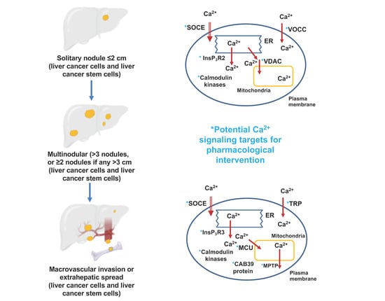
Figure 1. Schematic depiction of the arrangement of hepatocytes and other cell types in a liver lobule and the context of a liver lobule within the whole liver. Blood in the portal artery and portal vein is shown in red and blue, respectively and bile fluid and the bile collecting duct in green. The arrangements of hepatocytes, hepatic stellate cells and liver endothelial cells (LEC) is shown schematically. Taken from Ben-Moshe et al. 2013 [4], with permission.
Cancers 12 02755 g001
Figure 2. Ca2+-signaling pathways and hepatocellular carcinoma (HCC) cell proliferation; (A) Schematic representation of pathways involved in Ca2+-signaling in HCC cells leading to enhanced cell proliferation; (B) schematic representation of the roles of increased [Ca2+]cyt in activation of the PI3kinase/Akt/mTORC and ERK pathways in mediating enhanced cell proliferation.
Cancers 12 02755 g002
Figure 3. Ca2+-signaling pathways and HCC cell apoptosis. (A) Schematic representation of pathways involved in Ca2+-signaling in HCC cells leading to enhanced cell apoptosis. On one hand, a sustained increase in [Ca2+]cyt (e.g., from activation of Ca2+ entry) ultimately induces opening of the mitochondrial permeability pore and activation of the apoptotic pathway, while on the other hand sustained decrease in [Ca2+]cyt (e.g., from inhibition of Ca2+ entry) can lead to ER stress-induced apoptosis; (B) scheme showing in more detail the roles of [Ca2+]cyt, [Ca2+]MT, mitochondrial and ER Ca2+ transporters and channels and caspase enzymes in the apoptotic pathway activated by a sustained increase in [Ca2+]cyt. VDAC—voltage-dependent anion channel; MPTP—mitochondrial permeability transition pore.
Cancers 12 02755 g003
Figure 4. Schematic representation of the stages in the development of HCC. The scheme shows tumor burden, liver function and treatment options for each stage. Adapted from Villanueva 2019 [3].
Cancers 12 02755 g004
Figure 5. Gross anatomy and histology of HCC liver tumors. (A) Removal of an HCC tumor by liver resection. (Photo courteously provided by Dr. R. Padbury, S.A. Liver Transplant Unit, Flinders Medical Center, South Australia.); (B) representative micrographs (three separate images) of immunohistochemical analysis (400×) of the expression of STIM1 (identified by red labelling) in HCC tumor tissue and in adjacent non-tumor tissue. Cells enriched in STIM1 protein can be seen both in the tumor and in the invading edge of the tumor; (C) integrated optical density (IOD) of STIM1 against immunoglobulin G (IgG) in the tumor and in the invading edge. PVTT, portal vein tumor thrombus. Images in B and graph in C taken from Zhao et al. [61], with permission under Creative Commons License Deed CC BV 4.0), * p < 0.05 and ** p < 0.01.
Cancers 12 02755 g005
Figure 6. Schematic representation of current ideas on the origin, fate and roles in normal liver and in development of hepatocellular carcinoma and intrahepatic cholangiocarcinoma of liver stem cells/progenitor cells and liver cancer stem cells [7,8,9,10].
Cancers 12 02755 g006
Figure 7. Examples of a self-renewal property and “spontaneous” [Ca2+]cyt oscillations in liver cancer stem cells identified as such by expression of the α2δ1 protein (subunit of voltage-operated Ca2+ channels). (A,B) Representative phase-contrast micrographs showing spheroids formed by 1B50-1 positive cells, but not by 1B50-1 negative cells. The α2δ1 protein (antigen) is detected by the 1B50-1 antibody in a subset of liver stem cells (1B50-1 positive). (C,D) representative plots of [Ca2+]cyt (fluorescence of Ca2+ sensor) as a function of time for Hep-11 HCC cells (1B50-1 negative) and Hep-12 cells (IB50-1 positive), showing spontaneous oscillations in Ca2+ in IB50-1 positive cells (expressing the α2δ1 protein and thought to be liver cancer stem cells). Taken from Zhao et al. 2013 [13], with permission.
Cancers 12 02755 g007
Figure 8. Schematic representation of the proposed mechanism by which the hypoxic environment of HCC tumors is thought to lead to the activation of store-operated Ca2+ entry and subsequent increase in expression of STIM1, exacerbation of store-operated Ca2+ entry and enhanced tumor growth. It is proposed that hypoxia causes an initial increase in store-operated Ca2+ entry which then leads to an increase in [Ca2+]cyt, activation of CamKII, stabilization of transcription factor hypoxia-induced factor-1 (HIF1), binding of HIF-1 to p300 and the HRE response element of the STIM1 promoter, increased expression of STIM1 and enhanced activation of store-operated Ca2+ entry (green arrows). Adapted from Li et al. 2015 [95].
Cancers 12 02755 g008
Figure 9. Scheme showing the Ca2+-signaling pathways and proteins proposed to be involved in the mechanism by which increased expression and activity of the mitochondrial Ca2+ uniporter and mitochondrial uniporter regulator protein 1 lead to increased HCC cell proliferation, decreased apoptosis and increased potential for migration, invasion and metastasis [34,81,105].
Cancers 12 02755 g009
Table 1. Ca2+-signaling proteins so far identified as potential targets for the prevention or treatment of hepatocellular carcinoma.
Stage of Hepatocellular CarcinomaTargeted Ca2+ Signaling ProcessSpecific Protein TargetedProposed Pharmacological InterventionReferences
Liver cancer stem cells: initiation, promotion and resistance to systemic drugsCa2+ entryL- and N-type voltage operated Ca2+ channelsInhibition[13]
T-type voltage operated Ca2+ channelsActivation[14]
TRPV2 channelsActivation[15]
Ca2+ release from the endoplasmic reticulumInsP3R2Inhibition[16]
Ca2+-binding proteinsCaMKIIγActivation[15]
Hepatitis B and C infection: initiation and promotionCa2+ uptake by mitochondriaCa2+ transfer from endoplasmic reticulum to mitochondria (voltage-dependent anion channel, VDAC)Inhibition[17,18]
Ca2+ entryStore-operated Ca2+ entryInhibition[19]
Non-alcoholic fatty liver disease: initiation and promotionCa2+ entryOrai1Activation[5,20]
STIM1
Ca2+ entryVoltage-operated Ca2+ channels Inhibition
(verapamil)
[21]
Ca2+ uptake by the endoplasmic reticulumSERCA2bActivation[22,23]
Ca2+-binding proteinsCaMKIIInhibition[24]
CaMKK2Inhibition[25]
Progression, migration and metastasisCa2+ entryOrai1Inhibition[26,27]
STIM1
TRPC6Inhibition[28,29]
TRPV4Inhibition[30]
TRPV1Activation[31,32]
Ca2+ release from the endoplasmic reticulumInsP3R3Inhibition[33]
Uptake and release of Ca2+ from mitochondriaMitochondrial uniporter (MCU) and MCU regulator protein 1 (Ca2+ uptake to mitochondria)Inhibition[34]
Mitochondrial permeability transition pore (Ca2+ release from mitochondria)Activation[35]
Mitofusin-2Activation[36]
Progression, migration and metastasisCa2+-binding proteinsCaMKIIγ Inhibition
(tetrandrine, berbamine)
[15,37]
CaMKIVActivation[38]
Ca2+-binding protein 39Inhibition[39]
Increased [Ca2+]cyt (tuftelin1)Inhibition[39]
Table 2. Some models of HCC employed in studies of Ca2+-signaling targets in HCC. Liver cells in culture, liver cell lines derived from human hepatocellular carcinoma tumors and mouse liver tumor models.
ModelAttributes of the Model a
Isolated hepatocytes either freshly isolated or in cultureHepatocytes isolated from human non-diseased liver tissue, subsequently grown in culture for periods of about 1 h to 5 days.
Immortalized human liver cell lines derived from non-diseased human liverL01 and L02 (HL-7702) liver cells. Immortalized cells originally obtained from normal fetal or adult human liver.
Human HCC cells lines derived from human HCC tissueCommonly used: HepG2 cells and Huh-7 cells
Examples of other HCC cell lines include: MHCC97H, SK-Hep-1, SNU398, PLC/PRF/5, SMMC-7721.
Liver cancer stem cellsHep-12 cells which exhibit liver cancer stem cell marker proteins and Hep-11 control cells which do not exhibit liver cancer stem cell marker proteins;
Subsets of HCC cells (often HepG2 cells and Huh-7 cells) which exhibit liver cancer stem cell marker proteins.
Mouse liver HCC model in which HCC tumors are induced by a chemical mutagenDiethylnitrosamine (DEN)-induced liver tumors
Subcutaneous mouse xenograft modelsHCC cells (immortalized cell line or cells isolated from human liver HCC tissue) implanted subcutaneously into immunodeficient mice: Nude mice, severe combined immunodeficient (SCID) mice, and non-obese diabetic-severe combined immunodeficiency disease (NOD/SCID) mice
a Summaries of some human HCC cell lines and mouse models of HCC are provided by He et al. 2015 and Caruso et al. 2019 [70,71]. References to some specific human HCC cell lines are also given in the text.
Table 3. Mutations detected in Ca2+-signaling genes in human HCC tissue.
Ca2+-Signaling PathwayGeneProteinReferences
Ca2+ entry channels in plasma membraneCACNA1BVoltage-dependent N-type Ca2+ channel subunit α-1B[12]
CACNA1EVoltage-dependent R-type Ca2+ channel subunit α-1E
CACNA1HVoltage-dependent T-type Ca2+ channel subunit α-1 H[11]
CACNA1IVoltage-dependent T-type Ca2+ channel subunit α-1I[12]
CACNA1AVoltage-dependent P/Q-type Ca2+ channel subunit α-1A
CACNA1CVoltage-dependent L-type Ca2+ channel subunit α-1C
CACNA1DVoltage-dependent L-type Ca2+ channel subunit α-1D[11]
CACNA1GVoltage-dependent T-type Ca2+ channel subunit α-1G[11,12,74]
CACNA1SVoltage-dependent L-type Ca2+ channel subunit α-1S
ORAI1Calcium release-activated Ca2+ channel protein 1
Ca2+ transporters and exchange proteins in plasma membraneSLC8A1Na+ -Ca2+ exchanger 1[11,12,74]
SLC8A2Na+ -Ca2+ exchanger 2
ATP2B1Plasma membrane Ca2+-transporting ATPase 1 (PMCA1)
ATP2B2Plasma membrane Ca2+-transporting ATPase 2 (PMCA2)
ATP2B3Plasma membrane Ca2+-transporting ATPase 3 (PMCA3)
ATP2B4Plasma membrane Ca2+-transporting ATPase 4 (PMCA4)
Ca2+ channels and transporters in endoplasmic reticulumITPR1Inositol 1,4,5-trisphosphate receptor type 1 (IP3R 1)[12]
ITPR2Inositol 1,4,5-trisphosphate receptor type 2 (IP3R 2)[11,12]
ITPR3Inositol 1,4,5-trisphosphate receptor type 3 (IP3R 3)[33]
STIM1Stromal interaction molecule 1[11,12,33]
STIM2Stromal interaction molecule 2
RYR1Ryanodine receptor 1
RYR2Ryanodine receptor 2[11]
RYR3Ryanodine receptor 3[12]
ATP2A1Sarcoplasmic/endoplasmic reticulum Ca2+ATPase 1 (SERCA1)
ATP2A2Sarcoplasmic/endoplasmic reticulum Ca2+ATPase 2 (SERCA2)
ATP2A3Sarcoplasmic/endoplasmic reticulum Ca2+ ATPase 3 (SERCA3)
Ca2+-binding proteinsCALML3Calmodulin-like protein 3 (CaM-like protein)[12]
PTK2BCa2+-dependent tyrosine kinase 2β[12]
S100A8, S100A9, S100A11, S100PS100 Ca2+-binding protein A8, A9, A11 and P[75]
ITPKBInositol-trisphosphate 3-kinase B[12]
PDF1ACa2+/calmodulin-dependent 3’,5’-cyclic nucleotide phosphodiesterase 1A
PDE1CCa2+/calmodulin-dependent 3’,5’-cyclic nucleotide phosphodiesterase 1C
PDE1BCa2+/calmodulin-dependent 3’,5’-cyclic nucleotide phosphodiesterase 1B
PPP3CBCalmodulin-dependent calcineurin A subunitβ isoform
PPP3CCCalmodulin-dependent calcineurin A subunit γ isoform
Table 4. Ca2+-signaling genes under or overexpressed in human HCC tissue.
Ca2+ Signaling PathwayGeneProteinChange in Protein ExpressionReferences
Ca2+ channels and transporters in plasma membraneORAI1Orai 1Increased[27]
CACNA1H Voltage-operated Ca2+ channel subunit α-1 HIncreased[77]
TRPC6 Transient receptor potential cation channel subfamily C member 6Increased[28]
TRPM2Transient receptor potential cation channel subfamily M member 2Increased[78]
TRPV2Transient receptor potential cation channel subfamily V member 2Increased[79]
TRPV4Transient receptor potential cation channel subfamily V member 4Decreased[78]
Ca2+ channels and transporters in endoplasmic reticulumSTIM1 Stromal interaction molecule 1 Increased[80]
SERCA2Sarco/endoplasmic reticulum (Ca2+, Mg2+) ATP-aseDecreased (in non-alcoholic steatohepatitis-induced HCC)[23]
Ca2+ channels and transporters in mitochondriaMCUR1Mitochondrial Ca2+ uniporter regulator 1Increased[81]
Ca2+-binding proteinsHRCHistidine-rich Ca2+-binding proteinIncreased[82]
NCS1Neuronal Ca2+ sensor 1Increased[83]
CAB39Ca2+-binding protein 39Increased[39]
Table 5. Potential targets involving Ca2+-signaling pathways for therapeutic intervention directed to prevention of development of hepatocellular carcinoma in non-alcoholic fatty liver patients a.
Proposed Ca2+ Transporter, Channel or Ca2+-Binding ProteinProposed Intervention StrategyReferences
SERCA2bActivation using small molecule activator such as the allosteric activator CDN1163[22]
Activation by modification of ER membrane fluidity affected by altering thioesterase superfamily member 2/phosphatidyl transfer protein[90]
Increased expression induced by maresin 1 leading to increased AMPK activity[91]
Activation by modulation of the SERCA2b regulator protein Cisd2[23]
Ca2+ entryActivation of store-operated Ca2+ entry by small molecule activator of Orai1 or STIM1.[20]
Inhibition of PKC leading to de-phosphorylation and activation of Orai1[92]
Inhibition of Ca2+ entry using Ca2+-channel blockers verapamil and nifedipine[21,93]
Ryanodine receptors (RYR1 and RYR2)Activation using small molecules such as caffeine and caffeine analogs[11]
InsP3RInhibition using small molecule inhibitors such as heparin and caffeine[11,94]
CaMKIIInhibition by natural product tetrandrine of phosphorylation of CaMKII[37]
CaMKK2Inhibition using small molecule inhibitor such as STO-609[25]
a Taken from Ali et al. 2019 [5], with permission.

