Article

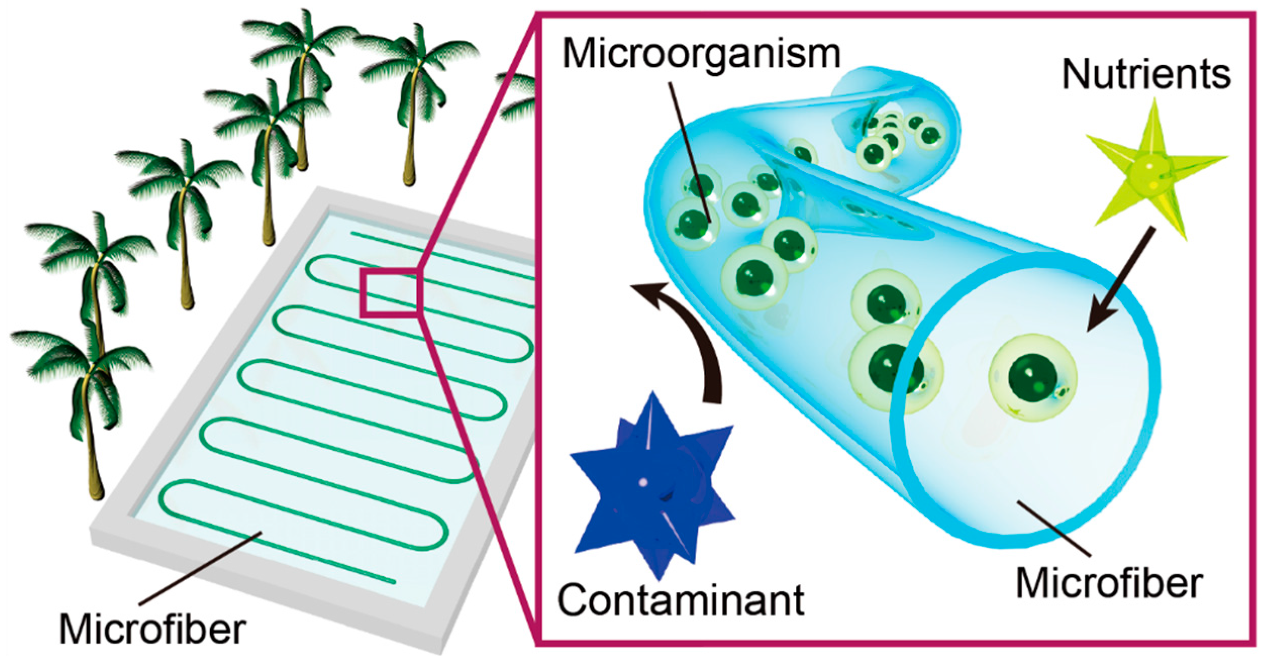
Hollow Hydrogel Microfiber Encapsulating Microorganisms for Mass-Cultivation in Open Systems

1
School of Integrated Design Engineering, Keio University, 3-14-1 Hiyoshi, Kohoku-ku, Yokohama, Kanagawa 223-8522, Japan
2
Department of Mechanical Engineering, Keio University, 3-14-1 Hiyoshi, Kohoku-ku, Yokohama, Kanagawa 223-8522, Japan
*
Author to whom correspondence should be addressed.
Micromachines 2017, 8(6), 176; https://doi.org/10.3390/mi8060176
Submission received: 25 April 2017 / Revised: 23 May 2017 / Accepted: 1 June 2017 / Published: 3 June 2017
(This article belongs to the Special Issue Biomedical Microfluidic Devices)

Abstract

:
Open cultivation systems to monoculture microorganisms are promising for the commercialization of low-value commodities because they reduce the cultivation cost. However, contamination from biological pollutants frequently impedes the process. Here we propose a cultivation method using hollow hydrogel microfibers encapsulating microorganisms. Due to the pore size, hydrogels allow nutrients and waste to pass through while preventing invading microorganisms from entering the microfiber. Experimental cultivation shows the growth of target bacteria inside the alginate hydrogel microfiber during exposure to invading bacteria. The membrane thickness of the microfiber greatly affects the bacterial growth due to changes in membrane permeability. The enhancement of mechanical toughness is also demonstrated by employing a double-network hydrogel for long-term cultivation. The hollow hydrogel microfiber has the potential to become a mainstream solution for mass-cultivation of microorganisms in an open system.

1. Introduction

Microorganisms, which utilize complex metabolic reactions made increasingly sophisticated by natural selection, have been powerful and useful chemical plants throughout human history. Due to the fast growth rate of microorganisms and the availability of inexpensive culture medium, their diverse metabolites have been used in various industries [1], including agriculture, food, wastewater treatment, pharmaceuticals, and biofuel. With the emergence of recombinant DNA technology, even eukaryotic biomolecules for therapeutics (e.g., insulin [2], human growth hormone [3]) and structural materials (e.g., spider silk [4], resilin [5]) have been produced in a microbial body.
To maximize the cost-effectiveness of white biotechnology, the methodology of microbial cultivation is critically important. Regarding mass-cultivation of microorganisms on an industrial scale, the cultivation systems are generally classified as closed or open [6]. The advantages of the closed cultivation system include good control of the cultivation condition and a reduced risk of contamination. However, the complex and expensive facility of the system increases the cost of the final product. On the other hand, an open cultivation system is cost-effective because it has fewer equipment requirements and can potentially be scaled up. Especially for the production of low-value commodities such as biofuels by photosynthetic microorganisms, cost is the most important dominant factor [7]. Therefore, an open cultivation system is the only choice for the cultivation of photosynthetic microorganisms [8].
For open cultivation systems, contamination from invading heterotrophs, pathogens, and grazers, as well as dust and sand, is the main restriction for commercial viability since the contaminants greatly reduce the growth rate of the target microorganisms [9]. Thus, many researchers have proposed open cultivation methods to overcome the problem of animate and inanimate contamination. One approach is to use chemical additives such as quinine sulfate [10,11], rotenone [12], and ammonium hydroxide [13]. Yet this method decreases the cell growth rate. Another approach is to use selective environments, such as high salinity or alkalinity [14]. However, this approach constrains the usable species (e.g., Dunaliella salina or Spirulina). The other solution to avoid the risk of contamination is to control the process for the desired culture condition, such as a temporary reduction in pH, thereby enabling a fast growth rate of the target microbes [15]. Although this strategy is applicable to various species, the additional costs necessary to install and operate a system for controlling the environment are prohibitive.
We previously introduced a concept for a new cultivation technique using hollow hydrogel microfibers to cultivate microorganisms in open cultivation systems [16]. In the present study, we conduct detailed analyses of the method, including enhancement of the mechanical toughness utilizing a double-network hydrogel. Figure 1 shows a conceptual schematic of the method; hollow hydrogel microfibers encapsulating the target microorganisms, which are produced using a three-dimensional co-axial microfluidic device, are immersed into open environments such as lakes and ponds. The pore size of the hydrogel fiber membrane allows nutrients, gases, and waste to pass through while preventing invading heterotrophs and viruses from entering the fiber [17]. This simple approach significantly reduces costs by using alginate, which is an earth-abundant biopolymer, and employing a microfluidic fabrication process, which is easy for scale-up and automation. In this study, we demonstrate cultivation of Corynebacterium glutamicum as the target microbe encapsulated in the microfiber for the proof of concept. First, the hollow hydrogel microfiber is formed by using a co-flowing microfluidic device. Second, growth rates of C. glutamicum with and without the hydrogel fiber are compared, employing Vibrio alginolyticus as an invading heterotroph. Third, the effect of the fiber wall thickness on the microbial growth rate is evaluated for characterization of the system. Finally, the applicability of the double-network hydrogel to increase the durability of the microfiber is assessed.

2. Experimental Section

2.1. Materials and Reagents

C. glutamicum (NBRC 12168) and V. alginolyticus (NBRC 15630) were used as the target bacterium and invading bacterium, respectively. The chemicals for cultivating the bacteria, including MgSO4·7H2O, ethanol, polypepton, yeast extract, and Daigo’s Artificial Seawater SP for Marine Microalgae Medium, were purchased from Wako Pure Chemical Industries, Japan. Deionized (DI) water was prepared using a Millipore system (Direct-Q3, Millipore, Bellerica, MA, USA).
For the materials to form the hollow hydrogel microfibers, sodium alginate (80–120 cP), and calcium chloride were purchased from Wako Pure Chemical Industries, Japan. For the double-network hydrogel, 30% w/v-Acrylamide/Bis Mixed Solution (29:1), ammonium peroxodisulfate (APS), and N,N,N′,N′-Tetramethylethylenediamine (TEMED) were purchased from Nacalai Tesque, Inc., Kyoto, Japan.
Blue fluorescent microspheres (F8814, 1.0 μm) and green fluorescent microspheres (G0100, 1.0 μm) for fluorescent imaging of the produced hollow microfibers were purchased from Thermo Fisher Scientific Inc., Waltham , MA, USA.

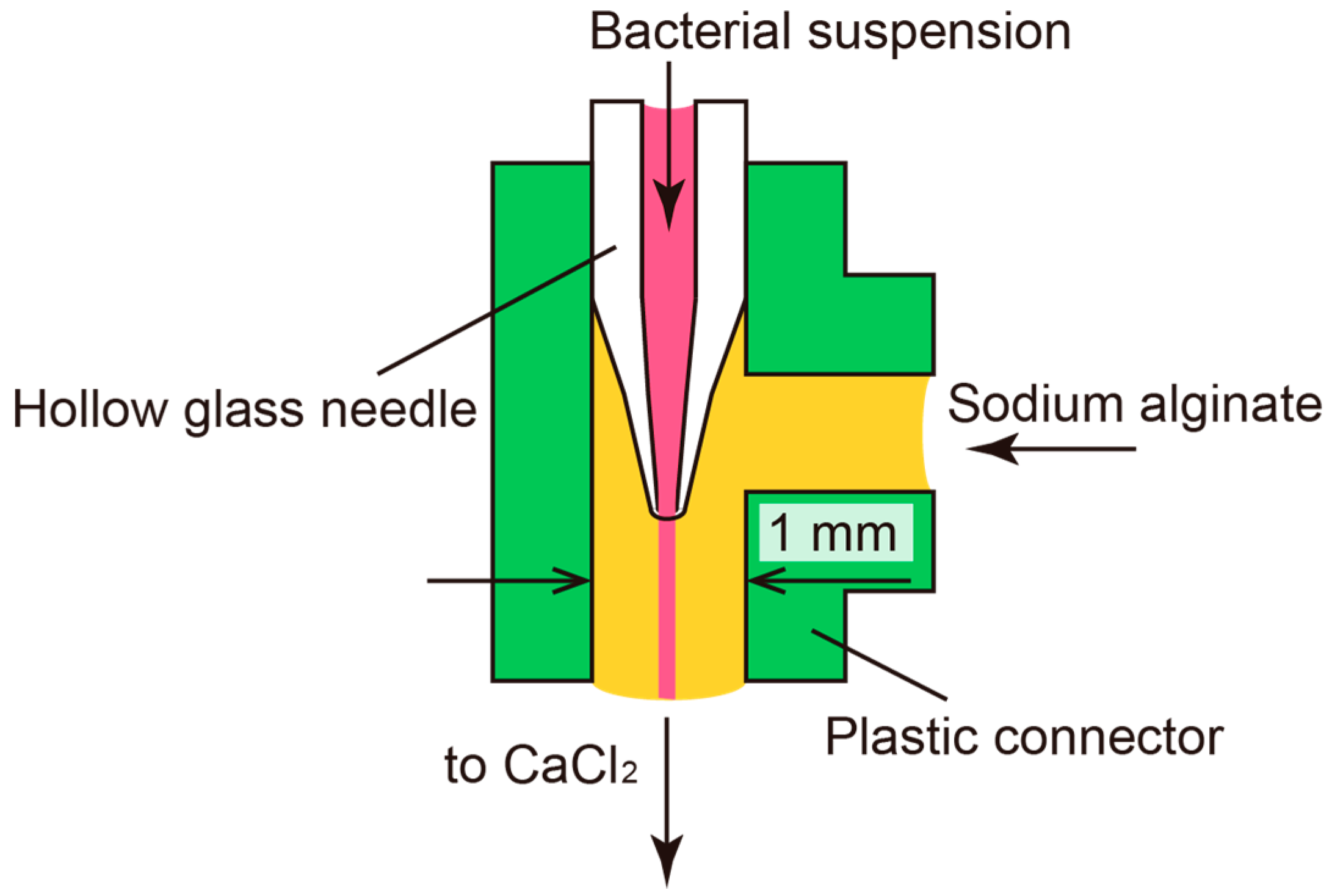
2.2. Microfluidic Formation of Hollow Hydrogel Microfiber

Figure 2 shows the co-flowing microfluidic device comprising a plastic connector (VTF 106, AS ONE, Osaka, Japan) with an inner diameter of 1.3 mm and a hollow glass needle. The design of the device is similar to designs reported elsewhere [18,19,20]. The glass needle was formed by pulling a hollow glass tube (Hirschmann Laboratory, Eberstadt, Germany) with an inner diameter of 0.47 mm and an outer diameter of 1.2 mm to have a sharp edge at the tip using a Puller PC-10 (Narishige Group, Tokyo, Japan). A coaxial laminar flow was formed by introducing a bacterial suspension from the inlet of the glass needle and sodium alginate solution (1.5% w/v) from the other inlet. The sodium alginate solidifies at the outlet when it encounters Ca2+ ions in a 150 mM calcium chloride solution. Thus, a hollow hydrogel microfiber encapsulating bacteria was obtained. The diameter of the microfiber can be controlled by changing the flow rates of the inner and outer flow. Syringe pumps (AS ONE) were used in the present study to control the flow rates.

2.3. Culture of Bacteria in Hollow Microfiber

As the proof-of-concept experiment, C. glutamicum was used as the target bacterium. This bacterium has an elliptical shape and produces lactic acid. First, we measured the bacterial growth rates with and without the microfiber. The microfiber containing C. glutamicum was immersed into the culture medium, and the cell density was measured every 3 h. The cell density in the microfiber was measured by visually counting the number of C. glutamicum extracted from the fiber by pipetting. For the control condition, C. glutamicum was cultivated in bulk culture medium composed of 10 g of polypepton, 2 g of yeast extract, 1 g of MgSO4·7H2O, and 1 L of DI water.
Next, we tested whether the microfiber can prevent other bacteria from entering the microfiber. As a control experiment, we cultured C. glutamicum in bulk culture medium containing V. alginolyticus. Then, we cultivated C. glutamicum in the microfiber in culture medium containing V. alginolyticus. The cell densities of the bacteria for both conditions were measured for 1 h. V. alginolyticus is motile, making it easy to distinguish both bacteria. Additionally, the effect of the membrane thickness of the microfiber on the cell growth rate was investigated by measuring the concentration of lactic acid, which is a by-product of C. glutamicum, inside the microfiber by pipetting. The measurement was conducted 2 h after cultivation in 20 mL of culture medium using an F-kit (l-lactic acid, Roche Diagnostics, Basel, Switzerland) and an ultraviolet-visible-near-infrared spectrophotometer (UV-Vis-NIR; U-3310, Hitachi, Tokyo, Japan).

2.4. Double-Network Hollow Hydrogel Microfiber

A double-network hydrogel is a material composed of two different polymer chains—one with rigid chains, and the other with flexible chains—that exhibits extremely high mechanical toughness and stretchability [21]. When the gel is stretched, the rigid polymer chain ruptures and dissipates energy. Since the alginate hydrogel is brittle and biodegradable, a tougher material could be suitable for long-term cultivation. We assessed the applicability of using a double-network hydrogel composed of alginate and polyacrylamide [22] in our system.
To prepare the double-network hydrogel microfiber, a bacterial suspension of C. glutamicum was introduced into the glass needle, and sodium alginate solution mixed with acrylamide/bis solution and APS was introduced into the other inlet channel. The coaxial flow was extruded into the mixed solution of calcium chloride and TEMED, resulting in formation of the hollow microfiber of the double-network hydrogel. Double-network hydrogel microfibers with six different mix ratios were prepared. For all samples, concentrations of APS and TEMED were fixed at 5.1 mM and 8.6 mM, respectively.
To assess its mechanical properties, the microfiber was stretched using a tensile machine (RSAIII, TA Instruments, New Castle, DE, USA). The outer diameter, membrane thickness, and length of the fiber were fixed at 1.5 mm, 100 μm, and 10 mm, respectively. The rate of stretch was kept constant at 0.1 mm/s.
To investigate the effect of polyacrylamide in the double-network hydrogel on the metabolism of C. glutamicum, the double-network hydrogel microfiber was immersed in the 20 mL culture medium for 3 h. After the cultivation, the amount of lactic acid was measured using an F-kit and UV-Vis-NIR spectrophotometer. All the membrane thicknesses of the fiber were 100 μm.

3. Results and Discussion

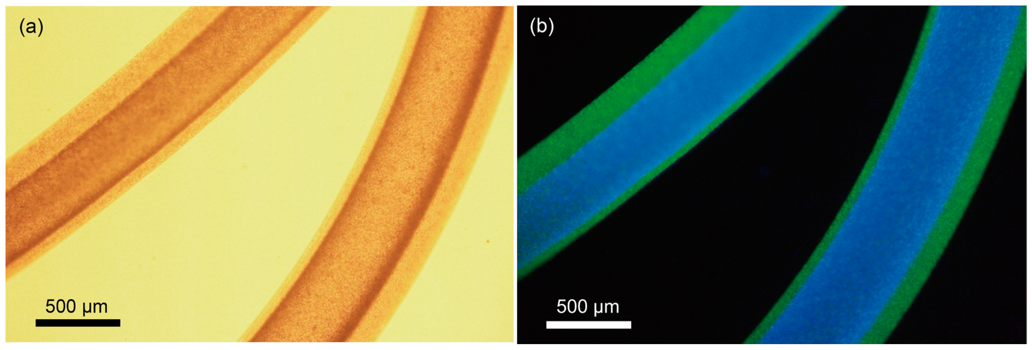
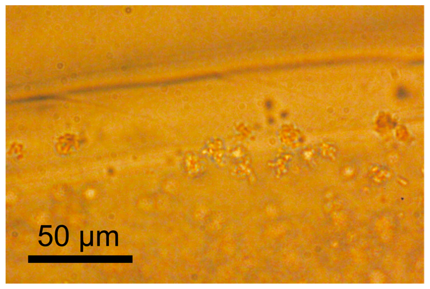
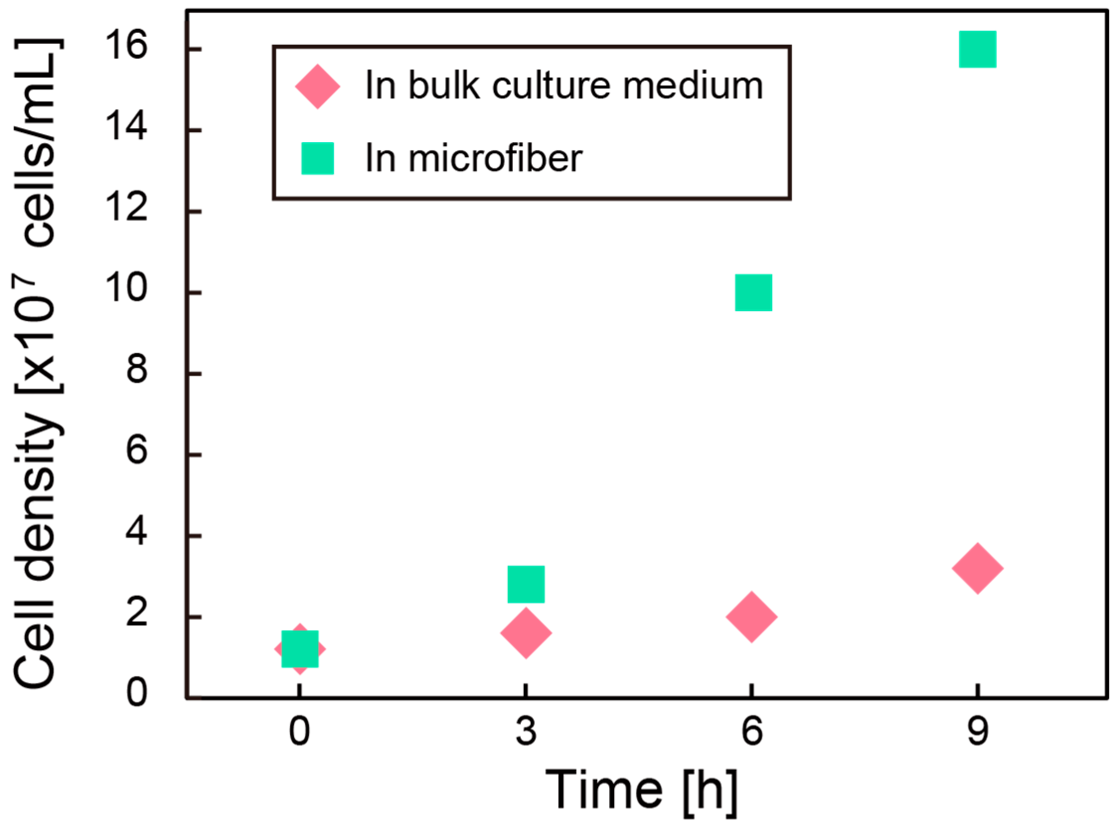
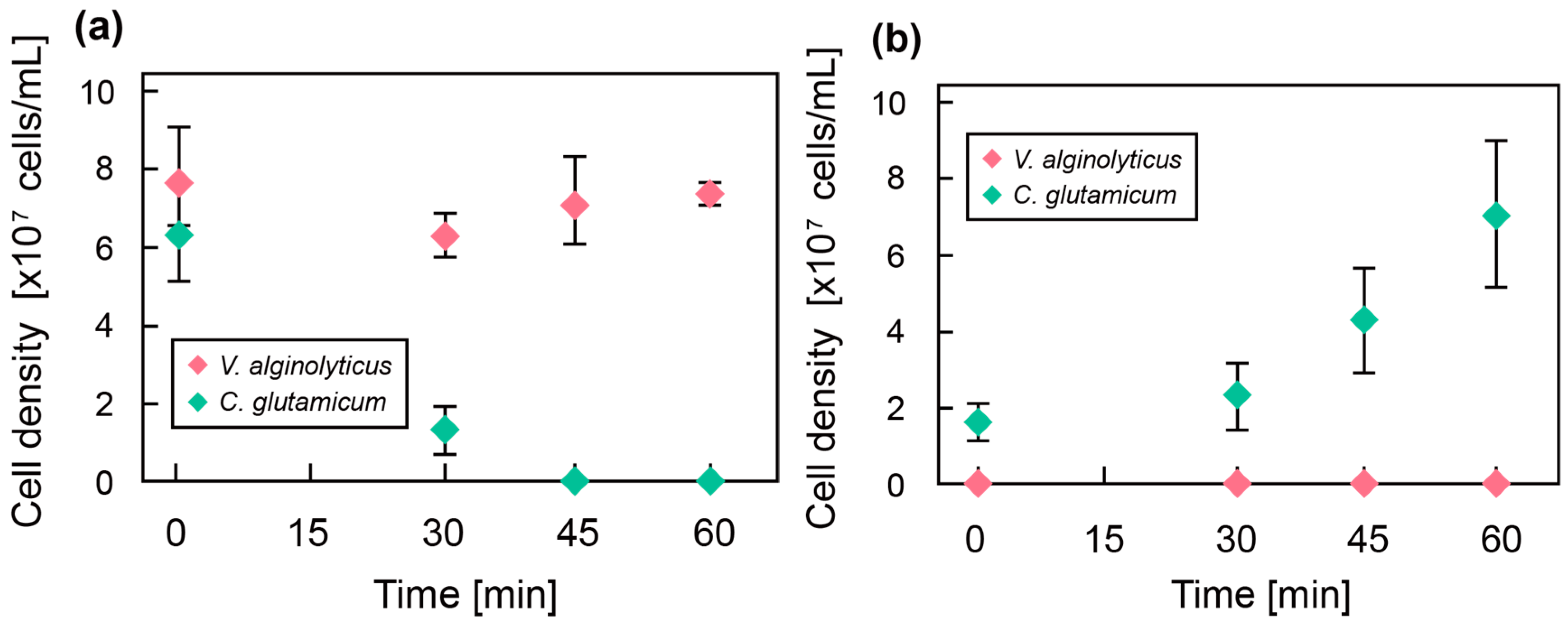
3.1. Culture of Bacteria in Hollow Microfiber

Figure 3 shows a micrograph of the hydrogel microfiber colored with fluorescent particles. This is a merged image of blue particles for the core region and green particles for the shell region, indicating the formation of the hollow hydrogel microfiber. The hollow microfiber encapsulating C. glutamicum was successfully formed, as shown in Figure 4. Figure 5 shows the cell density after cultivation in the bulk culture medium and the microfiber. The growth rate of the bacteria in the microfiber was faster than that without the microfiber. This is probably because the bacteria inside the microfiber were surrounded by sufficient bulk culture, enabling rapid exchange of oxygen, nutrients, and waste through the microfiber. In cases when the target microbes, such as Aurantiochytrium, internally store their by-products, it is necessary to extract the bacteria from the fiber. After cultivation for 9 h, the microfiber was dissolved with NaHCO3. Fluorescent observation using a green fluorescent nucleic acid stain (SYTO9) revealed that more than 80% of the bacteria remained alive.
Next, we co-cultured C. glutamicum and V. alginolyticus with and without the microfiber. Figure 6 shows the cell densities of the bacteria (a) in a bulk culture medium and (b) inside the microfiber during cultivation for 1 h. Without the microfiber, all C. glutamicum were dead in 45 min. On the other hand, C. glutamicum in the hydrogel microfiber grew exponentially while the cell density of V. alginolyticus remained at zero, indicating that there was no invasion of V. alginolyticus into the microfiber. Based on these results, the microfiber could successfully protect the target bacteria from invading bacteria.
The effect of membrane thickness of the microfiber on the bacterial growth rate was also evaluated (Figure 7). By controlling the flow rate, the desired membrane thickness can be achieved. However, it was found that the microfiber did not form when the ratio of the volumetric flow rate of the sodium alginate solution to the bacterial suspension exceeded 50%. Therefore, microfibers with different membrane thicknesses were prepared at ratios of the volumetric flow rate less than 50%. As shown in Figure 7, lactic acid production sharply decreased when the membrane thickness was greater than 120 μm. This is probably because the nutrient supply rate by diffusion through the hydrogel membrane decreased with the thicker membrane, thereby restricting bacterial growth. Therefore, it is preferable that the membrane thickness of the microfiber is less than 120 μm.

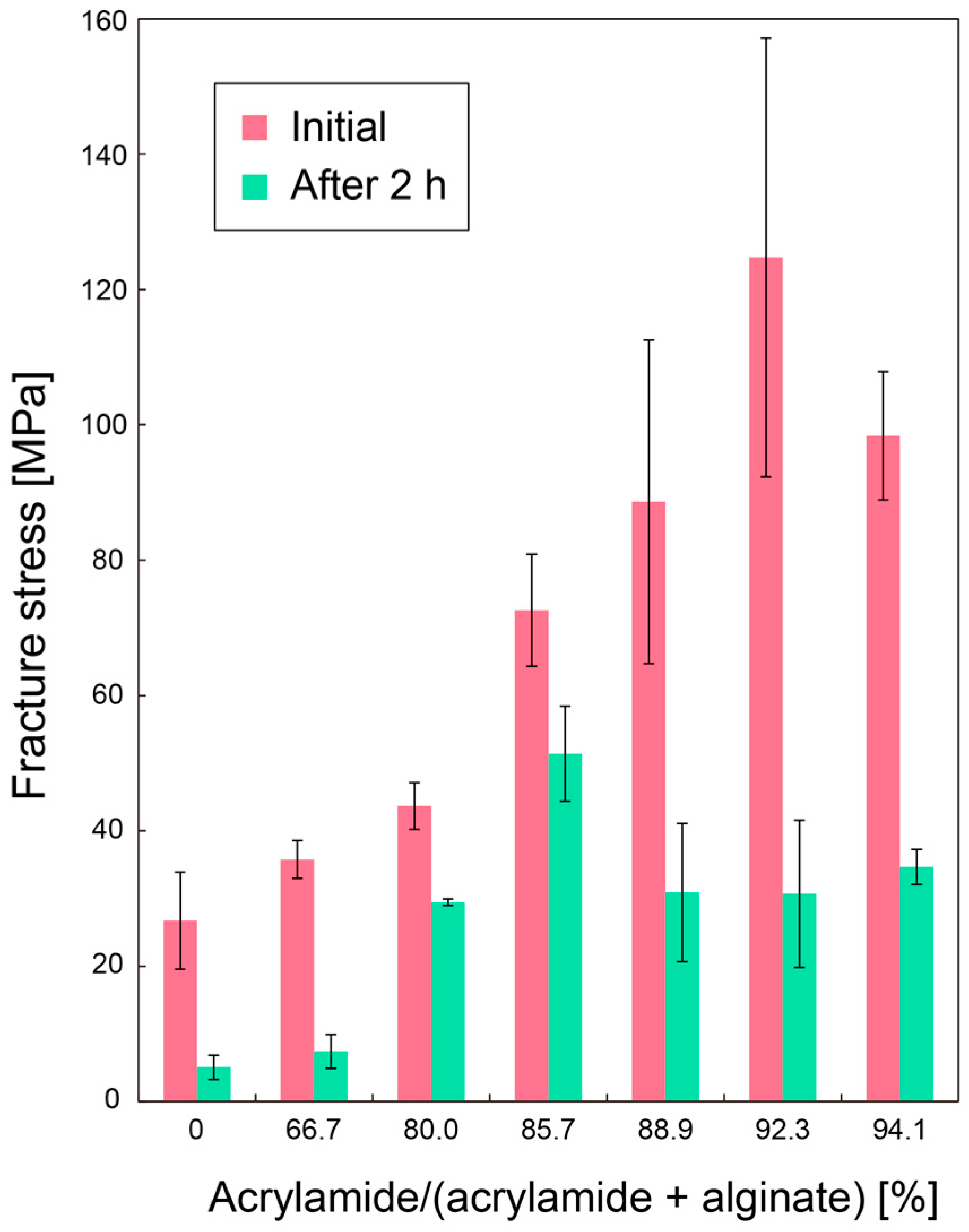
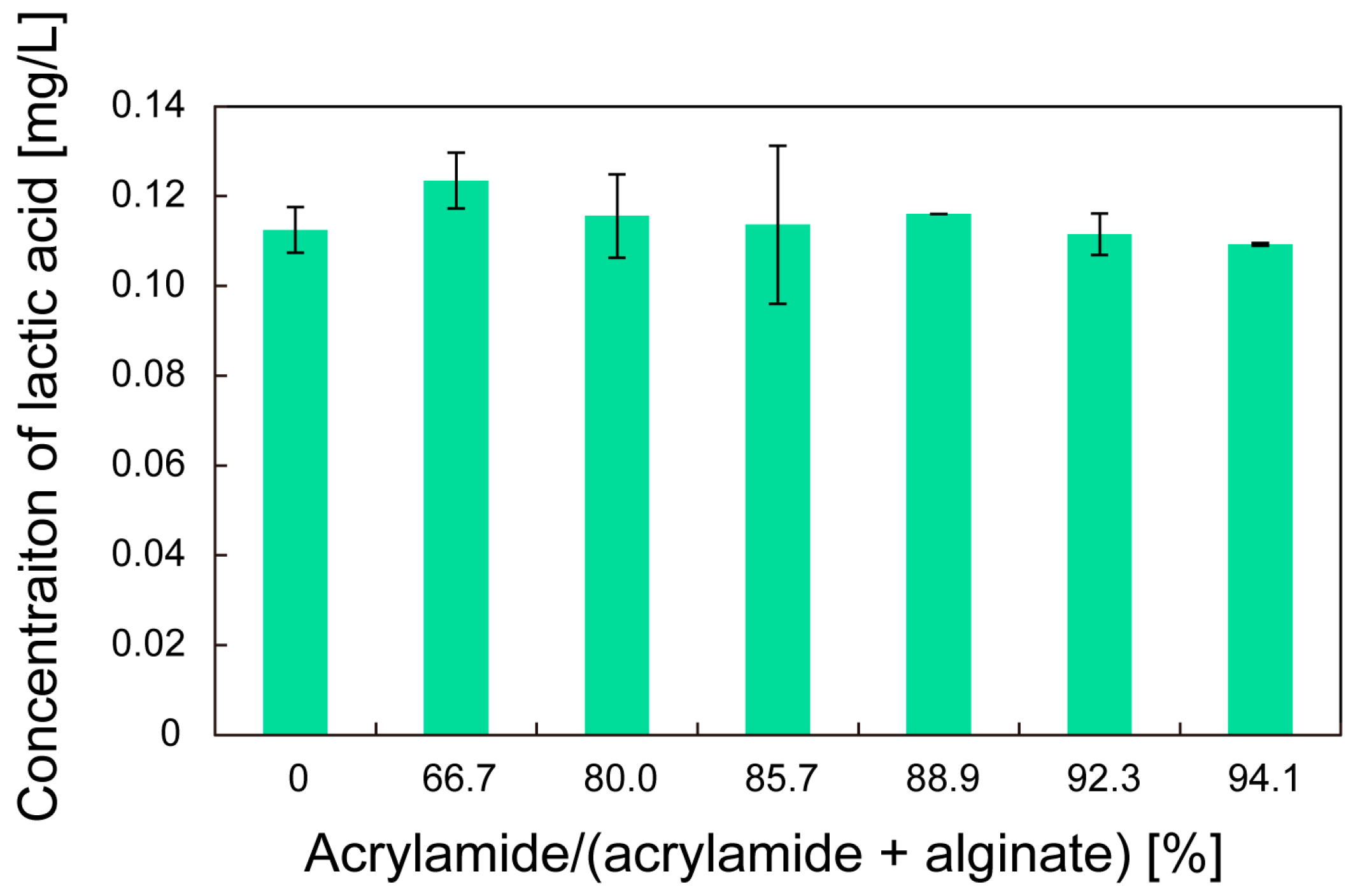
3.2. Double-Network Hollow Hydrogel Microfiber

Figure 8 shows the fracture stress of the microfiber due to elongation as a function of the composition ratio of the double-network hydrogel microfiber. The mechanical strength of the fiber was enhanced when the ratio for the initial condition was approximately 92.3. This result is consistent with the result of previous work [22]. However, all the fracture stresses were decreased after cultivation of C. glutamicum for 2 h. This is probably because the Ca2+ ions crosslinking alginate polymer chains were metabolized by the C. glutamicum, thereby reverting the alginate gel to the sol state. Nevertheless, the microfibers with composition ratios of 80–94.1% exhibited increased fracture stresses compared with the microfibers composed only of alginate after cultivation due to the remaining polyacrylamide. Therefore, the double-network hydrogel could be suitable for our system, especially for long-term cultivation.
Finally, the effect of the composition ratio on the growth rate of the bacteria was investigated. Figure 9 shows the concentration of lactic acid produced after cultivation for 3 h. For all the composition ratios, statistical analysis using analysis of variance did not show any significant differences. This indicates that the double-network hydrogels with different composition ratios had similar diffusion coefficients. Therefore, the double-network hydrogel has no effect on the cultivation of the bacteria.

4. Conclusions

We demonstrated a hollow hydrogel microfiber containing microorganisms for mass-cultivation in an open system. Microfibers with specific diameters and membrane thicknesses can be precisely formed using a simple microfluidic device. The target bacteria are cultivated in the microfiber while the invading bacteria are kept outside the microfiber. The membrane thickness of the microfiber greatly affects the growth rate of the bacteria; with the growth rate sharply decreasing at thicknesses exceeding 120 μm. However, a membrane that is too thin would reduce the mechanical toughness of the microfiber. The applicability of a double-network hydrogel to enhance the mechanical properties of the microfiber for our system was also assessed. The double-network hydrogel significantly increases the mechanical toughness of the microfiber without inhibiting bacterial growth. The hollow hydrogel microfiber has the potential to become a mainstream solution for mass-cultivation of microorganisms in an open cultivation system.

Acknowledgments

This work was supported by Japan Society for the Promotion of Science.

Author Contributions

K.H., H.O. and N.M. conceived the design of the design of the study. K.H., M.O. and K.F. fabricated microfibers. K.H. and M.O. designed all the experiments. M.O. performed the cultivation of C. glutamicum in bulk culture medium and the microfiber. M.O. performed the cultivation of C. glutamicum and V. alginolyticus. M.O. examined the effect of membrane thickness on the bacterial activity. M.O. and K.F. conducted the experiments of double-network hydrogel. H.O. provided considerable advice on the fabrication of hollow microfibers. K. H. wrote the paper. All authors discussed the results and commented on the manuscript.

Conflicts of Interest

The authors declare no conflict of interest.

References

  1. Najafpour, G. Biochemical Engineering and Biotechnology; Elsevier Science: Amsterdam, The Netherlands, 2006. [Google Scholar]
  2. Goeddel, D.V.; Kleid, D.G.; Bolivar, F.; Heyneker, H.L.; Yansura, D.G.; Crea, R.; Hirose, T.; Kraszewski, A.; Itakura, K.; Riggs, A.D. Expression in Escherichia-coli of chemically synthesized genes for human insulin. Proc. Natl. Acad. Sci. USA 1979, 76, 106–110. [Google Scholar] [CrossRef] [PubMed]
  3. Goeddel, D.V.; Heyneker, H.L.; Hozumi, T.; Arentzen, R.; Itakura, K.; Yansura, D.G.; Ross, M.J.; Miozzari, G.; Crea, R.; Seeburg, P.H. Direct expression in Escherichia-coli of a DNA-sequence coding for human growth-hormone. Nature 1979, 281, 544–548. [Google Scholar] [CrossRef] [PubMed]
  4. Lewis, R.V.; Hinman, M.; Kothakota, S.; Fournier, M.J. Expression and purification of a spider silk protein: A new strategy for producing repetitive proteins. Protein Expr. Purif. 1996, 7, 400–406. [Google Scholar] [CrossRef] [PubMed]
  5. Elvin, C.M.; Carr, A.G.; Huson, M.G.; Maxwell, J.M.; Pearson, R.D.; Vuocolo, T.; Liyou, N.E.; Wong, D.C.C.; Merritt, D.J.; Dixon, N.E. Synthesis and properties of crosslinked recombinant pro-resilin. Nature 2005, 437, 999–1002. [Google Scholar] [CrossRef] [PubMed]
  6. Mata, T.M.; Martins, A.A.; Caetano, N.S. Microalgae for biodiesel production and other applications: A review. Renew. Sustain. Energy Rev. 2010, 14, 217–232. [Google Scholar] [CrossRef]
  7. Lundquist, T.J.; Woertz, I.C.; Quinn, N.; Benemann, J.R. A realistic technology and engineering assessment of algae biofuel production. Energy Biosci. Inst. 2010, 1–178. [Google Scholar]
  8. Luque, R. Algal biofuels: The eternal promise? Energy Environ. Sci. 2010, 3, 254–257. [Google Scholar] [CrossRef]
  9. Wang, H.; Zhang, W.; Chen, L.; Wang, J.F.; Liu, T.Z. The contamination and control of biological pollutants in mass cultivation of microalgae. Bioresour. Technol. 2013, 128, 745–750. [Google Scholar] [CrossRef] [PubMed]
  10. Moreno-Garrido, I.; Canavate, J.P. Assessing chemical compounds for controlling predator ciliates in outdoor mass cultures of the green algae Dunaliella salina. Aquac. Eng. 2001, 24, 107–114. [Google Scholar] [CrossRef]
  11. Xu, C.Y.; Wu, K.Y.; Van Ginkel, S.W.; Igou, T.; Lee, H.J.; Bhargava, A.; Johnston, R.; Snell, T.; Chen, Y.S. The use of the schizonticidal agent quinine sulfate to prevent pond crashes for algal-biofuel production. Int. J. Mol. Sci. 2015, 16, 27450–27456. [Google Scholar] [CrossRef] [PubMed]
  12. Van Ginkel, S.W.; Bidwell, M.; Igou, T.; Gijon-Felix, R.; Salvi, E.; De Oliveira, S.H.R.; Duarte, L.H.K.; Steiner, D.; Hu, Z.X.; Johnston, R.; et al. The prevention of saltwater algal pond contamination using the electron transport chain disruptor, rotenone. Algal Res. 2016, 18, 209–212. [Google Scholar] [CrossRef]
  13. Lincoln, E.P.; Hall, T.W.; Koopman, B. Zooplankton control in mass algal cultures. Aquaculture 1983, 32, 331–337. [Google Scholar] [CrossRef]
  14. Brennan, L.; Owende, P. Biofuels from microalgae-a review of technologies for production, processing, and extractions of biofuels and co-products. Renew. Sustain. Energy Rev. 2010, 14, 557–577. [Google Scholar] [CrossRef]
  15. Huntley, M.E.; Redalje, D.G. CO2 mitigation and renewable oil from photosynthetic microbes: A new appraisal. Mitig. Adapt. Strateg. Glob. Chang. 2007, 12, 573–608. [Google Scholar] [CrossRef]
  16. Ogawa, M.; Higashi, K.; Miki, N. Development of hydrogel microtubes for microbe culture in open environment. In Proceedings of the 2015 37th Annual International Conference of the IEEE Engineering in Medicine and Biology Society, New York, NY, USA, 25–29 August 2015; pp. 5896–5899. [Google Scholar]
  17. Li, R.H.; Altreuter, D.H.; Gentile, F.T. Transport characterization of hydrogel matrices for cell encapsulation. Biotechnol. Bioeng. 1996, 50, 365–373. [Google Scholar] [CrossRef]
  18. Jeong, W.; Kim, J.; Kim, S.; Lee, S.; Mensing, G.; Beebe, D.J. Hydrodynamic microfabrication via “on the fly” photopolymerization of microscale fibers and tubes. Lab Chip 2004, 4, 576–580. [Google Scholar] [CrossRef] [PubMed]
  19. Shin, S.; Park, J.Y.; Lee, J.Y.; Park, H.; Park, Y.D.; Lee, K.B.; Whang, C.M.; Lee, S.H. “On the fly” continuous generation of alginate fibers using a microfluidic device. Langmuir 2007, 23, 9104–9108. [Google Scholar] [CrossRef] [PubMed]
  20. Lee, K.H.; Shin, S.J.; Park, Y.; Lee, S.H. Synthesis of cell-laden alginate hollow fibers using microfluidic chips and microvascularized tissue-engineering applications. Small 2009, 5, 1264–1268. [Google Scholar] [CrossRef] [PubMed]
  21. Gong, J.P.; Katsuyama, Y.; Kurokawa, T.; Osada, Y. Double-network hydrogels with extremely high mechanical strength. Adv. Mater. 2003, 15, 1155–1158. [Google Scholar] [CrossRef]
  22. Sun, J.Y.; Zhao, X.H.; Illeperuma, W.R.K.; Chaudhuri, O.; Oh, K.H.; Mooney, D.J.; Vlassak, J.J.; Suo, Z.G. Highly stretchable and tough hydrogels. Nature 2012, 489, 133–136. [Google Scholar] [CrossRef] [PubMed]
Figure 1. Conceptual schematic of the hollow hydrogel microfiber containing microorganisms in an open cultivation system. The pore size of the hydrogel allows nutrients to pass through the fiber but prevents contaminants from entering the fiber.
Micromachines 08 00176 g001
Figure 2. Formation of the microfiber using a co-flowing microfluidic device. The bacterial suspension is introduced from a hollow glass needle with an inner diameter of 100 μm at the tip. Sodium alginate solution is introduced from the other inlet. The produced coaxial laminar flow is extruded into a calcium chloride solution, which results in the formation of a hollow microfiber made of calcium alginate hydrogel.
Micromachines 08 00176 g002
Figure 3. Micrographs of the hollow hydrogel microfiber. (a) Optical micrograph. (b) Fluorescent micrograph corresponding to (a). The fiber is colored with blue and green fluorescent particles for the core and the shell region, respectively.
Micromachines 08 00176 g003
Figure 4. Micrograph showing C. glutamicum in the microfiber.
Micromachines 08 00176 g004
Figure 5. Cell density of C. glutamicum as a function of culture time. The measurements were conducted every 3 h for both conditions.
Micromachines 08 00176 g005
Figure 6. (a) Cell density of C. glutamicum and V. alginolyticus without the microfibers as a function of culture time. (b) Cell density of C. glutamicum in the microfiber exposed to a suspension containing V. alginolyticus as a function of culture time.
Micromachines 08 00176 g006
Figure 7. Concentration of the lactic acid produced by C. glutamicum as a function of the membrane thickness of the microfiber.
Micromachines 08 00176 g007
Figure 8. Fracture stress of the microfiber due to elongation as a function of the composition ratio of acrylamide and alginate.
Micromachines 08 00176 g008
Figure 9. Concentration of the lactic acid produced by C. glutamicum as a function of the composition ratio of acrylamide and alginate after cultivation for 3 h.
Micromachines 08 00176 g009

