
Photodetection Properties of CdS/Si Heterojunction Prepared by Pulsed Laser Ablation in DMSO Solution for Optoelectronic Application
 , , , , and
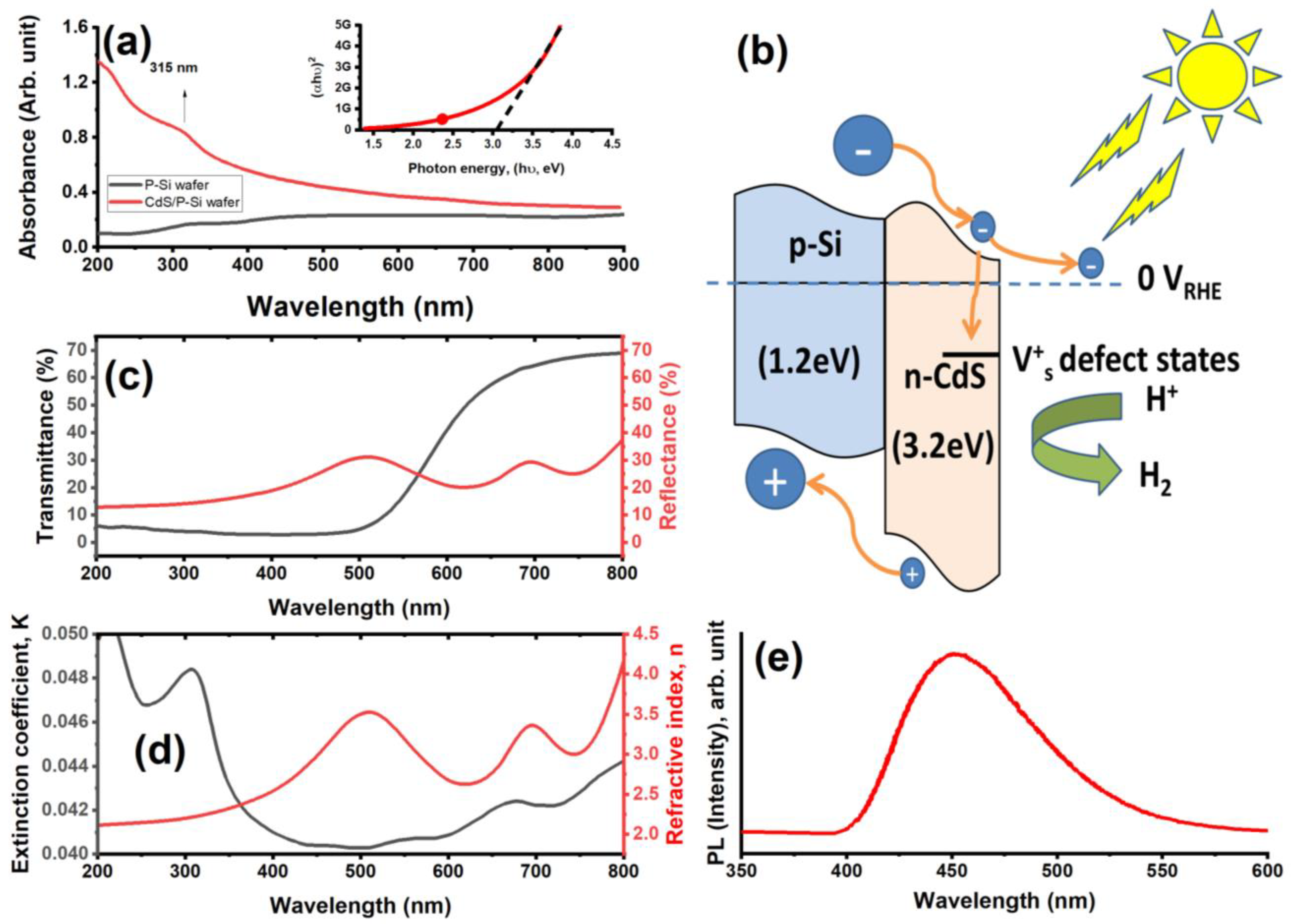
Abstract
:1. Introduction
2. Experimental Work
2.1. Materials
2.2. Preparation Method of CdS NPs
2.3. Preparation Method of CdS/Si Heterojunctions
2.4. Capacitance–Voltage Measurements
2.5. Characterization Techniques
3. Result and Discussion
3.1. Physicochemical Investigation
3.2. Optoelectronic Study
4. Conclusions
Author Contributions
Funding
Data Availability Statement
Acknowledgments
Conflicts of Interest
References
- Yao, J.; Zheng, Z.; Yang, G. Production of large-area 2D materials for high-performance photodetectors by pulsed-laser deposition. Prog. Mater. Sci. 2019, 106, 100573. [Google Scholar] [CrossRef]
- Cai, Y.; Zhang, K.; Feng, Q.; Zuo, Y.; Hu, Z.; Feng, Z.; Zhou, H.; Lu, X.; Zhang, C.; Tang, W.; et al. Tin-assisted growth of ε-Ga 2 O 3 film and the fabrication of photodetectors on sapphire substrate by PLD. Opt. Mater. Express 2018, 8, 3506–3517. [Google Scholar] [CrossRef]
- Zheng, Z.; Yao, J.; Yang, G. Growth of centimeter-scale high-quality In2Se3 films for transparent, flexible and high performance photodetectors. J. Mater. Chem. C 2016, 4, 8094–8103. [Google Scholar] [CrossRef]
- Li, C.; Fan, X.; Yu, L.; Cui, L.; Yin, M.; Li, Y.; Nan, N.; Liu, N. A resistive-type UV detector based on ZnO nanowalls decoated by Ag nanowires. Opt. Mater. 2020, 103, 109891. [Google Scholar] [CrossRef]
- Pedarnig, J.D.; Heitz, J.; Stehrer, T.; Praher, B.; Viskup, R.; Siraj, K.; Moser, A.; Vlad, A.; Bodea, M.A.; Bäuerle, D.; et al. Characterization of nano-composite oxide ceramics and monitoring of oxide thin film growth by laser-induced breakdown spectroscopy. Spectrochim. Acta Part B At. Spectrosc. 2008, 63, 1117–1121. [Google Scholar] [CrossRef]
- Detalle, V.; Héon, R.; Sabsabi, M.; St-Onge, L. An evaluation of a commercial Echelle spectrometer with intensified charge-coupled device detector for materials analysis by laser-induced plasma spectroscopy. Spectrochim. Acta Part B At. Spectrosc. 2001, 56, 1011–1025. [Google Scholar] [CrossRef]
- Elsalam, S.S.A.; Taha, R.H.; Tawfeik, A.M.; El-Monem, M.O.A.; Mahmoud, H.A. Antimicrobial Activity of Bio and Chemical Synthesized Cadmium Sulfide Nanoparticles. Egypt. J. Hosp. Med. 2018, 70, 1494–1507. [Google Scholar] [CrossRef] [Green Version]
- Darwish, A.M.; Eisa, W.H.; Shabaka, A.A.; Talaat, M.H. Investigation of factors affecting the synthesis of nano-cadmium sulfide by pulsed laser ablation in liquid environment. Spectrochim Acta A 2016, 153, 315–320. [Google Scholar] [CrossRef]
- Darwish, A.M.; Eisa, W.H.; Shabaka, A.A.; Talaat, M.H. Synthesis of nano-cadmium sulfide by pulsed laser ablation in liquid environment. Spectrosc. Lett. 2015, 48, 638–645. [Google Scholar] [CrossRef]
- Menazea, A. Pulsed laser ablation route assisted copper oxide nanoparticles doped in Polyethylene 325 Oxide/Polyvinyl pyrrolidone blend for enhancement the electrical conductivity. J. Mol. Struct. 2020, 1207, 127807. [Google Scholar] [CrossRef]
- Menazea, A. Femtosecond laser ablation-assisted synthesis of silver nanoparticles in organic and inorganic liquids medium and their antibacterial efficiency. Radiat. Phys. Chem. 2020, 168, 108616. [Google Scholar] [CrossRef]
- Altowyan, A.S.; Mostafa, A.M.; Ahmed, H.A. Effect of liquid media and laser energy on the preparation of Ag nanoparticles and their nanocomposites with Au nanoparticles via laser ablation for optoelectronic applications. Optik 2021, 241, 167217. [Google Scholar] [CrossRef]
- Shankar, P.; Ishak, M.H.; Padarti, J.K.; Mintcheva, N.; Iwamori, S.; Gurbatov, S.O.; Lee, J.H.; Kulinich, S.A. ZnO@ graphene oxide core@ shell nanoparticles prepared via one-pot approach based on laser ablation in water. Appl. Surf. Sci. 2020, 531, 147365. [Google Scholar] [CrossRef]
- Riahi, A.; Khamlich, S.; Balghouthi, M.; Khamliche, T.; Doyle, T.B.; Dimassi, W.; Guizani, A.; Maaza, M. Study of thermal conductivity of synthesized Al2O3-water nanofluid by pulsed laser ablation in liquid. J. Mol. Liq. 2020, 304, 112694. [Google Scholar] [CrossRef]
- Rawat, R.; Tiwari, A.; Arun, N.; Rao, S.N.; Pathak, A.; Rao, S.V.; Tripathi, A. Synthesis of CuO hollow nanoparticles using laser ablation: Effect of fluence and solvents. Appl. Phys. A 2020, 126, 226. [Google Scholar] [CrossRef]
- Mwafy, E.A.; Mostafa, A.M. Efficient removal of Cu (II) by SnO2/MWCNTs nanocomposite by pulsed laser ablation method. Nano-Struct. Nano-Objects 2020, 24, 100591. [Google Scholar] [CrossRef]
- Khokhra, R.; Bharti, B.; Lee, H.-N.; Kumar, R. Visible and UV photo-detection in ZnO nanostructured thin films via simple tuning of solution method. Sci. Rep. 2017, 7, 15032. [Google Scholar] [CrossRef] [Green Version]
- Slimen, F.B.; Zaaboub, Z.; Haouari, M.; Mohamed, N.B.H.; Ouada, H.B.; Chaussedent, S.; Gaumer, N. Effect of CdS nanocrystals on the photoluminescence of Eu 3+-doped silicophosphate sol gel glass. RSC Adv. 2017, 7, 14552–14561. [Google Scholar] [CrossRef] [Green Version]
- Salah, A.; Mansour, A.; Mohamed, M.B.; Azzouz, I.M.; Elnaby, S.; Badr, Y. Effects of nanoparticles size and concentration and laser power on nonlinear optical properties of Au and Au–CdSe nanocrystals. Appl. Surf. Sci. 2015, 353, 112–117. [Google Scholar] [CrossRef]
- Nan, F.; Liang, S.; Liu, X.-L.; Peng, X.-N.; Li, M.; Yang, Z.-J.; Zhou, L.; Hao, Z.-H.; Wang, Q.-Q. Sign-reversed and magnitude-enhanced nonlinear absorption of Au–CdS core–shell hetero-nanorods. Appl. Phys. Lett. 2013, 102, 163112. [Google Scholar] [CrossRef]
- Badawi, A.; Al-Hosiny, N.; Abdallah, S. The photovoltaic performance of CdS quantum dots sensitized solar cell using graphene/TiO2 working electrode. Superlattices Microstruct 2015, 81, 88–96. [Google Scholar] [CrossRef]
- Al-Baradi, A.M.; Altowairqi, F.A.; Atta, A.; Badawi, A.; Algarni, S.A.; Almalki, A.S.; Hassanien, A.M.; Alodhayb, A.; Kamal, A.M.; El-Nahass, M.M. Structural and optical characteristic features of RF sputtered CdS/ZnO thin films. Chin. Phys. B 2020, 29, 080702. [Google Scholar] [CrossRef]
- Li, W.; Li, M.; Xie, S.; Zhai, T.; Yu, M.; Liang, C.; Ouyang, X.; Lu, X.; Li, H.; Tong, Y. Improving the photoelectrochemical and photocatalytic performance of CdO nanorods with CdS decoration. CrystEngComm 2013, 15, 4212–4216. [Google Scholar] [CrossRef]
- Neelgund, G.M.; Oki, A.; Luo, Z. Antimicrobial activity of CdS and Ag2S quantum dots immobilized on poly (amidoamine) grafted carbon nanotubes. Colloids Surf. B Biointerfaces 2012, 100, 215–221. [Google Scholar] [CrossRef] [PubMed] [Green Version]
- Nyamen, L.D.; Pullabhotla, V.S.R.; Nejo, A.A.; Ndifon, P.; Revaprasadu, N. Heterocyclic dithiocarbamates: Precursors for shape controlled growth of CdS nanoparticles. New J. Chem. 2011, 35, 1133–1139. [Google Scholar] [CrossRef]
- Kalasad, M.; Rabinal, M.; Mulimani, B. Facile synthesis of bioconjugated fluorescent CdS nanoparticles of tunable light emission. J. Phys. D Appl. Phys. 2010, 43, 305301. [Google Scholar] [CrossRef]
- Mostafa, A.M.; Mwafy, E.A.; Hasanin, M.S. One-pot synthesis of nanostructured CdS, CuS, and SnS by pulsed laser ablation in liquid environment and their antimicrobial activity. Opt. Laser Tech. 2020, 121, 105824. [Google Scholar] [CrossRef]
- Sarkar, A.; Katiyar, A.K.; Mukherjee, S.; Singh, S.; Singh, S.K.; Das, A.K.; Ray, S.K. Geometry controlled white light emission and extraction in CdS/Black-Si conical heterojunctions. ACS Appl. Electron. Mater. 2018, 1, 25–33. [Google Scholar] [CrossRef]
- Isari, A.A.; Mehregan, M.; Mehregan, S.; Hayati, F.; Rezaei Kalantary, R.; Kakavandi, B. Sono-photocatalytic degradation of tetracycline and pharmaceutical wastewater using WO3/CNT heterojunction nanocomposite under US and visible light irradiations: A novel hybrid system. J. Hazard Mater. 2020, 390, 122050. [Google Scholar] [CrossRef]
- Hosseini, M.G.; Sefidi, P.Y.; Mert, A.M.; Kinayyigit, S. Investigation of solar-induced photoelectrochemical water splitting and photocatalytic dye removal activities of camphor sulfonic acid doped polyaniline-WO3-MWCNT ternary nanocomposite. J. Mater. Sci. Technol. 2020, 38, 7–18. [Google Scholar] [CrossRef]
- Wu, Y.; Wang, H.; Tu, W.; Liu, Y.; Tan, Y.Z.; Yuan, X.; Chew, J.W. Quasi-polymeric construction of stable perovskite-type LaFeO3/g-C3N4 heterostructured photocatalyst for improved Z-scheme photocatalytic activity via solid pn heterojunction interfacial effect. J. Hazard. Mater. 2018, 347, 412–422. [Google Scholar] [CrossRef] [PubMed]
- Zeng, C.; Huang, H.; Dong, F.; Ye, L.; Zhang, T.; Zhang, Y.; Guo, Y.; Liu, C.; Hu, Y. Dual redox couples Ag/Ag+ and I−/(IO3)− self-sacrificed transformation for realizing multiplex hierarchical architectures with universally powerful photocatalytic performance. Appl. Catal. B Environ. 2017, 200, 620–632. [Google Scholar] [CrossRef]
- Ghosh, A.; Mondal, A. Fabrication of stable, efficient and recyclable p-CuO/n-ZnO thin film heterojunction for visible light driven photocatalytic degradation of organic dyes. Mater. Lett. 2016, 164, 221–224. [Google Scholar] [CrossRef]


Disclaimer/Publisher’s Note: The statements, opinions and data contained in all publications are solely those of the individual author(s) and contributor(s) and not of MDPI and/or the editor(s). MDPI and/or the editor(s) disclaim responsibility for any injury to people or property resulting from any ideas, methods, instructions or products referred to in the content. |
© 2023 by the authors. Licensee MDPI, Basel, Switzerland. This article is an open access article distributed under the terms and conditions of the Creative Commons Attribution (CC BY) license (https://creativecommons.org/licenses/by/4.0/).
Share and Cite
Alkallas, F.H.; Alghamdi, S.M.; Al-Ahmadi, A.N.; Trabelsi, A.B.G.; Mwafy, E.A.; Elsharkawy, W.B.; Alsubhe, E.; Mostafa, A.M.; Rezk, R.A. Photodetection Properties of CdS/Si Heterojunction Prepared by Pulsed Laser Ablation in DMSO Solution for Optoelectronic Application. Micromachines 2023, 14, 1546. https://doi.org/10.3390/mi14081546
Alkallas FH, Alghamdi SM, Al-Ahmadi AN, Trabelsi ABG, Mwafy EA, Elsharkawy WB, Alsubhe E, Mostafa AM, Rezk RA. Photodetection Properties of CdS/Si Heterojunction Prepared by Pulsed Laser Ablation in DMSO Solution for Optoelectronic Application. Micromachines. 2023; 14(8):1546. https://doi.org/10.3390/mi14081546
Chicago/Turabian StyleAlkallas, Fatemah H., Shoug M. Alghamdi, Ameenah N. Al-Ahmadi, Amira Ben Gouider Trabelsi, Eman A. Mwafy, W. B. Elsharkawy, Emaan Alsubhe, Ayman M. Mostafa, and Reham A. Rezk. 2023. "Photodetection Properties of CdS/Si Heterojunction Prepared by Pulsed Laser Ablation in DMSO Solution for Optoelectronic Application" Micromachines 14, no. 8: 1546. https://doi.org/10.3390/mi14081546