


Design of Photonic Crystal Biosensors for Cancer Cell Detection
Abstract
:1. Introduction
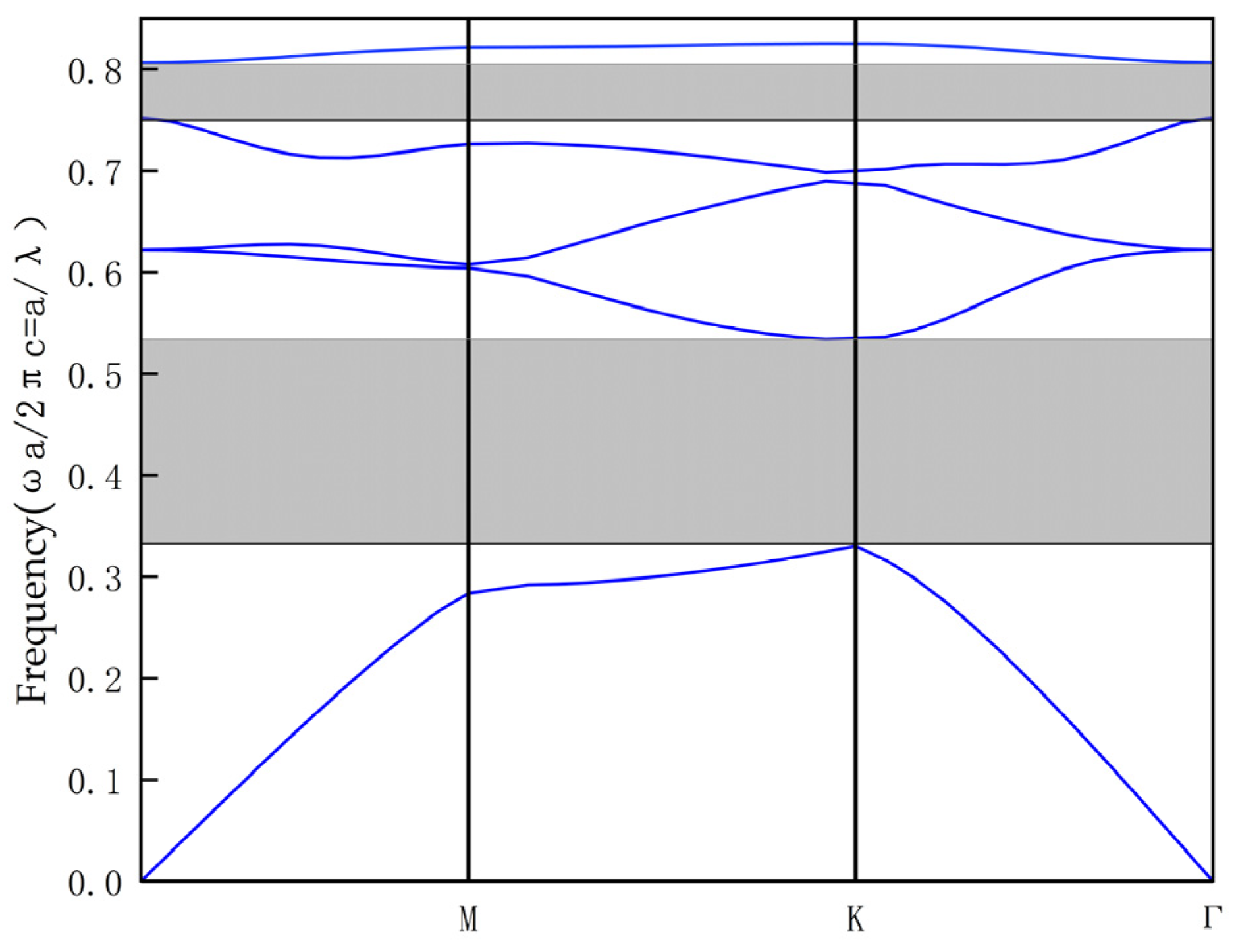
2. Sensor Performance Evaluation
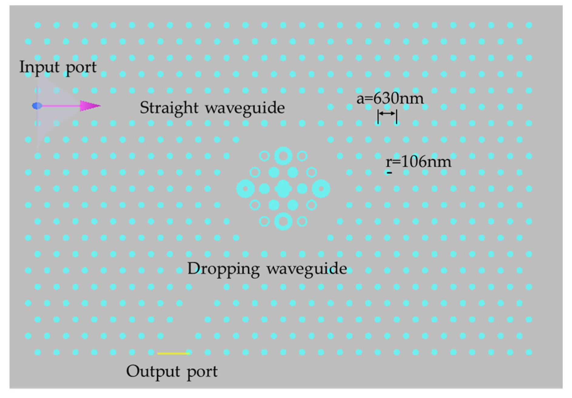
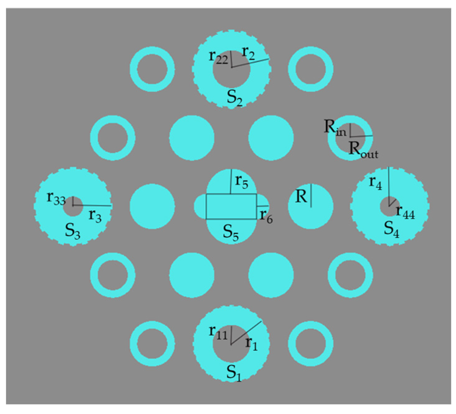
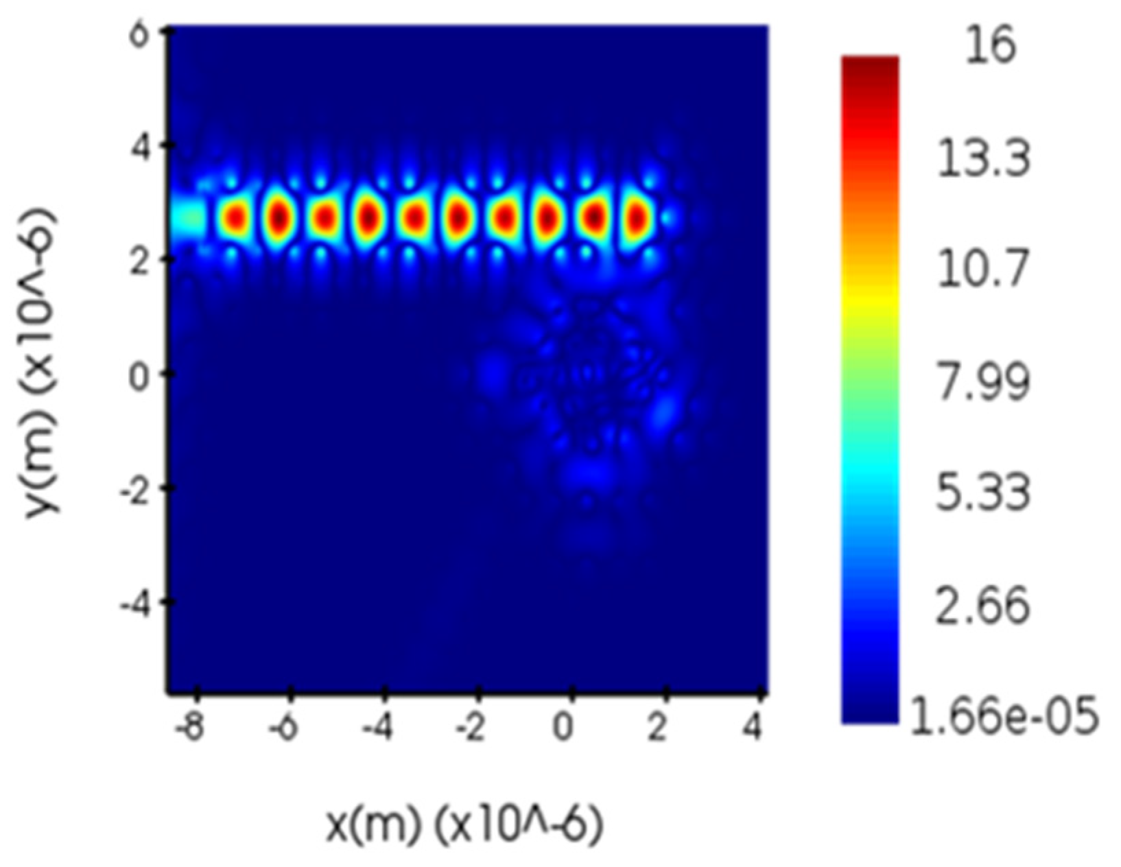
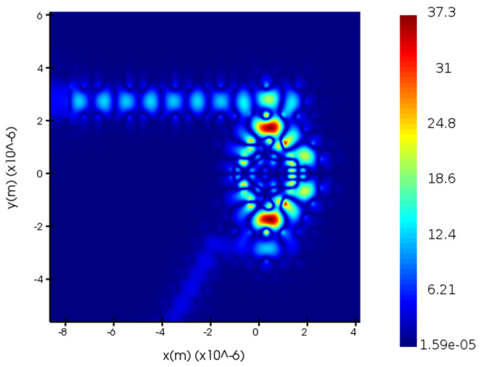
3. Design and Performance Analysis of Biosensors
4. Conclusions
Author Contributions
Funding
Data Availability Statement
Conflicts of Interest
References
- Sung, H.; Ferlay, J.; Siegel, R.L. Global cancer statistics 2020: Globocan estimates of incidence and mortality worldwide for 36 cancers in 185 countries. CA A Cancer J. Clin. 2021, 71, 209–249. [Google Scholar] [CrossRef]
- Udagawa, H.; Takahashi, S.; Hirao, M. Liposomal eribulin for advanced adenoid cystic carcinoma, gastric cancer, esophageal cancer, and small cell lung cancer. Cancer Med. 2023, 12, 1269–1278. [Google Scholar] [CrossRef]
- Nadig, V.; Herrmann, K.; Mottaghy, F.M. Hybrid total-body pet scanners—Current status and future perspectives. Eur. J. Nucl. Med. Mol. Imaging 2022, 49, 445–459. [Google Scholar] [CrossRef]
- Yan, S.M.; Nan, Y.; Li, X.T. The Association between the Differential Expression of lncRNA and Type 2 Diabetes Mellitus in People with Hypertriglyceridemia. Int. J. Mol. Sci. 2023, 24, 4279. [Google Scholar] [CrossRef]
- Yablonovitch, E. Inhibited spontaneous emission in solid-state physics and electronics. Phys. Rev. Lett. 1987, 58, 2059. [Google Scholar] [CrossRef] [Green Version]
- John, S. Strong localization of photons in certain disordered dielectric superlattices. Phys. Rev. Lett. 1987, 58, 2486. [Google Scholar] [CrossRef] [Green Version]
- Xu, X.Y.; Ren, G.H.; Feleppa, T. Self-calibrating programmable photonic integrated circuits. Nat. Photonics 2022, 16, 595–602. [Google Scholar] [CrossRef]
- Pang, Y.; Xu, Y.; Zhao, X. Stabilized narrow-linewidth Brillouin random fiber laser with a double-coupler fiber ring resonator. J. Light. Technol. 2022, 40, 2988–2995. [Google Scholar] [CrossRef]
- Yashaswini, P.R.; Gayathri, H.N.; Srikanth, P.C. Performance analysis of photonic crystal based biosensor for the detection of bio-molecules in urine and blood. Mater. Today Proc. 2023, 80, 2247–2254. [Google Scholar] [CrossRef]
- Malek, C.; Abdallah, S.A.O.; Awasthi, S.K. Biophotonic sensor for swift detection of malignant brain tissues by using nanocomposite YBa2Cu3O7/dielectric material as a 1D defective photonic crystal. Sci. Rep. 2023, 13, 8115. [Google Scholar] [CrossRef]
- Zouache, T.; Hocini, A.A. 2D photonic crystal indium arsenide based with dual micro-cavities coupled to a waveguide as a platform for a high sensitivity pressure sensor. Opt. Quantum Electron. 2023, 55, 238. [Google Scholar] [CrossRef]
- Kassa-Baghdouche, L. High-sensitivity spectroscopic gas sensor using optimized H1 photonic crystal microcavities. JOSA B 2020, 37, A277–A284. [Google Scholar] [CrossRef]
- Vijaya-Shanthi, K.; Robinson, S. Two-dimensional photonic crystal based sensor for pressure sensing. Photonic Sens. 2014, 4, 248–253. [Google Scholar] [CrossRef] [Green Version]
- Kassa-Baghdouche, L. Optical properties of a point-defect nanocavity-based elliptical-hole photonic crystal for mid-infrared liquid sensing. Phys. Scr. 2019, 95, 015502. [Google Scholar] [CrossRef]
- Kassa-Baghdouche, L.; Cassan, E. Mid-infrared gas sensor based on high-Q/V point-defect photonic crystal nanocavities. Opt. Quantum Electron. 2020, 52, 260. [Google Scholar] [CrossRef]
- Ineda, M.F.; Chan, L.L.Y.; Kuhlenschmidt, T. Rapid specific and label-free detection of porcine rotavirus using photonic crystal biosensors. IEEE Sens. J. 2009, 9, 470–477. [Google Scholar]
- Parandin, F.; Heidari, F.; Aslinezhad, M. Design of 2D photonic crystal biosensor to detect blood components. Opt. Quantum Electron. 2022, 54, 618. [Google Scholar] [CrossRef]
- Krishnamoorthi, B.; Elizabeth, C.B.; Michael, M. A novel rhombic shaped photonic crystal bio-sensor for identifying disorders in the blood samples. Opt. Quantum Electron. 2023, 55, 312. [Google Scholar] [CrossRef]
- Olyaee, S.; Selfouri, M.; Mohsenirad, H. Label-free detection of glycated haemoglobin in human blood using silicon-based photonic crystal nanocavity biosensor. J. Mod. Opt. 2016, 63, 1274–1279. [Google Scholar]
- Danie, M.; Kiani, B. Design of a label-free photonic crystal refractive index sensor for biomedical applications. Photonics Nanostruct.-Fundam. Appl. 2018, 31, 89–98. [Google Scholar] [CrossRef]
- Jindal, S.; Sobti, S.; Kumar, M. Nanocavity-coupled photonic crystal waveguide as highly sensitive platform for cancer detection. IEEE Sens. J. 2016, 16, 3705–3710. [Google Scholar] [CrossRef]
- Mohamadi, A.; Seifouri, M.; Karami, R. Proposal of a high-Q biosensor using a triangular photonic crystal filter. Opt. Quantum Electron. 2021, 53, 471. [Google Scholar] [CrossRef]
- Maache, M.; Fazea, Y.; Bilehassan, I. High-sensitivity capsule-shaped sensor based on 2D photonic crystals. Symmetry 2020, 12, 1480. [Google Scholar] [CrossRef]
- White, I.M.; Fan, X. On the performance quantification of resonant refractive index sensors. Opt. Express 2008, 16, 1020–1102. [Google Scholar] [CrossRef] [PubMed] [Green Version]
- Zhang, Y.N.; Zhao, Y.; Wu, D. Fiber Loop Ring-Down Refractive Index Sensor Based on High- Q Photonic Crystal Cavity. IEEE Sens. J. 2014, 14, 1878–1885. [Google Scholar] [CrossRef]
- Yang, D.; Tian, H.; Wu, N. Nanoscale torsion-free photonic crystal pressure sensor with ultra-high sensitivity based on side-coupled piston-type microcavity. Sens. Actuators A Phys. 2013, 199, 30–36. [Google Scholar] [CrossRef]
- Kassa-Baghdouche, L.; Boumaza, T.; Cassan, E. Enhancement of Q-factor in SiN-based planar photonic crystal L3 nanocavity for integrated photonics in the visible-wavelength range. Optik 2015, 126, 3467–3471. [Google Scholar] [CrossRef]
- Daher, M.G.; Taya, S.A.; Colak, I. Design of a nano-sensor for cancer cell detection based on a ternary photonic crystal with high sensitivity and low detection limit. Chin. J. Phys. 2022, 77, 1168–1181. [Google Scholar] [CrossRef]
- Sajan, S.C.; Singh, A.; Sharma, P.K. Silicon Photonics Biosensors for Cancer Cells Detection-A Review. IEEE Sens. J. 2023, 23, 3366–3377. [Google Scholar] [CrossRef]
- Sani, M.H.; Ghanbari, A.; Saghaei, H. High-sensitivity biosensor for simultaneous detection of cancer and diabetes using photonic crystal microstructure. Opt. Quantum Electron. 2022, 54, 1–14. [Google Scholar] [CrossRef]
- Baratye, F.; Hamedi, S. Label-Free cancer cell biosensor based on photonic crystal ring resonator. Results Phys. 2023, 46, 106317. [Google Scholar] [CrossRef]
- Ali, L.; Mohammed, M.U.; Khan, M. High-quality optical ring resonator-based biosensor for cancer detection. IEEE Sens. J. 2019, 20, 1867–1875. [Google Scholar] [CrossRef]
- Asuvaran, A.; Elathrasan, G. Design of two-dimensional photonic crystal-based biosensor for abnormal tissue analysis. Silicon 2022, 14, 7203–7210. [Google Scholar] [CrossRef]
- Kumar, R.; Bharti, G.K.; Bindal, R.K. Modeling and simulation of an optical sensor for cancer cell detection. Int. J. Electr. Electron. Res. 2022, 10, 792–795. [Google Scholar] [CrossRef]


| r1 (um) | r2 (um) | r3 (um) | r4 (um) | Resonant Wavelength (nm) | Q | Transmittance (%) |
|---|---|---|---|---|---|---|
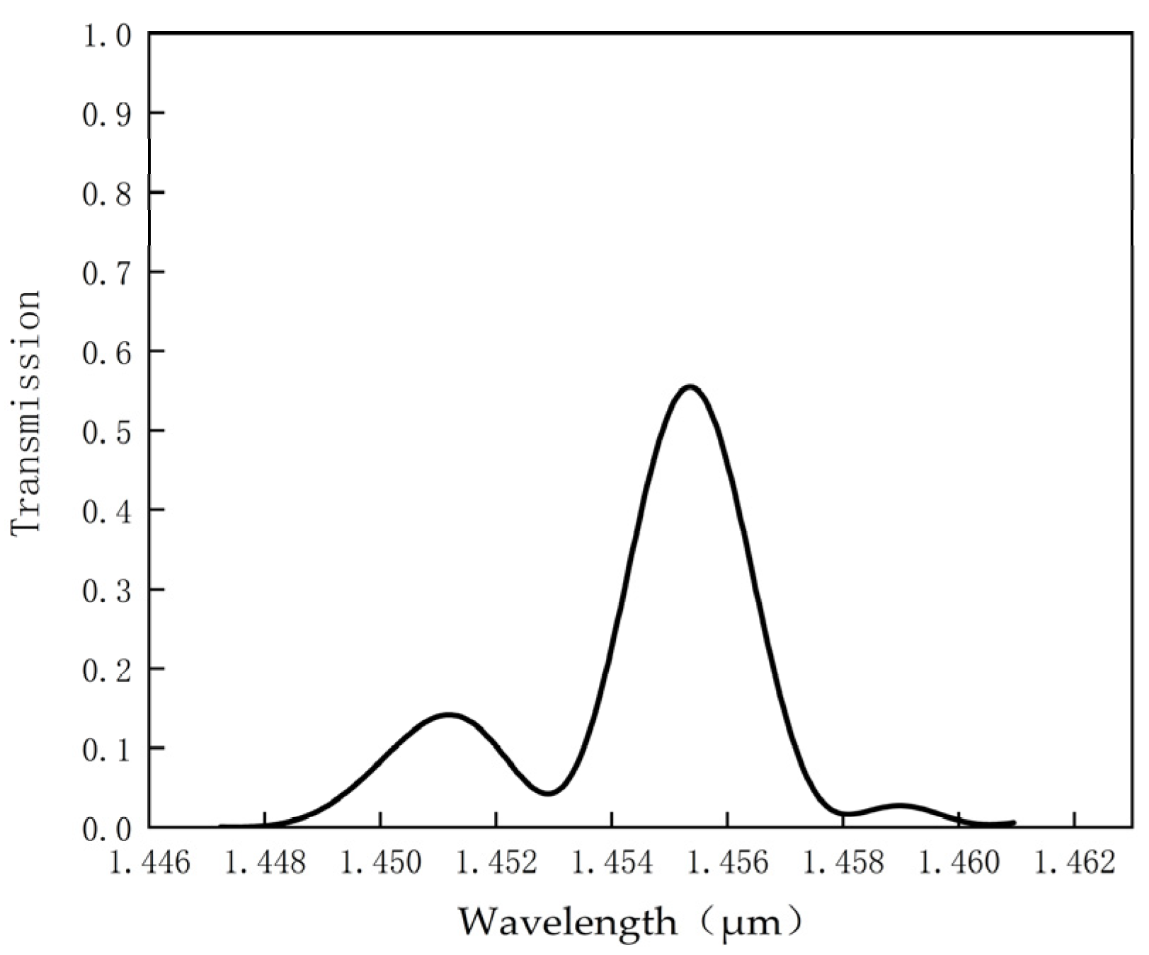
| 0.2 | 0.3 | 0.3 | 0.3 | 1502.07 | 639 | 71.2 |
| 0.25 | 0.3 | 0.3 | 0.3 | 1455.29 | 537 | 55.8 |
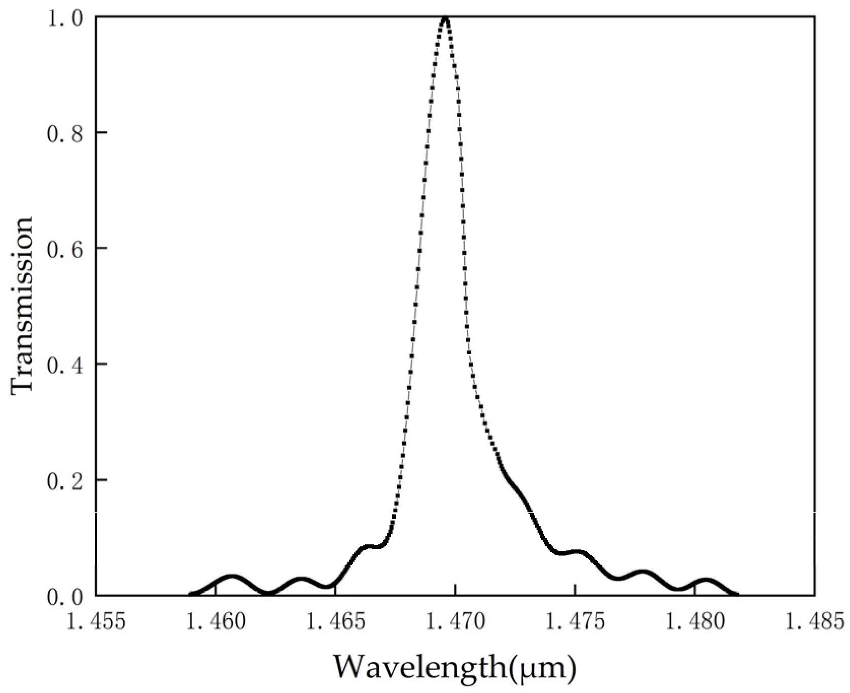
| 0.3 | 0.3 | 0.3 | 0.3 | 1470.81 | 647 | 97.6 |
| 0.35 | 0.3 | 0.3 | 0.3 | 1493.28 | 496 | 88.26 |
| 0.4 | 0.3 | 0.3 | 0.3 | 1508.44 | 603 | 69.5 |
| 0.3 | 0.2 | 0.3 | 0.3 | 1524.16 | 554 | 47.8 |
| 0.3 | 0.25 | 0.3 | 0.3 | 1459.22 | 583 | 87.8 |
| 0.3 | 0.35 | 0.3 | 0.3 | 1493.55 | 551 | 99.96 |
| 0.3 | 0.4 | 0.3 | 0.3 | 1516.45 | 417 | 61 |
| 0.3 | 0.3 | 0.2 | 0.3 | 1491.76 | 438 | 93 |
| 0.3 | 0.3 | 0.25 | 0.3 | 1478.74 | 573 | 97 |
| 0.3 | 0.3 | 0.35 | 0.3 | 1488.78 | 633 | 98.74 |
| 0.3 | 0.3 | 0.4 | 0.3 | 1470.65 | 570 | 89.2 |
| 0.3 | 0.3 | 0.3 | 0.2 | 1490.15 | 545.84 | 88 |
| 0.3 | 0.3 | 0.3 | 0.25 | 1478.47 | 555.81 | 95.36 |
| 0.3 | 0.3 | 0.3 | 0.35 | 1503.24 | 538 | 55 |
| 0.3 | 0.3 | 0.3 | 0.4 | 1469.58 | 980 | 99.62 |
| Detection Source | Refractive Index | Resonant Wavelength (nm) | Q | Wavelength Shift (nm) | S (nm/RIU) | DL (RIU) |
|---|---|---|---|---|---|---|
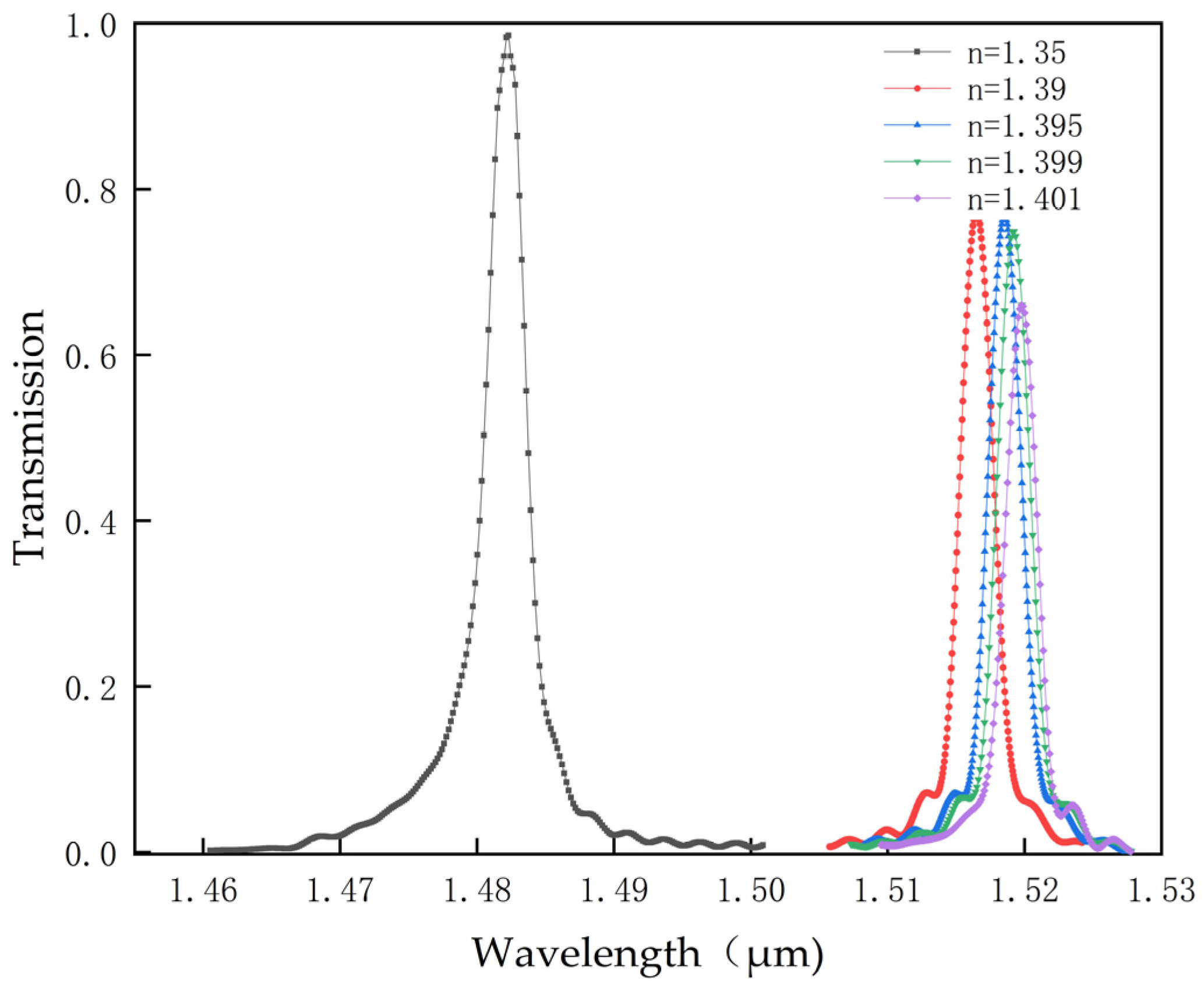
| S1 as the detection source. | 1.35 | 1481 | 510 | - | - | - |
| 1.39 | 1517.63 | 702 | 36.63 | 915.75 | 0.000236 | |
| 1.395 | 1518.95 | 534.84 | 37.95 | 843.3 | 0.000336 | |
| 1.399 | 1519.84 | 512 | 38.84 | 792 | 0.0003748 | |
| 1.401 | 1520.89 | 531 | 39.89 | 782.15 | 0.000366 | |
| S2 as the detection source. | 1.35 | 1519.84 | 853 | - | - | - |
| 1.39 | 1547.45 | 650.2 | 27.61 | 690.25 | 0.000344 | |
| 1.395 | 1548.09 | 586 | 28.25 | 627.8 | 0.000421 | |
| 1.399 | 1548.61 | 543 | 28.77 | 587.14 | 0.000485 | |
| 1.401 | 1549.3 | 564 | 29.46 | 577.64 | 0.000475 | |
| S3 as the detection source. | 1.35 | 1457.65 | 681 | - | - | - |
| 1.39 | 1469.87 | 639 | 12.22 | 305.5 | 0.000792 | |
| 1.395 | 1472.49 | 779 | 14.84 | 329.77 | 0.000573 | |
| 1.399 | 1474.49 | 708 | 16.84 | 343.63 | 0.00061 | |
| 1.401 | 1475.29 | 602 | 17.64 | 345.88 | 0.000708 | |
| S4 as the detection source. | 1.35 | 1469.37 | 489 | - | - | - |
| 1.39 | 1493.73 | 682 | 24.36 | 609 | 0.00036 | |
| 1.395 | 1495.03 | 695 | 25.66 | 570.22 | 0.00038 | |
| 1.399 | 1495.73 | 688 | 26.36 | 537.95 | 0.000404 | |
| 1.401 | 1496.05 | 715 | 26.68 | 523.13 | 0.0004 |
| References | Sample Detection | Q | S (nm/RIU) | Transmission Power (%) | DL |
|---|---|---|---|---|---|
| Baratye et al. [31] | Cancer cell | 3803.55 | 308.5 | 98.78 | - |
| Khan et al. [32] | Cancer cell | 1200 | 227 | - | - |
| Asuvarana et al. [33] | Cancer cell | 573 | 4615 | 95 | 0.0013 |
| Bindal et al. [34] | Cancer cell | 650 | 850 | 70 | - |
| This work | Cancer cell | 980 | 915.75 | 99.62 | 0.000236 |
Disclaimer/Publisher’s Note: The statements, opinions and data contained in all publications are solely those of the individual author(s) and contributor(s) and not of MDPI and/or the editor(s). MDPI and/or the editor(s) disclaim responsibility for any injury to people or property resulting from any ideas, methods, instructions or products referred to in the content. |
© 2023 by the authors. Licensee MDPI, Basel, Switzerland. This article is an open access article distributed under the terms and conditions of the Creative Commons Attribution (CC BY) license (https://creativecommons.org/licenses/by/4.0/).
Share and Cite
Yang, Y.; Xiang, Y.; Qi, X. Design of Photonic Crystal Biosensors for Cancer Cell Detection. Micromachines 2023, 14, 1478. https://doi.org/10.3390/mi14071478
Yang Y, Xiang Y, Qi X. Design of Photonic Crystal Biosensors for Cancer Cell Detection. Micromachines. 2023; 14(7):1478. https://doi.org/10.3390/mi14071478
Chicago/Turabian StyleYang, Yang, Yang Xiang, and Xubin Qi. 2023. "Design of Photonic Crystal Biosensors for Cancer Cell Detection" Micromachines 14, no. 7: 1478. https://doi.org/10.3390/mi14071478