
Lateral Flow Assay for Hepatitis B Detection: A Review of Current and New Assays

Abstract
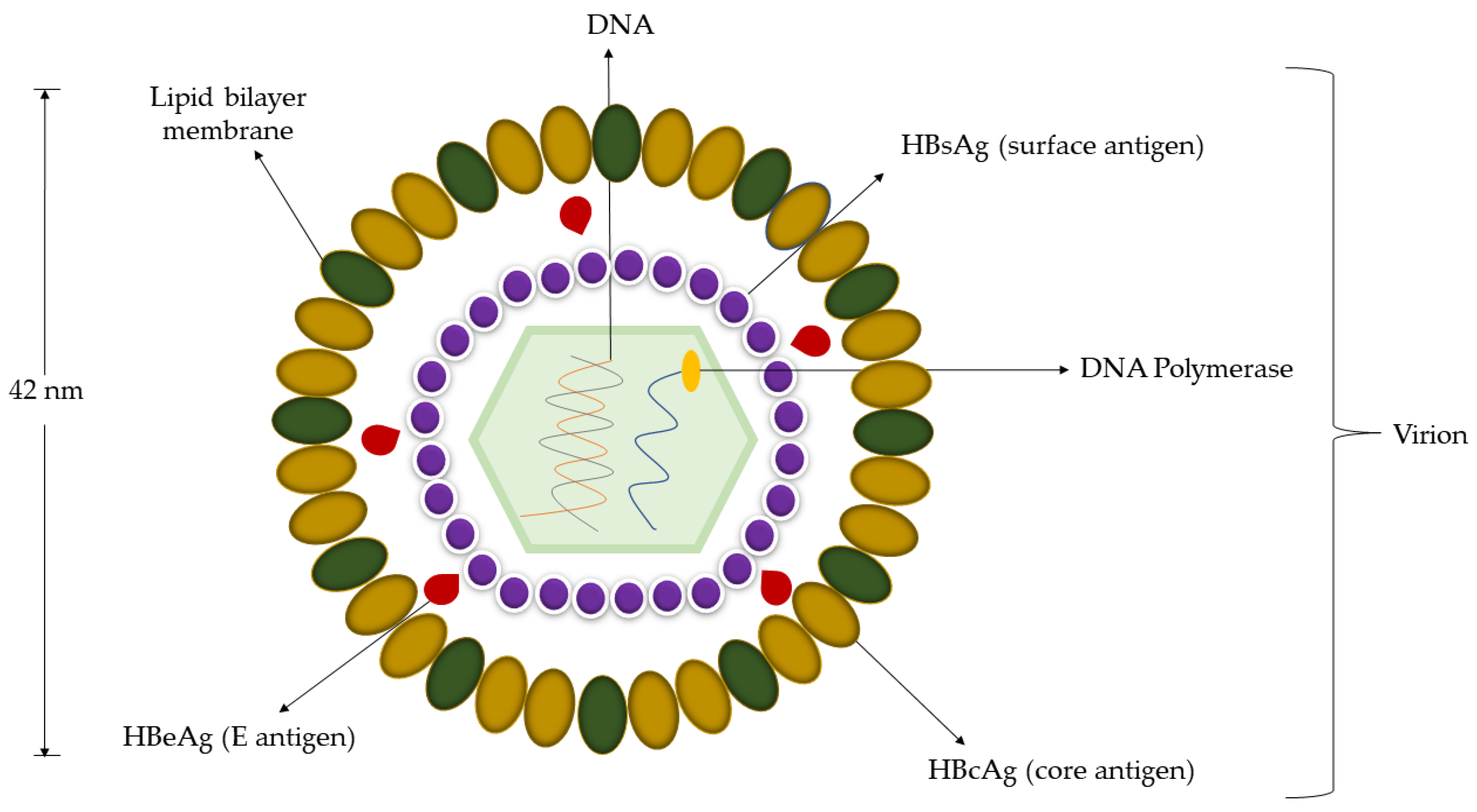
:1. Introduction
2. Diagnosis of HBV
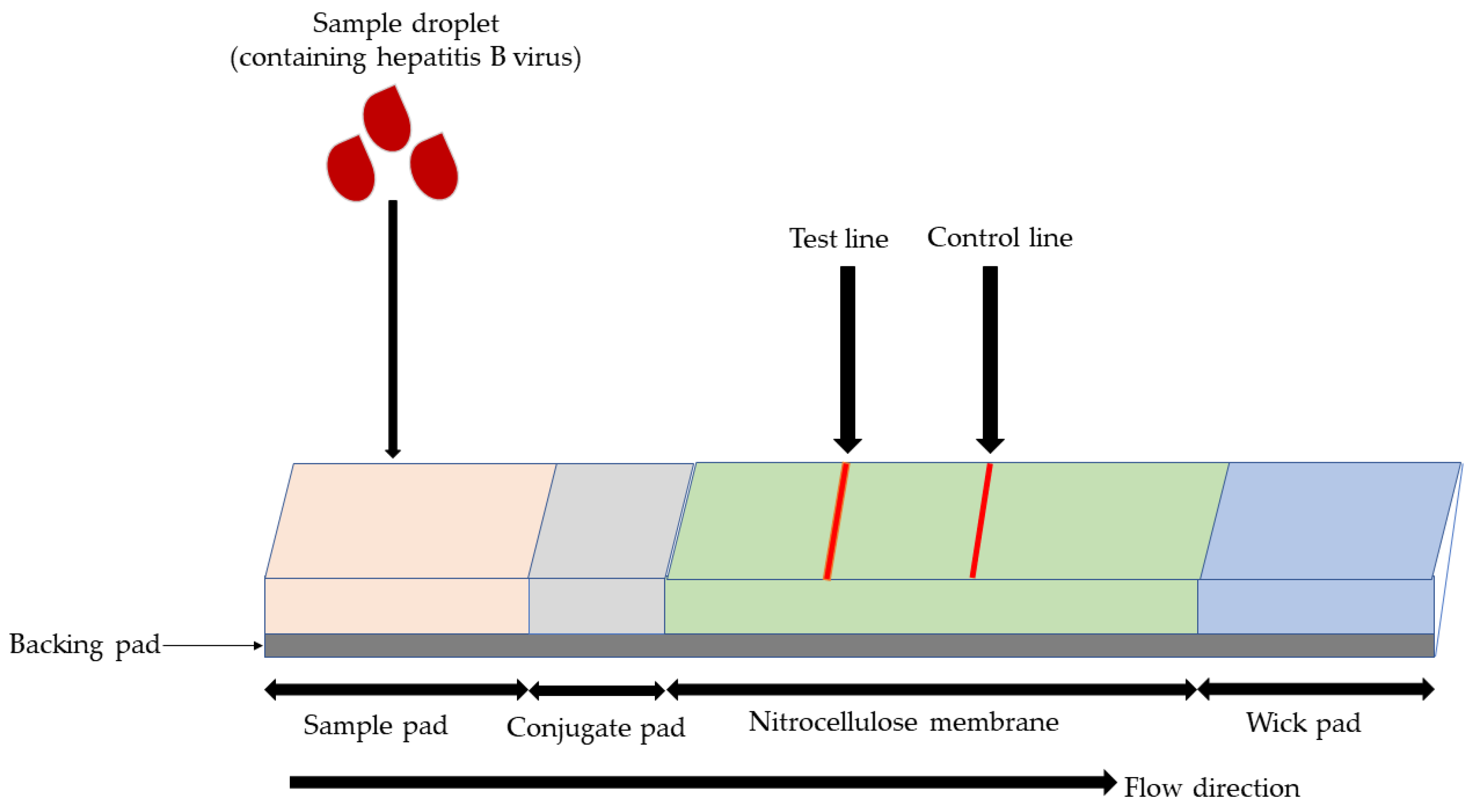
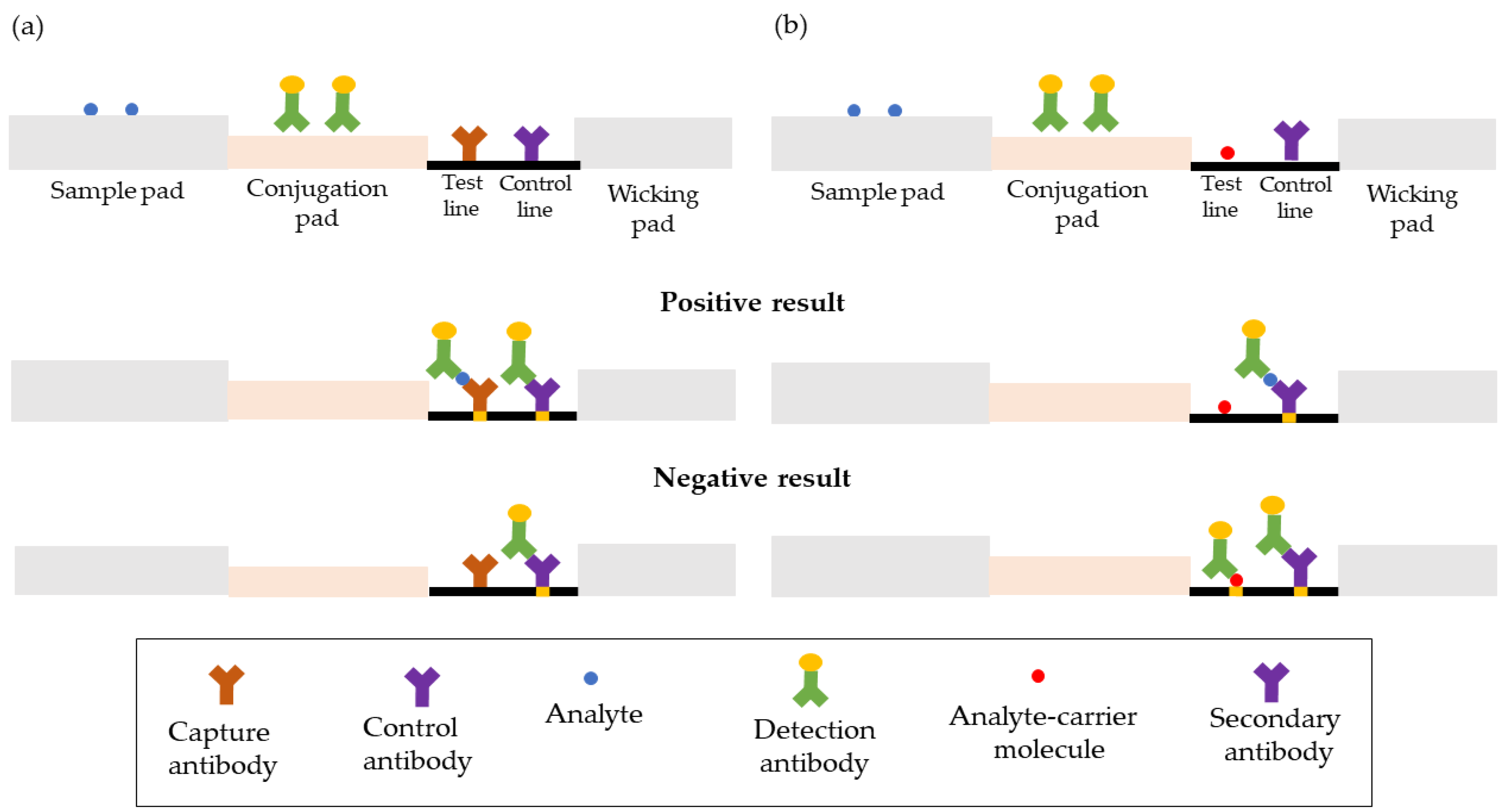
3. Principle of LFA
3.1. The Membrane as an Analytical Region
3.2. The Sample Pad
3.3. The Conjugate Pad
3.4. The Wicking Pad
3.5. The Backing Pad
4. Commercial LFA for the HBV Detection
5. Emerging LFA Techniques for HBV Detection
5.1. Probe Signal Enhancement for Serological Detection
5.2. Sample Amplification Techniques in Molecular Detection
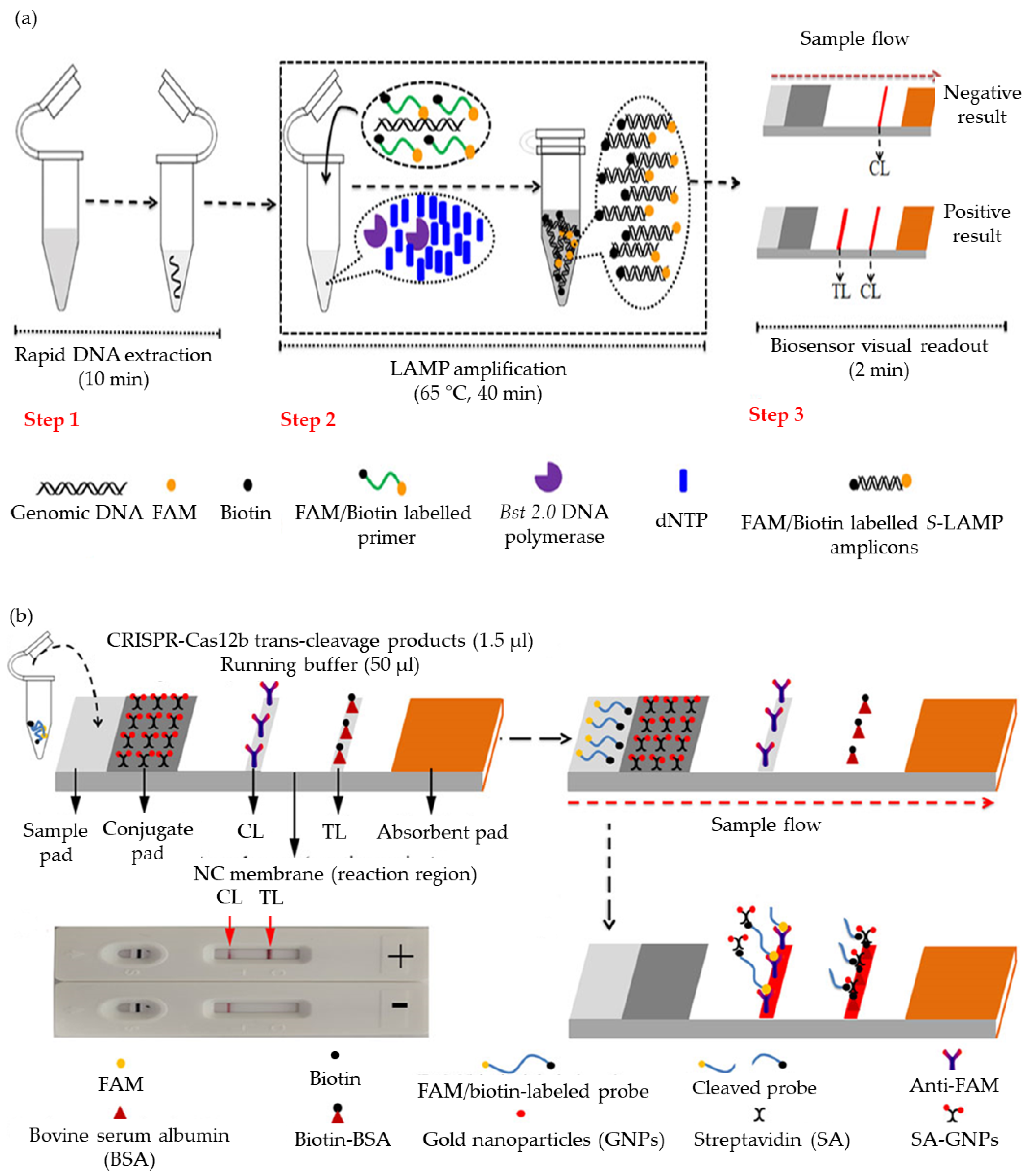
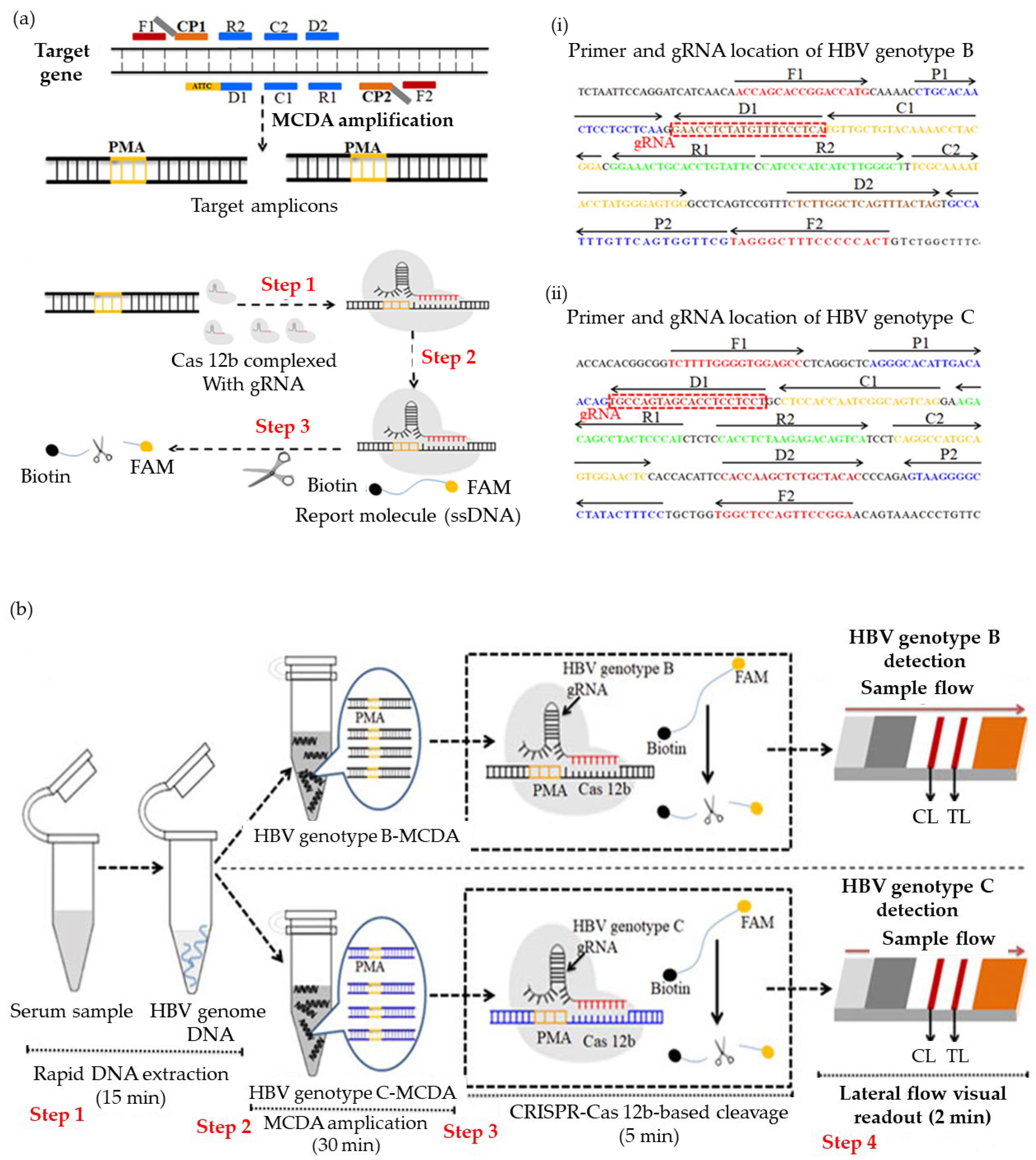
5.2.1. Architecture Modification
5.2.2. PCR Replacement Technique
5.2.3. Detection Label
6. Conclusions and Future Perspectives
Author Contributions
Funding
Data Availability Statement
Conflicts of Interest
References
- World Health Organization. Global health sector strategy on viral hepatitis 2016–2021. Glob. Hepat. Program. Dep. HIV/AIDS 2016, 56, 53. [Google Scholar]
- Nguyen, M.H.; Wong, G.; Gane, E.; Kao, J.-H.; Dusheiko, G. Hepatitis B Virus: Advances in prevention, diagnosis, and theraphy. Clin. Microbiol. Rev. 2020, 33, e00046-19. [Google Scholar] [CrossRef] [PubMed]
- Popping, S.; Bade, D.; Boucher, C.; Van Der Valk, M.; El-Sayed, M.; Sigurour, O.; Sypsa, V.; Morgan, T.; Gamkrelidze, A.; Mukabatsinda, C.; et al. The global campaign to eliminate HBV and HCV infection: International Viral Hepatitis Elimination Meeting and core indicators for development towards the 2030 elimination goals. J. Virus Erad. 2019, 5, 60–66. [Google Scholar] [CrossRef] [PubMed]
- Pawlotsky, J. The Importance of Point-of-Care Tests for HCV and HBV in the Implementation of WHO Strategy of Viral Hepatitis Elimination by 2030. 2015, 41, 2015–2017. [Google Scholar]
- Al-Shaban, Z.; Abdel-Hamid, A.Z.; Hock, T.T.; Hassan, R. Comparison between RT-PCR and ELISA for the detection of HBV in blood donors. Biohealth Sci. Bull. 2010, 2010, 5–7. [Google Scholar]
- Kim, S.H. ELISA for quantitative determination of hepatitis b virus surface antigen. Immune Netw. 2017, 17, 451–459. [Google Scholar] [CrossRef] [Green Version]
- Kimura, T.; Rokuhara, A.; Sakamoto, Y.; Yagi, S.; Tanaka, E.; Kiyosawa, K.; Maki, N. Sensitive enzyme immunoassay for hepatitis B virus core-related antigens and their correlation to virus load. J. Clin. Microbiol. 2002, 40, 439–445. [Google Scholar] [CrossRef] [Green Version]
- McCready, J.A.; Fields, H.A.; Coleman, P.J.; Kane, M.; Schatz, G.; Morens, D. Evaluation of enzyme immunoassay (EIA) as a screening method for hepatitis B markers in an open population. Epidemiol. Infect. 1991, 107, 673–684. [Google Scholar] [CrossRef] [Green Version]
- Deguchi, M.; Yamashita, N.; Kagita, M.; Asari, S.; Iwatani, Y.; Tsuchida, T.; Iinuma, K.; Mushahwar, I.K. Quantitation of hepatitis B surface antigen by an automated chemiluminescent microparticle immunoassay. J. Virol. Methods 2004, 115, 217–222. [Google Scholar] [CrossRef]
- Kohmoto, M.; Enomoto, M.; Tamori, A.; Habu, D.; Takeda, T.; Kawada, N.; Sakaguchi, H.; Seki, S.; Shiomi, S.; Nishiguchi, S. Quantitative detection of hepatitis B surface antigen by chemiluminescent microparticle immunoassay during lamivudine treatment of chronic hepatitis B virus carriers. J. Med. Virol. 2005, 75, 235–239. [Google Scholar] [CrossRef]
- İnan, N.; Demirel, A.; Kabakoğlu Ünsur, E.; Görmüş, U.; Sönmez, E.; Tabak, F.; Arısoy, A. Comparison of Chemiluminescence Microparticle Immunoassay and Electrochemiluminescence Immunoassay for Detection of HBsAg. Viral Hepatit. Derg. 2014, 20, 101–105. [Google Scholar] [CrossRef]
- Zhang, Z.Q.; Wang, Y.B.; Lu, W.; Liu, D.P.; Shi, B.S.; Zhang, X.N.; Huang, D.; Li, X.F.; Zhou, X.L.; Ding, R.R. Performance of hepatitis B core-related antigen versus hepatitis B surface antigen and hepatitis B Virus DNA in predicting HBeAg-positive and HBeAg-negative chronic hepatitis. Ann. Lab. Med. 2018, 39, 67–75. [Google Scholar] [CrossRef]
- Wu, Y.; Ling, X.; Yu, G.; Zhu, H.; Hu, W.; Pu, C.; Zhu, F. The efficacy of HBsAg detection using electro-chemiluminescence immunoassay for blood donor screening in China. Ann. Blood 2019, 4, 30. [Google Scholar] [CrossRef]
- Shahid, M.; Sami, H.; Sharma, S.; Kumar, A.; Khan, P.A.; Khan, H.M. Comparative analysis of Electro-Chemiluminescence Immunoassay (ECLIA), ELISA and Rapid Diagnostic Test (RDT) for detection of Hepatitis B Surface Antigen (HBSAG). Pathology 2020, 52, S126–S127. [Google Scholar] [CrossRef] [Green Version]
- Villar, L.M.; Cruz, H.M.; Deodato, R.M.; Miguel, J.C.; Da Silva, E.F.; Flores, G.L.; Lewis-Ximenez, L.L. Usefulness of automated assays for detecting hepatitis B and C markers in dried blood spot samples. BMC Res. Notes 2019, 12, 10–13. [Google Scholar] [CrossRef]
- Chen, Y.; Wang, J.; Liu, Z.; Wang, X.; Li, X.; Shan, G. A simple and versatile paper-based electrochemiluminescence biosensing platform for hepatitis B virus surface antigen detection. Biochem. Eng. J. 2018, 129, 1–6. [Google Scholar] [CrossRef]
- Kurdi, M.; Abughararah, M.; Mulike, M.; Yamani, O.; Bugdady, M.; Noor, M. Molecular detection of hepatitis B virus (HBV) among voluntary ELISA positive blood donors in Almadinah Almunawwarah. J. Taibah Univ. Med. Sci. 2014, 9, 166–170. [Google Scholar] [CrossRef] [Green Version]
- Tyas, A.A.; Raeni, S.F.; Sakti, S.P.; Sabarudin, A. Recent Advances of Hepatitis B Detection towards Paper-Based Analytical Devices. Sci. World J. 2021, 2021, 6643573. [Google Scholar] [CrossRef]
- Kaneko, S.; Miller, R.H.; Di Bisceglie, A.; Feinstone, S.M.; Hoofnagle, J.H.; Purcell, R.H. Hepatitis B virus DNA detection and comparison with hepatitis B surface antigen. Gastroenterol. Jpn 1990, 25, 57–61. [Google Scholar] [CrossRef]
- Obiomah, C.; Amilo, G.; Ndulue, I. Evaluation of HBsAg Quantification as Surrogate to HBV DNA Viral Load in Hepatitis B Infected Patients in Anambra State, Nigeria. Am. J. Mol. Biol. 2020, 10, 129–140. [Google Scholar] [CrossRef]
- Liu, C.; Chang, L.; Jia, T.; Guo, F.; Zhang, L.; Ji, H.; Zhao, J.; Wang, L. Real-time PCR assays for hepatitis B virus DNA quantification may require two different targets. Virol. J. 2017, 14, 94. [Google Scholar] [CrossRef] [PubMed] [Green Version]
- Mendy, M.E.; Kaye, S.; Van Der Sande, M.; Rayco-Solon, P.; Waight, P.A.; Shipton, D.; Awi, D.; Snell, P.; Whittle, H.; McConkey, S.J. Application of real-time PCR to quantify hepatitis B virus DNA in chronic carriers in The Gambia. Virol. J. 2006, 3, 23. [Google Scholar] [CrossRef] [PubMed] [Green Version]
- Amini, A.; Varsaneux, O.; Kelly, H.; Tang, W.; Chen, W.; Boeras, D.I.; Falconer, J.; Tucker, J.D.; Chou, R.; Ishizaki, A.; et al. Diagnostic accuracy of tests to detect hepatitis B surface antigen: A systematic review of the literature and meta-analysis. BMC Infect. Dis. 2017, 17, 19–37. [Google Scholar] [CrossRef] [PubMed]
- Sarin, S.K.; Kumar, M.; Eslam, M.; George, J.; Al Mahtab, M.; Akbar, S.M.F.; Jia, J.; Tian, Q.; Aggarwal, R.; Muljono, D.H.; et al. Liver diseases in the Asia-Pacific region: A Lancet Gastroenterology & Hepatology Commission. Lancet Gastroenterol. Hepatol. 2020, 5, 167–228. [Google Scholar] [CrossRef] [PubMed] [Green Version]
- Easterbrook, P.J.; Roberts, T.; Sands, A.; Peeling, R. Diagnosis of viral hepatitis. Curr. Opin. HIV AIDS 2017, 12, 302–314. [Google Scholar] [CrossRef]
- Marcuccilli, F.; Chevaliez, S.; Muller, T.; Colagrossi, L.; Abbondanza, G.; Beyser, K.; Wlassow, M.; Ortonne, V.; Perno, C.F.; Ciotti, M. Multicenter evaluation of the Cepheid Xpert® HBV viral load test. Diagnostics 2021, 11, 297. [Google Scholar] [CrossRef]
- Boehringer, H.R.; O’farrell, B.J. Lateral Flow Assays in Infectious Disease Diagnosis. Clin. Chem. 2022, 68, 52–58. [Google Scholar] [CrossRef]
- Wang, D.; He, S.; Wang, X.; Yan, Y.; Liu, J.; Wu, S.; Liu, S.; Lei, Y.; Chen, M.; Li, L.; et al. Rapid Lateral Flow Immunoassay for the Fluorescence Detection of SARS-CoV-2 RNA. Nat. Biomed. Eng. 2020, 4, 1150–1158. [Google Scholar] [CrossRef]
- Liu, Y.; Zhan, L.; Qin, Z.; Sackrison, J.; Bischof, J.C. Ultrasensitive and Highly Specific Lateral Flow Assays for Point-of-Care Diagnosis. ACS Nano 2021, 15, 3593–3611. [Google Scholar] [CrossRef]
- Cadet, M.J. Screening for Hepatitis B: Serology Markers Interpretation. Work. Health Saf. 2018, 2018, 94707. [Google Scholar] [CrossRef] [Green Version]
- Liang, T.J. Hepatitis B: The Virus and Disease. Hepatology 2009, 49, S13–S21. [Google Scholar] [CrossRef] [Green Version]
- Kim, H.; Ko, C.; Lee, J.; Kim, M. Current Progress in the Development of Hepatitis B Virus Mode-of-Action and Efficacy. Molecules 2021, 26, 7420. [Google Scholar] [CrossRef]
- Dane, D.S.; Cameron, C.H.; Briggs, M. Virus-Like Particles in Serum of Patients With Australia-Antigen-Associated Hepatitis. Lancet 1970, 295, 695–698. [Google Scholar] [CrossRef]
- Short, J.M.; Chen, S.; Roseman, A.M.; Butler, P.J.G.; Crowther, R.A. Structure of Hepatitis B Surface Antigen from Subviral Tubes Determined by Electron Cryomicroscopy. J. Mol. Biol. 2009, 390, 135–141. [Google Scholar] [CrossRef]
- Venkatakrishnan, B.; Zlotnick, A. The Structural Biology of Hepatitis B Virus: Form and Function. Annu. Rev. Virol. 2016, 3, 429–451. [Google Scholar] [CrossRef] [Green Version]
- Gish, R.G.; Given, B.D.; Lai, C.; Locarnini, S.A.; Lau, J.Y.N.; Lewis, D.L.; Schluep, T. Chronic hepatitis B: Virology, natural history, current management and a glimpse at future opportunities. Antivir. Res. 2015, 121, 47–58. [Google Scholar] [CrossRef]
- Marchetti, A.L.; Guo, H. New Insights on Molecular Mechanism of Hepatitis B Virus Covalently Closed Circular DNA Formation. Cells 2020, 9, 2430. [Google Scholar] [CrossRef]
- Villar, L.M.; Cruz, H.M.; Barbosa, J.R.; Bezerra, C.S.; Portilho, M.M.; Scalioni, L. de P. Update on hepatitis B and C virus diagnosis. World J. Virol. 2015, 4, 323. [Google Scholar] [CrossRef]
- CDC. Epidemiology Vaccine-Preventable, 14th ed.; Hall, E., Wodi, A.P., Hamborsky, J., Eds.; Public Health Foundation: Washington, DC, USA, 2021. [Google Scholar]
- Rong, X.; Ailing, F.; Xiaodong, L.; Jie, H.; Min, L. Monitoring hepatitis B by using point-of-care testing: Biomarkers, current technologies, and perspectives. Expert Rev. Mol. Diagn. 2021, 21, 195–211. [Google Scholar] [CrossRef]
- Song, J.E.; Kim, D.Y. Diagnosis of hepatitis B. Ann. Transl. Med. 2016, 4, 338. [Google Scholar] [CrossRef] [Green Version]
- Du, X.; Liu, Y.; Ma, L.; Lu, J.; Jin, Y.; Ren, S.; He, Z.; Chen, X. Virological and serological features of acute hepatitis B in adults. Medicine 2017, 96, e6088. [Google Scholar] [CrossRef] [PubMed]
- Mohammed, E.H.; Saud, M.R.; Yemane, S.; Isam Mohammed, E.; Freweini, T. Serological and virological profile of patients with chronic hepatitis B infection in Eritrea. Int. J. Clin. Virol. 2020, 4, 95–101. [Google Scholar] [CrossRef]
- Yu, W.; Goddard, C.; Clearfield, E.; Mills, C.; Xiao, T.; Guo, H.; Morrey, J.D.; Motter, N.E.; Zhao, K.; Block, T.M.; et al. Design, synthesis, and biological evaluation of triazolo-pyrimidine derivatives as novel inhibitors of hepatitis B virus surface antigen (HBsAg) secretion. J. Med. Chem. 2011, 54, 5660–5670. [Google Scholar] [CrossRef] [PubMed] [Green Version]
- Kuhns, M.C.; Holzmayer, V.; McNamara, A.L.; Anderson, M.; Cloherty, G.A. Hepatitis B seroconversion revisited: New insights into the natural history of acute hepatitis B virus (HBV) infection from quantitative and highly sensitive assays and novel biomarkers. Virol. J. 2021, 18, 235. [Google Scholar] [CrossRef]
- Kafeero, H.M.; Ndagire, D.; Ocama, P.; Kato, C.D.; Wampande, E.; Walusansa, A.; Kajumbula, H.; Kateete, D.; Sendagire, H. Hepatitis B virus (HBV) serological patterns among the HBsAg negative hospital attendees screened for immunization. Sci. Rep. 2022, 12, 7425. [Google Scholar] [CrossRef]
- Li, A.; Yuan, Q.; Huang, Z.; Fan, J.; Guo, R.; Lou, B.; Zheng, Q.; Ge, S.; Chen, Y.; Su, Z.; et al. Novel double-antigen sandwich immunoassay for human hepatitis B core antibody. Clin. Vaccine Immunol. 2010, 17, 464–469. [Google Scholar] [CrossRef] [Green Version]
- Zhang, W.; Aryan, M.; Qian, S.; Cabrera, R.; Liu, X. A Focused Review on Recent Advances in the Diagnosis and Treatment of Viral Hepatitis. Gastroenterol. Res. 2021, 14, 139–156. [Google Scholar] [CrossRef]
- Mak, W.C.; Beni, V.; Turner, A.P.F. Lateral-flow technology: From visual to instrumental. TrAC Trends Anal. Chem. 2016, 79, 297–305. [Google Scholar] [CrossRef]
- Bishop, J.D.; Hsieh, H.V.; Gasperino, D.J.; Weigl, B.H. Sensitivity enhancement in lateral flow assays: A systems perspective. Lab Chip 2019, 19, 2486–2499. [Google Scholar] [CrossRef] [Green Version]
- Koczula, K.M.; Gallotta, A. Lateral flow assays. Essays Biochem. 2016, 60, 111–120. [Google Scholar] [CrossRef]
- Bahadır, E.B.; Sezgintürk, M.K. Lateral flow assays: Principles, designs and labels. TrAC Trends Anal. Chem. 2016, 82, 286–306. [Google Scholar] [CrossRef]
- Kasetsirikul, S.; Shiddiky, M.J.A.; Nguyen, N.-T. Challenges and perspectives in the development of paper-based lateral flow assays. Microfluid. Nanofluidics 2020, 24, 17. [Google Scholar] [CrossRef]
- Wang, R.; Ongagna-Yhombi, S.Y.; Lu, Z.; Centeno-Tablante, E.; Colt, S.; Cao, X.; Ren, Y.; Cárdenas, W.B.; Mehta, S.; Erickson, D. Rapid Diagnostic Platform for Colorimetric Differential Detection of Dengue and Chikungunya Viral Infections. Anal. Chem. 2019, 91, 5415–5423. [Google Scholar] [CrossRef]
- Ghasemi, A.; Rabiee, N.; Ahmadi, S.; Hashemzadeh, S.; Lolasi, F.; Bozorgomid, M.; Kalbasi, A.; Nasseri, B.; Shiralizadeh Dezfuli, A.; Aref, A.R.; et al. Optical assays based on colloidal inorganic nanoparticles. Analyst 2018, 143, 3249–3283. [Google Scholar] [CrossRef]
- Dzantiev, B.B.; Byzova, N.A.; Urusov, A.E.; Zherdev, A.V. Immunochromatographic methods in food analysis. TrAC Trends Anal. Chem. 2014, 55, 81–93. [Google Scholar] [CrossRef]
- Zhu, W.; Meng, K.; Zhang, Y.; Bu, Z.; Zhao, D.; Meng, G. Lateral Flow Assay for the Detection of African Swine Fever Virus Antibodies Using Gold Nanoparticle-Labeled Acid-Treated p72. Front. Chem. 2022, 9, 804981. [Google Scholar] [CrossRef]
- Roberts, A.; Prakashan, D.; Dhanze, H.; Gandham, R.K.; Gandhi, S.; Sharma, G.T. Immuno-chromatic probe based lateral flow assay for point-of-care detection of Japanese encephalitis virus NS1 protein biomarker in clinical samples using a smartphone-based approach. Nanoscale Adv. 2022, 4, 3966–3977. [Google Scholar] [CrossRef]
- Huang, C.; Wen, T.; Shi, F.J.; Zeng, X.Y.; Jiao, Y.J. Rapid Detection of IgM Antibodies against the SARS-CoV-2 Virus via Colloidal Gold Nanoparticle-Based Lateral-Flow Assay. ACS Omega 2020, 5, 12550–12556. [Google Scholar] [CrossRef]
- Hamad, E.M.; Hawamdeh, G.; Jarrad, N.A.; Yasin, O.; Al-Gharabli, S.I.; Shadfan, R. Detection of Human Chorionic Gonadotropin (hCG) Hormone using Digital Lateral Flow Immunoassay. In Proceedings of the 2018 40th Annual International Conference of the IEEE Engineering in Medicine and Biology Society (EMBC), Institute of Electrical and Electronics Engineers, Honolulu, HI, USA, 18–21 July 2018; pp. 3845–3848. [Google Scholar] [CrossRef]
- Chen, L.; Wang, H.; Guo, T.; Xiao, C.; Liu, L.; Zhang, X.; Liu, B.; Li, P.; Liu, A.; Li, B.; et al. A rapid point-of-care test for dengue virus-1 based on a lateral flow assay with a near-infrared fluorescent dye. J. Immunol. Methods 2018, 456, 23–27. [Google Scholar] [CrossRef]
- Rohrman, B.A.; Leautaud, V.; Molyneux, E.; Richards-Kortum, R.R. A Lateral Flow Assay for Quantitative Detection of Amplified HIV-1 RNA. PLoS ONE 2012, 7, 45611. [Google Scholar] [CrossRef] [Green Version]
- Borse, V.; Srivastava, R. Process parameter optimization for lateral flow immunosensing. Mater. Sci. Energy Technol. 2019, 2, 434–441. [Google Scholar] [CrossRef]
- Altschuh, P.; Kunz, W.; Bremerich, M.; Reiter, A.; Selzer, M.; Nestler, B. Wicking in Porous Polymeric Membranes: Determination of an Effective Capillary Radius to Predict the Flow Behavior in Lateral Flow Assays. Membranes 2022, 12, 638. [Google Scholar] [CrossRef] [PubMed]
- Juntunen, E. Lateral Flow Immunoassays with Fluorescent Reporter Technologies; University of Turku: Turku, Finland, 2018; ISBN 9789512971268. [Google Scholar]
- Zhu, M.; Zhang, W.N.; Tian, J.Y.; Zhao, W.Y.; Chen, Z.Q.; Sun, L.H.; Xue, F.; Liu, Y.; Tan, X.Q.; Wang, L.M.; et al. Development of a lateral-flow assay (LFA) for rapid detection of Soybean mosaic virus. J. Virol. Methods 2016, 235, 51–57. [Google Scholar] [CrossRef] [PubMed]
- Jang, M.; Kim, S.J.; Song, J.; Kim, S. Rapid and simple detection of influenza virus via isothermal amplification lateral flow assay. Anal. Bioanal. Chem. 2022, 414, 4685–4696. [Google Scholar] [CrossRef]
- Farrell, B.O.; Immunoassays, C. Lateral Flow Immunoassay; Springer Science & Business Media: Berlin/Heidelberg, Germany, 2009; ISBN 9781597452403. [Google Scholar]
- Posthuma-Trumpie, G.A.; Korf, J.; Van Amerongen, A. Lateral flow (immuno)assay: Its strengths, weaknesses, opportunities and threats. A literature survey. Anal. Bioanal. Chem. 2009, 393, 569–582. [Google Scholar] [CrossRef] [Green Version]
- Fu, X.; Cheng, Z.; Yu, J.; Choo, P.; Chen, L.; Choo, J. A SERS-based lateral flow assay biosensor for highly sensitive detection of HIV-1 DNA. Biosens. Bioelectron. 2016, 78, 530–537. [Google Scholar] [CrossRef]
- Mansfield, M.A. The Use of Nitrocellulose Membranes in Lateral-Flow Assays. In Drugs of Abuse; Springer International Publishing: Cham, Switzerland, 2007; pp. 71–85. [Google Scholar] [CrossRef]
- European Commission. 2009/886/EC: Commission Decision of 27 November 2009 Amending Decision 2002/364/EC on Common Technical Specifications for In Vitro Diagnostic Medical Devices (Notified under Document C (2009) 9464) (Text with EEA Relevance); European Commission: Brussels, Belgium, 2009; pp. 1–20.
- WHO. WHO Prequalification of In Vitro Diagnostics Public Report Product: Determine HBsAg 2; WHO: Geneva, Switzerland, 2018; pp. 1–16.
- WHO. WHO Prequalification of In Vitro Diagnostics Public Report Product: Bioline HBs Ag WB; WHO: Geneva, Switzerland, 2018.
- Programme, D.; Virus, I.; Test, R.; Blood, W. List of HIV Diagnostic Test Kits and Equipments Classified according to the Global Fund Quality Assurance Policy; Global Fund: Geneva, Switzerland, 2015; pp. 1–59. [Google Scholar]
- World Health Organization. WHO List of Prequalified Diagnostic Products; WHO: Geneva, Switzerland, 2013; p. 4. Available online: http://www.who.int/diagnostics_laboratory/evaluations/PQ_list/en/ (accessed on 27 May 2023).
- Avellon, A.; Ala, A.; Diaz, A.; Domingo, D.; Gonzalez, R.; Hidalgo, L.; Kooner, P.; Loganathan, S.; Martin, D.; McPherson, S.; et al. Clinical performance of Determine HBsAg 2 rapid test for Hepatitis B detection. J. Med. Virol. 2020, 92, 3403–3411. [Google Scholar] [CrossRef] [Green Version]
- Dembele, B.; Affi-Aboli, R.; Kabran, M.; Sevede, D.; Goha, V.; Adiko, A.C.; Kouamé, R.; Allah-Kouadio, E.; Inwoley, A. Evaluation of Four Rapid Tests for Detection of Hepatitis B Surface Antigen in Ivory Coast. J. Immunol. Res. 2020, 2020, 6315718. [Google Scholar] [CrossRef]
- Chevaliez, S.; Roudot-Thoraval, F.; Hézode, C.; Pawlotsky, J.M.; Njouom, R. Performance of rapid diagnostic tests for hepatitis B surface antigen detection in serum or plasma. Diagn. Microbiol. Infect. Dis. 2021, 100, 115353. [Google Scholar] [CrossRef]
- Chaurasia, D.; Shrivastava, R.K. Evaluation of rapid diagnostic test compared with ELISA for detection of hepatitis B surface antigen. Indian J. Microbiol. Res. 2020, 7, 203–206. [Google Scholar] [CrossRef]
- Prabha, P.; Saikeerthana, D.; Vijayashree, V.; Gogan, M. A Comparison of Rapid Screening Test and ELISA for the Diagnosis of Hepatitis B Surface Antigen in Patients Attending a Tertiary Care Hospital, Tamil Nadu, India. Natl. J. Lab. Med. 2022, 11, 22–25. [Google Scholar] [CrossRef]
- Saboor Soomro, R.; Shah, I.A.; Saboor, A.; Bhutto, A.U.B.; Memon, S. Sensitivity and Specificity of Hepatitis B Virus Screening via Rapid Immunoassay Chromatographic Test. Cureus 2021, 13, 10–13. [Google Scholar] [CrossRef]
- Hayder, I.; Ahmed, W.; Alam, S.E. Comparison of Different ICT Kits for HBsAg and Anti HCV Using Gold Standard ELISA. Pakistan J. Med. Res. Pak. J. Med. Res. 2012, 51, 72–76. [Google Scholar]
- Al-Matary, A.M.; Al Gashaa, F.A.S. Comparison of different rapid screening tests and ELISA for HBV, HCV, and HIV among healthy blood donors and recipients at Jibla University Hospital Yemen. J. Med. Life 2022, 15, 1403–1408. [Google Scholar] [CrossRef]
- Navvabi, N.; Khadem Ansari, M.H.; Navvabi, A.; Chalipa, H.R.; Zitricky, F. Comparative assessment of immunochromatography and ELISA diagnostic tests for HBsAg detection in PCR-confirmed HBV infection. Rev. Gastroenterol. Mex. 2022, 87, 176–180. [Google Scholar] [CrossRef]
- Momose, H.; Murayama, A.; Yamada, N.; Matsubayashi, K.; Matsuoka, S.; Ikebe, E.; Kuramitsu, M.; Muramatsu, M.; Kato, T.; Hamaguchi, I. Performance evaluation of in vitro diagnostic kits for hepatitis B virus infection using the regional reference panel of Japan. Virol. J. 2023, 20, 93. [Google Scholar] [CrossRef]
- Shen, J.; Zhou, Y.; Fu, F.; Xu, H.; Lv, J.; Xiong, Y.; Wang, A. Immunochromatographic assay for quantitative and sensitive detection of hepatitis B virus surface antigen using highly luminescent quantum dot-beads. Talanta 2015, 142, 145–149. [Google Scholar] [CrossRef]
- Gish, R.G.; Gutierrez, J.A.; Navarro-Cazarez, N.; Giang, K.; Adler, D.; Tran, B.; Locarnini, S.; Hammond, R.; Bowden, S. A simple and inexpensive point-of-care test for hepatitis B surface antigen detection: Serological and molecular evaluation. J. Viral Hepat. 2014, 21, 905–908. [Google Scholar] [CrossRef]
- Choi, D.H.; Lee, S.K.; Oh, Y.K.; Bae, B.W.; Lee, S.D.; Kim, S.; Shin, Y.B.; Kim, M.G. A dual gold nanoparticle conjugate-based lateral flow assay (LFA) method for the analysis of troponin I. Biosens. Bioelectron. 2010, 25, 1999–2002. [Google Scholar] [CrossRef]
- Lou, S.; Ye, J.Y.; Li, K.Q.; Wu, A. A gold nanoparticle-based immunochromatographic assay: The influence of nanoparticulate size. Analyst 2012, 137, 1174–1181. [Google Scholar] [CrossRef]
- Kim, D.S.; Kim, Y.T.; Hong, S.B.; Kim, J.; Huh, N.S.; Lee, M.K.; Lee, S.J.; Kim, B.I.; Kim, I.S.; Huh, Y.S.; et al. Development of lateral flow assay based on size-controlled gold nanoparticles for detection of hepatitis B surface antigen. Sensors 2016, 16, 2154. [Google Scholar] [CrossRef] [PubMed]
- Li, J.; Duan, H.; Xu, P.; Huang, X.; Xiong, Y. Effect of different-sized spherical gold nanoparticles grown layer by layer on the sensitivity of an immunochromatographic assay. RSC Adv. 2016, 6, 26178–26185. [Google Scholar] [CrossRef]
- Chen, X.; Leng, Y.; Hao, L.; Duan, H.; Yuan, J.; Zhang, W.; Huang, X.; Xiong, Y. Self-assembled colloidal gold superparticles to enhance the sensitivity of lateral flow immunoassays with sandwich format. Theranostics 2020, 10, 3737–3748. [Google Scholar] [CrossRef] [PubMed]
- Shen, Y.; Shen, G. Signal-Enhanced Lateral Flow Immunoassay with Dual Gold Nanoparticle Conjugates for the Detection of Hepatitis B Surface Antigen. ACS Omega 2019, 4, 5083–5087. [Google Scholar] [CrossRef] [Green Version]
- Zou, Z.; Du, D.; Wang, J.; Smith, J.N.; Timchalk, C.; Li, Y.; Lin, Y. Quantum dot-based immunochromatographic fluorescent biosensor for biomonitoring trichloropyridinol, a biomarker of exposure to chlorpyrifos. Anal. Chem. 2010, 82, 5125–5133. [Google Scholar] [CrossRef]
- Rong, Z.; Wang, Q.; Sun, N.; Jia, X.; Wang, K.; Xiao, R.; Wang, S. Smartphone-based fluorescent lateral flow immunoassay platform for highly sensitive point-of-care detection of Zika virus nonstructural protein 1. Anal. Chim. Acta 2019, 1055, 140–147. [Google Scholar] [CrossRef]
- Li, X.; Li, W.; Yang, Q.; Gong, X.; Guo, W.; Dong, C.; Liu, J.; Xuan, L.; Chang, J. Rapid and quantitative detection of prostate specific antigen with a quantum dot nanobeads-based immunochromatography test strip. ACS Appl. Mater. Interfaces 2014, 6, 6406–6414. [Google Scholar] [CrossRef]
- Hu, J.; Zhang, Z.L.; Wen, C.Y.; Tang, M.; Wu, L.L.; Liu, C.; Zhu, L.; Pang, D.W. Sensitive and Quantitative Detection of C-Reaction Protein Based on Immunofluorescent Nanospheres Coupled with Lateral Flow Test Strip. Anal. Chem. 2016, 88, 6577–6584. [Google Scholar] [CrossRef]
- Duan, H.; Huang, X.; Shao, Y.; Zheng, L.; Guo, L.; Xiong, Y. Size-Dependent Immunochromatographic Assay with Quantum Dot Nanobeads for Sensitive and Quantitative Detection of Ochratoxin A in Corn. Anal. Chem. 2017, 89, 7062–7068. [Google Scholar] [CrossRef]
- Shen, H.; Yuan, H.; Niu, J.Z.; Xu, S.; Zhou, C.; Ma, L.; Li, L.S. Phosphine-free synthesis of high-quality reverse type-I ZnSe/CdSe core with CdS/CdxZn1—XS/ZnS multishell nanocrystals and their application for detection of human hepatitis B surface antigen. Nanotechnology 2011, 22, 375602. [Google Scholar] [CrossRef]
- Zhang, P.; Lu, H.; Chen, J.; Han, H.; Ma, W. Simple and Sensitive Detection of HBsAg by Using a Quantum Dots Nanobeads Based Dot-Blot Immunoassay. Theranostics 2014, 4, 307. [Google Scholar] [CrossRef] [Green Version]
- Rong, Z.; Xiao, R.; Peng, Y.; Zhang, A.; Wei, H.; Ma, Q.; Wang, D.; Wang, Q.; Bai, Z.; Wang, F.; et al. Integrated fluorescent lateral flow assay platform for point-of-care diagnosis of infectious diseases by using a multichannel test cartridge. Sens. Actuators B Chem. 2021, 329, 129193. [Google Scholar] [CrossRef]
- An, B.G.; Kim, H.R.; Kang, M.J.; Park, J.G.; Chang, Y.W.; Pyun, J.C. Chemiluminescent lateral-flow immunoassays by using in-situ synthesis of CdS NW photosensor. Anal. Chim. Acta 2016, 927, 99–106. [Google Scholar] [CrossRef]
- Xia, X.; Xu, Y.; Zhao, X.; Li, Q. Lateral flow immunoassay using europium chelate-loaded silica nanoparticles as labels. Clin. Chem. 2009, 55, 179–182. [Google Scholar] [CrossRef]
- Zhang, X.; Jiang, L.; Zhang, C.; Li, D.; Wang, C.; Gao, F.; Cui, D. A silicon dioxide modified magnetic nanoparticles-labeled lateral flow strips for HBs antigen. J. Biomed. Nanotechnol. 2011, 7, 776–781. [Google Scholar] [CrossRef]
- Cai, Y.; Yan, J.; Zhu, L.; Wang, H.; Lu, Y. A Rapid Immunochromatographic Method Based on a Secondary Antibody-Labelled Magnetic Nanoprobe for the Detection of Hepatitis B preS2 Surface Antigen. Biosensors 2020, 10, 161. [Google Scholar] [CrossRef]
- Si, J.; Li, J.; Zhang, L.; Zhang, W.; Yao, J.; Li, T.; Wang, W.; Zhu, W.; Allain, J.P.; Fu, Y.; et al. A signal amplification system on a lateral flow immunoassay detecting for hepatitis e-antigen in human blood samples. J. Med. Virol. 2019, 91, 1301–1306. [Google Scholar] [CrossRef]
- Li, M.R.; Lu, J.H.; Ye, L.H.; Sun, X.L.; Zheng, Y.H.; Liu, Z.Q.; Zhang, H.C.; Liu, Y.Y.; Lv, Y.; Huang, Y.; et al. Quantitative hepatitis B core antibody level is associated with inflammatory activity in treatment-naïve chronic hepatitis B patients. Medcine 2016, 95, e4422. [Google Scholar] [CrossRef]
- Li, M.R.; Zheng, H.W.; Lu, J.H.; Ma, S.M.; Ye, L.H.; Liu, Z.Q.; Zhang, H.C.; Liu, Y.Y.; Lv, Y.; Huang, Y.; et al. Serum hepatitis B core antibody titer use in screening for significant fibrosis in treatment-naïve patients with chronic hepatitis B. Oncotarget 2017, 8, 11063–11070. [Google Scholar] [CrossRef] [Green Version]
- Liang, R.L.; Deng, Q.T.; Chen, Z.H.; Xu, X.P.; Zhou, J.W.; Liang, J.Y.; Dong, Z.N.; Liu, T.C.; Wu, Y.S. Europium (III) chelate microparticle-based lateral flow immunoassay strips for rapid and quantitative detection of antibody to hepatitis B core antigen. Sci. Rep. 2017, 7, 14093. [Google Scholar] [CrossRef] [Green Version]
- Jin, B.; Yang, Y.; He, R.; Park, Y.I.; Lee, A.; Bai, D.; Li, F.; Lu, T.J.; Xu, F.; Lin, M. Lateral flow aptamer assay integrated smartphone-based portable device for simultaneous detection of multiple targets using upconversion nanoparticles. Sensors Actuators B Chem. 2018, 276, 48–56. [Google Scholar] [CrossRef]
- Martiskainen, I.; Talha, S.M.; Vuorenpää, K.; Salminen, T.; Juntunen, E.; Chattopadhyay, S.; Kumar, D.; Vuorinen, T.; Pettersson, K.; Khanna, N.; et al. Upconverting nanoparticle reporter–based highly sensitive rapid lateral flow immunoassay for hepatitis B virus surface antigen. Anal. Bioanal. Chem. 2021, 413, 967–978. [Google Scholar] [CrossRef] [PubMed]
- Li, L.; Zhou, L.; Yu, Y.; Zhu, Z.; Lin, C.; Lu, C.; Yang, R. Development of up-converting phosphor technology-based lateral-flow assay for rapidly quantitative detection of hepatitis B surface antibody. Diagn. Microbiol. Infect. Dis. 2009, 63, 165–172. [Google Scholar] [CrossRef] [PubMed]
- Choi, J.R.; Liu, Z.; Hu, J.; Tang, R.; Gong, Y.; Feng, S.; Ren, H.; Wen, T.; Yang, H.; Qu, Z.; et al. Polydimethylsiloxane-Paper Hybrid Lateral Flow Assay for Highly Sensitive Point-of-Care Nucleic Acid Testing. Anal. Chem. 2016, 88, 6254–6264. [Google Scholar] [CrossRef] [PubMed]
- Choi, J.R.; Yong, K.W.; Tang, R.; Gong, Y.; Wen, T.; Yang, H.; Li, A.; Chia, Y.C.; Pingguan-Murphy, B.; Xu, F. Lateral Flow Assay Based on Paper–Hydrogel Hybrid Material for Sensitive Point-of-Care Detection of Dengue Virus. Adv. Healthc. Mater. 2017, 6, 920. [Google Scholar] [CrossRef]
- Tang, R.; Yang, H.; Gong, Y.; Liu, Z.; Li, X.J.; Wen, T.; Qu, Z.G.; Zhang, S.; Mei, Q.; Xu, F. Improved Analytical Sensitivity of Lateral Flow Assay using Sponge for HBV Nucleic Acid Detection. Sci. Rep. 2017, 7, 1360. [Google Scholar] [CrossRef] [Green Version]
- Toley, B.J.; Mckenzie, B.; Liang, T.; Buser, J.R.; Yager, P.; Fu, E. Tunable-Delay Shunts for Paper Microfluidic Devices. Anal. Chem. 2013, 85, 11545–11552. [Google Scholar] [CrossRef] [Green Version]
- Zhang, B.; Zhu, Z.; Li, F.; Xie, X.; Ding, A. Rapid and sensitive detection of hepatitis B virus by lateral flow recombinase polymerase amplification assay. J. Virol. Methods 2021, 291, 114094. [Google Scholar] [CrossRef]
- Yi, T.; Zhang, H.; Liang, H.; Gong, G.; Cai, Y. Betaine-assisted recombinase polymerase assay for rapid hepatitis B virus detection. Biotechnol. Appl. Biochem. 2020, 68, 469–475. [Google Scholar] [CrossRef]
- Mayran, C.; Foulongne, V.; Van de Perre, P.; Fournier-Wirth, C.; Molès, J.P.; Cantaloube, J.F. Rapid Diagnostic Test for Hepatitis B Virus Viral Load Based on Recombinase Polymerase Amplification Combined with a Lateral Flow Read-Out. Diagnostics 2022, 12, 621. [Google Scholar] [CrossRef]
- Lu, Y.; Li, M.; Liu, H.; Lin, S.; Zhao, X.; Liu, Z.; Zhao, L.; Wan, K.L. Detecting Mycobacterium tuberculosis complex and rifampicin resistance via a new rapid multienzyme isothermal point mutation assay. Anal. Biochem. 2021, 630, 114341. [Google Scholar] [CrossRef]
- Bai, X.; Ma, X.; Li, M.; Li, X.; Fan, G.; Zhang, R.; Wang, R.; Duan, Q.; Shen, X.; Xie, Y.; et al. Field applicable detection of hepatitis B virus using internal controlled duplex recombinase-aided amplification assay and lateral flow dipstick assay. J. Med. Virol. 2020, 92, 3344–3353. [Google Scholar] [CrossRef]
- Sun, M.L.; Lai, H.Y.; Chong, N.Y.; Liu, D.F.; Zhang, Z.Y.; Pang, B.; Yao, J. Simple and Feasible Detection of Hepatitis B Virus via Combination of Multienzyme Isothermal Rapid Amplification and Lateral Flow Dipstick Strip. Front. Mol. Biosci. 2021, 8, 763079. [Google Scholar] [CrossRef]
- Liu, W.; Yuan, C.; Zhang, L.; Feng, Y. Development of isothermal amplification methods for rapid and sensitive detection of heat-labile enterotoxin producing Escherichia coli. J. Microbiol. Methods 2019, 161, 47–55. [Google Scholar] [CrossRef]
- Nyan, D.C.; Ulitzky, L.E.; Cehan, N.; Williamson, P.; Winkelman, V.; Rios, M.; Taylor, D.R. Bonifait Rapid Detection of Hepatitis B Virus in Blood Plasma by a Specific and Sensitive Loop_Mediated Isothermal Amplification Assay. Clin. Infect. Dis. 2015, 59, 16–23. [Google Scholar] [CrossRef] [Green Version]
- Chen, X.; Wang, S.; Tan, Y.; Huang, J.; Yang, X.; Li, S. Nanoparticle-Based Lateral Flow Biosensors Integrated With Loop-Mediated Isothermal Amplification for the Rapid and Visual Diagnosis of Hepatitis B Virus in Clinical Application. Front. Bioeng. Biotechnol. 2021, 9, 731415. [Google Scholar] [CrossRef]
- Luu, L.D.W.; Payne, M.; Zhang, X.; Luo, L.; Lan, R. Development and comparison of novel multiple cross displacement amplification (MCDA) assays with other nucleic acid amplification methods for SARS-CoV-2 detection. Sci. Rep. 2021, 11, 1873. [Google Scholar] [CrossRef]
- Zhang, X.; Payne, M.; Wang, Q.; Sintchenko, V.; Lan, R. Highly Sensitive and Specific Detection and Serotyping of Five Prevalent Salmonella Serovars by Multiple Cross-Displacement Amplification. J. Mol. Diagn. 2020, 22, 708–719. [Google Scholar] [CrossRef]
- Jiao, W.W.; Wang, G.R.; Sun, L.; Xiao, J.; Li, J.Q.; Wang, Y.C.; Quan, S.T.; Huang, H.R.; Shen, A.D. Multiple Cross Displacement Amplification Combined With Real-Time Polymerase Chain Reaction Platform: A Rapid, Sensitive Method to Detect Mycobacterium tuberculosis. Front. Microbiol. 2021, 12, 812690. [Google Scholar] [CrossRef]
- Wang, L.; Li, Y.; Chu, J.; Xu, Z.; Zhong, Q. Development and application of a simple loop-mediated isothermal amplification method on rapid detection of Listeria monocytogenes strains. Mol. Biol. Rep. 2012, 39, 445–449. [Google Scholar] [CrossRef]
- Wang, Y.; Wang, Y.; Ma, A.; Li, D.; Ye, C. Rapid and sensitive detection of Listeria monocytogenes by cross-priming amplification of lmo0733 gene. FEMS Microbiol. Lett. 2014, 361, 43–51. [Google Scholar] [CrossRef] [PubMed] [Green Version]
- Wang, Y.; Wang, Y.; Ma, A.J.; Li, D.X.; Luo, L.J.; Liu, D.X.; Jin, D.; Liu, K.; Ye, C.Y. Rapid and Sensitive Isothermal Detection of Nucleic-Acid Sequence by Multiple Cross Displacement Amplification. Sci. Rep. 2015, 5, 11902. [Google Scholar] [CrossRef] [PubMed] [Green Version]
- Notomi, T.; Okayama, H.; Masubuchi, H.; Yonekawa, T.; Watanabe, K.; Amino, N.; Hase, T. Loop-Mediated Isothermal Amplification of DNA. Nucleic Acids Res. 2000, 28, e63. [Google Scholar] [CrossRef] [PubMed] [Green Version]
- Inácio, J.; Flores, O.; Spencer-Martins, I. Efficient identification of clinically relevant Candida yeast species by use of an assay combining panfungal loop-mediated isothermal DNA amplification with hybridization to species-specific oligonucleotide probes. J. Clin. Microbiol. 2008, 46, 713–720. [Google Scholar] [CrossRef] [PubMed] [Green Version]
- Chen, X.; Zhou, Q.; Dong, S.; Wang, S.; Liu, R.; Wu, X.; Li, S. Multiple cross displacement amplification linked with nanoparticles-based lateral flow biosensor in screening of hepatitis B virus in clinical application. Infect. Drug Resist. 2021, 14, 1219–1229. [Google Scholar] [CrossRef]
- Lin, L.; Guo, J.; Liu, H.; Jiang, X. Rapid Detection of Hepatitis B Virus in Blood Samples Using a Combination of Polymerase Spiral Reaction With Nanoparticles Lateral-Flow Biosensor. Front. Mol. Biosci. 2021, 7, 578892. [Google Scholar] [CrossRef]
- Chen, X.; Tan, Y.; Wang, S.; Wu, X.; Liu, R.; Yang, X.; Wang, Y.; Tai, J.; Li, S. A CRISPR-Cas12b–Based Platform for Ultrasensitive, Rapid, and Highly Specific Detection of Hepatitis B Virus Genotypes B and C in Clinical Application. Front. Bioeng. Biotechnol. 2021, 9, 743322. [Google Scholar] [CrossRef]
- Ding, R.; Long, J.; Yuan, M.; Zheng, X.; Shen, Y.; Jin, Y.; Yang, H.; Li, H.; Chen, S.; Duan, G. Crispr/cas12-based ultra-sensitive and specific point-of-care detection of HBV. Int. J. Mol. Sci. 2021, 22, 4842. [Google Scholar] [CrossRef]
- Goda, T.; Tabata, M.; Miyahara, Y. Electrical and electrochemical monitoring of nucleic acid amplification. Front. Bioeng. Biotechnol. 2015, 3, 29. [Google Scholar] [CrossRef] [Green Version]
- Srisomwat, C.; Yakoh, A.; Chuaypen, N.; Tangkijvanich, P.; Vilaivan, T.; Chailapakul, O. Amplification-free DNA Sensor for the One-Step Detection of the Hepatitis B Virus Using an Automated Paper-Based Lateral Flow Electrochemical Device. Anal. Chem. 2021, 93, 2879–2887. [Google Scholar] [CrossRef]
- Gong, Y.; Zheng, Y.; Jin, B.; You, M.; Wang, J.; Li, X.J.; Lin, M.; Xu, F.; Li, F. A portable and universal upconversion nanoparticle-based lateral flow assay platform for point-of-care testing. Talanta 2019, 201, 126–133. [Google Scholar] [CrossRef]
- Song, L.W.; Wang, Y.B.; Fang, L.L.; Wu, Y.; Yang, L.; Chen, J.Y.; Ge, S.X.; Zhang, J.; Xiong, Y.Z.; Deng, X.M.; et al. Rapid fluorescent lateral-flow immunoassay for hepatitis B virus genotyping. Anal. Chem. 2015, 87, 5173–5180. [Google Scholar] [CrossRef]
- Qiu, X.; Song, L.; Yang, S.; Guo, M.; Yuan, Q.; Ge, S.; Min, X.; Xia, N. A fast and low-cost genotyping method for hepatitis B virus based on pattern recognition in point-of-care settings. Sci. Rep. 2016, 6, 28274. [Google Scholar] [CrossRef] [Green Version]
- Liu, Y.; Le, C.; Tyrrell, D.L.; Le, X.C.; Li, X.F. Aptamer Binding Assay for the e Antigen of Hepatitis B Using Modified Aptamers with G-Quadruplex Structures. Anal. Chem. 2020, 92, 6495–6501. [Google Scholar] [CrossRef] [Green Version]
- Zou, X.; Wu, J.; Gu, J.; Shen, L.; Mao, L. Application of aptamers in virus detection and antiviral therapy. Front. Microbiol. 2019, 10, 1462. [Google Scholar] [CrossRef] [Green Version]
- Liu, J.; Yang, Y.; Hu, B.; Ma, Z.Y.; Huang, H.P.; Yu, Y.; Liu, S.P.; Lu, M.J.; Yang, D.L. Development of HBsAg-binding aptamers that bind HepG2.2.15 cells via HBV surface antigen. Virol. Sin. 2010, 25, 27–35. [Google Scholar] [CrossRef]
- Zhang, Z.; Zhang, J.; Pei, X.; Zhang, Q.; Lu, B.; Zhang, X.; Liu, J. An aptamer targets HBV core protein and suppresses HBV replication in HepG2.2.15 cells. Int. J. Mol. Med. 2014, 34, 1423–1429. [Google Scholar] [CrossRef] [Green Version]
- Huang, R.; Xi, Z.; Deng, Y.; He, N. Fluorescence based Aptasensors for the determination of hepatitis B virus e antigen. Sci. Rep. 2016, 6, 31103. [Google Scholar] [CrossRef] [Green Version]
- Xi, Z.; Gong, Q.; Wang, C.; Zheng, B. Highly sensitive chemiluminescent Aptasensor for detecting HBV infection based on rapid magnetic separation and double-functionalized gold nanoparticles. Sci. Rep. 2018, 8, 9444. [Google Scholar] [CrossRef] [Green Version]


| Markers | Clinical Interpretation |
|---|---|
| HBV DNA | HBV DNA levels are detected 30 days after infection, often peak during acute hepatitis, and then progressively decline and vanish as the illness goes away on its own. HBV DNA can be found roughly 21 days before HBsAg generally manifests in the blood. |
| HBsAg | Present during acute infection and continues to be detectable during the chronic infection, when the HBsAg remains detectable for greater than six months. If negative, chronic HBV infection is typically ruled out. |
| Anti-HBs | A protein produced by the body’s immune system in response to the presence of HBsAg. If negative, the patient has no apparent immunity to HBV. |
| HBcAg | A marker of HBV infection and can be used in diagnostic tests to detect the presence of the virus. It is not infectious on its own and does not cause disease, but it is an indicator of active HBV replication. |
| Anti-HBc | For acute infection, the IgM subtype of anti-HBc is seen. If negative, past infection with HBV is typically ruled out. |
| HBeAg | The secretory form of the nucleocapsid of the HBV can be detected in the serum of a patient in the immune tolerance of the reactivation phase. This antigen can be used to track the development of chronic HBV selectively. |
| HBeAb | An antibody produced by the body’s immune system in response to HBeAg. It is a marker of the resolution of illness. On rare occasions, carriers can show both HBeAg and an anti-HBe. |
| Product Name | Manufacturer | Volume/Assay | Sensitivity% (95% CI) | Specificity% (95% CI) | Invalid Rate | Inter-Reader Variability |
|---|---|---|---|---|---|---|
| DetermineTM HBsAg 2 | Abbott Diagnostics Medical Co., Ltd., Chiba, Japan | 50 µL | 100 (98.2–100) | 100 (98.8–100) | 0.12% | 0 |
| BIOLINE HBsAg WB | Abbott Diagnostics Korea Inc., Giheung-gu, Korea | 100 µL | 100 (98.1–100) | 99.0 (97.2–99.8) | 0.2% | 0.2% |
| Study Site | LFA-Tested | Reference Kit | Number of Samples | Sensitivity% (95% CI) | Specificity% (95% CI) |
|---|---|---|---|---|---|
| Multiple sites in Europe [77] | DetermineTM HBsAg 2 | Abbott ARCHITECT quantitative HBsAg (cut-off 0.13 IU/mL) | 348 fingerstick whole blood | 97.2 (93.1, 99.2) (15 min) 97.2 (93.1, 99.2) (30 min) | 100.0 (98.2, 100.0) (15 min) 100.0 (98.2, 100.0) (30 min) |
| 348 venous whole blood | 97.2 (93.1, 99.2) (15 min) 97.2 (93.1, 99.2) (30 min) | 100.0 (98.2, 100.0) (15 min) 100.0 (98.2, 100.0) (30 min) | |||
| 347 plasma | 98.6 (95.1, 99.8) (15 min) 100.0 (97.5, 100.0) | 100.0 (98.2, 100.0) (15 min) 100.0 (98.2, 100.0) | |||
| 348 serum | 97.9 (94.1, 99.6) (15 min) 100.0 (97.5, 100.0) | 99.5 (97.3, 100.0) (15 min) 99.5 (97.3, 100.0) | |||
| Ivory Coast [78] | Determine™ HBsAg (Alere International Limited, Ballybrit Galway, Ireland) | Dia.Pro HBsAg® one version ULTRA (Diagnostic Bio Probes Srl, Milano, Italy) Monolisa™ HBsAg ULTRA (BIO-RAD, Marnes-la-coquette, France) | 699 serum and plasma 405 whole blood | 100 (99.17–100) | 100 (99.17–100) |
| Vikia HBsAg® tests (Biomérieux, Marcy l’étoile, France) | 100 (99.17–100) | 100 (99.17–100) | |||
| SD Bioline HBsAg WB® (Standard Diagnostics Inc., Korea) | 99.46 (98.33–99.89) | 99.82 (98.88–100) | |||
| Standard Q HBsAg® (SD Biosensor, India) | 97.1 (95.30–98.24) | 99.82 (98.88–100) | |||
| Creteil, France and Yaounde, Cameroon [79] | SD Bioline HBsAg (Abbott) | Architect automated device (Architext HBsAg Qualitative assay, Abbott Diagnostics, Chicago, IL, USA) Cobas AmpliPrep/Cobas TaqMan HBV test, version 2.0 (CAP/CTM, Roche Molecular Systems, Pleasanton, CA, USA) | 209 serum 250 plasma | 98.3 (96.0−99.4) ‘Pooled’ | 99.4 (96.8−100) ‘Pooled’ |
| Hexagon HBsAg (Human Diagnostics) | 251 serum 250 plasma | 98.3 (96.2−99.4) ‘Pooled’ | 99.5 (97.2−100) ‘Pooled’ | ||
| First Response HBsAg Card Test (Premier Medical Corporation) | 99.0 (97.1−99.8) ‘Pooled’ | 99.0 (96.4−99.9) ‘Pooled’ | |||
| HBsAg Card (Cypress Diagnostics) | 98.3 (96.2−99.4) ‘Pooled’ | 98.0 (94.9−99.4) ‘Pooled’ | |||
| Toyo HBsAg rapid test (Turklab) | 98.3 (96.2−99.4) ‘Pooled’ | 99.5 (97.2−100) ‘Pooled’ | |||
| VIKIA HBsAg (bioMerieux) | 99.3 (97.6−99.9) ‘Pooled’ | 99.0 (96.4−99.9) ‘Pooled’ | |||
| Central India [80] | Meriscreen HBsAg test (Meril Diagnostics) | SD HBsAg ELISA 3.0 (SD Biostandard Diagnostics Private Ltd., Haryana, India) | 526 samples | 96.8 | 99.7 |
| Coimbatore, Tamil Nadu, India [81] | HEPAVIEW (Viola Diagnostic System, Diagnostics Pvt., Ltd.) | Merilisa HBsAg (Meril Diagnostics Pvt., Ltd., Gujarat, India) | 200 serum | 83.4 | 100.0 |
| Sukkur, Pakistan [82] | Determine (Abbott, Chicago, IL, USA) | PCR (Macrogen, Seoul, Korea) | 151 samples | 91.43 | 98.28 |
| Karachi, Pakistan [83] | Acon USA | 4th generation ELISA | 400 samples | 95.0 | 100.0 |
| Intec Chin | 98.0 | 100.0 | |||
| Determine Abbot | 98.0 | 100.0 | |||
| Yemen [84] | INTEC | ELISA | 400 blood | 75.0 | 98.0 |
| SD | 25.0 | 98.5 | |||
| ABON | 62.5 | 99.7 | |||
| CLUN | 75.0 | 95.9 | |||
| Iran [85] | Ab Core Cassette (Rojan Azma Company) | Gene kit (HBV RQ Ref. #HBV0913A- V3.2) | 200 samples | 97.0 | 91.0 |
| Japan [86] | Determine HBsAg (Abbott Diagnostics Medical Co., Tokyo, Japan) | 4th WHO IS for HBV DNA (NIBSC code: 10/266) | 144 plasma samples | 1 IU/mL | 93.8 |
| DetermineTM HBsAg 2 (Abbott Diagnostics Medical Co., Chiba, Japan) | 0.1 IU/mL | 100.0 |
| Applied Technology | Biomarker | Modification | Sensitivity | Limit of Detection | Number of Samples |
|---|---|---|---|---|---|
| Tagged Probe [90] | HBsAg | Size of AuNP | N | 20 µg/mL | N |
| Tagged Probe [92] | HBsAg | Size of AuNP | 13.8-fold high | 0.46 ng/mL | N |
| Tagged Probe [93] | HBsAg | Dual AuNP | 30-fold high | 0.06 ng/mL | N |
| Tagged Probe [100] | HBsAg | QD | 30-fold high | 0.06 ng/mL | N |
| Tagged Probe [101] | HBsAg | QD beads | 10-fold high | 75 pg/mL | 96 serum |
| Tagged Probe [102] | HBsAg | CdSe/ZnS QD nanobeads | 9.09 times | 0.22 IU/mL | N |
| Tagged Probe [103] | HBsAg | CdS nanowires | 10-fold high | 0.5 ng/mL | N |
| Tagged Probe [104] | HBsAg | Eu chelate-loaded SiNP | 100 times | 0.03 µg/L | 286 serum |
| Tagged Probe [105] | HBsAg | SiO2/Fe3O4 nanocomposite | 100% | 0.1 pg/mL | 100 serum |
| Tagged Probe [106] | preS2Ag | Magnetic NP | 93.3% | 3.6 ng/mL | 25 serum |
| Tagged Probe [107] | HBeAg | 40 nm AuNP | 27-fold high | 9 ng/mL | 420 serum and plasma |
| Tagged Probe [110] | Anti-HBc | Eu (III) Chelate microparticle | 95.9% | 0.31 IU/mL | 231 serum |
| Tagged Probe [113] | HBsAb | UCNP phosphor | 99.19% | 20 mIU/mL | 306 serum |
| RPA [118] | DNA | Isothermal amplification | 100% | 10 copies/reaction | 85 serum |
| RPA [119] | DNA | Betaine-assisted RPA | 90% | 20 copies/µL | 40 serum |
| RPA [120] | DNA | DNA extraction | 98% | 1.17 × 10⁴ IU/mL | 89 plasma |
| MIRA [123] | X and S gene | Isothermal amplification | 10 pg | 100 fg | 45 blood |
| RAA [122] | DNA | Isothermal amplification | 95.7% | 1.8 × 10 IU/mL | 157 serum |
| LAMP [125] | DNA | Isothermal amplification | 10 IU/reaction | 7–10 IU/rxn | 182 plasma |
| LAMP [126] | S gene | LAMP- LFA | 10-fold | 7.5 IU/test | 115 serum |
| MCDA [135] | S gene | Isothermal amplification | 5 IU/reaction | 5 IU | 136 serum |
| PSR [136] | S gene | FITC-labeled DNA probe | 10 times | 5.4 copies/mL | 82 plasma |
| Architecture [117] | DNA | Sponge shunt | 10-fold high | 103 copies/mL | 12 blood |
| Electrochemical [140] | DNA | Pyrrolidinyl peptide nucleic acid | 10 copies/µL | 7.23 pM | N |
| Tagged Probe [141] | DNA | UCNP | 10-fold high | 0.103 nM | N |
| CRISPR/Cas [137] | S gene | MCDA & CRISPR/Cas12b | 10 copies/ test | 1 × 10⁸ copies/µL | 114 serum |
Disclaimer/Publisher’s Note: The statements, opinions and data contained in all publications are solely those of the individual author(s) and contributor(s) and not of MDPI and/or the editor(s). MDPI and/or the editor(s) disclaim responsibility for any injury to people or property resulting from any ideas, methods, instructions or products referred to in the content. |
© 2023 by the authors. Licensee MDPI, Basel, Switzerland. This article is an open access article distributed under the terms and conditions of the Creative Commons Attribution (CC BY) license (https://creativecommons.org/licenses/by/4.0/).
Share and Cite
Abu, N.; Mohd Bakhori, N.; Shueb, R.H. Lateral Flow Assay for Hepatitis B Detection: A Review of Current and New Assays. Micromachines 2023, 14, 1239. https://doi.org/10.3390/mi14061239
Abu N, Mohd Bakhori N, Shueb RH. Lateral Flow Assay for Hepatitis B Detection: A Review of Current and New Assays. Micromachines. 2023; 14(6):1239. https://doi.org/10.3390/mi14061239
Chicago/Turabian StyleAbu, Norhidayah, Noremylia Mohd Bakhori, and Rafidah Hanim Shueb. 2023. "Lateral Flow Assay for Hepatitis B Detection: A Review of Current and New Assays" Micromachines 14, no. 6: 1239. https://doi.org/10.3390/mi14061239