
RETRACTED: Preparation and Analysis of Structured Color Janus Droplets Based on Microfluidic 3D Droplet Printing
 , , and
Abstract
:1. Introduction
2. Materials and Methods
2.1. Materials
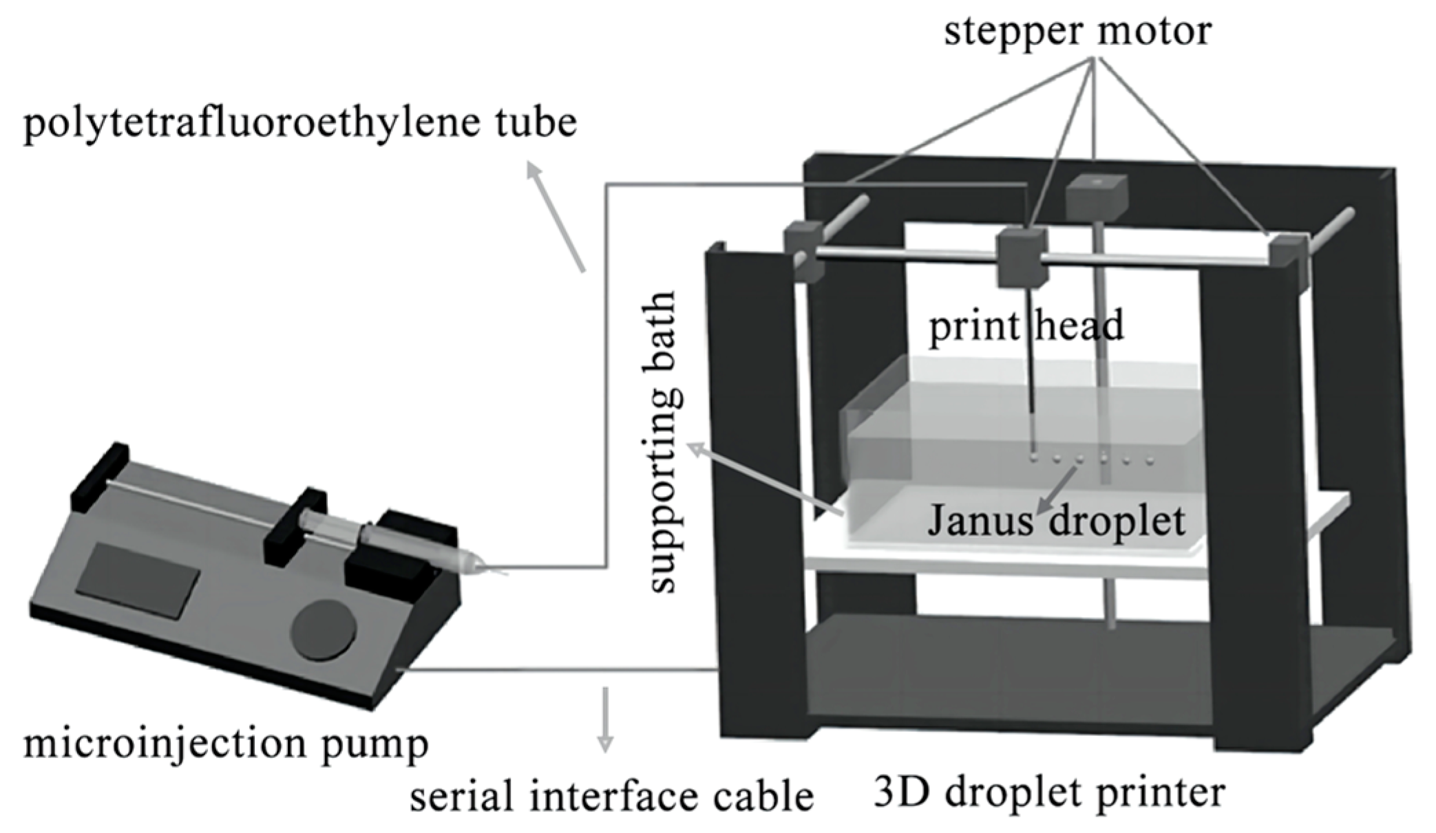
2.2. Methods
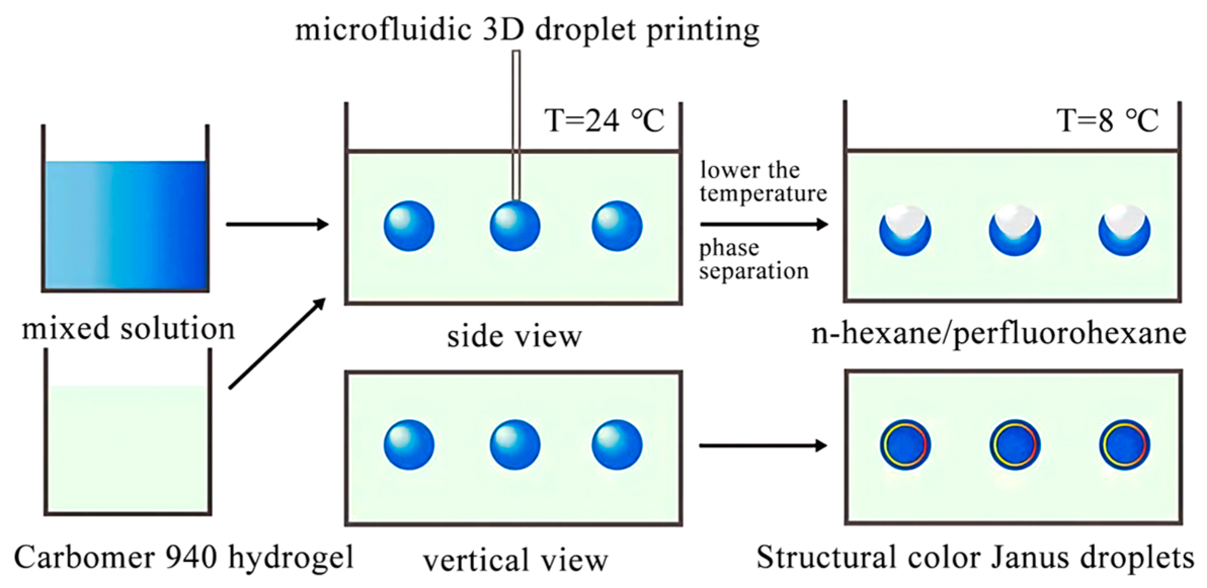
2.2.1. Preparation of Janus Droplets with Structural Color
2.2.2. Influence of Volume Ratio of the Oil Phase
2.2.3. Influence of Droplet Size
2.2.4. Influence of Surfactant Ratio
2.2.5. Statistical Analysis
3. Results and Discussion
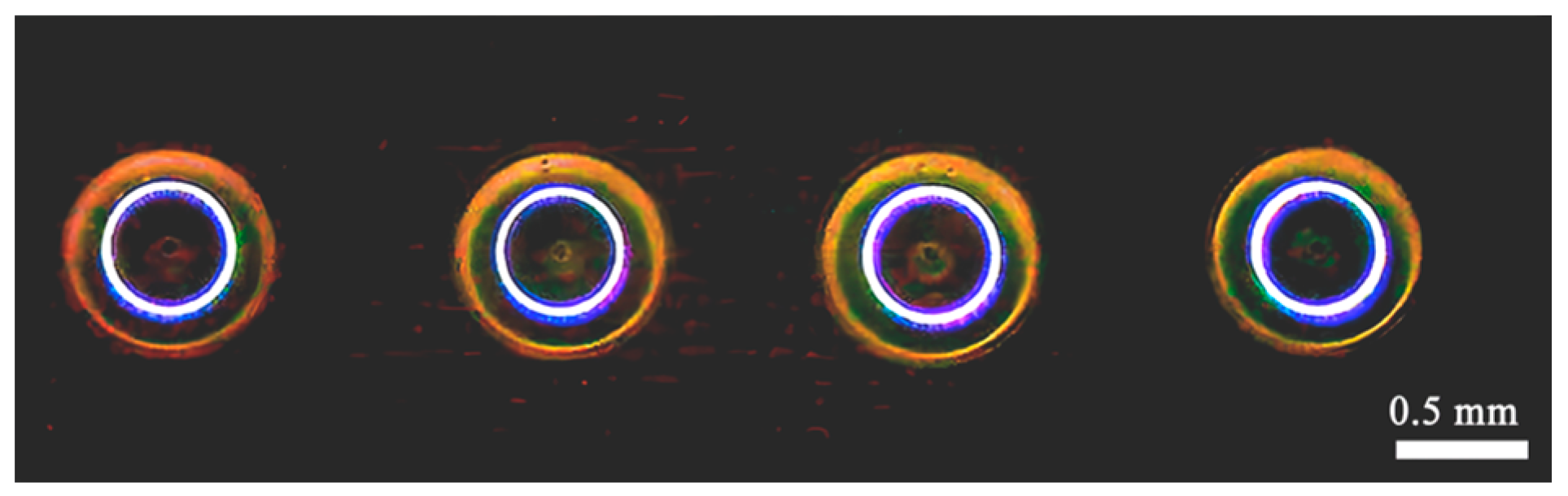
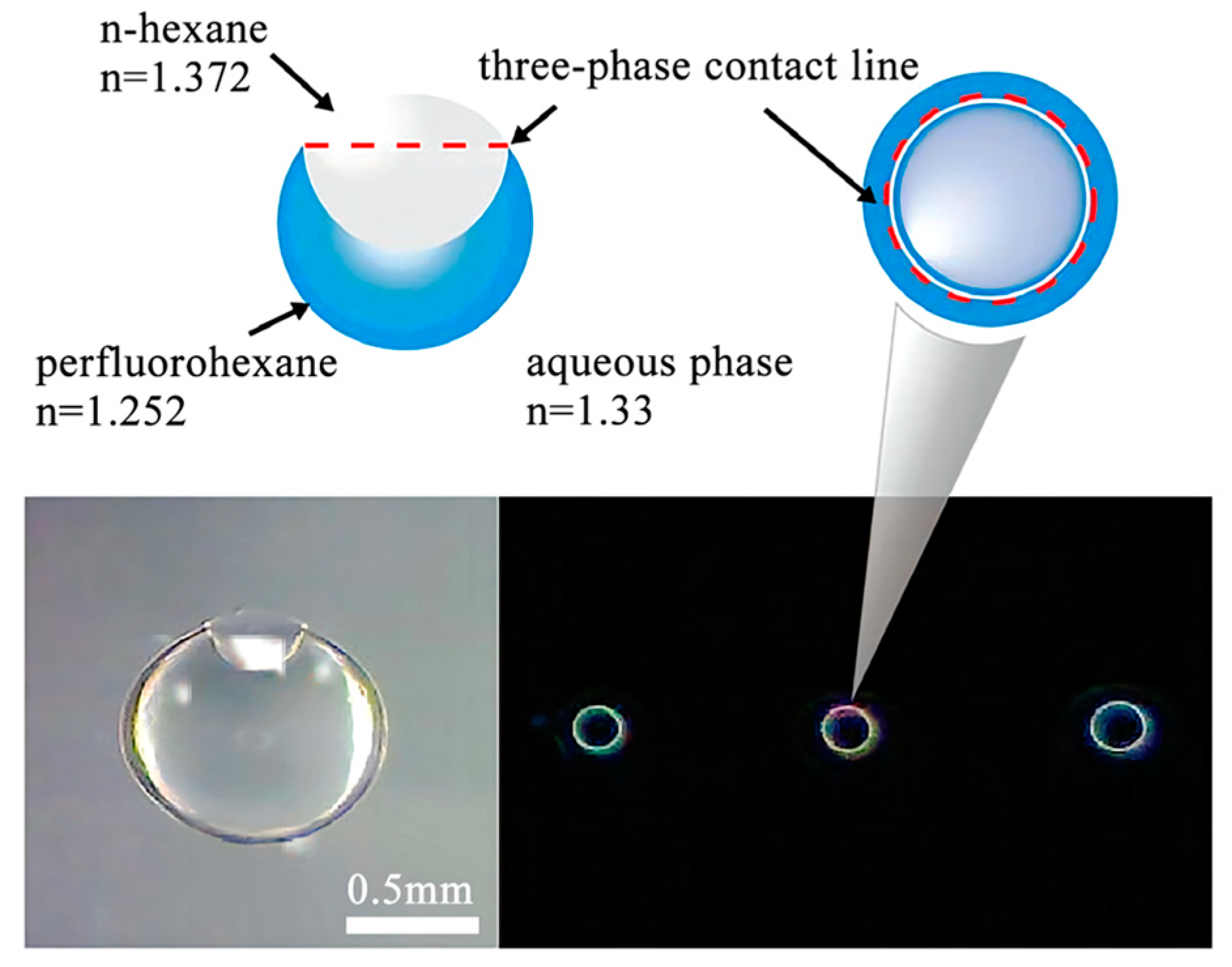
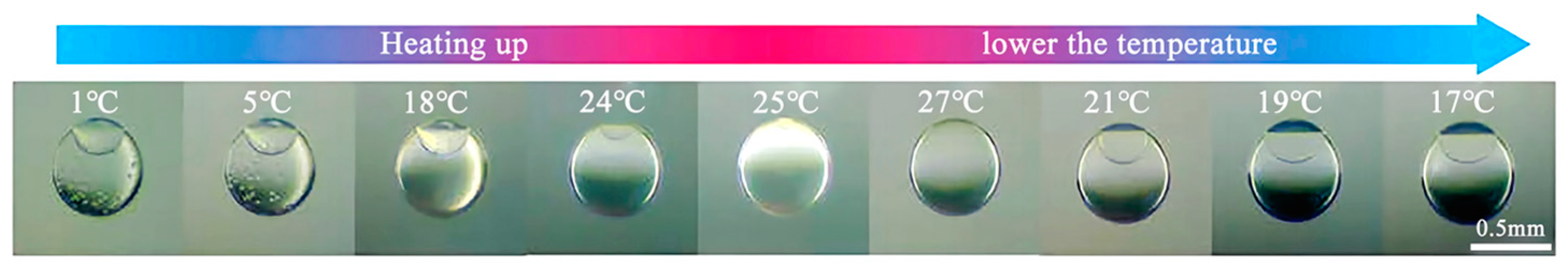
3.1. Structural Color of Janus Droplets
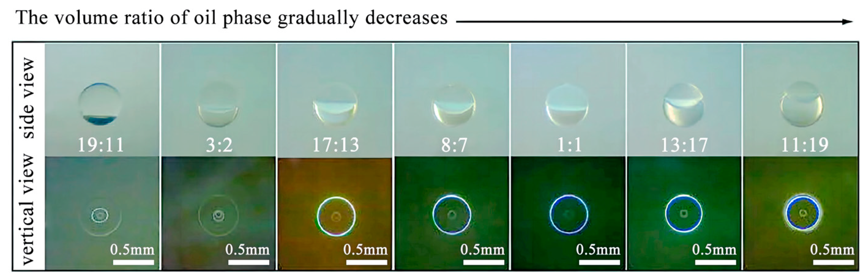
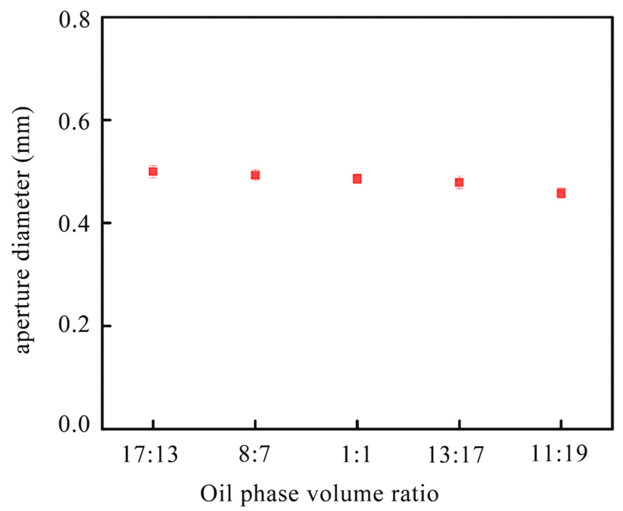
3.2. Effect of Oil Phase Volume Ratio on Janus Droplets
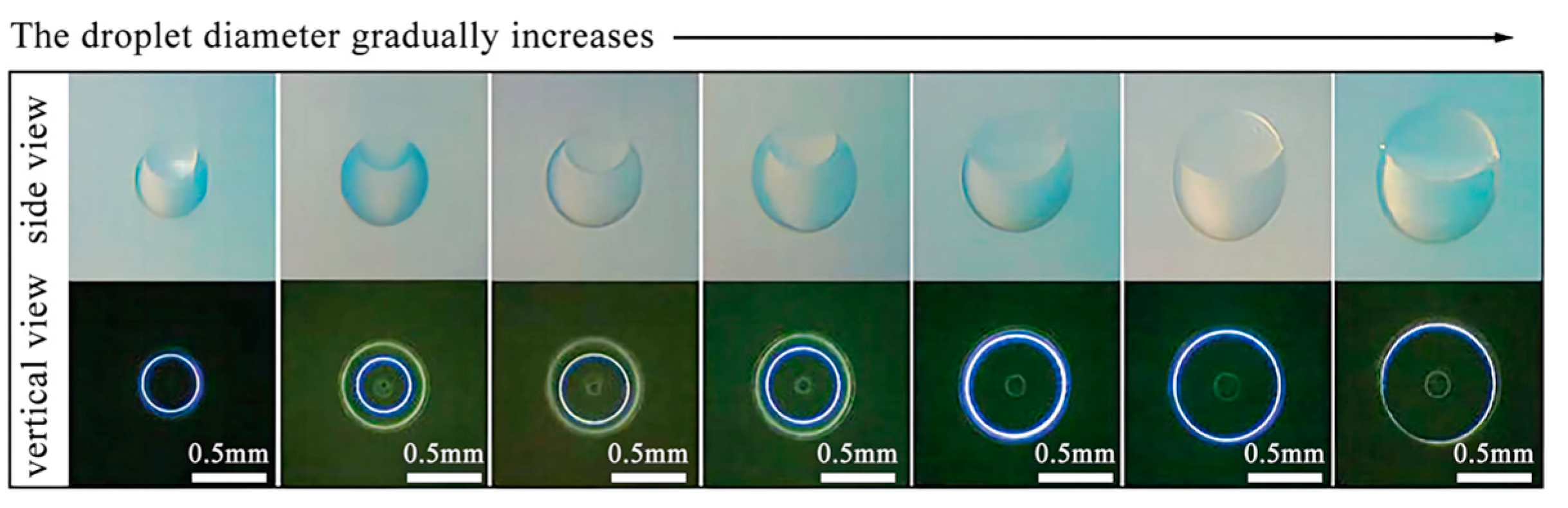
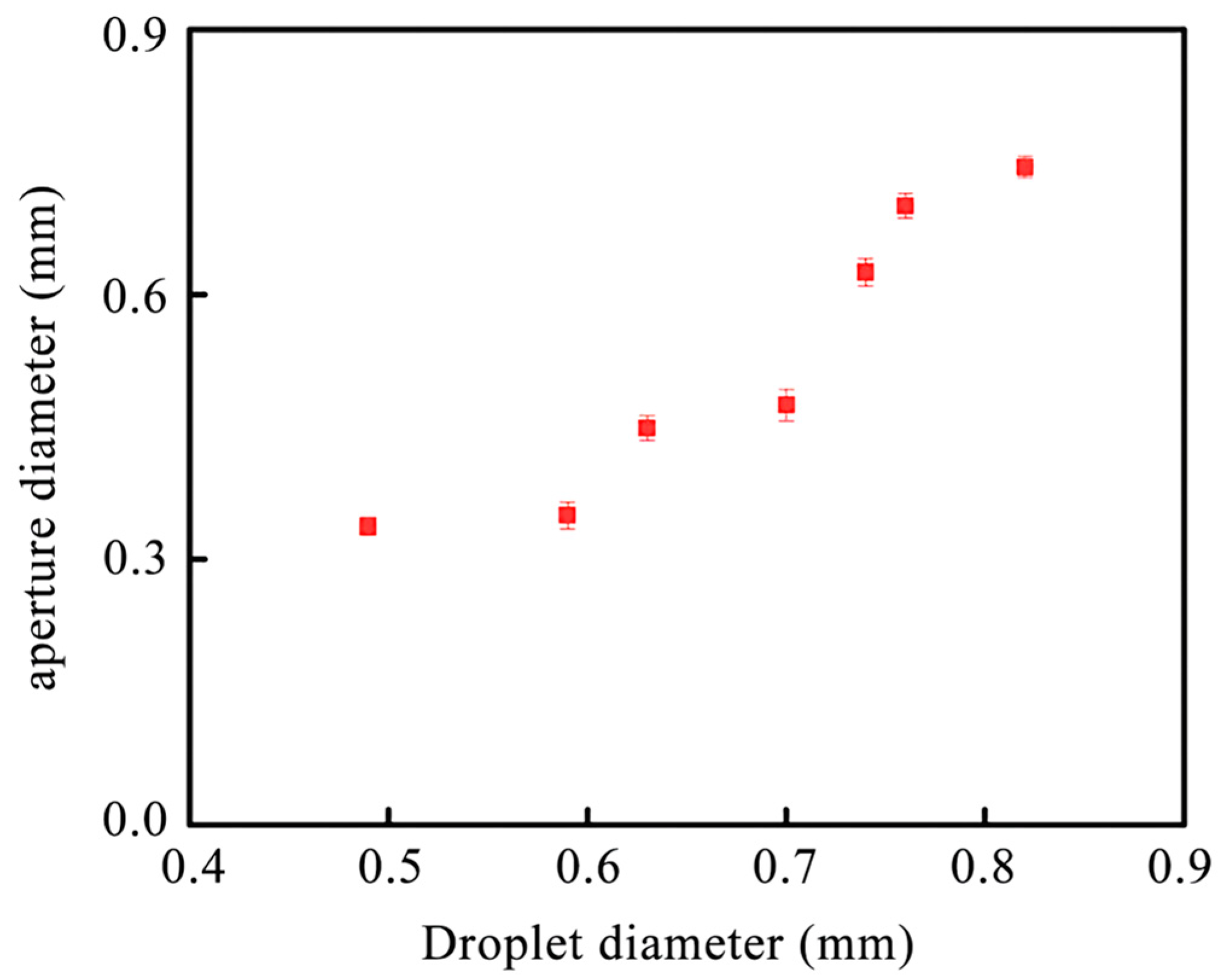
3.3. Influence of Droplet Size on Structure Color Aperture Diameter
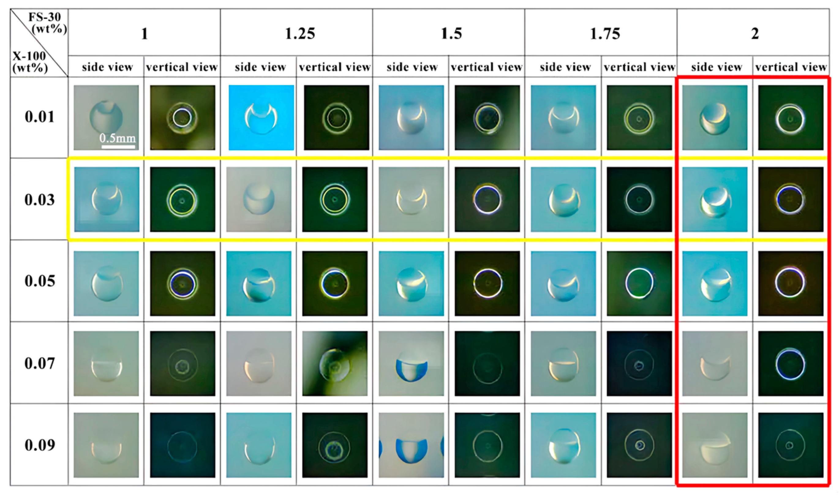
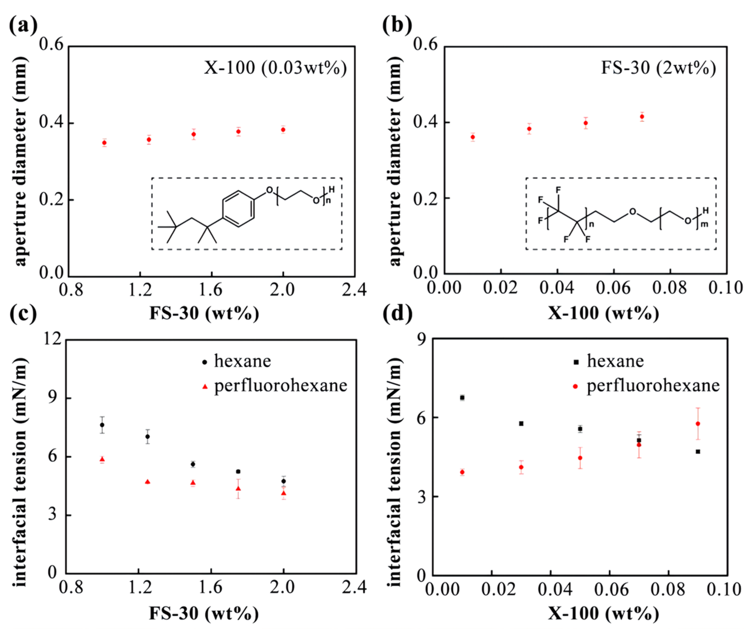
3.4. Influence of Surfactant Ratio on Janus Droplets
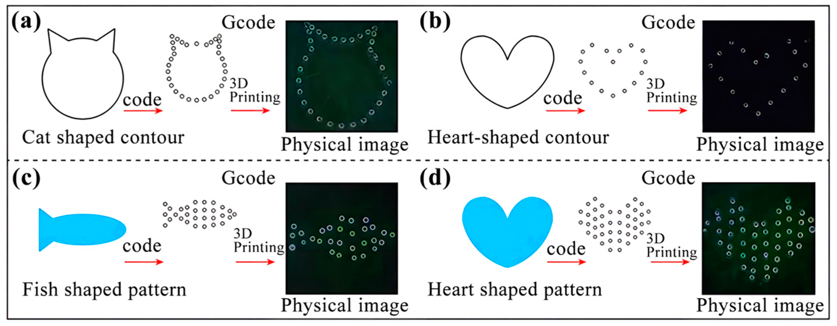
3.5. Patterned Printing
4. Conclusions
Author Contributions
Funding
Data Availability Statement
Conflicts of Interest
References
- Chen, J.; Liu, X.; Tian, Y.; Zhu, W.; Yan, C.; Shi, Y.; Kong, L.B.; Qi, H.J.; Zhou, K. 3D-Printed Anisotropic Polymer Materials for Functional Applications. Adv. Mater. 2022, 34, e2102877. [Google Scholar] [CrossRef]
- Finny, A.S.; Popoola, O.; Andreescu, S. 3D-Printable Nanocellulose-Based Functional Materials: Fundamentals and Applications. Nanomaterials 2021, 11, 2358. [Google Scholar] [CrossRef] [PubMed]
- Maslen, C.; Gholamipour-Shirazi, A.; Butler, M.D.; Kropacek, J.; Rehor, I.; Montenegro-Johnson, T. A New Class of Single-Material, Non-Reciprocal Microactuators. Macromol. Rapid Commun. 2023, 44, e2200842. [Google Scholar] [CrossRef] [PubMed]
- Lu, F.; Neal, E.A.; Nakanishi, T. Self-Assembled and Nonassembled Alkylated-Fullerene Materials. Acc. Chem. Res. 2019, 52, 1834–1843. [Google Scholar] [CrossRef] [PubMed]
- Wang, J.; Zhu, Z.; Liu, P.; Yi, S.; Peng, L.; Yang, Z.; Tian, X.; Jiang, L. Magneto-Responsive Shutter for On-Demand Droplet Manipulation. Adv. Sci. 2021, 8, e2103182. [Google Scholar] [CrossRef]
- Guo, Q.; Li, Y.; Liu, Q.; Li, Y.; Song, D.P. Janus Photonic Microspheres with Bridged Lamellar Structures via Droplet-Confined Block Copolymer Co-Assembly. Angew. Chem. 2022, 61, e202113759. [Google Scholar] [CrossRef]
- Qi, Y.; Zhou, C.; Qiu, Y.; Cao, X.; Niu, W.; Wu, S.; Zheng, Y.; Ma, W.; Ye, H.; Zhang, S. Biomimetic Janus photonic soft actuator with structural color self-reporting. Mater. Horiz. 2022, 9, 1243–1252. [Google Scholar] [CrossRef]
- Liu, M.; Fu, J.; Yang, S.; Wang, Y.; Jin, L.; Nah, S.H.; Gao, Y.; Ning, Y.; Murray, C.B.; Yang, S. Janus Microdroplets with Tunable Self-Recoverable and Switchable Reflective Structural Colors. Adv. Mater. 2023, 35, e2207985. [Google Scholar] [CrossRef]
- Sun, Q.; Hu, X.; Xu, B.; Lin, S.; Deng, X.; Zhou, S. Janus Charged Droplet Manipulation Mediated by Invisible Charge Walls. Adv. Sci. 2022, 9, e2204382. [Google Scholar] [CrossRef]
- Zhang, H.; Wang, F.; Nestler, B. Janus Droplet Formation via Thermally Induced Phase Separation: A Numerical Model with Diffusion and Convection. Langmuir ACS J. Surf. Colloids 2022, 38, 6882–6895. [Google Scholar] [CrossRef]
- Saqib, M.; Tran, P.A.; Ercan, B.; Erdem, E.Y. Microfluidic Methods in Janus Particle Synthesis. Int. J. Nanomed. 2022, 17, 4355–4366. [Google Scholar] [CrossRef]
- Zhang, H.; Qu, T.; Wang, H.; Wu, W.; Lu, F.; Ou, J.; Zhu, G.; Gao, L.; Cheng, L. Preparation of asymmetric Janus hollow silica microparticle and its application on oily wastewaters. Sci. Rep. 2023, 13, 4135–4147. [Google Scholar] [CrossRef] [PubMed]
- Kim, J.B.; Kim, J.W.; Kim, M.; Kim, S.H. Dual-Colored Janus Microspheres with Photonic and Plasmonic Faces. Small 2022, 18, e2201437. [Google Scholar] [CrossRef] [PubMed]
- Raju, R.R.; Koetz, J. Pickering Janus Emulsions Stabilized with Gold Nanoparticles. Langmuir ACS J. Surf. Colloids 2022, 38, 147–155. [Google Scholar] [CrossRef]
- Wei, X.; Cai, L.; Chen, H.; Shang, L.; Zhao, Y.; Sun, W. Noninvasive Multiplexed Analysis of Bladder Cancer-Derived Urine Exosomes via Janus Magnetic Microspheres. Anal. Chem. 2022, 94, 18034–18041. [Google Scholar] [CrossRef] [PubMed]
- Montalbán, M.G.; Collado-González, M.; Lozano-Pérez, A.A.; Baños, F.G.D.; Víllora, G. Density and refractive index data of binary and ternary mixtures of imidazolium-based ionic liquids, n-hexane and organic compounds involved in the kinetic resolution of rac-2-pentanol. Data Brief 2018, 19, 134–144. [Google Scholar] [CrossRef] [PubMed]
- Liu, Y.; Chen, F.; Guo, D.; Ma, Y. One-dimensional assembly of β-form anhydrous guanine microrods. Soft Matter 2021, 17, 1955–1962. [Google Scholar] [CrossRef] [PubMed]
- Lee, Y.; Duy, P.K.; Chung, H. Incorporating Non-NIR Absorbing Agent into Packed Powder Samples in Diffuse Reflectance NIR Measurement to Improve Representation of Sample Composition and Accuracy of Concentration Determination. Anal. Chem. 2020, 92, 1016–1023. [Google Scholar] [CrossRef]
- Teng, Z.; Wang, R.; Zhou, Y.; Kolios, M.; Wang, Y.; Zhang, N.; Wang, Z.; Zheng, Y.; Lu, G. A magnetic droplet vaporization approach using perfluorohexane-encapsulated magnetic mesoporous particles for ultrasound imaging and tumor ablation. Biomaterials 2017, 134, 43–50. [Google Scholar] [CrossRef]
- Kikkawa, Y.; Tsuzuki, S. Analysis of intermolecular interactions of n-perfluoroalkanes with circumcoronene using dispersion-corrected DFT calculations: Comparison with those of n-alkanes. Phys. Chem. Chem. Phys. 2023, 25, 11331–11337. [Google Scholar] [CrossRef]
- Morgado, P.; Garcia, A.R.; Martins, L.F.G.; Ilharco, L.M.; Filipe, E.J.M. Alkane Coiling in Perfluoroalkane Solutions: A New Primitive Solvophobic Effect. Langmuir ACS J. Surf. Colloids 2017, 33, 11429–11435. [Google Scholar] [CrossRef] [PubMed]
- Warr, C.A.; Hinnen, H.S.; Avery, S.; Cate, R.J.; Nordin, G.P.; Pitt, W.G. 3D-Printed Microfluidic Droplet Generator with Hydrophilic and Hydrophobic Polymers. Micromachines 2021, 12, 91. [Google Scholar] [CrossRef] [PubMed]
- Weigel, N.; Männel, M.J.; Thiele, J. Flexible Materials for High-Resolution 3D Printing of Microfluidic Devices with Integrated Droplet Size Regulation. ACS Appl. Mater. Interfaces 2021, 13, 31086–31101. [Google Scholar] [CrossRef] [PubMed]
- Ying, Y.; Huang, Z.; Tu, Y.; Wu, Q.; Li, Z.; Zhang, Y.; Yu, H.; Zeng, A.; Huang, H.; Ye, J.; et al. A shear-thinning, ROS-scavenging hydrogel combined with dental pulp stem cells promotes spinal cord repair by inhibiting ferroptosis. Bioact. Mater. 2023, 22, 274–290. [Google Scholar] [CrossRef] [PubMed]
- Hayati, F.; Ghamsari, S.M.; Dehghan, M.M.; Oryan, A. Effects of carbomer 940 hydrogel on burn wounds: An in vitro and in vivo study. J. Dermatol. Treat. 2018, 29, 593–599. [Google Scholar] [CrossRef]
- Huang, Y.; Shi, F.; Wang, L.; Yang, Y.; Khan, B.M.; Cheong, K.L.; Liu, Y. Preparation and evaluation of Bletilla striata polysaccharide/carboxymethyl chitosan/Carbomer 940 hydrogel for wound healing. Int. J. Biol. Macromol. 2019, 132, 729–737. [Google Scholar] [CrossRef]
- Lasky, J.A.; Thannickal, V.J. NOTCH-ing up Surface Tension in the Fibrotic Lung. Am. J. Respir. Crit. Care Med. 2023, 207, 235–236. [Google Scholar] [CrossRef]
- Lin, J.J.; Kristensen, T.B.; Calderón, S.M.; Malila, J.; Prisle, N.L. Effects of surface tension time-evolution for CCN activation of a complex organic surfactant. Environ. Science. Process. Impacts 2020, 22, 271–284. [Google Scholar] [CrossRef]
- Hsieh, T.L.; Law, S.; Garoff, S.; Tilton, R.D. pH-Dependent Interfacial Tension and Dilatational Modulus Synergism of Oil-Soluble Fatty Acid and Water-Soluble Cationic Surfactants at the Oil/Water Interface. Langmuir ACS J. Surf. Colloids 2021, 37, 11573–11581. [Google Scholar] [CrossRef]
- Strickley, R.G.; Lambert, W.J. A review of Formulations of Commercially Available Antibodies. J. Pharm. Sci. 2021, 110, 2590–2608. [Google Scholar] [CrossRef]
- Lbadaoui-Darvas, M.; Idrissi, A.; Jedlovszky, P. Computer Simulation of the Surface of Aqueous Ionic and Surfactant Solutions. J. Phys. Chem. B 2022, 126, 751–765. [Google Scholar] [CrossRef] [PubMed]
- Tucker, I.M.; Burley, A.; Petkova, R.E.; Hosking, S.L.; Webster, J.R.P.; Li, P.X.; Ma, K.; Doutch, J.; Penfoldoo, J.; Thomas, R.K. Self-assembly in escin-nonionic surfactant mixtures: From micelles to vesicles. J. Colloid Interface Sci. 2022, 626, 305–313. [Google Scholar] [CrossRef] [PubMed]
- Nguyen, T.X.D.; Razavi, S.; Papavassiliou, D.V. Janus Nanoparticle and Surfactant Effects on Oil Drop Migration in Water under Shear. J. Phys. Chem. B 2022, 126, 6314–6323. [Google Scholar] [CrossRef]
- Lin, X.; Wu, H.; Zeng, S.; Peng, T.; Zhang, P.; Wan, X.; Lang, Y.; Zhang, B.; Jia, Y.; Shen, R.; et al. A self-designed device integrated with a Fermat spiral microfluidic chip for ratiometric and automated point-of-care testing of anthrax biomarker in real samples. Biosens. Bioelectron. 2023, 230, 115283. [Google Scholar] [CrossRef] [PubMed]
- Yin, B.; Yue, W.; Sohan, A.; Wan, X.; Zhou, T.; Shi, L.; Qian, C.; Lin, X. Construction of a desirable hyperbolic microfluidic chip for ultrasensitive determination of PCT based on chemiluminescence. J. Mater. Chem. B 2023, 11, 1978–1986. [Google Scholar] [CrossRef] [PubMed]
- Zeng, S.; Sun, X.; Wan, X.; Qian, C.; Yue, W.; Sohan, A.; Lin, X.; Yin, B. A cascade Fermat spiral microfluidic mixer chip for accurate detection and logic discrimination of cancer cells. The Analyst 2022, 147, 3424–3433. [Google Scholar] [CrossRef]
- Yin, B.; Wan, X.; Sohan, A.; Lin, X. Microfluidics-Based POCT for SARS-CoV-2 Diagnostics. Micromachines 2022, 13, 1238. [Google Scholar] [CrossRef]
- Yin, B.; Wan, X.; Qian, C.; Sohan, A.; Zhou, T.; Yue, W. Enzyme Method-Based Microfluidic Chip for the Rapid Detection of Copper Ions. Micromachines 2021, 12, 1380. [Google Scholar] [CrossRef]
- Zhang, K.; Ren, Y.; Jiang, T.; Jiang, H. Flexible fabrication of lipophilic-hydrophilic micromotors by off-chip photopolymerization of three-phase immiscible flow induced Janus droplet templates. Anal. Chim. Acta 2021, 1182, 338955. [Google Scholar] [CrossRef]
- Jiang, S.; Hu, Y.; Wu, H.; Zhang, Y.; Zhang, Y.; Wang, Y.; Zhang, Y.; Zhu, W.; Li, J.; Wu, D.; et al. Multifunctional Janus Microplates Arrays Actuated by Magnetic Fields for Water/Light Switches and Bio-Inspired Assimilatory Coloration. Adv. Mater. 2019, 31, e1807507. [Google Scholar] [CrossRef]
- Bai, F.; Zhang, H.; Li, X.; Li, F.; Joo, S.W. Generation and Dynamics of Janus Droplets in Shear-Thinning Fluid Flow in a Double Y-Type Microchannel. Micromachines 2021, 12, 149. [Google Scholar] [CrossRef]
- Sun, X.T.; Guo, R.; Wang, D.N.; Wei, Y.Y.; Yang, C.G.; Xu, Z.R. Microfluidic preparation of polymer-lipid Janus microparticles with staged drug release property. J. Colloid Interface Sci. 2019, 553, 631–638. [Google Scholar] [CrossRef]
- Chen, C.M.; Chang, C.H.; Chao, C.H.; Wang, M.H.; Yeh, T.F. Biophysical and chemical stability of surfactant/budesonide and the pulmonary distribution following intra-tracheal administration. Drug Deliv. 2019, 26, 604–611. [Google Scholar] [CrossRef]
- Milionis, A.; Antonini, C.; Jung, S.; Nelson, A.; Schutzius, T.M.; Poulikakos, D. Contactless Transport and Mixing of Liquids on Self-Sustained Sublimating Coatings. Langmuir ACS J. Surf. Colloids 2017, 33, 1799–1809. [Google Scholar] [CrossRef]
- Li, H.; Fauquignon, M.; Haddou, M.; Schatz, C.; Chapel, J.P. Interfacial Behavior of Solid- and Liquid-like Polyelectrolyte Complexes as a Function of Charge Stoichiometry. Polymers 2021, 13, 3848. [Google Scholar] [CrossRef]


| Group | Oil Phase Volume Ratio (n-Hexane/Perfluorohexane) | Diameter (mm) | FS-30 (wt%) | X-100 (wt%) |
|---|---|---|---|---|
| 1 | 19:11 (1.727) | 0.569 | 1.5 | 0.05 |
| 2 | 3:2 (1.5) | 0.569 | 1.5 | 0.05 |
| 3 | 17:13 (1.308) | 0.569 | 1.5 | 0.05 |
| 4 | 8:7 (1.143) | 0.569 | 1.5 | 0.05 |
| 5 | 1:1 (1.0) | 0.569 | 1.5 | 0.05 |
| 6 | 13:17 (0.765) | 0.569 | 1.5 | 0.05 |
| 7 | 11:19 (0.579) | 0.569 | 1.5 | 0.05 |
| Group | Diameter (mm) | Oil Phase Volume Ratio (n-Hexane/Perfluorohexane) | FS-30 (wt%) | X-100 (wt%) |
|---|---|---|---|---|
| 1 | 0.487 | 3:7 | 1.5 | 0.05 |
| 2 | 0.589 | 3:7 | 1.5 | 0.05 |
| 3 | 0.638 | 3:7 | 1.5 | 0.05 |
| 4 | 0.715 | 3:7 | 1.5 | 0.05 |
| 5 | 0.745 | 3:7 | 1.5 | 0.05 |
| 6 | 0.770 | 3:7 | 1.5 | 0.05 |
| 7 | 0.825 | 3:7 | 1.5 | 0.05 |
Disclaimer/Publisher’s Note: The statements, opinions and data contained in all publications are solely those of the individual author(s) and contributor(s) and not of MDPI and/or the editor(s). MDPI and/or the editor(s) disclaim responsibility for any injury to people or property resulting from any ideas, methods, instructions or products referred to in the content. |
© 2023 by the authors. Licensee MDPI, Basel, Switzerland. This article is an open access article distributed under the terms and conditions of the Creative Commons Attribution (CC BY) license (https://creativecommons.org/licenses/by/4.0/).
Share and Cite
Wu, C.; Jia, H.; Almuaalemi, H.Y.M.; Sohan, A.S.M.M.F.; Yin, B. RETRACTED: Preparation and Analysis of Structured Color Janus Droplets Based on Microfluidic 3D Droplet Printing. Micromachines 2023, 14, 1911. https://doi.org/10.3390/mi14101911
Wu C, Jia H, Almuaalemi HYM, Sohan ASMMF, Yin B. RETRACTED: Preparation and Analysis of Structured Color Janus Droplets Based on Microfluidic 3D Droplet Printing. Micromachines. 2023; 14(10):1911. https://doi.org/10.3390/mi14101911
Chicago/Turabian StyleWu, Chuang, Hanqi Jia, Haithm Yahya Mohammed Almuaalemi, A. S. M. Muhtasim Fuad Sohan, and Binfeng Yin. 2023. "RETRACTED: Preparation and Analysis of Structured Color Janus Droplets Based on Microfluidic 3D Droplet Printing" Micromachines 14, no. 10: 1911. https://doi.org/10.3390/mi14101911