
Micro/Nano Periodic Surface Structures and Performance of Stainless Steel Machined Using Femtosecond Lasers

Abstract
:1. Introduction
2. Preparation of Micro/Nano Periodic Surface Structures
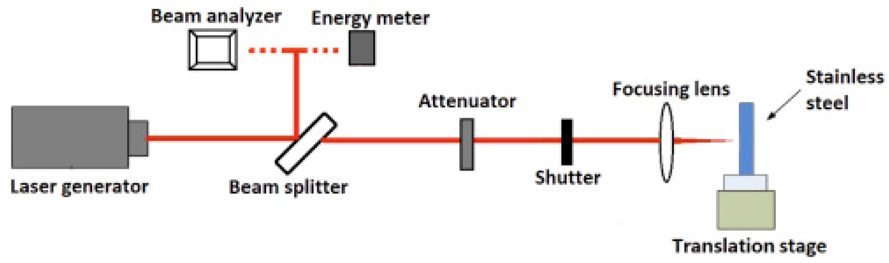
2.1. Machines, Equipment, and Type of Laser
2.2. Micro/Nanoripple Structure
2.2.1. Ablation Threshold and Laser Fluence
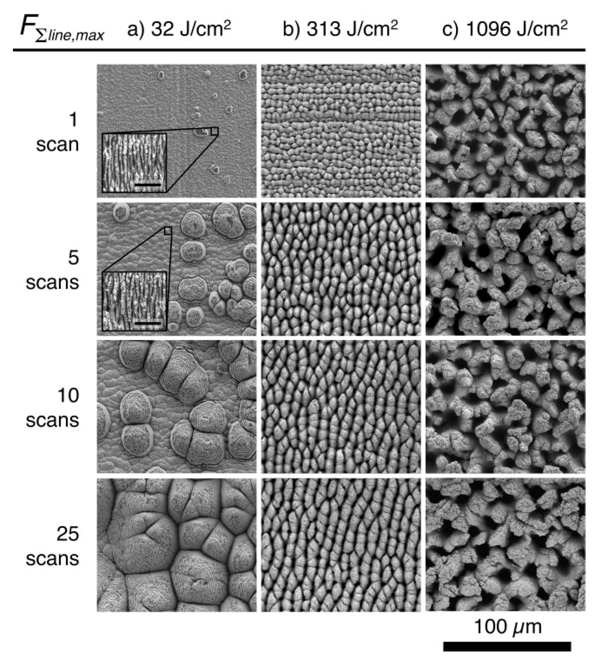
2.2.2. Pulse Number/Scan Number
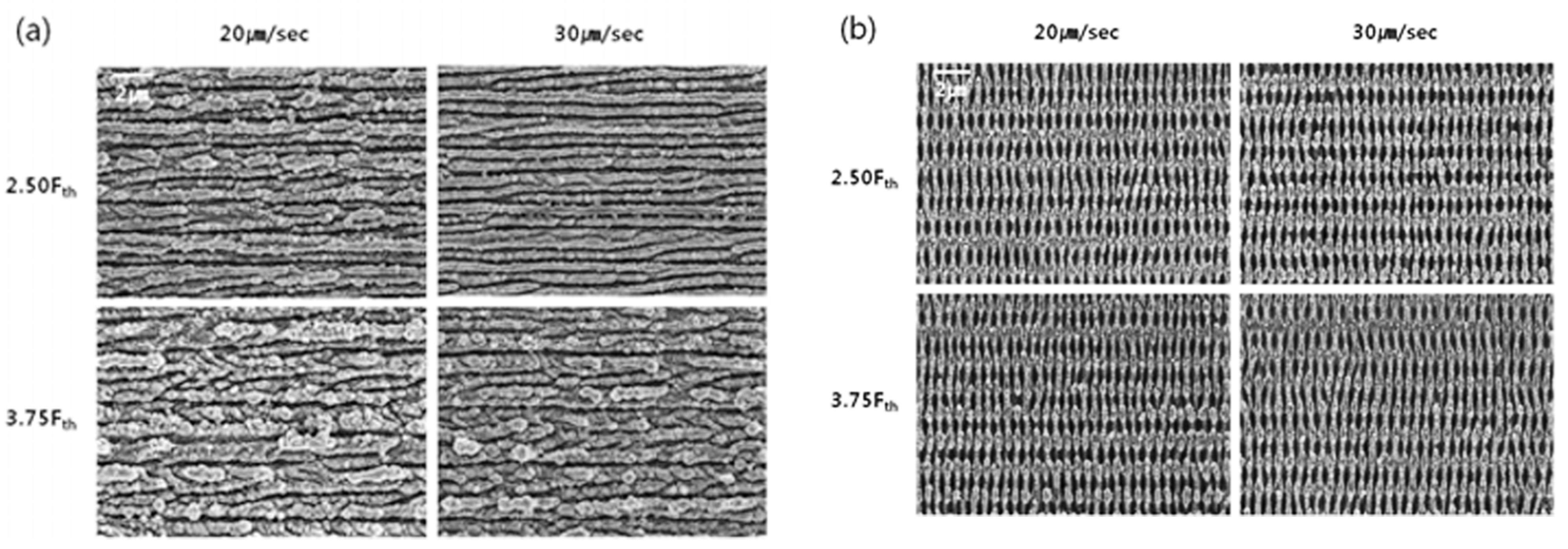
2.2.3. Scan Speed
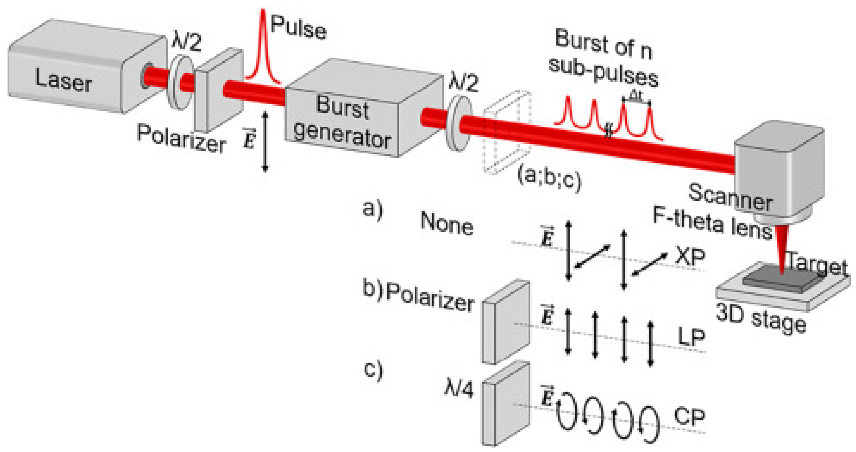
2.2.4. Pulse Bursts
2.2.5. Experimental Environment
2.3. Other Structures
3. Micro/Nano Periodic Surface Structure Performance
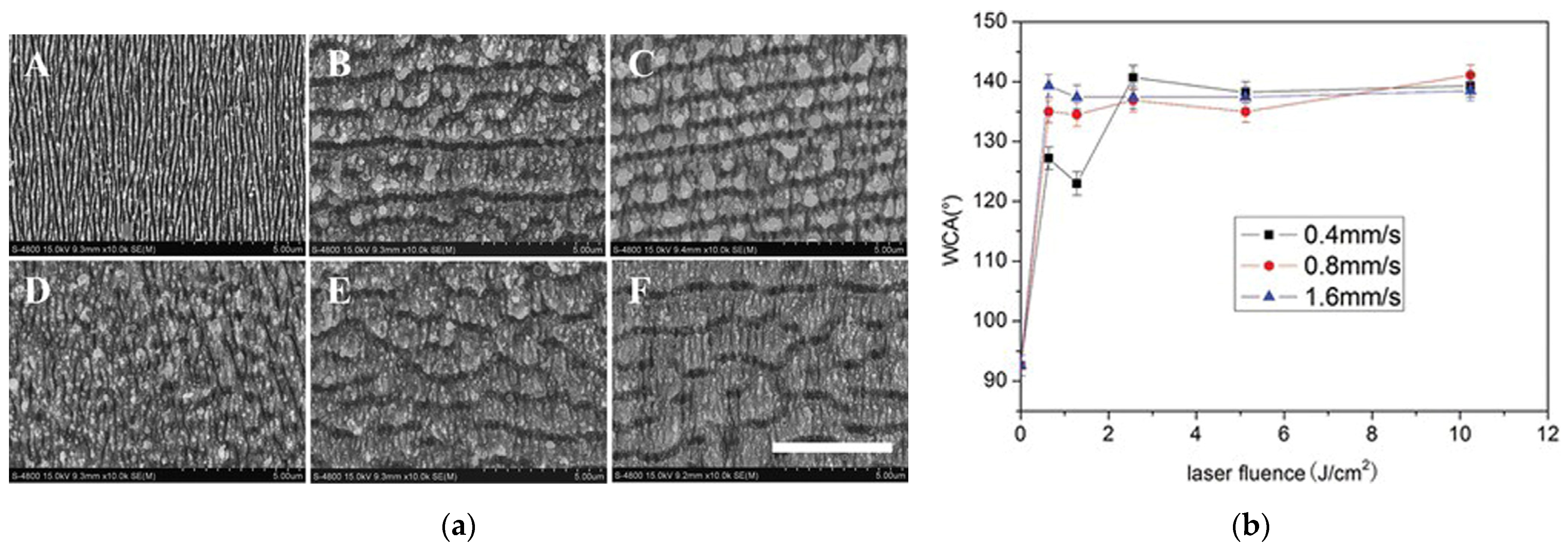
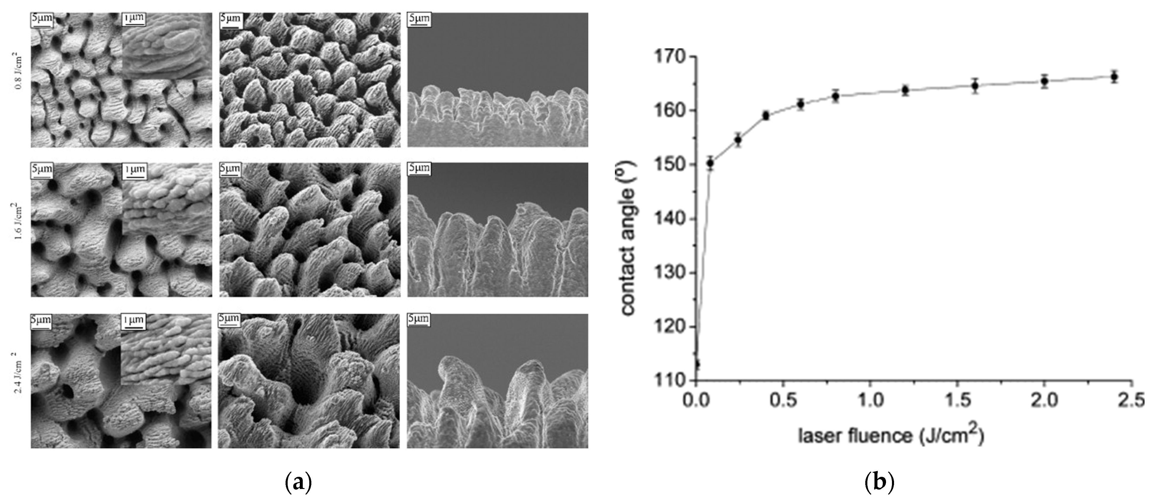
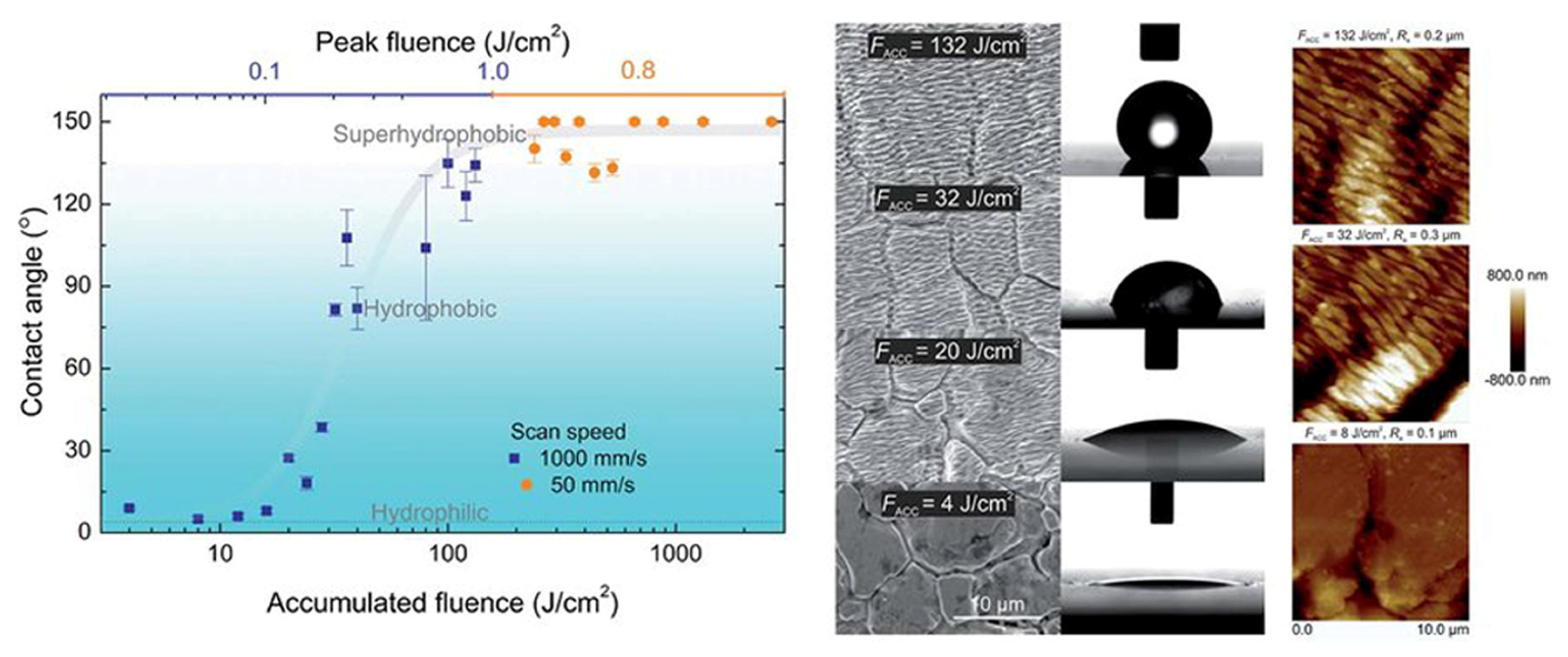
3.1. Wetting Performance
3.2. Other Performances
4. Several Laws and Challenges Revealed by the Research
4.1. Micro/Nano Periodic Structure Evolution
4.2. Effects of the Femtosecond Laser Parameters
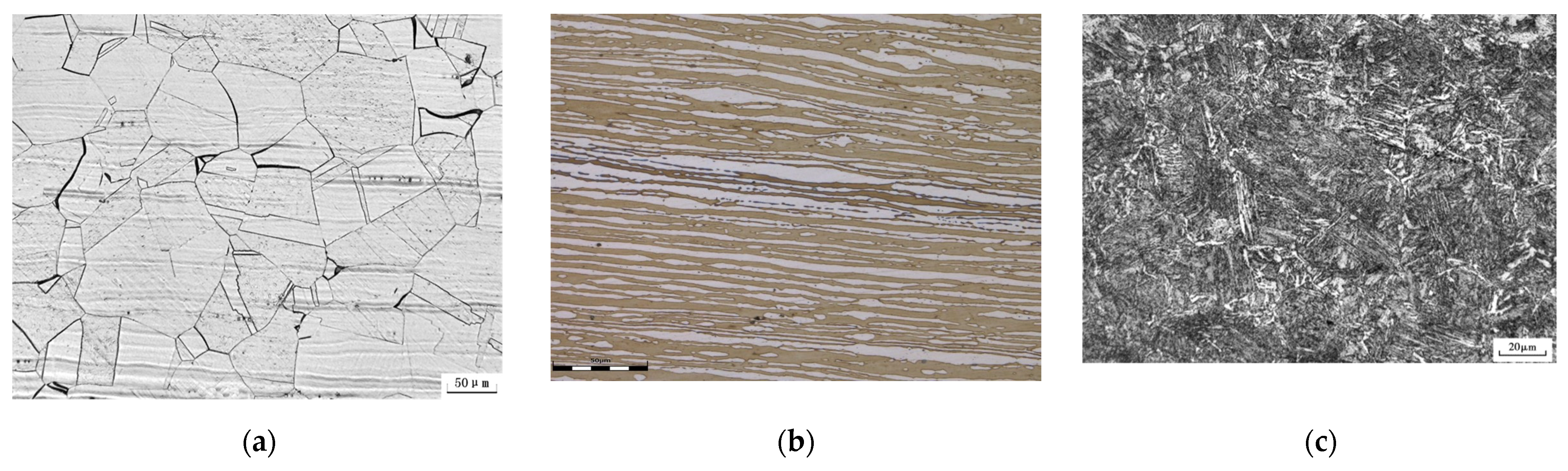
4.3. Influence of Materials’ Condition and Microstructure
5. Conclusions
- (1)
- The shape, size, and period of the micro/nano periodic structures produced on the surface of femtosecond laser ablation materials are not only closely related to the properties of the material itself, but also depend on the experimental research environment and the laser parameters used. Surface structures with different morphology and periods have been prepared under different experimental environments and laser-processing parameters. The researchers focused on the laser fluence, pulse number, scanning speed, bursts of pulses, and processing environment, and gained some regularity of understanding.
- (2)
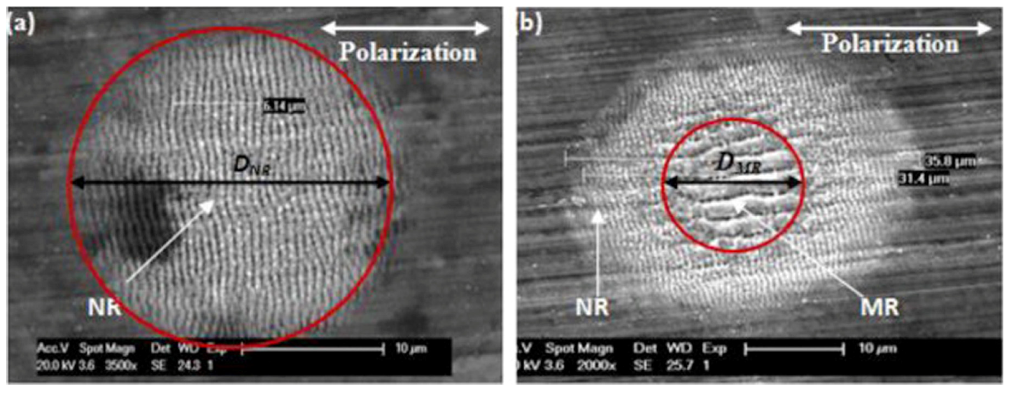
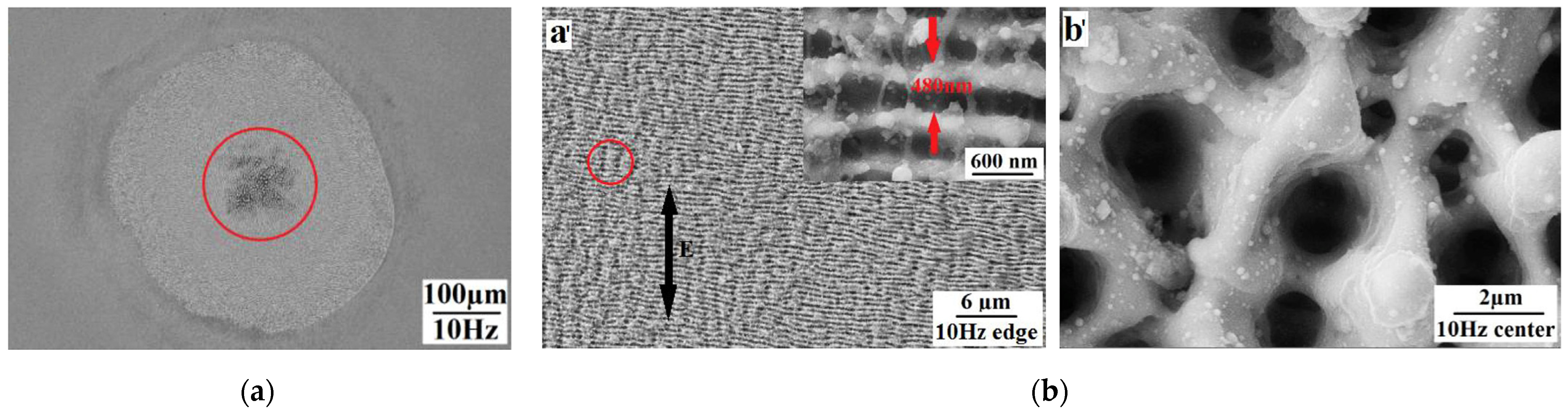
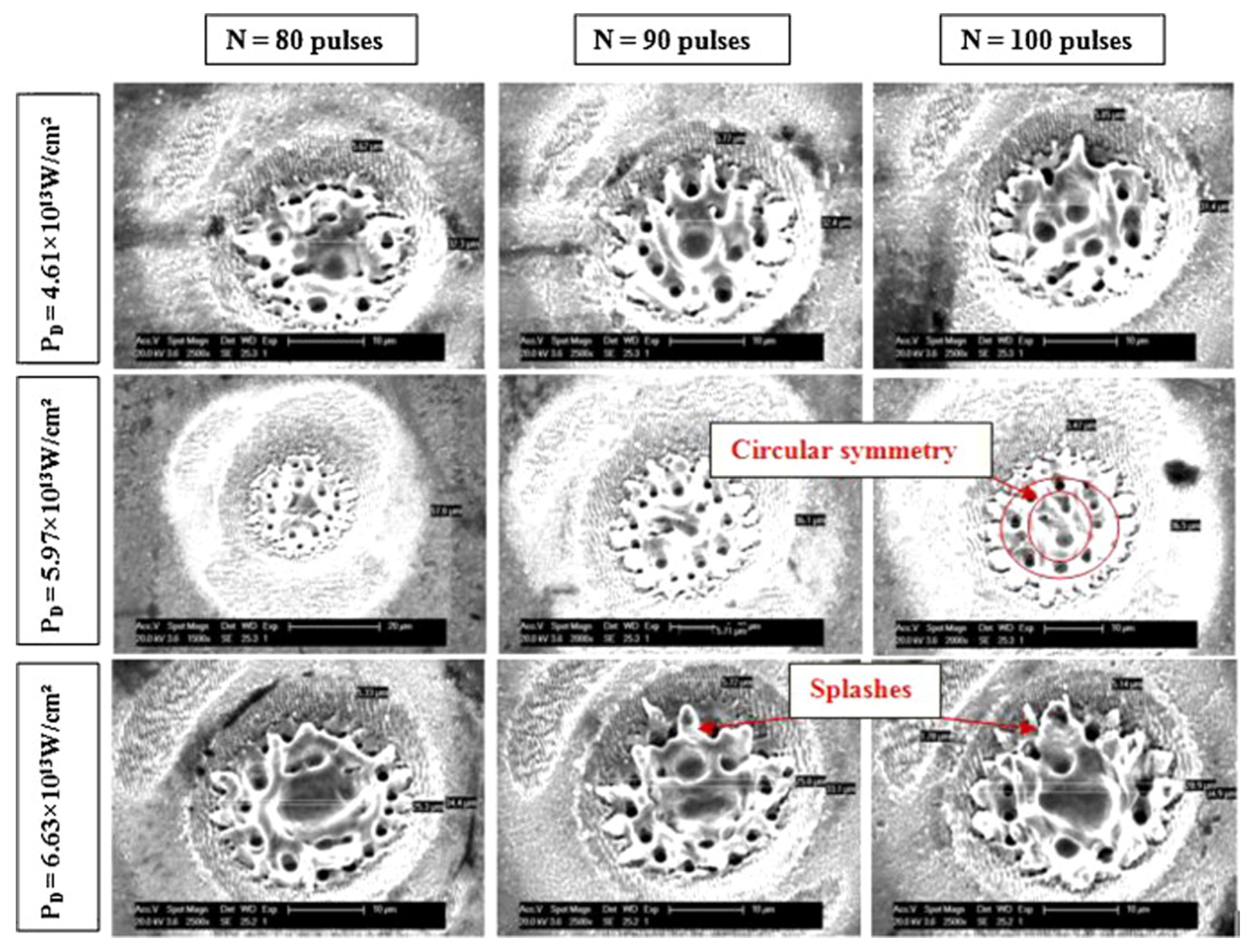
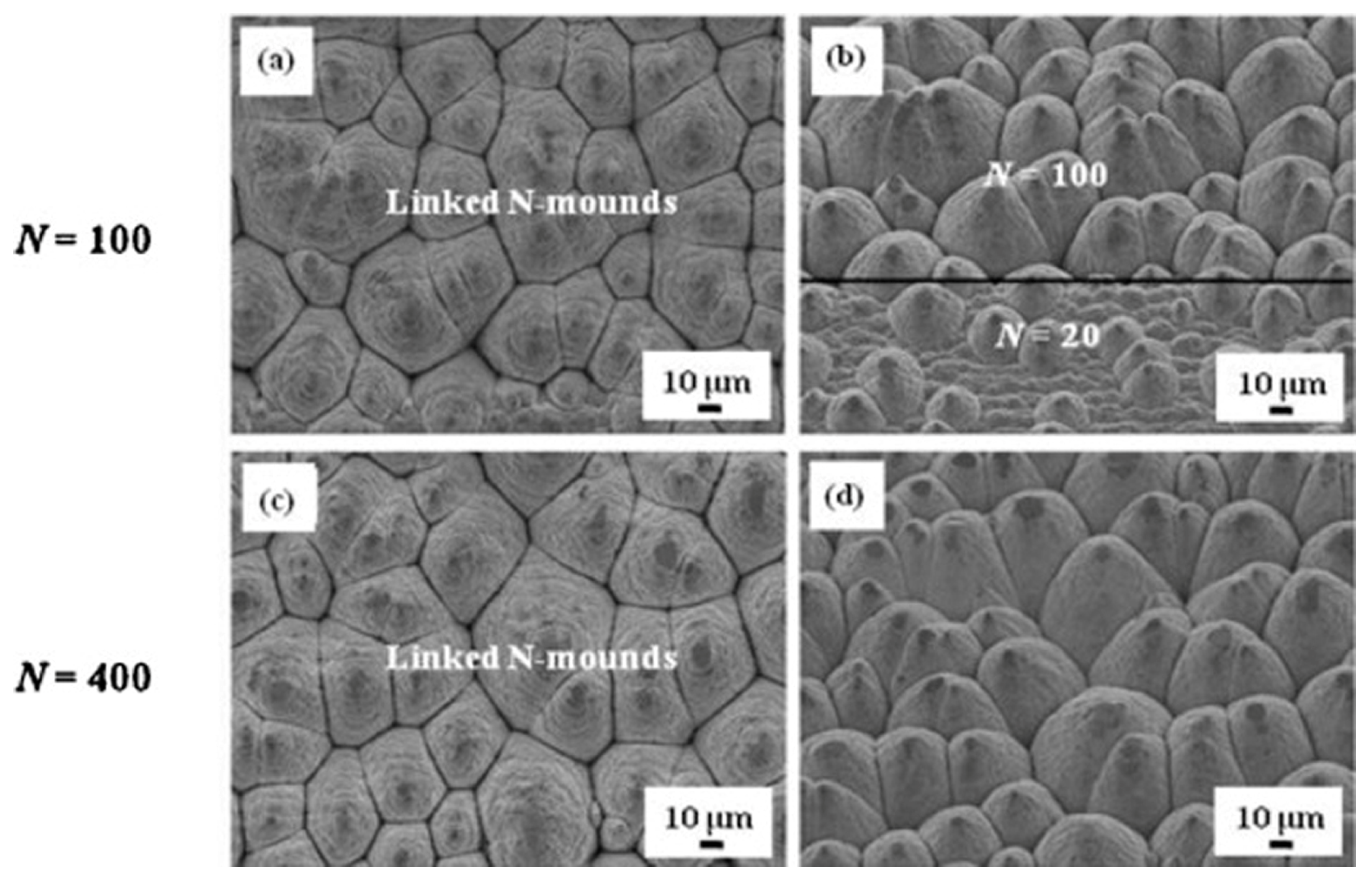
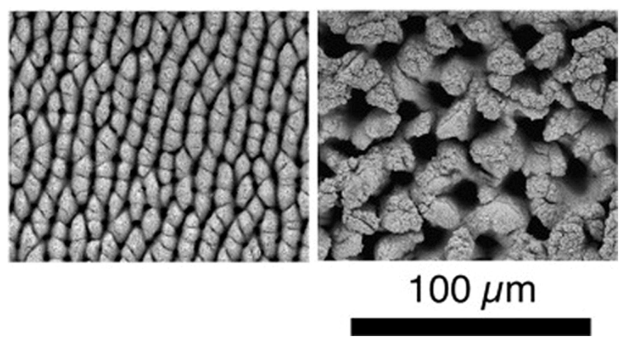
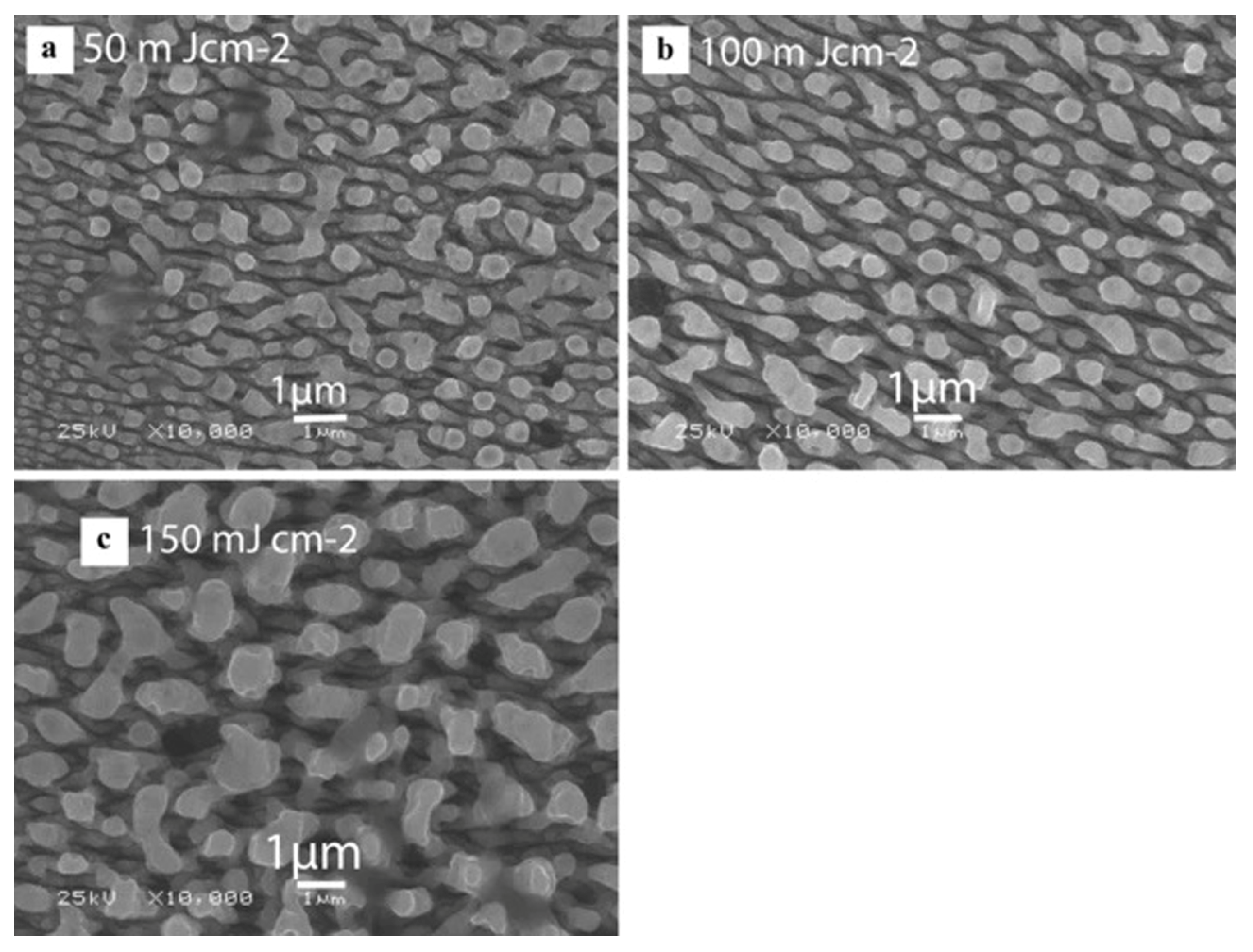
- In a lower laser fluence condition, the nanoscale LIPSS structure and the microscale groove structure can be obtained. The period and direction of the ripple structure will change regularly with the laser parameters. The ablation threshold of nano HSFLs (∥) is the lowest, and with an increase in the laser fluence, will gradually change to nano LSFLs (⊥) and then to microripples/grooves (∥). At a higher laser fluence, micro and nano hierarchical structures can be obtained, such as the moth-eye structure, porous mesh structure, mound/conical structure, and columnar structure.
- (3)
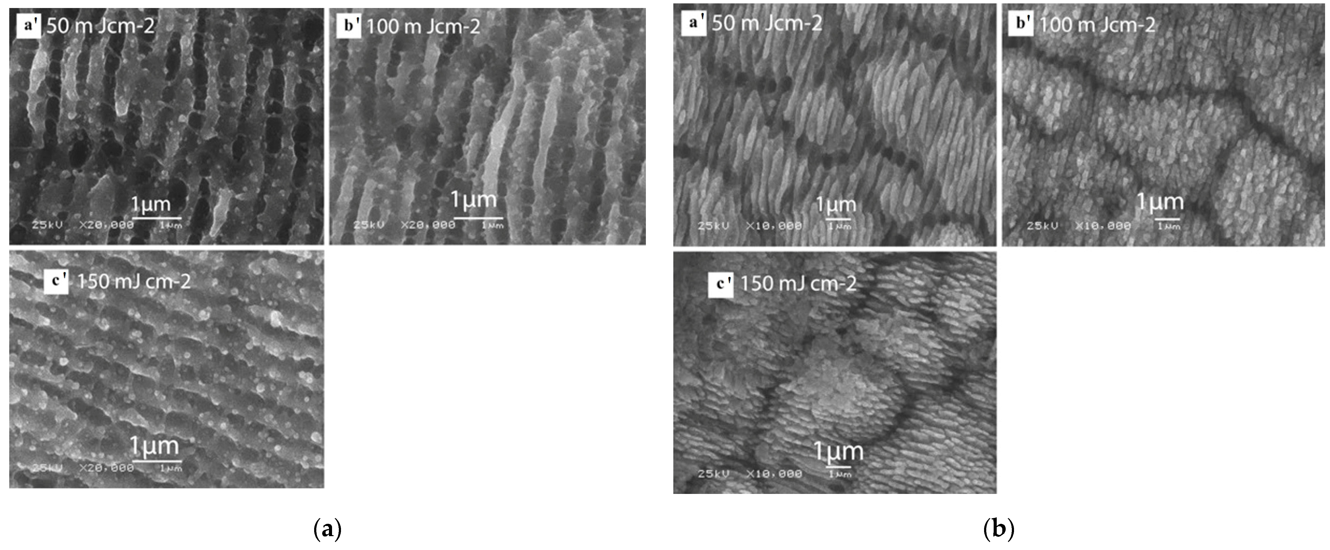
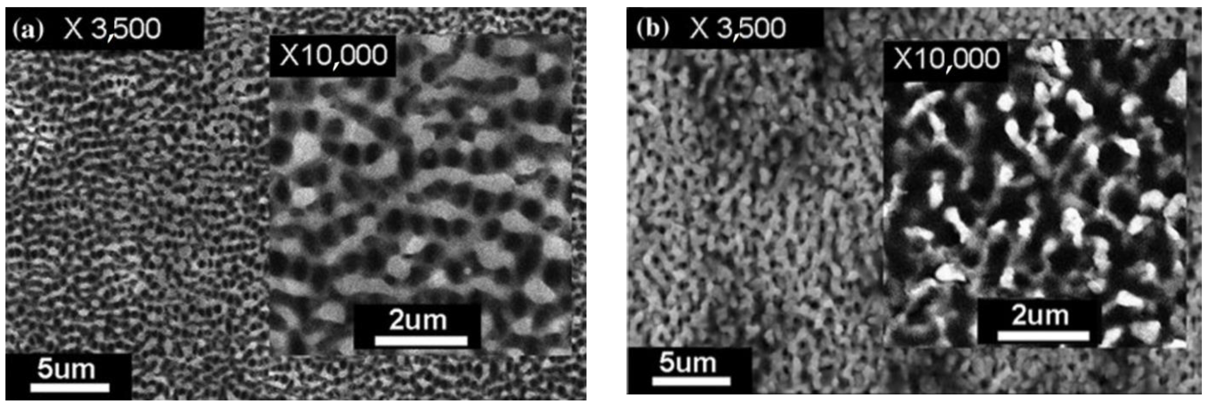
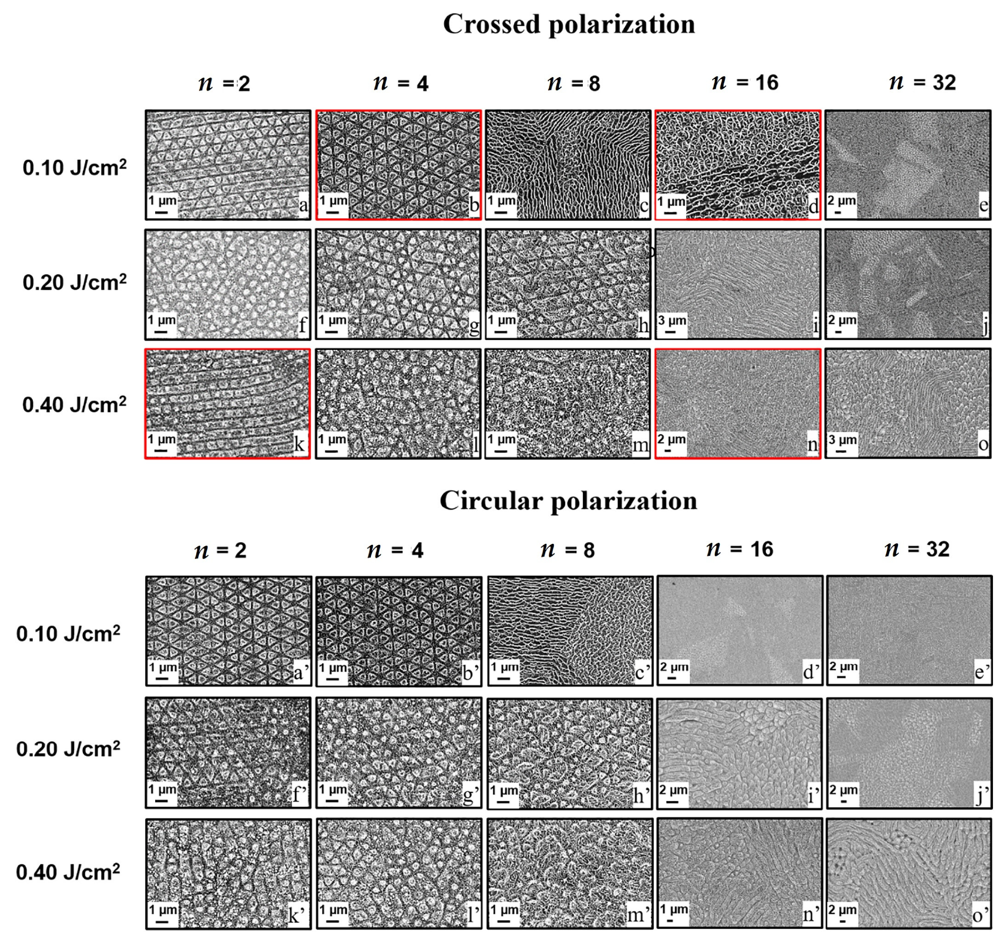
- The ripple structures formed in an air environment are relatively continuous and neat, while the ripple structures formed in a liquid environment such as deionized water and ethanol become discontinuous due to the uneven energy distribution and hydrodynamic effects, and are cut into short strips and dumbbell structures. Cross-polarization and circular-polarization femtosecond lasers are used to prepare a more complicated and changeable structure.
- (4)
- Compared with an ordinary material surface, the femtosecond-laser-induced micro/nano periodic surface structure shows different physical and chemical properties. Focusing on wetting performance, corrosion performance, and friction properties, the research confirmed that micro/nano multiple-roughness hierarchical structures showed excellent properties and have a bright application prospect. However, there are still some challenges in practical engineering applications. For example, the surface state (roughness) and microstructure (crystal boundary distribution) of metal structural materials have an important impact on the preparation and performance of micro/nano periodic surface structures, and require further in-depth research and exploration.
Author Contributions
Funding
Conflicts of Interest
References
- Mao, L.; Gao, H.; Yao, H.; Liu, L.; Cölfen, H.; Liu, G.; Chen, S.; Li, S.; Yan, Y.; Liu, Y.; et al. Synthetic nacre by predesigned matrix-directed mineralization. Science 2016, 354, 107–110. [Google Scholar] [CrossRef] [PubMed] [Green Version]
- Koch, K.; Bhushan, B.; Barthlott, W. Multifunctional surface structures of plants: An inspiration for biomimetics. Prog. Mater. Sci. 2009, 54, 137–178. [Google Scholar] [CrossRef]
- Ahmmed, K.M.T.; Grambow, C.; Kietzig, A.-M. Fabrication of micro/nano structures on metals by femtosecond laser micromachining. Micromachines 2014, 5, 1219–1253. [Google Scholar] [CrossRef]
- Szczepanski, C.R.; Guittard, F.; Darmanin, T. Recent advances in the study and design of parahydrophobic surfaces: From natural examples to synthetic approaches. Adv. Colloid Interface Sci. 2017, 241, 37–61. [Google Scholar] [CrossRef] [PubMed]
- Liu, S.Q.; Hu, J.; Zhao, M.J. Femtosecond laser-induced periodic surface structure and its applications. Sci. China Press 2016, 61, 1560–1573. [Google Scholar]
- Bonse, J.; Höhm, S.; Rosenfeld, A.; Krueger, J. Sub-100-nm laser-induced periodic surface structures upon irradiation of titanium by Ti: Sapphire femtosecond laser pulses in air. Appl. Phys. A Mater. Sci. Process. 2013, 110, 547–551. [Google Scholar] [CrossRef]
- Simon, P.; Ihlemann, J. Machining of submicron structures on metals and semiconductors by ultrashort UV-laser pulses. Appl. Phys. A 1996, 63, 505–508. [Google Scholar] [CrossRef]
- Deng, G.; Feng, G.; Liu, K.; Zhou, S. Temperature dependence of laser-induced micro/nanostructures for femtosecond laser irradiation of silicon. Appl. Opt. 2014, 53, 3004–3009. [Google Scholar] [CrossRef]
- Birnbaum, M. Semiconductor surface damage produced by ruby lasers. J. Appl. Phys. 1965, 36, 3688–3689. [Google Scholar] [CrossRef]
- Van Driel, H.M.; Sipe, J.E.; Young, J.F. Laser-induced periodic surface structure on solids: A universal phenomenon. Phys. Rev. Lett. 1982, 49, 1955–1958. [Google Scholar] [CrossRef]
- Albu, C.; Dinescu, A.; Filipescu, M.; Ulmeanu, M.; Zamfirescu, M. Periodical structures induced by femtosecond laser on metals in air and liquid environments. Appl. Surf. Sci. 2013, 278, 347–351. [Google Scholar] [CrossRef]
- Costache, F.; Henyk, M.; Reif, J. Surface patterning on insulators upon femtosecond laser ablation. Appl. Surf. Sci. 2003, 208–209, 486–491. [Google Scholar] [CrossRef]
- Jia, T.Q.; Chen, H.X.; Huang, M.; Zhao, F. Formation of nanogratings on the surface of a ZnSe crystal irradiated by femtosecond laser pulses. Phys. Rev. B Condens. Matter Mater. Phys. 2005, 72, 125429. [Google Scholar] [CrossRef]
- Zhang, L.; Cao, X.; Xiang, R.; Li, S.; Wang, L.; Sun, H. Superhydrophobic structure fabricated by femtosecond laser on nickel surface. Adv. Mater. Res. 2014, 1004–1005, 1311–1315. [Google Scholar] [CrossRef]
- Long, J.; Fan, P.; Zhong, M.; Zhang, H.; Xie, Y.; Lin, C. Superhydrophobic and colorful copper surfaces fabricated by picosecond laser induced periodic nanostructures. Appl. Surf. Sci. 2014, 311, 461–467. [Google Scholar] [CrossRef]
- Zhang, Y.; Zou, G.; Liu, L.; Zhao, Y.; Liang, Q.; Wu, A.; Zhou, Y. Time-dependent wettability of nanopatterned surfaces fabricated by femtosecond laser with high efficiency. Appl. Surf. Sci. 2016, 389, 554–559. [Google Scholar] [CrossRef]
- Grewal, H.; Pendyala, P.; Shin, H.; Cho, I.; Yoon, E. Nanotribological behavior of bioinspired textured surfaces with directional characteristics. Wear 2017, 384–385, 151–158. [Google Scholar] [CrossRef]
- Wang, X.; Giovannini, M.; Xing, Y.; Kang, M. Fabrication and tribological behaviors of corner-cube-like dimple arrays produced by laser surface texturing on medical needles. Tribol. Int. 2015, 92, 553–558. [Google Scholar] [CrossRef]
- Bonse, J.; Koter, R.; Hartelt, M.; Spaltmann, D.; Pentzien, S.; Höhm, S.; Rosenfeld, A.; Krüger, J. Tribological performance of femtosecond laser-induced periodic surface structures on titanium and a high toughness bearing steel. Appl. Surf. Sci. 2015, 336, 21–27. [Google Scholar] [CrossRef]
- Liu, Y.; Li, S.; Niu, S.; Cao, X.; Han, Z.; Ren, L. Bio-inspired micro/nano structured surface with structural color and anisotropic wettability on Cu substrate. Appl. Surf. Sci. 2016, 379, 230–237. [Google Scholar] [CrossRef]
- Hwang, T.Y.; Vorobyev, A.Y.; Guo, C. Enhanced efficiency of solar-driven thermoelectric generator with femtosecond laser-textured metals. Opt. Express 2011, 19, A824–A829. [Google Scholar] [CrossRef] [PubMed]
- Singh, N.; Alexander, D.R.; Schiffern, J.; Doerr, D. Femtosecond laser production of metal surfaces having unique surface structures that are broadband absorbers. J. Laser Appl. 2006, 18, 242–244. [Google Scholar] [CrossRef]
- Crick, C.R.; Parkin, I.P. Preparation and Characterisation of Super-Hydrophobic Surfaces. Chemistry 2010, 16, 3568–3588. [Google Scholar] [CrossRef] [PubMed]
- Wang, Z. Slanted functional gradient micropillars for optimal bioinspired dry adhesion. ACS Nano 2018, 12, 1273–1284. [Google Scholar] [CrossRef]
- Vorobyev, A.Y.; Guo, C. Colorizing Metals with Femtosecond Laser Pulses. Appl. Phys. Lett. 2008, 92, 041914. [Google Scholar] [CrossRef]
- Jin, X.; Zhang, X.; Peng, Y.; Cao, M.; Liu, H.; Pei, X.; Liu, K.; Jiang, L. Multifunctional engineering aluminum surfaces for self-propelled anti-condensation. Adv. Eng. Mater. 2015, 17, 961–968. [Google Scholar] [CrossRef]
- Jin, X.; Shi, B.; Zheng, L.; Pei, X.; Zhang, X.; Sun, Z.; Du, Y.; Kim, J.; Wang, X.; Dou, S.; et al. Bio-inspired multifunctional metallic foams through the fusion of different biological solutions. Adv. Funct. Mater. 2014, 24, 2721–2726. [Google Scholar] [CrossRef]
- Liu, H.; Jin, X.; Zhou, D.; Yang, Q.; Li, L. Potential application of functional micro/nano structures in petroleum. Pet. Explor. Dev. 2018, 45, 698–704. [Google Scholar] [CrossRef]
- Shirk, M.D.; Molian, P.A. A review of ultrashort pulsed laser ablation of materials. J. Laser Appl. 1998, 10, 18–28. [Google Scholar] [CrossRef]
- Cheng, J.; Liu, C.; Shang, S.; Liu, D.; Perrie, W.; Dearden, G.; Watkins, K. A review of ultrafast laser materials micromachining. Opt. Laser Technol. 2013, 46, 88–102. [Google Scholar] [CrossRef]
- Zhu, Z.; Wu, J.; Wu, Z.; Wu, T.; He, Y.; Yin, K. Femtosecond laser micro/nano fabrication for bioinspired superhydrophobic or underwater. J. Cent. South Univ. 2021, 28, 3882–3906. [Google Scholar] [CrossRef]
- Yao, C.; Xu, S.; Ye, Y.; Jiang, Y.; Ding, R.; Gao, W.; Yuan, X. The influence of femtosecond laser repetition rates and pulse numbers on the formation of micro/nano structures on stainless steel. J. Alloys Compd. 2017, 722, 235–241. [Google Scholar] [CrossRef]
- Yao, T.; Wu, P.; Wu, T.; Cheng, C.; Yang, S. Fabrication of anti-reflective structures using hot embossing with a stainless steel template irradiated by femtosecond laser. Microelectron. Eng. 2011, 88, 2908–2912. [Google Scholar] [CrossRef]
- Giannuzzi, G.; Gaudiuso, C.; Mundo, R.D.; Mirenghi, L.; Fraggelakis, F.; Kling, R.; Lugarà, P.; Ancona, A. Short and long term surface chemistry and wetting behaviour of stainless steel with 1D and 2D periodic structures induced by bursts of femtosecond laser pulses. Appl. Surf. Sci. 2019, 494, 1055–1065. [Google Scholar] [CrossRef]
- Qi, L.; Nishii, K.; Namba, Y. Regular subwavelength surface structures induced by femtosecond laser pulses on stainless steel. Opt. Lett. 2009, 34, 1846–1848. [Google Scholar] [CrossRef]
- Bizi-Bandoki, P.; Valette, S.; Audouard, E.; Benayoun, S. Effect of stationary femtosecond laser irradiation on substructures’ formation on a mold stainless steel surface. Appl. Surf. Sci. 2013, 270, 197–204. [Google Scholar] [CrossRef]
- Choi, S.H.; Sohn, I.B.; Lee, H. Femtosecond laser-induced line structuring on mold stainless steel STAVAX with various scanning speeds and two polarization configurations. Int. J. Precis. Eng. Manuf. 2012, 13, 845–854. [Google Scholar] [CrossRef]
- Bashir, S.; Rafique, M.S.; Ajami, A.; Husinsky, W.; Kalsoom, U. The growth of nanoscale periodic and dot-like structures on the surface of stainless steel with femtosecond laser pulses in the dry and wet ambient environment. Appl. Phys. A Mater. Sci. Process. 2013, 113, 673–681. [Google Scholar] [CrossRef]
- Maharjan, N.; Zhou, W.; Zhou, Y. Surface ablation of 52100 bearing steel using femtosecond laser irradiation. Surf. Rev. Lett. 2019, 26, 1850227. [Google Scholar] [CrossRef]
- Cui, Z.; Li, Y.; Wang, W.; Lin, C.; Xu, B. Effect of environmental media on ablation rate of stainless steel under femtosecond laser multiple raster scans. Chin. Opt. Lett. 2015, 13, 011402. [Google Scholar]
- Razi, S.; Varlamova, O.; Reif, J.; Bestehorn, M.; Varlamov, S.; Mollabashi, M.; Madanipour, K.; Ratzke, M. Birth of periodic Micro/Nano structures on 316L stainless steel surface following femtosecond laser irradiation; single and multi scanning study. Opt. Laser Technol. 2018, 104, 8–16. [Google Scholar] [CrossRef]
- Liu, Y.H.; Tseng, Y.K.; Cheng, C.W. Direct fabrication of rotational femtosecond laser-induced periodic surface structure on a tilted stainless steel surface. Opt. Laser Technol. 2021, 134, 106648. [Google Scholar] [CrossRef]
- Fraggelakis, F.; Mincuzzi, G.; Manek-Hönninger, I.; Lopez, J.; Kling, R. Generation of micro- and nano-morphologies on a stainless steel surface irradiated with 257nm femtosecond laser pulses. RSC Adv. 2018, 8, 16082–16087. [Google Scholar] [CrossRef] [PubMed] [Green Version]
- Bian, H.; Yang, Q.; Liu, H.; Chen, F.; Du, G.; Si, J.; Hou, X. A facile preparation route for netlike microstructures on a stainless steel using an ethanol-mediated femtosecond laser irradiation. Mater. Sci. Eng. C 2013, 33, 663–667. [Google Scholar] [CrossRef] [PubMed]
- Ageev, E.; Veiko, V.; Vlasova, E.; Karlagina, Y.; Krivonosov, A.; Moskvin, M.; Odintsova, G.; Pshenichnov, V.; Romanov, V.; Yatsuk, R. Controlled nanostructures formation on stainless steel by short laser pulses for products protection against falsification. Opt. Express 2018, 26, 2117–2122. [Google Scholar] [CrossRef]
- Armbruster, O.; Naghilou, A.; Kitzler, M.; Kautek, W. Spot size and pulse number dependence of femtosecond laser ablation thresholds of silicon and stainless steel. Appl. Surf. Sci. 2017, 396, 1736–1740. [Google Scholar] [CrossRef]
- Xu, S.; Ding, R.; Yao, C.; Liu, H.; Wan, Y.; Wang, J.; Ye, Y.; Yuan, X. Effects of pulse durations and environments on femtosecond laser ablation of stainless steel. Appl. Phys. A 2018, 124, 310. [Google Scholar] [CrossRef]
- Yasumaru, N.; Sentoku, E.; Miyazaki, K.; Kiuchi, J. Femtosecond-laser-induced nanostructure formed on nitrided stainless steel. Appl. Surf. Sci. 2013, 264, 611–615. [Google Scholar] [CrossRef]
- Hou, S.; Huo, Y.; Xiong, P.; Zhang, Y.; Zhang, S.; Jia, T.; Sun, Z.; Qiu, J.; Xu, Z. Formation of long- and short-periodic nanoripples on stainless steel irradiated by femtosecond laser pulses. J. Phys. D Appl. Phys. 2011, 44, 505401. [Google Scholar] [CrossRef]
- Liu, B.; Wang, W.; Jiang, G.; Mei, X.; Wang, K.; Wang, J.; Wang, Z. Evolution of nano-ripples on stainless steel irradiated by picosecond laser pulses. J. Laser Appl. 2014, 26, 012001. [Google Scholar] [CrossRef]
- Wang, X.; Li, C.; Ma, C.; Feng, J.; Hong, W.; Zhang, Z. Formation of laser induced periodic structures on stainless steel using multi-burst picosecond pulses. Opt. Express 2018, 26, 6325–6330. [Google Scholar] [CrossRef] [PubMed]
- Giannuzzi, G.; Gaudiuso, C.; Franco, C.D.; Scamarcio, G.; Lugarà, P.; Ancona, A. Large area laser-induced periodic surface structures on steel by bursts of femtosecond pulses with picosecond delays. Opt. Lasers Eng. 2019, 114, 15–21. [Google Scholar] [CrossRef]
- Mannion, P.T.; Magee, J.; Coyne, E.; O’Connor, G.; Glynn, T. The effect of damage accumulation behaviour on ablation thresholds and damage morphology in ultrafast laser micro-machining of common metals in air. Appl. Surf. Sci. 2004, 233, 275–287. [Google Scholar] [CrossRef]
- Yahng, J.S.; Nam, J.R.; Jeoung, S.C. The influence of substrate temperature on femtosecond laser micro-processing of silicon, stainless steel and glass. Opt. Lasers Eng. 2009, 47, 815–820. [Google Scholar] [CrossRef]
- Wang, X.; Li, C.; Hong, W.; Ma, C.; Xing, Y.; Feng, J. Fabrication of ordered hierarchical structures on stainless steel by picosecond laser for modified wettability applications. Opt. Express 2018, 26, 18998–19008. [Google Scholar] [CrossRef] [PubMed]
- Wu, B.; Zhou, M.; Li, J.; Ye, X.; Li, G.; Cai, L. Superhydrophobic surfaces fabricated by microstructuring of stainless steel using a femtosecond laser. Appl. Surf. Sci. 2009, 256, 61–66. [Google Scholar] [CrossRef]
- Jia, T.Q.; Chen, H.X.; Huang, M.; Zhao, F.; Li, X.; Xu, S.; Sun, H.; Feng, D.; Li, C.; Wang, X.; et al. Ultraviolet-infrared femtosecond laser-induced damage in fused silica and CaF2 crystals. Phys. Rev. B Condens. Matter Mater. Phys. 2006, 73, 054105. [Google Scholar] [CrossRef]
- Levy, Y.; Derrien, T.J.; Bulgakova, N.M.; Gurevich, E.; Mocek, T. Relaxation dynamics of femtosecond-laser-induced temperature modulation on the surfaces of metals and semiconductors. Appl. Surf. Sci. 2016, 374, 157–164. [Google Scholar] [CrossRef]
- Li, Y.; Cui, Z.; Wang, W.; Lin, C.; Tsai, H. Formation of linked nanostructure-textured mound-shaped microstructures on stainless steel surface via femtosecond laser ablation. Appl. Surf. Sci. 2015, 324, 775–783. [Google Scholar] [CrossRef]
- Ling, E.J.Y.; Saïd, J.; Brodusch, N.; Gauvin, R.; Servio, P.; Kietzig, A. Investigating and understanding the effects of multiple femtosecond laser scans on the surface topography of stainless steel 304 and titanium. Appl. Surf. Sci. 2015, 353, 512–521. [Google Scholar] [CrossRef] [Green Version]
- Eichstädt, J.; Römer, G.R.B.E.; Huis, A.J. Determination of irradiationparameters for laser-induced periodic surface structures. Appl. Surf. Sci. 2013, 264, 79–87. [Google Scholar] [CrossRef]
- Spielmann, C.; Burnett, N.H.; Sartania, S.; Koppitsch, R.; Schnürer, M.; Kan, C.; Lenzner, M.; Wobrauschek, P.; Krausz, F. Generation of coherent x-rays in the water window using 5-femtosecond laser pulses. Science 1997, 278, 661–664. [Google Scholar] [CrossRef]
- Liang, C.; Li, B.; Wang, H.; Li, B.; Yang, J.; Zhou, L.; Li, H.; Wang, X.; Li, C. Preparation of hydrophobic and oleophilic surface of 316 L stainless steel by femtosecond laser irradiation in Water. J. Dispers. Sci. Technol. 2014, 35, 1345–1350. [Google Scholar] [CrossRef]
- Martínez-Calderona, M.; Rodrígueza, A.; Dias-Pontea, A.; Morant-Minana, M.; Gomez-Aranzadi, M.; Olaizola, S. Femtosecond laser fabrication of highly hydrophobic stainless steel surface with hierarchical structures fabricated by combining ordered microstructures and LIPSS. Appl. Surf. Sci. 2016, 374, 81–89. [Google Scholar] [CrossRef]
- Florian, C.; Skoulas, E.; Puerto, D.; Mimidis, A.; Stratakis, E.; Solis, J.; Siegel, J. Controlling the wettability of steel surfaces processed with femtosecond laser pulses. ACS Appl. Mater. Interfaces 2018, 10, 36564–36571. [Google Scholar] [CrossRef] [PubMed]
- Moradi, S.; Kamal, S.; Englezos, P.; Hatzikiriakos, S. Femtosecond laser irradiation of metallic surfaces: Effects of laser parameters on superhydrophobicity. Nanotechnology 2013, 24, 415302. [Google Scholar] [CrossRef]
- Hu, S.; Reddyhoff, T.; Puhan, D.; Vladescu, S.; Shi, X.; Dini, D.; Peng, Z. Droplet manipulation of hierarchical steel surfaces using femtosecond laser fabrication. Appl. Surf. Sci. 2020, 521, 146474. [Google Scholar] [CrossRef]
- Žemaitis, A.; Mimidis, A.; Papadopoulos, A.; Gecys, P.; Raciukaitis, G.; Stratakis, E.; Gedvilas, M. Controlling the wettability of stainless steel from highly-hydrophilic to super-hydrophobic by femtosecond laser-induced ripples and nanospikes. RSC Adv. 2020, 10, 37956–37961. [Google Scholar] [CrossRef]
- Yao, C.; Ye, Y.; Jia, B.; Li, Y.; Ding, R.; Jiang, Y.; Wang, Y.; Yuan, X. Polarization and fluence effects in femtosecond laser induced micro/nano structures on stainless steel with antireflection property. Appl. Surf. Sci. 2017, 425, 1118–1124. [Google Scholar] [CrossRef]
- Razi, S.; Madanipour, K.; Mollabashi, M. Laser surface texturing of 316L stainless steel in air and water: A method for increasing hydrophilicity via direct creation of microstructures. Opt. Laser Technol. 2016, 80, 237–246. [Google Scholar] [CrossRef]
- Singh, A.K.; Kumar, B.S.; Jha, P.; Mahanti, A.; Singh, K.; Kain, V.; Sinha, S. Surface micro-structuring of type 304 stainless steel by femtosecond pulsed laser: Effect on surface wettability and corrosion resistance. Appl. Phys. A 2018, 124, 846. [Google Scholar] [CrossRef]
- Wenzel, R.N. Resistance of solid surfaces to wetting by water. Trans. Faraday Soc. 1936, 28, 988–994. [Google Scholar] [CrossRef]
- Cassie, A.B.D.; Baxter, S. Wettability of porous surfaces. Trans. Faraday Soc. 1944, 40, 546–551. [Google Scholar] [CrossRef]
- Trdan, U.; Hočevar, M.; Gregorčič, P. Transition from superhydrophilic to superhydrophobic state of laser textured stainless steel surface and its effect on corrosion resistance. Corros. Sci. 2017, 123, 21–26. [Google Scholar] [CrossRef] [Green Version]
- Wang, Z.; Li, Y.B.; Bai, F.; Wang, C.; Zhao, Q. Angle-dependent lubricated tribological properties of stainless steel by femtosecond laser surface texturing. Opt. Laser Technol. 2016, 81, 60–66. [Google Scholar] [CrossRef]
- Wang, Z.; Zhao, Q.; Wang, C. Reduction of friction of metals using laser-induced periodic surface nanostructures. Micromachines 2015, 6, 1606–1616. [Google Scholar] [CrossRef]
- Yang, L.; Ding, Y.; Cheng, B.; He, J.; Wang, G.; Wang, Y. Investigations on femtosecond laser modified micro-textured surface with anti-friction property on bearing steel GCr15. Appl. Surf. Sci. 2018, 434, 831–842. [Google Scholar] [CrossRef]
- Gnilitskyi, I.; Rota, A.; Gualtieri, E.; Valeri, S.; Orazi, L. Tribological properties of high-speed uniform femtosecond laser patterning on stainless steel. Lubricants 2019, 7, 83. [Google Scholar] [CrossRef] [Green Version]
- Emmony, D.; Howson, R.; Willis, L. Laser mirror damage in germanium at 10.6 μm. Appl. Phys. Lett. 1973, 23, 598–600. [Google Scholar] [CrossRef]
- Isenor, N.R. CO2 laser-produced ripple patterns on NixP1-x surfaces. Appl. Phys. Lett. 1977, 31, 148–150. [Google Scholar] [CrossRef]
- Sipe, J.E.; Young, J.F.; Preston, J.S.; van Driel, H.M. Laser-induced periodic surface structure. I. Theory. Phys. Rev. B Condens. Matter 1983, 27, 1141–1154. [Google Scholar] [CrossRef]
- Reif, J.; Costache, F.; Varlamova, O.; Jia, G.; Ratzke, M. Self-organized regular surface patterning by pulsed laser ablation. Phys. Status Solidi C 2009, 6, 681–686. [Google Scholar] [CrossRef]
- Harzic, R.L.; Dörr, D.; Sauer, D.; Stracke, F.; Zimmermann, H. Generation of high spatial frequency ripples on silicon under ultrashort laser pulses irradiation. Appl. Phys. Lett. 2011, 98, 211905. [Google Scholar] [CrossRef]
- Derrien, T.J.; Koter, R.; Krüger, J.; Höhm, S.; Rosenfeld, A.; Bonse, J. Plasmonic formation mechanism of periodic 100-nm-structures upon femtosecond laser irradiation of silicon in water. J. Appl. Phys. 2014, 116, 074902. [Google Scholar] [CrossRef]
- Garrelie, F.; Colombier, J.P.; Pigeon, F.; Tonchev, S.; Faure, N.; Bounhalli, M.; Reynaud, S.; Parriaux, O. Evidence of surface plasmon resonance in ultrafast laser-induced ripples. Opt. Express 2011, 19, 9035–9043. [Google Scholar] [CrossRef]
- Li, S.; Li, S.; Zhang, F.; Dunli, L.; Li, H.; Tian, D.; Jiang, Y.; Chen, A.; Jin, M. Possible evidence of coulomb explosion in the femtosecond laser ablation of metal at low laser fluence. Appl. Surf. Sci. 2015, 355, 681–685. [Google Scholar] [CrossRef]


| Material | Laser Wavelength λ nm | Pulse Width t fs a | Repetition Frequency f kHz | Laser Fluence F J/cm2 | Processing Environment | Period of Ripples Λ nm b | Direction | Reference |
|---|---|---|---|---|---|---|---|---|
| AISI 420 AISI 304 | 780 | 164 | 1 | 1.32 | Air | 526 | c | [35] |
| AISI 420 AISI 304 | 780 | 164 | 1 | 0.67 | Air | 310 | c | [35] |
| X40Cr14 | 800 | 125 | 5 | 0.10~1.66 | Air | 500~680 | ⊥ | [36] |
| X40Cr14 | 800 | 125 | 5 | 1.51~4.98 | Air | 1.6~4.2 μm | ∥ | [36] |
| 304 | 800 | 35 | 0.004~1 | 0.70 | Air | 480~620 | ⊥ | [32] |
| 304 | 800 | 35 | 0.004~1 | 0.70 | Air | 1.4~2.3 μm | ∥ | [32] |
| SS304 | 800 | 35~260 | 0.1 | 0.15~0.37 | Air | 540~700 | c | [47] |
| AISI 52100 | 790 | 130 | 1 | 0.32 | Air | 590~630 | c | [39] |
| STAVAX | 800 | 185 | 1 | 0.21 | Air | 400 | ⊥ | [37] |
| AISI 443 | 800 | 120 | 1 | 1.33 | Air, water, and methanol | <1 μm | ⊥ | [40] |
| AISI 304 | 800 | 30 | 1 | 0.55 | Air | 550 | c | [44] |
| AISI 304 | 800 | 30 | 1 | 0.55 | Ethanol | 270~340 | ⊥ | [44] |
| AISI 304 | 800 | 25 | 1 | 0.05~0.15 | Air | 400~500 | c | [38] |
| AISI 304 | 800 | 25 | 1 | 0.05~0.15 | Air | 200~250 | c | [38] |
| AISI 304 | 800 | 25 | 1 | 0.05~0.15 | Water | 200~400 | c | [38] |
| AISI 304 | 800 | 25 | 1 | 0.05~0.15 | Ethanol | 200~400 | c | [38] |
| AISI 304 | 532 | 10 ps | 10 | 0.25~1.44 | Air | 450 | ⊥ | [51] |
| AISI 304 | 532 | 10 ps | 10 | 0.25~1.44 | Air | 100~130 | ∥ | [51] |
| AISI 304 | 532 | 10 ps | 10 | 0.54~1.44 | Air | 1.2 μm | ∥ | [51] |
| A stainless steel | 1064 | 10 ps | 1 | 0.27 | Air | 100~200 | ∥ | [50] |
| A stainless steel | 1064 | 10 ps | 1 | 0.27 | Air | 800 | ⊥ | [50] |
| A stainless steel | 1064 | 10 ps | 1 | 0.27 | Air | 250~400 | ⊥ | [50] |
| A stainless steel | 800 | 50 | 0.001~1 | 0.07~0.18 | Air | 400~600 | ⊥ | [49] |
| A stainless steel | 800 | 50 | 0.001~1 | 0.07~0.13 | Air | 270~310 | ⊥ | [49] |
| 316L | 800 | 100 | 1 | 0.41~0.52 | Air | 530~720 | ⊥ | [41] |
| AISI 304 | 532 | 10 ps | 50 | 0.32 | Air | 450 | ⊥ | [55] |
| AISI 316L | 800 | 130 | 1 | 0.08~0.20 | Vacuum | 500 | ⊥ | [56] |
| Nitrided 304 | 800 | 180 | 1 | 0.08~0.22 | Air | 250~670 | ⊥ | [48] |
| 316L | 257 | 350 | 250 | 0.11 | Air | 76 ± 2 | ∥ | [43] |
| 316L | 257 | 350 | 250 | 0.11 | Air | 153 ± 4 | ⊥ | [43] |
| 316L | 257 | 350 | 250 | 0.11 | Air | 426 ± 7 | ∥ | [43] |
| Material | Laser Wavelength λ nm | Pulse Width t fs | Repetition Frequency f kHz | Laser Fluence F J/cm2 | Processing Environment | Typical Structures | Reference |
|---|---|---|---|---|---|---|---|
| X40Cr14 | 800 | 125 | 5 | 2.49~ | Air | Porous structure Hole size: 1~4 μm | [36] |
| X40Cr14 | 800 | 125 | 5 | 4.15~ | Air | Splashlike structure | [36] |
| 304 | 800 | 35 | 0.004~1 | 0.7 | Air | Moth-eye-like structure Hole size: 1.7~5 μm | [32] |
| AISI 443 | 800 | 120 | 1 | 1.33 | Air | N-mounds/cones | [40] |
| AISI 443 | 800 | 120 | 1 | 1.33 | Methanol | Ripple-textured microprotrusions | [40] |
| AISI 304 | 800 | 30 | 1 | 0.55 | Water | Ripple-covered random microstructures | [44] |
| AISI 304 | 800 | 30 | 1 | 0.55 | Ethanol | Gratinglike structure Hole size: 280~320 nm | [44] |
| AISI 304 | 800 | 30 | 1 | 0.55 | Ethanol | Porous netlike structure Nanorods: 100~200 nm × 1~2 μm | [44] |
| AISI 304 | 800 | 25 | 1 | 0.05~0.15 | Ethanol | Dumbbell-shaped structure | [38] |
| AISI 443 | 800 | 120 | 1 | 0.70~1.66 | Air | N-mounds/cones | [59] |
| 304 | 800 | <100 | 10 | 313 a | Air | Columnar structure | [60] |
| 304 | 800 | <100 | 10 | 1096 a | Air | Chaotic structure | [60] |
| AISI 301 | 1030 | 200 | 200 | 0.1~0.4 | Air | Triangular bushlike structures | [34] |
| Material | Laser Wavelength λ nm | Pulse Width t fs | Repetition Frequency f kHz | Laser Fluence F J/cm2 | Scan Speed υ mm/s | Processing Environment | Surface Structure | Main Property | Reference |
|---|---|---|---|---|---|---|---|---|---|
| 316L | 800 | 50 | 1 | 0.64~10.24 | 0.4~1.6 | Water | Ripples, holes | Hydrophobic and oleophilic | [63] |
| AISI 304 | 532 | 10 ps | 50 | 0.32, 5.1 | 40 | Air | Ripples, micromatrix | Hydrophobic | [55] |
| AISI 316L | 800 | 130 | 1 | 0.08~2.4 | 1 | Vacuum | Ripples, cones | Superhydrophobic | [56] |
| 304 | 800 | 35 | 0.1 | 0.3~1.0 | 0.36 | Air | Ripples | Hydrophilic | [69] |
| 304SS | 800 | 50 | 3 | 0.9 | 0.4 | Air | Microprotrusions | Hydrophilic→ hydrophobic | [71] |
| 1.4301 | 1026 | 170 | 100 | 0.08~0.84 | 1~50 | Air | Ripples, spikes | Hydrophilic→ Superhydrophobic | [68] |
| AISI 304 | 800 | 130 | 1 | 25.2 | 1 | Air | Ripples, micromatrix | Hydrophilic→ superhydrophobic | [64] |
| 316L | 800 | 150 | 1 | 1.5~480 | 0.25~1.85 | Air | Ripples, columnar protrusions, cones | Superhydrophobic | [66] |
| AISI 316L | 1064 | 40ns | 25 | 62.28 | 150 | Air | Micro grating structure | Superhydrophobic, corrosion-resistant | [74] |
| AISI 304L | 800 | 130 | 1 | >0.1 | / | Air | Microgrooves | Wear-resistant | [75] |
| AISI 304L | 800 | 130 | 1 | 0.36 | 0.2 | Air | Nanoripples | Wear-resistant | [76] |
| GCr15 | 1030 | 255 | 50~200 | >0.52 | 1~2 | Air | Circle, triangle, square, rhombus | Wear-resistant | [77] |
| X5CrNi1810 | 1030 | 213 | 600 | 0.51 | 3000 | Air | Ripples | Wear-resistant | [78] |
Publisher’s Note: MDPI stays neutral with regard to jurisdictional claims in published maps and institutional affiliations. |
© 2022 by the authors. Licensee MDPI, Basel, Switzerland. This article is an open access article distributed under the terms and conditions of the Creative Commons Attribution (CC BY) license (https://creativecommons.org/licenses/by/4.0/).
Share and Cite
Xu, X.; Cheng, L.; Zhao, X.; Wang, J.; Chen, X. Micro/Nano Periodic Surface Structures and Performance of Stainless Steel Machined Using Femtosecond Lasers. Micromachines 2022, 13, 976. https://doi.org/10.3390/mi13060976
Xu X, Cheng L, Zhao X, Wang J, Chen X. Micro/Nano Periodic Surface Structures and Performance of Stainless Steel Machined Using Femtosecond Lasers. Micromachines. 2022; 13(6):976. https://doi.org/10.3390/mi13060976
Chicago/Turabian StyleXu, Xiaofeng, Laifei Cheng, Xiaojiao Zhao, Jing Wang, and Xinyi Chen. 2022. "Micro/Nano Periodic Surface Structures and Performance of Stainless Steel Machined Using Femtosecond Lasers" Micromachines 13, no. 6: 976. https://doi.org/10.3390/mi13060976