
Femtosecond-Laser Assisted Surgery of the Eye: Overview and Impact of the Low-Energy Concept


Abstract
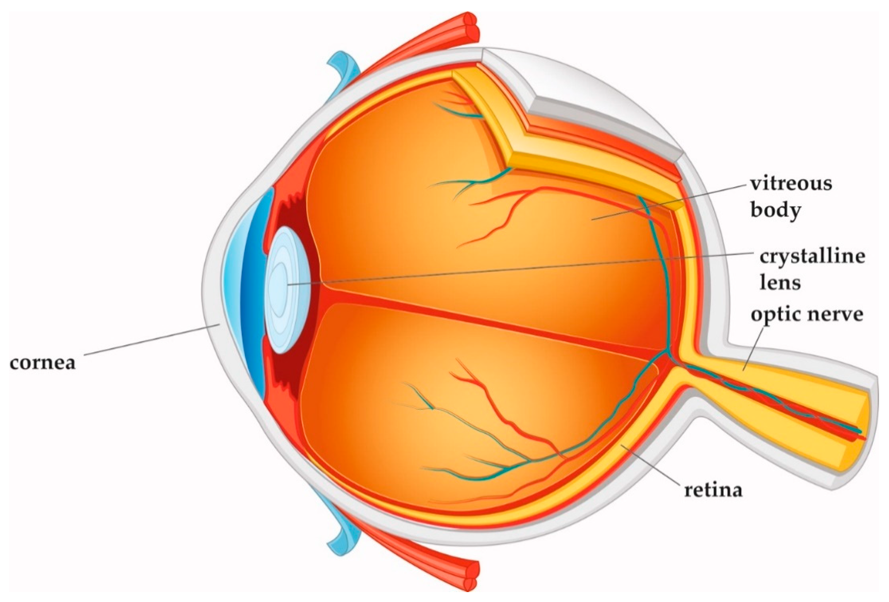
:1. Introduction
2. Laser Technology
2.1. Solid-State Lasers in Ophthalmology
2.1.1. Nd:YAG Laser with Ns Pulse Durations
2.1.2. Femtosecond Lasers
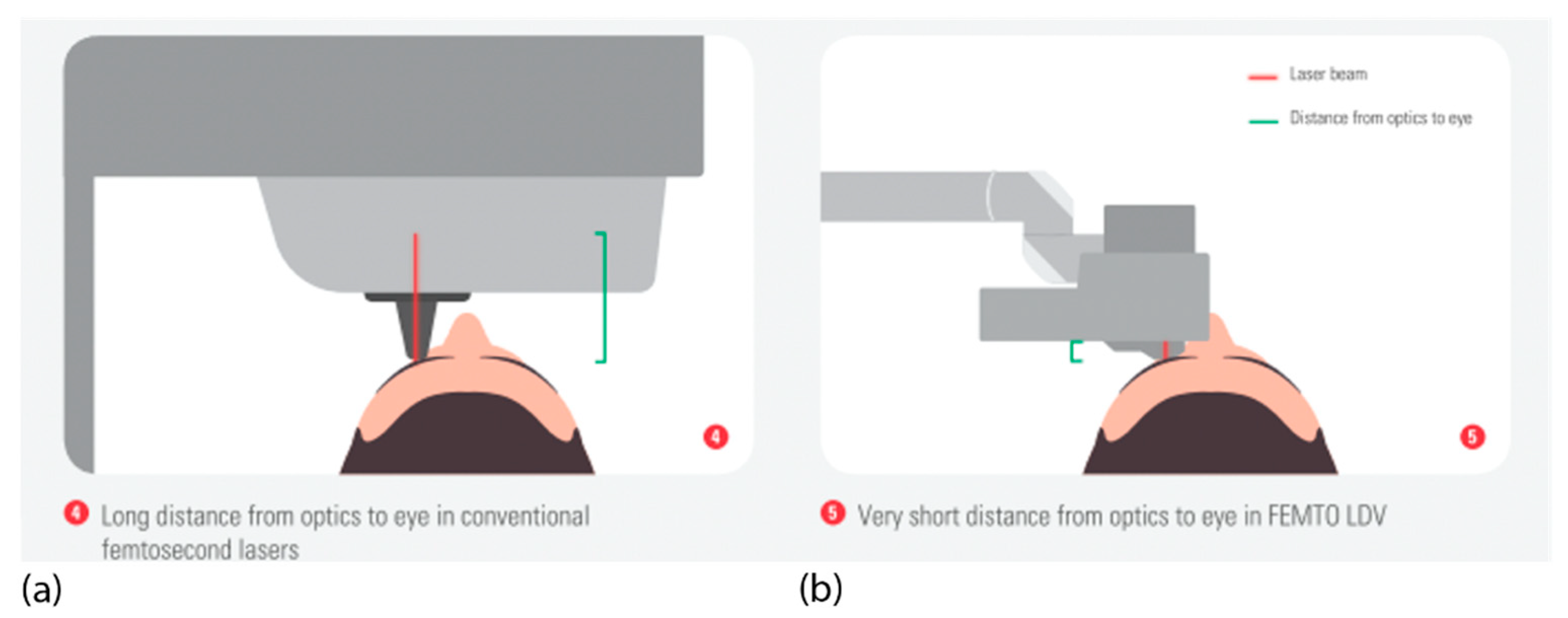
2.1.3. Modern Low Pulse Energy High Repetition Rate Fs Lasers
2.2. Femtosecond Laser–Tissue Interaction
2.3. Supporting Technology Needed in Ophthalmic Fs-Laser Systems
2.3.1. OCT Imaging
2.3.2. Vacuum Docking Interfaces
3. Clinical Applications
3.1. Refractive Surgery
3.1.1. Fs Flap Creation for Refractive Surgery
- LASIK
- Stromal keratophakia (additive refractive surgery)
3.1.2. Intrastromal Pockets
- Corneal stromal lenticule extraction
- Intrastromal corneal ring segments
3.1.3. Intrastromal and Trans-Stromal Cuts for Astigmatic Correction
3.2. Corneal Surgery
3.2.1. Penetrating Keratoplasty
- Background
- Trephination
- Sidecuts
3.2.2. Lamellar Keratoplasty
- Background
- Deep anterior lamellar keratoplasty (DALK)
- Posterior lamellar keratoplasty
3.3. Cataract Surgery
3.3.1. Capsulotomy
3.3.2. Nucleus Fragmentation
3.3.3. Corneal Incisions
- Full-thickness incisions
- Partial-thickness incisions
3.3.4. Future Applications
4. Summary
Author Contributions
Funding
Conflicts of Interest
References
- Welch, A.J.; van Gemert, M. Optical-Thermal Response of Laser-Irradiated Tissue; Springer: Berlin/Heidelberg, Germany, 2011. [Google Scholar]
- Kaschke, M.; Donnerhacke, K.-H.; Rill, M.S. Optical Devices in Ophthalmology and Optometry: Technology, Design Principles and Clinical Applications; WILEY-VCH Verlag GmbH & Co. KGaA: Weinheim, Germany, 2014. [Google Scholar]
- Aron-Rosa, D.; Aron, J.J.; Griesemann, M.; Thyzel, R. Use of the neodymium-yag laser to open the posterior capsule after lens implant surgery: A preliminary report. Am. Intra-Ocular Implant. Soc. J. 1980, 6, 352–354. [Google Scholar] [CrossRef]
- Stern, D.; Schoenlein, R.W.; Puliafito, C.A.; Dobi, E.T.; Birngruber, R.; Fujimoto, J.G. Corneal Ablation by Nanosecond, Picosecond, and Femtosecond Lasers at 532 and 625 nm. Arch. Ophthalmol. 1989, 107, 587–592. [Google Scholar] [CrossRef] [PubMed]
- Huang, D.; Swanson, E.A.; Lin, C.P.; Schuman, J.S.; Stinson, W.G.; Chang, W.; Hee, M.R.; Flotte, T.; Gregory, K.; Puliafito, C.A.; et al. Optical Coherence Tomography. Science 1991, 254, 1178–1181. [Google Scholar] [CrossRef] [Green Version]
- Bhargava, R.; Kumar, P.; Phogat, H.; Chaudhary, K.P. Neodymium-yttrium aluminium garnet laser capsulotomy energy levels for posterior capsule opacification. J. Ophthalmic Vis. Res. 2015, 10, 37–42. [Google Scholar] [CrossRef] [PubMed]
- Vogel, A.; Noack, J.; Hüttman, G.; Paltauf, G. Mechanisms of femtosecond laser nanosurgery of cells and tissues. Appl. Phys. A 2005, 81, 1015–1047. [Google Scholar] [CrossRef]
- Heisterkamp, A.; Ripken, T.; Lubatschowski, H.; Welling, H.; Luetkefels, E.; Drommer, W.; Ertmer, W. Intrastromal cutting effects in rabbit cornea using femtosecond laser pulses. In Optical Biopsy and Tissue Optics; International Society for Optics and Photonics: Bellingham, WA, USA, 2000; pp. 52–60. [Google Scholar]
- Pepose, J.S.; Lubatschowski, H. Comparing Femtosecond Lasers. Cataract Refract. Surg. Today 2008, 10, 45–51. [Google Scholar]
- Vogel, A.; Busch, R. Shock Wave Emission and Cavitation Bubble Generation by Picosecond and Nanosecond Optical Breakdown in Water. J. Acoust. Soc. Am. 1996, 100, 148–165. [Google Scholar] [CrossRef]
- Vogel, A.; Venugopalan, V. Mechanisms of Pulsed Laser Ablation of Biological Tissues. Chem. Rev. 2003, 103, 577–644. [Google Scholar] [CrossRef] [Green Version]
- Tinne, N.; Knoop, G.; Kallweit, N.; Veith, S.; Bleeker, S.; Lubatschowski, H.; Krüger, A.; Ripken, T. Effects of cavitation bubble interaction with temporally separated fs-laser pulses. J. Biomed. Opt. 2014, 19, 48001. [Google Scholar] [CrossRef] [Green Version]
- Lubatschowski, H.; Maatz, G.; Heisterkamp, A.; Hetzel, U.; Drommer, W.; Welling, H.; Ertmer, W. Application of ultrashort laser pulses for intrastromal refractive surgery. Graefe’s Arch. Clin. Exp. Ophthalmol. 2000, 238, 33–39. [Google Scholar] [CrossRef]
- Ratkay-Traub, I.; Juhasz, T.; Horvath, C.; Suarez, C.; Kiss, K.; Ferincz, I.; Kurtz, R. Ultra-short pulse (femtosecond) laser surgery: Initial use in LASIK flap creation. Ophthalmol. Clin. N. Am. 2001, 14, 347–355. [Google Scholar]
- Nagy, Z.Z.; McAlinden, C. Femtosecond laser cataract surgery. Eye Vis. 2015, 2, 1–8. [Google Scholar] [CrossRef] [PubMed] [Green Version]
- Nagy, Z.; Takacs, A.; Filkorn, T.; Sarayba, M. Initial Clinical Evaluation of an Intraocular Femtosecond Laser in Cataract Surgery. J. Refract. Surg. 2009, 25, 1053–1060. [Google Scholar] [CrossRef] [PubMed] [Green Version]
- Ostovic, M.; Klaproth, O.K.; Hengerer, F.H.; Mayer, W.J.; Kohnen, T. Light Microscopy and Scanning Electron Microscopy Analysis of Rigid Curved Interface Femtosecond Laser-Assisted and Manual Anterior Capsulotomy. J. Cataract Refract. Surg. 2013, 39, 1587–1592. [Google Scholar] [CrossRef]
- Lubatschowski, H. Overview of Commercially Available Femtosecond Lasers in Refractive Surgery. J. Refract. Surg. 2008, 24, S102–S107. [Google Scholar] [CrossRef]
- Riau, A.K.; Liu, Y.C.; Lwin, N.C.; Ang, H.P.; Tan, N.Y.; Yam, G.H.; Tan, D.T.; Mehta, J.S. Comparative Study of Nj- and Muj-Energy Level Femtosecond Lasers: Evaluation of Flap Adhesion Strength, Stromal Bed Quality, and Tissue Responses. Invest. Ophthalmol. Vis. Sci. 2014, 55, 3186–3194. [Google Scholar] [CrossRef] [Green Version]
- Mayer, W.J.; Klaproth, O.K.; Ostovic, M.; Terfort, A.; Vavaleskou, T.; Hengerer, F.H.; Kohnen, T. Cell Death and Ultrastructural Morphology of Femtosecond Laser–Assisted Anterior Capsulotomy. Investig. Opthalmol. Vis. Sci. 2014, 55, 893–898. [Google Scholar] [CrossRef] [Green Version]
- Schultz, T.; Joachim, S.C.; Stellbogen, M.; Dick, H.B. Prostaglandin Release During Femtosecond Laser-Assisted Cataract Surgery: Main Inducer. J. Refract. Surg. 2015, 31, 78–81. [Google Scholar] [CrossRef]
- Schwarzenbacher, L.; Schartmueller, D.; Leydolt, C.; Menapace, R. Intra-individual comparison of cytokine and prostaglandin levels with and without low-energy, high-frequency femtosecond laser cataract pretreatment following single-dose topical NSAID application. J. Cataract Refract. Surg. 2020, 46, 1086–1091. [Google Scholar] [CrossRef]
- Fercher, A.F.; Drexler, W.; Hitzenberger, C.K.; Lasser, T. Optical Coherence Tomography—Principles and Applications. Rep. Prog. Phys. 2003, 66, 239–303. [Google Scholar] [CrossRef]
- Schuman, J.S.; Puliafito, C.A.; Fujimoto, J.G.; Duker, J.S. Optical Coherence Tomography of Ocular Diseases; Slack Inc.: San Francisco, CA, USA, 2012. [Google Scholar]
- Wojtkowski, M.; Leitgeb, R.A.; Kowalczyk, A.; Bajraszewski, T.; Fercher, A.F. In vivo human retinal imaging by Fourier domain optical coherence tomography. J. Biomed. Opt. 2002, 7, 457–463. [Google Scholar] [CrossRef] [PubMed]
- Izatt, J.A.; Hee, M.R.; Swanson, E.A.; Lin, C.P.; Huang, D.; Schuman, J.S.; Puliafito, C.A.; Fujimoto, J.G. Micrometer-Scale Resolution Imaging of the Anterior Eye in Vivo with Optical Coherence Tomography. Arch. Ophthalmol. 1994, 112, 1584–1589. [Google Scholar] [CrossRef] [PubMed]
- Kermani, O.; Fabian, W.; Lubatschowski, H. Real-Time Optical Coherence Tomography-Guided Femtosecond Laser Sub-Bowman Keratomileusis on Human Donor Eyes. Am. J. Ophthalmol. 2008, 146, 42–45. [Google Scholar] [CrossRef] [PubMed]
- Chang, J.S.; Chen, I.N.; Chan, W.M.; Ng, J.C.; Chan, V.K.; Law, A.K. Initial evaluation of a femtosecond laser system in cataract surgery. J. Cataract Refract. Surg. 2014, 40, 29–36. [Google Scholar] [CrossRef] [PubMed]
- Grewal, D.S.; Schultz, T.; Basti, S.; Dick, H.B. Femtosecond laser–assisted cataract surgery—Current status and future directions. Surv. Ophthalmol. 2016, 61, 103–131. [Google Scholar] [CrossRef]
- Talamo, J.H.; Gooding, P.; Angeley, D.; Culbertson, W.W.; Schuele, G.; Andersen, D.; Marcellino, G.; Essock-Burns, E.; Batlle, J.; Feliz, R.; et al. Optical patient interface in femtosecond laser–assisted cataract surgery: Contact corneal applanation versus liquid immersion. J. Cataract Refract. Surg. 2013, 39, 501–510. [Google Scholar] [CrossRef]
- Mirshahi, A.; Kohnen, T. Effect of Microkeratome Suction During LASIK on Ocular Structures. Ophthalmology 2005, 112, 645–649. [Google Scholar] [CrossRef]
- Flaxel, C.J.; Choi, Y.H.; Sheety, M.; Oeinck, S.C.; Lee, J.Y.; McDonnell, P.J. Proposed Mechanism for Retinal Tears after Lasik: An Experimental Model. Ophthalmology 2004, 111, 24–27. [Google Scholar] [CrossRef]
- Kanclerz, P.; Grzybowski, A. Does Corneal Refractive Surgery Increase the Risk of Retinal Detachment? A Literature Review and Statistical Analysis. J. Refract. Surg. 2019, 35, 517–524. [Google Scholar] [CrossRef]
- Toth, C.A.; Mostafavi, R.; Fekrat, S.; Kim, T. LASIK and vitreous pathology after LASIK. Ophthalmology 2002, 109, 624–625. [Google Scholar] [CrossRef]
- Kezirian, G.M.; Stonecipher, K.G. Comparison of the IntraLase femtosecond laser and mechanical keratomes for laser in situ keratomileusis. J. Cataract Refract. Surg. 2004, 30, 804–811. [Google Scholar] [CrossRef] [PubMed]
- Chen, S.; Feng, Y.; Stojanovic, A.; Jankov, M.R., II; Wang, Q. Intralase Femtosecond Laser Vs Mechanical Microkeratomes in Lasik for Myopia: A Systematic Review and Meta-Analysis. J. Refract. Surg. 2012, 28, 15–24. [Google Scholar] [CrossRef] [PubMed] [Green Version]
- Barraquer, J.I. The History and Evolution of Keratomileusis. Int. Ophthalmol. Clin. 1996, 36, 1–7. [Google Scholar] [CrossRef] [PubMed]
- Riau, A.K.; Liu, Y.-C.; Yam, G.H.; Mehta, J.S. Stromal keratophakia: Corneal inlay implantation. Prog. Retin. Eye Res. 2020, 75, 100780. [Google Scholar] [CrossRef]
- Wei, S.; Wang, Y. Comparison of corneal sensitivity between FS-LASIK and femtosecond lenticule extraction (ReLEx flex) or small-incision lenticule extraction (ReLEx smile) for myopic eyes. Graefe’s Arch. Clin. Exp. Ophthalmol. 2013, 251, 1645–1654. [Google Scholar] [CrossRef]
- Ang, M.; Farook, M.; Htoon, H.M.; Mehta, J.S. Randomized Clinical Trial Comparing Femtosecond LASIK and Small-Incision Lenticule Extraction. Ophthalmology 2020, 127, 724–730. [Google Scholar] [CrossRef]
- Ang, M.; Mehta, J.S.; Chan, C.; Htoon, H.M.; Koh, J.C.; Tan, D. Refractive lenticule extraction: Transition and comparison of 3 surgical techniques. J. Cataract Refract. Surg. 2014, 40, 1415–1424. [Google Scholar] [CrossRef]
- Riau, A.K.; Angunawela, R.I.; Chaurasia, S.S.; Lee, W.S.; Tan, D.T.; Mehta, J.S. Early Corneal Wound Healing and Inflammatory Responses after Refractive Lenticule Extraction (ReLEx). Investig. Opthalmol. Vis. Sci. 2011, 52, 6213–6221. [Google Scholar] [CrossRef] [Green Version]
- Vestergaard, A.H.; Grauslund, J.; Ivarsen, A.; Hjortdal, J. Efficacy, safety, predictability, contrast sensitivity, and aberrations after femtosecond laser lenticule extraction. J. Cataract Refract. Surg. 2014, 40, 403–411. [Google Scholar] [CrossRef]
- Sandoval, H.P.; Donnenfeld, E.D.; Kohnen, T.; Lindstrom, R.L.; Potvin, R.; Tremblay, D.M.; Solomon, K.D. Modern laser in situ keratomileusis outcomes. J. Cataract Refract. Surg. 2016, 42, 1224–1234. [Google Scholar] [CrossRef]
- Hashemi, H.; Alvani, A.; Seyedian, M.A.; Yaseri, M.; Khabazkhoob, M.; Esfandiari, H. Appropriate Sequence of Combined Intracorneal Ring Implantation and Corneal Collagen Cross-Linking in Keratoconus: A Systematic Review and Meta-Analysis. Cornea 2018, 37, 1601–1607. [Google Scholar] [CrossRef] [PubMed]
- Lans, L.J. Experimentelle Untersuchungen über Entstehung von Astigmatismus Durch Nicht-Perforirende Corneawunden. Albrecht Graefes Arch. Ophthalmol. 1898, 45, 117–152. [Google Scholar] [CrossRef]
- Osher, R.H. Paired transverse relaxing keratotomy: A combined technique for reducing astigmatism. J. Cataract Refract. Surg. 1989, 15, 32–37. [Google Scholar] [CrossRef]
- Thornton, S.P.; Sanders, D.R. Graded Nonintersecting Transverse Incisions for Correction of Idiopathic Astigmatism. J. Cataract Refract. Surg. 1987, 13, 27–31. [Google Scholar] [CrossRef]
- Chang, J.S.M. Femtosecond laser-assisted astigmatic keratotomy: A review. Eye Vis. 2018, 5, 6. [Google Scholar] [CrossRef] [PubMed] [Green Version]
- Mirshahi, A.; Latz, C. Femtosecond laser-assisted astigmatic keratotomy. Ophthalmologe 2020, 117, 415–423. [Google Scholar] [CrossRef]
- Chan, T.C.-Y.; Ng, A.L.; Cheng, G.P.; Wang, Z.; Woo, V.C.; Jhanji, V. Corneal Astigmatism and Aberrations After Combined Femtosecond-Assisted Phacoemulsification and Arcuate Keratotomy: Two-Year Results. Am. J. Ophthalmol. 2016, 170, 83–90. [Google Scholar] [CrossRef]
- Tan, D.; Dart, J.K.G.; Holland, E.J.; Kinoshita, S. Corneal transplantation. Lancet 2012, 379, 1749–1761. [Google Scholar] [CrossRef]
- Seitz, B.; Langenbucher, A.; Hager, T.; Janunts, E.; El-Husseiny, M.; Szentmary, N. Penetrating Keratoplasty for Keratoconus—Excimer Versus Femtosecond Laser Trephination. Open Ophthalmol. J. 2017, 11, 225–240. [Google Scholar] [CrossRef] [Green Version]
- Seitz, B.; Hager, T.; Langenbucher, A.; Naumann, G.O.H. Reconsidering Sequential Double Running Suture Removal after Penetrating Keratoplasty: A Prospective Randomized Study Comparing Excimer Laser and Motor Trephination. Cornea 2018, 37, 301–306. [Google Scholar] [CrossRef]
- Tóth, G.; Szentmáry, N.; Langenbucher, A.; Akhmedova, E.; El-Husseiny, M.; Seitz, B. Comparison of Excimer Laser Versus Femtosecond Laser Assisted Trephination in Penetrating Keratoplasty: A Retrospective Study. Adv. Ther. 2019, 36, 3471–3482. [Google Scholar] [CrossRef] [PubMed]
- Boden, K.T.; Schlosser, R.; Boden, K.; Januschowski, K.; Szurman, P.; Rickmann, A. Novel Liquid Interface for Femtosecond Laser-Assisted Penetrating Keratoplasty. Curr. Eye Res. 2020, 45, 1051–1057. [Google Scholar] [CrossRef] [PubMed]
- Maier, P.; Böhringer, D.; Birnbaum, F.; Reinhard, T. Improved Wound Stability of Top-Hat Profiled Femtosecond Laser-Assisted Penetrating Keratoplasty In Vitro. Cornea 2012, 31, 963–966. [Google Scholar] [CrossRef] [PubMed]
- Seitz, B.; Brünner, H.; Viestenz, A.; Hofmann-Rummelt, C.; Schlötzer-Schrehardt, U.; Naumann, G.O.; Langenbucher, A. Inverse Mushroom-Shaped Nonmechanical Penetrating Keratoplasty Using a Femtosecond Laser. Am. J. Ophthalmol. 2005, 139, 941–944. [Google Scholar] [CrossRef] [PubMed]
- Maier, P.C.; Birnbaum, F.; Reinhard, T. Therapeutic Applications of the Femtosecond Laser in Corneal Surgery. Klin. Monbl. Augenheilkd. 2010, 227, 453–459. [Google Scholar] [CrossRef] [PubMed]
- Reinhart, W.J.; Musch, D.C.; Jacobs, D.S.; Lee, W.B.; Kaufman, S.C.; Shtein, R.M. Deep Anterior Lamellar Keratoplasty as an Alternative to Penetrating Keratoplasty a Report by the American Academy of Ophthalmology. Ophthalmology 2011, 118, 209–218. [Google Scholar] [CrossRef] [PubMed]
- Anwar, M.; Teichmann, K.D. Big-Bubble Technique to Bare Descemet’s Membrane in Anterior Lamellar Keratoplasty. J. Cataract Refract. Surg. 2002, 28, 398–403. [Google Scholar] [CrossRef]
- De Macedo, J.P.; de Oliveira, L.A.; Hirai, F.; de Sousa, L.B. Femtosecond Laser-Assisted Deep Anterior Lamellar Keratoplasty in Phototherapeutic Keratectomy Versus the Big-Bubble Technique in Keratoconus. Int. J. Ophthalmol. 2018, 11, 807–812. [Google Scholar]
- Buzzonetti, L.; Laborante, A.; Petrocelli, G. Standardized Big-Bubble Technique in Deep Anterior Lamellar Keratoplasty Assisted by the Femtosecond Laser. J. Cataract Refract. Surg. 2010, 36, 1631–1636. [Google Scholar] [CrossRef]
- Buzzonetti, L.; Petrocelli, G.; Valente, P.; Petroni, S.; Parrilla, R.; Iarossi, G. Refractive Outcome of Keratoconus Treated by Big-Bubble Deep Anterior Lamellar Keratoplasty in Pediatric Patients: Two-Year Follow-up Comparison between Mechanical Trephine and Femtosecond Laser Assisted Techniques. Eye Vis. 2019, 6, 1. [Google Scholar] [CrossRef] [Green Version]
- Stuart, A.J.; Romano, V.; Virgili, G.; Shortt, A.J. Descemet’s Membrane Endothelial Keratoplasty (Dmek) Versus Descemet’s Stripping Automated Endothelial Keratoplasty (Dsaek) for Corneal Endothelial Failure. Cochrane Database Syst. Rev. 2018, 6, d012097. [Google Scholar] [CrossRef] [PubMed]
- Einan-Lifshitz, A.; Sorkin, N.; Boutin, T.; Showail, M.; Borovik, A.; Alobthani, M.; Chan, C.C.; Rootman, D.S. Comparison of Femtosecond Laser-Enabled Descemetorhexis and Manual Descemetorhexis in Descemet Membrane Endothelial Keratoplasty. Cornea 2017, 36, 767–770. [Google Scholar] [CrossRef] [PubMed]
- Sorkin, N.; Mednick, Z.; Einan-Lifshitz, A.; Trinh, T.; Santaella, G.; Telli, A.; Chan, C.C.; Rootman, D.S. Three-Year Outcome Comparison Between Femtosecond Laser-Assisted and Manual Descemet Membrane Endothelial Keratoplasty. Cornea 2019, 38, 812–816. [Google Scholar] [CrossRef]
- Sorkin, N.; Mimouni, M.; Santaella, G.; Trinh, T.; Cohen, E.; Einan-Lifshitz, A.; Chan, C.C.; Rootman, D.S. Comparison of Manual and Femtosecond Laser-Assisted Descemet Membrane Endothelial Keratoplasty for Failed Penetrating Keratoplasty. Am. J. Ophthalmol. 2020, 214, 1–8. [Google Scholar] [CrossRef] [PubMed]
- Bille, J.F.; Schanzlin, D. Method for Removing Cataractous Material. US Patent US5246435A, 1993. [Google Scholar]
- Boden, K.T.; Szurman, P. Current Value of Femtosecond Laser-Assisted Cataract Surgery. Ophthalmologe 2020, 117, 405–414. [Google Scholar] [CrossRef]
- Wang, J.; Su, F.; Wang, Y.; Chen, Y.; Chen, Q.; Li, F. Intra and Post-Operative Complications Observed with Femtosecond Laser-Assisted Cataract Surgery Versus Conventional Phacoemulsification Surgery: A Systematic Review and Meta-Analysis. BMC Ophthalmol. 2019, 19, 177. [Google Scholar] [CrossRef] [Green Version]
- Day, A.C.; Gore, D.M.; Bunce, C.; Evans, J.R. Laser-assisted cataract surgery versus standard ultrasound phacoemulsification cataract surgery. Cochrane Database Syst. Rev. 2016, 7, CD010735. [Google Scholar] [CrossRef]
- Schweitzer, C.; Brezin, A.; Cochener, B.; Monnet, D.; Germain, C.; Roseng, S.; Sitta, R.; Maillard, A. Femtosecond Laser-Assisted Versus Phacoemulsification Cataract Surgery (Femcat): A Multicentre Participant-Masked Randomised Superiority and Cost-Effectiveness Trial. Lancet 2020, 395, 212–224. [Google Scholar] [CrossRef]
- Kanclerz, P.; Alio, J.L. The benefits and drawbacks of femtosecond laser-assisted cataract surgery. Eur. J. Ophthalmol. 2020, 1–10. [Google Scholar] [CrossRef]
- Hooshmand, J.; Vote, B.J. Femtosecond Laser-Assisted Cataract Surgery, Technology, Outcome, Future Directions and Modern Applications. Asia Pac. J. Ophthalmol. 2017, 6, 393–400. [Google Scholar]
- Sanders, D.R.; Higginbotham, R.W.; Opatowsky, I.E.; Confino, J. Hyperopic shift in refraction associated with implantation of the single-piece Collamer intraocular lens. J. Cataract Refract. Surg. 2006, 32, 2110–2112. [Google Scholar] [CrossRef] [PubMed]
- Baumeister, M.; Bühren, J.; Kohnen, T. Tilt and decentration of spherical and aspheric intraocular lenses: Effect on higher-order aberrations. J. Cataract Refract. Surg. 2009, 35, 1006–1012. [Google Scholar] [CrossRef] [PubMed]
- Friedman, N.J.; Palanker, D.; Schuele, G.; Andersen, D.; Marcellino, G.; Seibel, B.S.; Batlle, J.; Feliz, R.; Talamo, J.H.; Blumenkranz, M.S.; et al. Femtosecond laser capsulotomy. J. Cataract Refract. Surg. 2011, 37, 1189–1198. [Google Scholar] [CrossRef] [PubMed]
- Kránitz, K.; Miháltz, K.; Sándor, G.L.; Takacs, A.; Knorz, M.C.; Nagy, Z.Z. Intraocular Lens Tilt and Decentration Measured By Scheimpflug Camera Following Manual or Femtosecond Laser–created Continuous Circular Capsulotomy. J. Refract. Surg. 2012, 28, 259–263. [Google Scholar] [CrossRef] [PubMed] [Green Version]
- Kránitz, K.; Takacs, A.; Miháltz, K.; Kovács, I.; Knorz, M.C.; Nagy, Z.Z. Femtosecond Laser Capsulotomy and Manual Continuous Curvilinear Capsulorrhexis Parameters and Their Effects on Intraocular Lens Centration. J. Refract. Surg. 2011, 27, 558–563. [Google Scholar] [CrossRef] [PubMed] [Green Version]
- Nagy, Z.Z.; Kránitz, K.; Takacs, A.I.; Miháltz, K.; Kovács, I.; Knorz, M.C. Comparison of Intraocular Lens Decentration Parameters After Femtosecond and Manual Capsulotomies. J. Refract. Surg. 2011, 27, 564–569. [Google Scholar] [CrossRef] [Green Version]
- Latz, C.; Mirshahi, A. Double Ring Sign of the Lens Capsule: Intraoperative Observation During Cataract Surgery. Ophthalmologe 2020. [Google Scholar] [CrossRef]
- Abouzeid, H.; Ferrini, W. Femtosecond-laser assisted cataract surgery: A review. Acta Ophthalmol. 2014, 92, 597–603. [Google Scholar] [CrossRef]
- Holland, D.; Rufer, F. New Intraocular Lens Designs for Femtosecond Laser-Assisted Cataract Operations: Chances and Benefits. Ophthalmologe 2020, 117, 424–430. [Google Scholar] [CrossRef]
- Murano, N.; Ishizaki, M.; Sato, S.; Fukuda, Y.; Takahashi, H. Corneal Endothelial Cell Damage by Free Radicals Associated with Ultrasound Oscillation. Arch. Ophthalmol. 2008, 126, 816–821. [Google Scholar] [CrossRef] [Green Version]
- Abell, R.G.; Kerr, N.M.; Vote, B.J. Toward Zero Effective Phacoemulsification Time Using Femtosecond Laser Pretreatment. Ophthalmology 2013, 120, 942–948. [Google Scholar] [CrossRef] [PubMed]
- Conrad-Hengerer, I.; Al Juburi, M.; Schultz, T.; Hengerer, F.H.; Dick, B.H. Corneal endothelial cell loss and corneal thickness in conventional compared with femtosecond laser–assisted cataract surgery: Three-month follow-up. J. Cataract Refract. Surg. 2013, 39, 1307–1313. [Google Scholar] [CrossRef] [PubMed]
- Takacs, A.I.; Kovacs, I.; Mihaltz, K.; Filkorn, T.; Knorz, M.C.; Nagy, Z.Z. Central Corneal Volume and Endothelial Cell Count Following Femtosecond Laser-Assisted Refractive Cataract Surgery Compared to Conventional Phacoemulsification. J. Refract. Surg. 2012, 28, 387–391. [Google Scholar] [CrossRef] [PubMed] [Green Version]
- Masket, S.; Sarayba, M.; Ignacio, T.; Fram, N. Femtosecond laser-assisted cataract incisions: Architectural stability and reproducibility. J. Cataract Refract. Surg. 2010, 36, 1048–1049. [Google Scholar] [CrossRef] [PubMed]
- Palanker, D.V.; Blumenkranz, M.S.; Andersen, D.; Wiltberger, M.; Marcellino, G.; Gooding, P.; Angeley, D.; Schuele, G.; Woodley, B.; Simoneau, M.; et al. Femtosecond Laser-Assisted Cataract Surgery with Integrated Optical Coherence Tomography. Sci. Transl. Med. 2010, 2, 58ra85. [Google Scholar] [CrossRef] [Green Version]
- Uy, H.S.; Shah, S.; Packer, M. Comparison of Wound Sealability Between Femtosecond Laser–Constructed and Manual Clear Corneal Incisions in Patients Undergoing Cataract Surgery: A Pilot Study. J. Refract. Surg. 2017, 33, 744–748. [Google Scholar] [CrossRef]
- Yoo, A.; Yun, S.; Kim, J.Y.; Kim, M.J.; Tchah, H. Femtosecond Laser-assisted Arcuate Keratotomy Versus Toric IOL Implantation for Correcting Astigmatism. J. Refract. Surg. 2015, 31, 574–578. [Google Scholar] [CrossRef]
- Dick, H.B. Future Perspectives of the Femtosecond Laser in Anterior Segment Surgery. Ophthalmologe 2020, 117, 431–436. [Google Scholar] [CrossRef]


| IntraLase (AMO, USA) | Wavelight FS200 (Alcon, USA) | LenSx (Alcon, USA) | LensAR (LensAR, Topcon, USA) | Catalys (Johnson and Johnson, USA) | Victus (Bausch and Lomb, Germany) | VisuMax (Zeiss Meditec, Germany) | LDV Z8 (Ziemer, Switzer-land) | Atos * (Schwind, Germany) | |
|---|---|---|---|---|---|---|---|---|---|
| Pulse repetition rate (kHz) | 30–150 | 200 | 60 | 80 | 120 | 80/160 | 500 | 10,000 | <to 4000 |
| Pulse duration (fs) | >500 | 350 | 600–800 | 500 | <600 | 290–550 | 220–580 | 250 | <295 |
| Pulse energy (µJ) | Ca. 1 | <1.5 | >15 | 7–15 | 3–10 | 6–10 | <1 | <1 | <1 |
| Applications: | |||||||||
| LASIK flaps | x | x | x | x | x | x | x | ||
| Refractive Lenticules | x | x | x | ||||||
| Cornea Surgery | x | x | x | x | |||||
| Cataract Surgery | x | x | x | x | x | ||||
| Patient interface | Flat applan. interface | Flat applan. interface | Curved softfit interface | Fluid filled interface | Liquid interface | Semiliquid curved interface | Curved interface | Liquid and flat interfaces | Curved interface |
Publisher’s Note: MDPI stays neutral with regard to jurisdictional claims in published maps and institutional affiliations. |
© 2021 by the authors. Licensee MDPI, Basel, Switzerland. This article is an open access article distributed under the terms and conditions of the Creative Commons Attribution (CC BY) license (http://creativecommons.org/licenses/by/4.0/).
Share and Cite
Latz, C.; Asshauer, T.; Rathjen, C.; Mirshahi, A. Femtosecond-Laser Assisted Surgery of the Eye: Overview and Impact of the Low-Energy Concept. Micromachines 2021, 12, 122. https://doi.org/10.3390/mi12020122
Latz C, Asshauer T, Rathjen C, Mirshahi A. Femtosecond-Laser Assisted Surgery of the Eye: Overview and Impact of the Low-Energy Concept. Micromachines. 2021; 12(2):122. https://doi.org/10.3390/mi12020122
Chicago/Turabian StyleLatz, Catharina, Thomas Asshauer, Christian Rathjen, and Alireza Mirshahi. 2021. "Femtosecond-Laser Assisted Surgery of the Eye: Overview and Impact of the Low-Energy Concept" Micromachines 12, no. 2: 122. https://doi.org/10.3390/mi12020122