Article

Selection for Sustainable Preservation through In Vitro Propagation of Mature Pyrus spinosa Genotypes Rich in Total Phenolics and Antioxidants

by
Styliani Alexandri
1,
Maria Tsaktsira
1,
Stefanos Hatzilazarou
2,
Stefanos Kostas
2,*,
Irini Nianiou-Obeidat
3,
Athanasios Economou
2,
Apostolos Scaltsoyiannes
1 and
Parthena Tsoulpha
1,*
1
Laboratory of Forest Genetics and Plant Breeding, School of Forestry and Natural Environment, Aristotle University, 54124 Thessaloniki, Greece
2
Laboratory of Floriculture, School of Agriculture, Aristotle University, 54124 Thessaloniki, Greece
3
Laboratory of Genetics and Plant Breeding, School of Agriculture, Aristotle University, 54124 Thessaloniki, Greece
*
Authors to whom correspondence should be addressed.
Sustainability 2023, 15(5), 4511; https://doi.org/10.3390/su15054511
Submission received: 22 December 2022 / Revised: 17 February 2023 / Accepted: 26 February 2023 / Published: 2 March 2023

Abstract

:
Pyrus spinosa Forssk. (almond-leaved pear) is a wild-growing native tree of the hilly countryside of Greece, which has recently gained interest for its valuable endogenous substances. In the present work, the determination of the total phenol content (TPC) and antioxidant capacity (AC) was carried out during 2021 and 2022 in leaves from 32 genotypes of mature wild-growing trees at four different locations of Central Macedonia District, Greece: Agia Anastasia, Chalkidiki, Lagadas and Pieria. The measurements taken from the leaf samples of all genotypes revealed a strong positive linear correlation between TPC and AC (R2 = 0.772). Two genotypes from Agia Anastasia, coded as AA2 and AA3, were comparatively the richest in TPC (44.86 for AA2 and 46.32 mg GAE/g fresh weight for AA3) and AC (70.31 and 71.21 μg AAE/g fresh weight for the same genotypes). For these two genotypes of high TPC and AC, an efficient micropropagation protocol was developed to preserve and multiply this valuable germplasm. Newly emerged shoot tips were excised from winter shoots and, after disinfestation, they were established on a modified Murashige and Skoog (MS) nutrient medium with 5 μΜ of 6-Benzyloaminopurine (BAP). For shoot multiplication, explants from clean cultures were transferred to Pear Medium 1 with 5 μΜ of BAP. The effect of the nutrient medium was tested by using five different nutrient media (modified MS, Pear Medium 1, Pear Medium 2, DKW and WPM) supplemented with 5 μΜ of BAP. Pear Medium 1 was the most effective in shoot formation. Among the four different BAP concentrations (0, 5, 10 and 20 μΜ) used in Pear Medium 1, 5 μΜ of BAP resulted in the production of the significantly highest number of shoots (22.7 shoots per explant, 2.4 cm long). The exposure of cultures to 10 μmol·m−2·s−1 for one week under light irradiance followed by 35 μmol·m−2·s−1 for four weeks increased both the number and length of the shoots produced. A 20-s dip of the shoot bases into 49.0 μΜ of Indole-3-butyric acid (IBA) in EtOH equally affected rooting in a modified rooting MS (79%) and Pear Medium 1 (80%). The root system developed better in the modified rooting MS medium (mean number of roots of 2.7 with a length of 3.9 cm). The acclimatization of the plantlets was successfully carried out (high survival rates five weeks after their establishment) on a perlite substrate under controlled environmental conditions.

1. Introduction

Pyrus spinosa Forssk. is a small, deciduous wild-growing tree belonging to the Rosaceae, which is widely spread in the Mediterranean basin and the Greek countryside [1,2]. Previously, the species was known as P. amygdaliformis Vill for the characteristic almond-shaped form of its leaves. Traditionally, the species is known for its ripe fruits that are consumed fresh or dry, but also as a tree with ornamental and/or medicinal value.
Recent research work on P. spinosa for TPC and AC has been focused mainly on its fruits. Tzanakis et al. [3] investigated the TPC and AC of almond-leaved pear fruits (mature and immature). Manolaraki et al. [4] studied the anthelmintic properties of seven Mediterranean P. spinosa plants due to their abundance in tannins, and found that the leaf extracts contained 2.4 g-equivalent tannic acid/100 g DP in total phenols, while Mihajilov-Krstev et al. [5] studied the fruit antioxidant and antimicrobial properties. Kundaković et al. [6] investigated the cytotoxicity of P. spinosa leaf and bark extracts on melanoma cell lines and healthy human embryonic lung fibroblasts. The latter study mainly demonstrated the antibacterial activity of almond-leaved pear extracts against all bacterial species tested, as well as antifungal activities, which were all attributed to the presence of a polyphenol (arbutin).
A good working tool to preserve and reproduce plant genotypes high in valuable natural substances is the asexual (clonal) propagation method. However, P. spinosa is a difficult species to propagate through shoot cuttings [7,8,9]. Thus, an alternative method for the asexual propagation of this species could be the in vitro culture technique. The species-specific literature on in vitro propagation of P. spinosa is rather limited. Dolcet-Sanjuan et al. [10] were the first to study the in vitro culture of this particular species under Fe-limited conditions, among three other Pyrus genotypes and Cydonia oblonga Mill. The most recent study focused on the critical factors affecting the micropropagation of P. spinosa from juvenile plant material [2].
The benefits to human health of phenolic-rich foods are widely acknowledged due to their antioxidant properties. Consumers increasingly appreciate the use of novel plant-based products from natural resources, even for serious health issues [11,12,13]. The TPC content and AC of the leaves of almond-leaved pear is an almost-unexplored area of research with great potential for the introduction of novel chemical-free health products. In this framework, the objective of the present study was to first quantify the total leaf phenols and antioxidants from P. spinosa wild-growing populations, then select the distinct mature genotypes with the highest contents and to finally develop a micropropagation protocol aiming at the preservation and future utilization of the characterized selections.

2. Materials and Methods

2.1. Plant Material

Three shoots (30 cm long) from eight mature wild-growing P. spinosa trees (20–30 years of age) were collected in mid-June from each of four different locations of Central Macedonia District, Greece: (a) Agia Anastasia, (b) Chalkidiki, (c) Lagadas and (d) Pieria. All trees had morphologically typical leaf (almond-shaped) and characteristic fruit traits. To avoid measurement discrepancies due to environmental conditions or the developmental stages of the trees, all collections were conducted early in the morning from the east side of the habitat where each genotype was found and from the lower branches of the crown. The coding of the selected individuals and their geographical coordinates are in Table 1.

2.2. Plant Material Preparation and Sample Extraction

After transferring the plant material to the Laboratory of Floriculture (School of Agriculture, Aristotle University), the largest leaves were excised from the shoots, weighed separately from each shoot and stored at 4 °C until extraction was performed later on the same day. The leaves (0.7 g) were ground to a powder in liquid nitrogen, and the samples were extracted with 7 mL of methanol at room temperature. After being vortexed (2 min), the extracts were centrifuged for 10 min at 10,000 rpm in an Eppendorf™ Centrifuge 5810/5810 R (Eppendorf, Hamburg, Germany).

2.3. Total Phenol Content (TPC)

For each sample, the TPC was determined by the Folin–Ciocalteu method [14] as follows: In 0.5 mL of extracted sample dissolved in methanol (1:50 v/v), 2.5 mL of Folin–Ciocalteu (1:10 v/v) solution was added. After 6 min, 2 mL of Νa2CO3 (7.5% w/v) solution was also added and vortexed. Finally, the sample mixture remained for 5 min at 50 °C in a digital water bath (LabTech, Seoul, Korea). The absorbance measurements were conducted using a Helios Alpha Spectrophotometer (Thermo Spectronic, Cambridge, UK) for each sample at 760 nm. The TPC was based on the gallic acid standard calibration curve (100, 50, 25, 12.5 and 0 μg·mL−1) and was expressed in milligrams of gallic acid equivalent per gram of fresh weight (mg GAE/g f.w.). The average content of TPC for each genotype was derived from three separate measurements.

2.4. Determination of Antioxidant Capacity (AC)

The free radical 2.2-Diphenyl-l-picry1hydrazyl method (DPPH) [15] was used for the determination of antioxidants as follows: In 0.2 mL of dissolved sample (1:20 v/v) in 80% (v/v) methanol, 2.8 mL of 4% (w/v) DPPH was added. The final sample mixtures were vortexed and kept in total darkness for 30 min under ambient temperature. The antioxidants were determined using the Helios Alpha Spectrophotometer at 517 nm by means of an ascorbic acid standard calibration curve (0.1, 0.05, 0.025, 0.01 and 0 μg·mL−1) and were expressed in micrograms of ascorbic acid equivalent per gram of fresh weight (μg AAE/g f.w.). Pure methanol was used as a blank. The final content of AC for each genotype was determined as the average of three separate measurements.
Using the data from the TPC and AC of P. spinosa leaves, a PCA plot was constructed with the XLSTAT program to group the genotypes in order to draw useful conclusions regarding their diversity.

2.5. Establishment of In Vitro Cultures and Shoot Proliferation

The plant material of the AA2 and AA3 genotypes from Agia Anastasia that were richest in total phenols and antioxidants was used for the in vitro experiments. Winter shoots of the following year (February) were collected and kept in vessels with tap water for 20 days under controlled environmental conditions in a plant growth chamber (16 h photoperiod, temperature 24 ± 1 °C and light irradiance of 35 μmol·m−2·s−1). The shoot tips of the newly emerged shoots (1.5–2.0 cm long) from the lateral buds were isolated and used for in vitro culture initiation after disinfection by following the procedure of Tsoulpha et al. [2].
Cultures were then established in vitro in glass culture tubes (height 15 cm, diameter 2.5 cm) in a modified Murashige and Skoog (MS) nutrient medium [16] (thiamine-HCl 1 mg·L−1, nicotinic acid 1 mg·L−1 and pyridoxine-HCl 1 mg·L−1) [10] supplemented with 5 μΜ of 6-Benzylaminopurine (BAP) (Sigma-Aldrich, St. Louis, MI, USA) and 7.1 g·L−1 agar (Sigma-Aldrich, St. Louis, MI, USA). The pH was adjusted to 5.7. All cultures were maintained for 4 weeks in a plant growth chamber under controlled environmental conditions, as described previously, using OSRAM L 58W/31-830 Lumilux Plus Eco lamps (OSRAM GmbH, Munich, Germany) for lighting.
The clean explants were transferred for shoot multiplication in MagentaTM B-cap vessels (Sigma-Aldrich, St. Louis, MI, USA) (height 9.85 cm and diameter 5 cm). Pear Medium 1 [17] supplemented with 5 μΜ BAP, 3.5 g·L−1 agar and 1.45 g·L−1 PhytagelTM (Sigma-Aldrich, St. Louis, MI, USA) was used in all experiments for shoot multiplication and rooting, unless otherwise stated. Shoot multiplication was based on axillary shoot formation and subculturing was conducted every five weeks. During subculturing, the explants (2.5–3.0 cm long) were placed horizontally on the nutrient medium after the removal of the apical part (0.3–0.5 cm) [2] and were used for testing the effects of different nutrient media, BAP concentrations and light irradiances on shoot formation. All cultures, except for those for the experiment of different light irradiances, were maintained for five weeks in a plant growth chamber at 24 ± 1 °C temperature and 16 h photoperiod under 10 μmol·m−2·s−1 of PPFD for the first week, followed by 35 μmol·m−2·s−1 for the next four weeks.

2.6. Effect of Nutrient Medium, BAP Concentration and Light Irradiance

Five different nutrient media, each supplemented with 5 μΜ of BAP, were tested in the multiplication stage, namely (a) Modified MS [16], as proposed by Dolcet-Sanjuan et al. [10], (b) Pear Medium 1 [17], (c) Pear Medium 2 [15], (d) Woody Plant Medium (WPM) [18] and (e) Driver and Kuniyuki Walnut medium (DKW) [19].
As Pear Medium 1 was the most suitable for the proliferation of P. spinosa explants in the previous experiment, it was used for testing the effects of different BAP concentrations on shoot multiplication and elongation. The tested four BAP concentrations were 0, 5, 10 and 20 μΜ.
To test the effect of light irradiance on shoot formation, four different treatments of PPFD were investigated: (a) 10 μmol·m−2·s−1 for the first week and then 35 μmol·m−2·s−1 for another four weeks, and five weeks for each of the other three treatments: (b) 10 μmol·m−2·s−1, (c) 35 μmol·m−2·s−1 and (d) 100 μmol·m−2·s−1. A Sekonic L-398 Photometer (Seconic Corporation, Tokyo, Japan) was used to measure the PPFD.

2.7. In Vitro Shoot Rooting and Plantlet Acclimatization

The elongated shoots (3–4 cm long) excised from the in vitro cultures of the different BAP concentrations were randomized for the rooting experiments. After removing the lower leaves and creating slight wounding cuts in this part, the basal parts of the shoots (1 cm) were dipped for 20 s into different IBA concentrations: 0, 2.46, 4.90, 24.60 or 49.00 μM. Their rooting ability was tested in vitro both in modified rooting MS nutrient medium (½ NH4NO3 and ½ KNO3) [10] and in Pear Medium 1. The root zone of the shoots was protected from immediate light by cardboard coverings for five weeks during rooting. The environmental conditions of the plant growth chamber were the same as those in the multiplication stage.
The rooted shoots were planted in a sterilized perlite substrate in 35-cell propagation trays (cell dimensions 5 × 5 × 10 cm) and were placed in a small transparent Plexiglas structure (46.5 × 26.5 × 30 cm), which then was moved to the plant growth chamber with controlled environmental conditions (temperature 24 ± 1 °C, photoperiod 16 h and PPFD 10 μmol·m−2·s−1) for plantlet acclimatization. During acclimatization, the young plantlets received a liquid solution of MS macroelements (with ½ NH4NO3 and ½ KNO3) every 20 days. Each plantlet was covered with a transparent hard plastic vessel (10 × 10 × 9 cm) for the first three of the five weeks of acclimatization in order to maintain high levels of relative humidity.

2.8. Data Recording and Statistical Analysis

For each genotype, three separate measurements were taken for the TPC and AC. All the data were analyzed by ANOVA. Mean comparisons were performed with Duncan’s multiple range test at p ≤ 0.05. Pearson’s correlation coefficient r was used for expressing the correlation between the TPC and AC values measured in all genotypes.
A completely randomized design was applied for all in vitro experiments on pooled plant material of both studied genotypes (AA2 and AA3). In the shoot multiplication experiments, 30 explants were used for each treatment, whereas, for the in vitro rooting trials, 20 shoots were employed. The number and length of the newly developed axillary shoots on the explants, as well as the root number and length formed on the rooted shoots (with at least one root being ≥ 0.5 cm) were recorded after five weeks of culture establishment. For the statistical analysis, all data were subjected to analysis of variance (ANOVA). For rooting data in percentages, arcsine transformation was performed prior to statistical analysis and the data were transformed back to percentages for presentation in tables. Means were compared by using Duncan’s multiple range test at p ≤ 0.05. The effect of rooting treatments and their interactions were tested and significant differences between means were determined by using the Bonferroni test at p ≤ 0.05. For all data, the statistical analyses were conducted by using SPSS v.27 (SPSS, Inc., Statistical Package for the Social Sciences, Chicago, IL, USA).

3. Results and Discussion

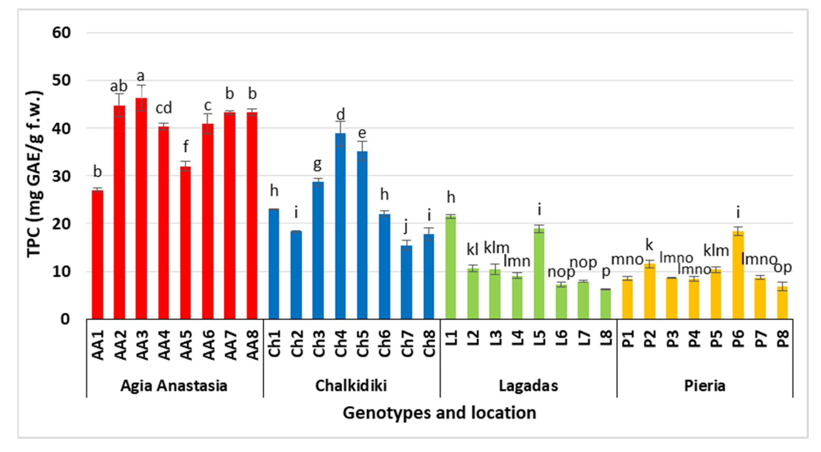
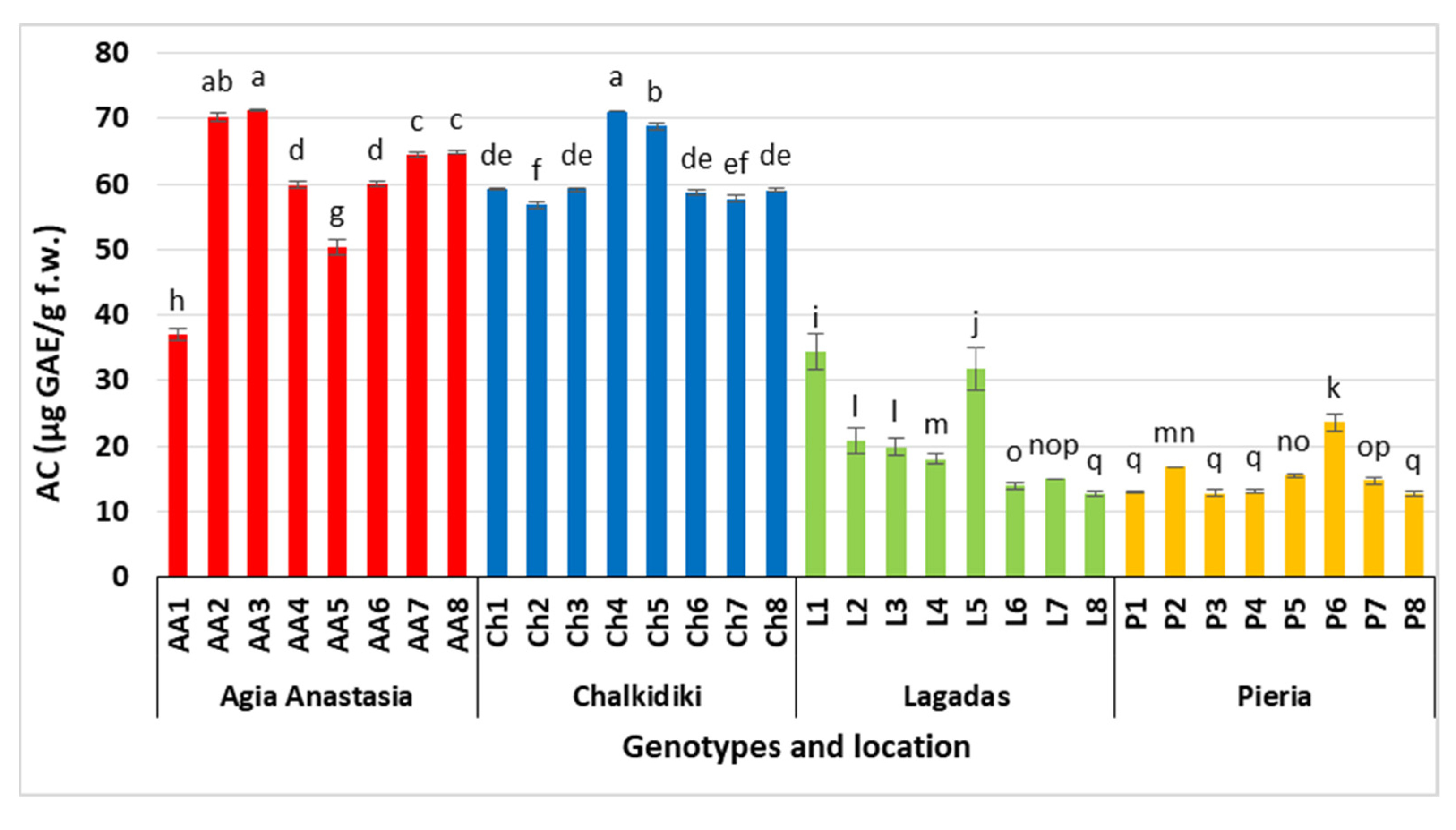
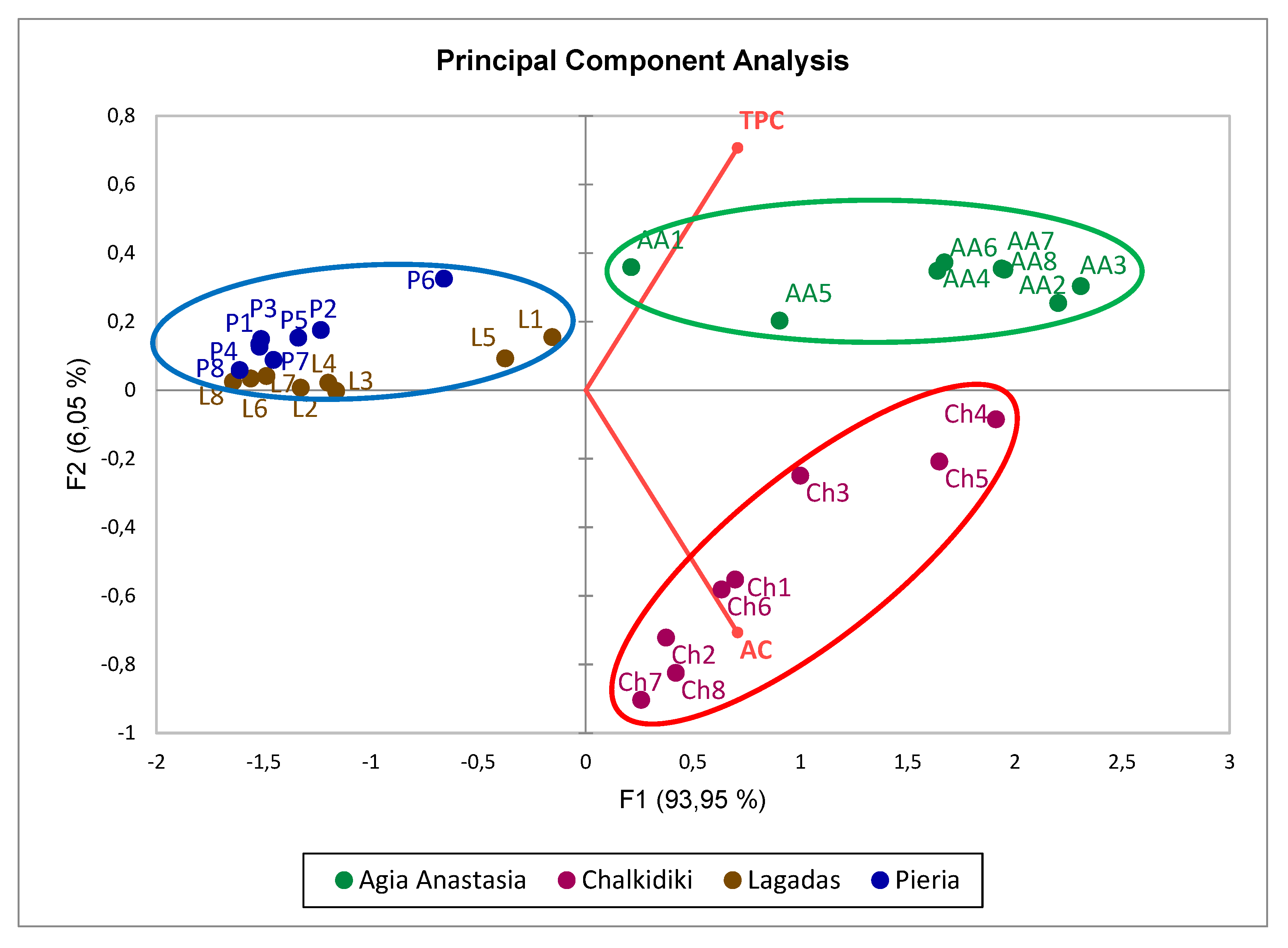
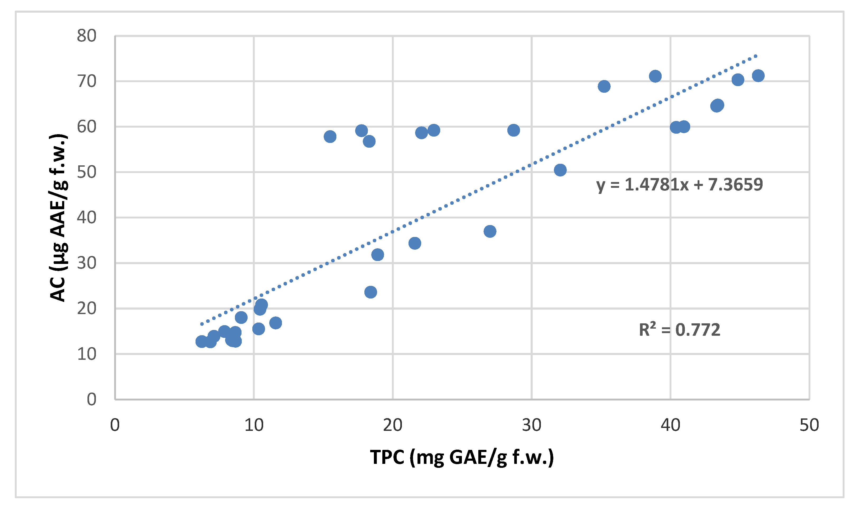
3.1. Total Phenol Content (TPC) and Antioxidant Capacity (AC)

The findings of the TPCs in fresh leaves of the selected P. spinosa genotypes from the four different locations in Greece are shown in Figure 1. Two of them from Agia Anastasia, coded as AA2 and AA3, had significantly the highest content of total phenols (44.86 and 46.32 mg GAE/g f.w., respectively), followed by AA7 and AA8 from the same location. All of the genotypes of Pieria and the majority of those from Lagadas had the lowest contents of total phenols. From Chalkidiki, only Ch4 and Ch5 showed higher TPCs in comparison with the other selected individuals of the same location. In general, the total phenols ranged from 6.25 mg GAE/g f.w. (genotype L8) to 46.32 mg GAE/g f.w. (genotype AA3).
Concerning AC, the genotypes AA3 and AA2 displayed the significantly highest values (71.21 μg AAΕ/g f.w. and 70.31 μg AAΕ/g f.w., respectively), both situated at Agia Anastasia, along with a selection from Chalkidiki under the code Ch4 (71.11 μg AAΕ/g f.w.), followed by Ch5 of the same location. In general, the antioxidant activity ranged from 12.66 μg AAΕ/g f.w. (P8) to 71.21 μg AAΕ/g f.w. (AA3) (Figure 2).
According to the PCA (Figure 3), in which the genotypes are distributed in relation to their productivity in TPC and AC of P. spinosa leaves, they seem to follow the population pattern. This applies to the Chalkidiki and Agia Anastasia populations (red and green circles, respectively), which stand out in relation to the rest of the genotypes. On the contrary, the populations of Pieria and Lagadas (blue circle) seem to have a similar profile in relation to their productivity in TPC and AC, probably due to the similarity of their genetic material.
In Figure 4, a strong positive linear correlation between TPC and AC (R2 = 0.772) can be seen according to Ratner [20], with Pearson’s correlation coefficient being r = 0.879 (p ≤ 0.01). This result indicated that the antioxidant capacity was mainly attributed to the content of total phenols of the studied plant material of P. spinosa. These findings confirmed those of other works regarding the positive correlation between TPC and AC. In particular, He et al. [21] found a strong positive relationship between the two factors in edible flowers of Pyrus pashia Buch.-Ham. ex D. Don. For Diospyros virginiana L., Grygorieva et al. [22] reported similar results, while for 33 different fruits, the same strong correlation of TPC and AC was confirmed by Chena et al. [23].
Tzanakis et al. [3] investigated the TPC and AC of fruits (peel and pulp) of P. amygdaliformis from the region of Argos, Greece. By applying the method of Folin–Ciocalteu [14], they reported that the TPCs of the dry weight of mature and immature fruits reached 0.222% and 0.489%, respectively. Furthermore, they found that, among the five species examined for their edible fruits, those of P. spinosa were the second-rich in phenolics after pomegranate (Punica granatum). In addition, the antioxidant activity determined by the DPPH method was reported to reach 54.69% in mature and 53.13% in immature fruits when extracted by n-propanol. Among seven different forest species examined, Manolaraki et al. [4] found, by using the same method of Folin–Ciocalteu, that the TPC in P. spinosa leaves, collected in autumn, may reach 2.4 g-equivalent tannic acid/100 g dry sample, a quantity which is almost half of that in the current research, as reported for the best genotype collected in the summer. Ekin et al. [24], evaluating the antioxidant activity of the leaf extracts of various Pyrus species, including P. spinosa, found extremely low levels compared with the findings of the present work.
Unlike the fact that the literature on P. spinosa is rather limited to date in this field, many other Pyrus species, and especially P. communis L., have been widely analyzed for phenolics and antioxidants. Thus, according to Zahid et al. [25], the total phenolics of P. communis may be determined to be 1.45 mg GAE/g dry plant extract or 1.52 mg GAE/g dry plant extract by using methanol or ethanol as a solvent, respectively. Yang et al. [26], determining the TPC and AC from leaf extracts of P. pyrifolia (Burm. f.) Nakai and P. ussuriensis Maxim. with ethanol collected in June from wild-growing populations, reported lower relative TPC values compared with our findings (28.65 and 22.87 μg·mg−1 extract, respectively) and high percentages of antioxidant inhibitory activity for both species (81.10 and 80.98%, respectively). In addition, the correlation between the TPC and AC in the latter study was not very high, in contrast to the results of the present work. In fact, for some of the extracts found with low total phenols, their AC was very high, and the reverse was recorded for some other samples. On the other hand, Pandey and Pant [27], working with the oriental edible P. pashia collected during August and September, determined high levels of TPC on leaf extracts coupled with relatively high AC values by the DPPH method. The high correlation coefficient (R2 = 0.772) between TPC and AC obtained in the present work (Figure 4) suggests that some specific phenolic compounds may have contributed to the antioxidant capacity, with some P. spinosa genotypes tested showing higher values than others for both parameters evaluated. The variations in the TPC and AC that occurred in the wild-sourced genotypes among the four locations examined herein could be attributed to genetic factors and different environmental conditions. The high levels of TPC and AC found in a number of P. spinosa genotypes make this selected plant material noteworthy for conservation and future exploitation through vegetative propagation.

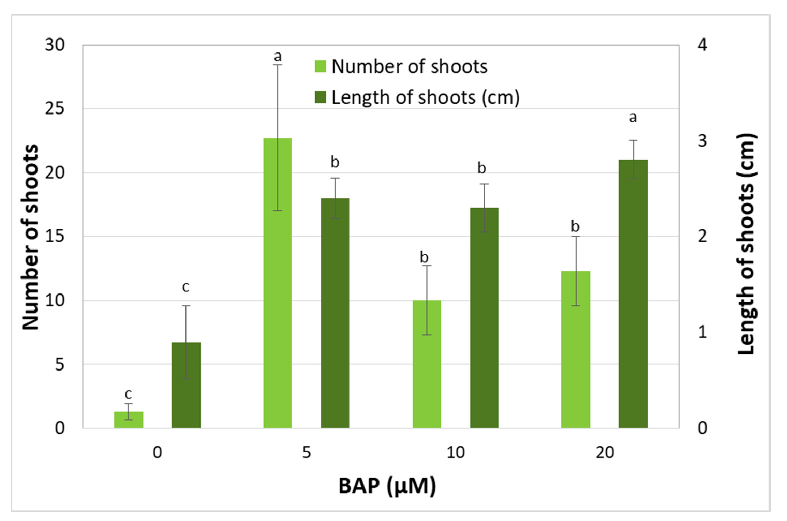
3.2. Effect of BAP Concentration on Shoot Multiplication

Among the different BAP concentrations tested, 5 μΜ BAP was the most effective for new shoot production and elongation (Figure 5). In contrast, the other two BAP treatments (10 and 20 μM) restricted the production of shoots, but not their lengthening (Figure 4). The control treatment, as expected, resulted in the smallest number and length of shoots produced. These results are in contrast to those of Dolcet-Sanjuan et al. [10] for P. amygdaliformis, who reported increasing new shoot production per explant with increasing BAP, from 5 μΜ to 10 and 20 μΜ, combined with very high PPFD (135 μmol·m−2·s−l), reaching up to 27 shoots per explant. This could be attributed to the different plant species used in combination with the different light irradiance levels applied.
Other researchers also reported increasing production of shoots with gradual increases in the BAP levels in various Pyrus species and varieties, but they observed the simultaneous appearance of morphological disorders, e.g., vitrification [28,29,30]. To alleviate the problem, the same or similar BAP concentrations to that used herein were proposed as the most efficient in the case of other Pyrus species: P. calleryana Decne. [31], P. syrica Boiss. [32] and P. communis var. pyraster [33]. In our work, however, for the greater BAP concentrations used, no sign of malformation appeared, which was probably due to good aeration conditions applied to the cultures.

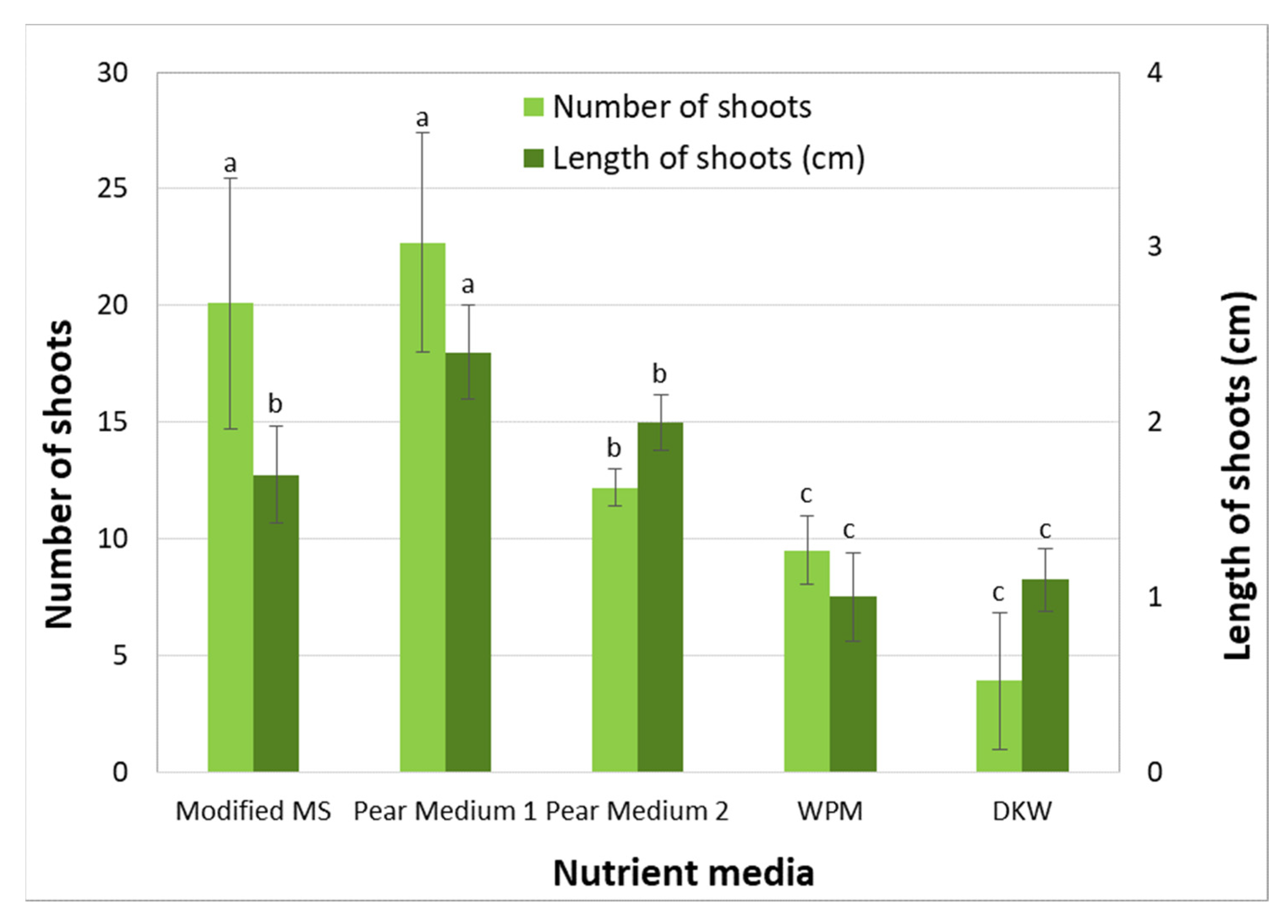
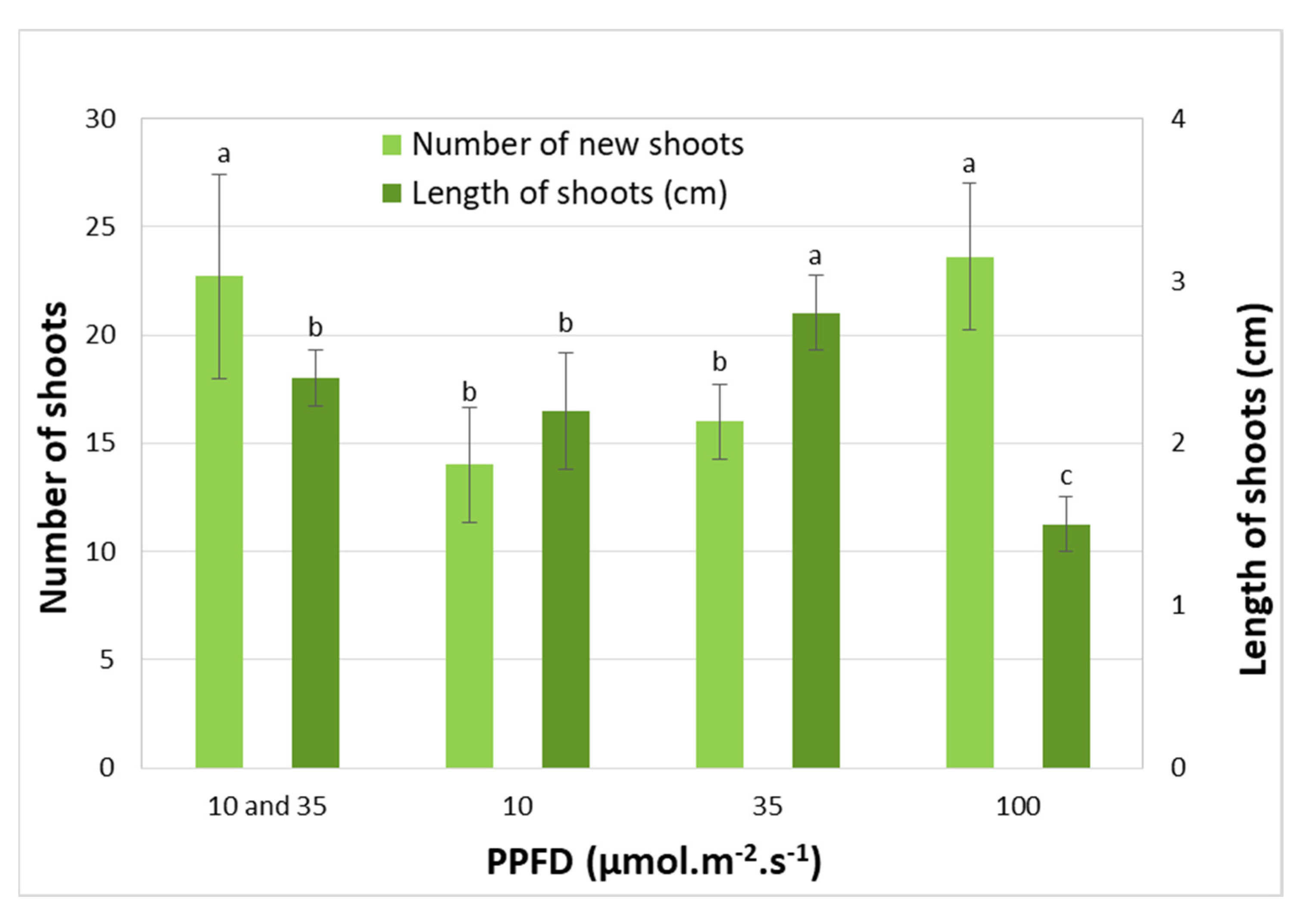
3.3. Effect of Nutrient Medium and Light Irradiance

In this experiment, the most typical nutrient media (modified MS, WPM and DKW) were tested on P. spinosa tissue cultures and were compared with two types (Pear Medium 1 and 2) of a new nutrient medium specially designed for Pyrus species. The explants multiplied and elongated better on Pear medium 1 compared with all of the other tested media (22.7 shoots per explant, 2.4 long) (Figure 6). In general, the nutrient media tested showed the following hierarchical sequence according to their suitability (from highest to lowest effect): Pear Medium 1 > modified MS > Pear Medium 2 > WPM > DKW (Figure 6).
P. spinosa, like most other Pyrus species, is a difficult plant in in vitro manipulation, as observed in our work, with the nutrient medium playing a key role. Similarly, Reed et al. [17], after meticulous research, proved the significance of the increased concentration of the MS meso nutrients, such as CaCl2·2H2O, KH2PO4 and MgSO4, for in vitro shoot multiplication, growth and quality by examining five different genotypes of three Pyrus species. Thus, they designed a suitable nutrient medium specially for Pyrus spp., which was also confirmed by our findings as the most appropriate for all P. spinosa material. In another study, Wada et al. [34] reported on the crucial influence of the same meso components (CaCl2·2H2O, KH2PO4 and MgSO4) on improving the micropropagation of ten different genotypes from six Pyrus species. Poothong and Reed [35] also proved that increased meso nutrients in five red raspberry cultivars may have significantly influenced the explant response, multiplication and shoot length of all cultivars tested. However, Bell et al. [36] found that the nutrient media DKW and QL [37] were more effective compared with MS and WPM for axillary shoot formation in two European pear cultivars, which is in disagreement with our results. Special in vitro mineral nutrification requirements have been reported by other researchers for various species, such as olive [38], passion fruit [39], eucalypts [40], hazelnuts [41] and bromeliads [42].
In our study, the illumination of cultures with PPFD of 10 μmol·m−2·s−1 for one week followed by 35 μmol·m−2·s−1 for the next four weeks and illumination with 100 μmol·m−2·s−1 resulted in the production of significantly higher numbers of shoots per explant (22.7 and 23.6, respectively) compared with all other treatments. Illumination with 10 μmol·m−2·s−1 or 35 μmol·m−2·s−1 applied for five weeks resulted in fewer numbers of shoots (12.0 and 14.0, respectively) (Figure 7 and Figure 8A). The longest shoots were obtained under PPFD of 35 μmol·m−2·s−1 (2.8 cm), followed by the illumination of the combination of 10 and 35 μmol·m−2·s−1 (2.4 cm), as well as the illumination of 10 μmol·m−2·s−1 for five weeks (2.2 cm). Under the PPFD of 100 μmol·m−2·s−1, the shortest shoots were formed (1.5 cm), which could not be easily handled for the next stage of rooting. The light treatment of 10 μmol·m−2·s−1 for one week followed by 35 μmol·m−2·s−1 over the next four weeks seemed to combine a high rate of shoot production and satisfactory length of shoots, and the results indicated that it was a determining factor of P. spinosa explant proliferation and shoot quality. These findings confirm the results of earlier research on juvenile individuals of P. spinosa, as the same combined treatment of 10 μmol·m−2·s−1 followed by 35 μmol·m−2·s−1 was the most efficient [2]. In line with our results, Yeo and Reed [43] reported increased new shoot production per explant under constant low light irradiance of 25 μmol·m−2·s−1 for three different Pyrus rootstocks. On the contrary, Dolcet-Sanjuan et al. [10], working on P. amygdaliformis, found that the best new shoot formation may have been promoted by a very high PPFD of 135 μmol·m−2·s−l on a modified MS nutrient medium enriched with 20 μΜ of BAP. These results are partially consistent with our findings in the highest PPFD treatment of 100 μmol·m−2·s−1 applied and only in terms of the number of shoots produced per explant, which were, however, short and not much acceptable for the stage of rooting.

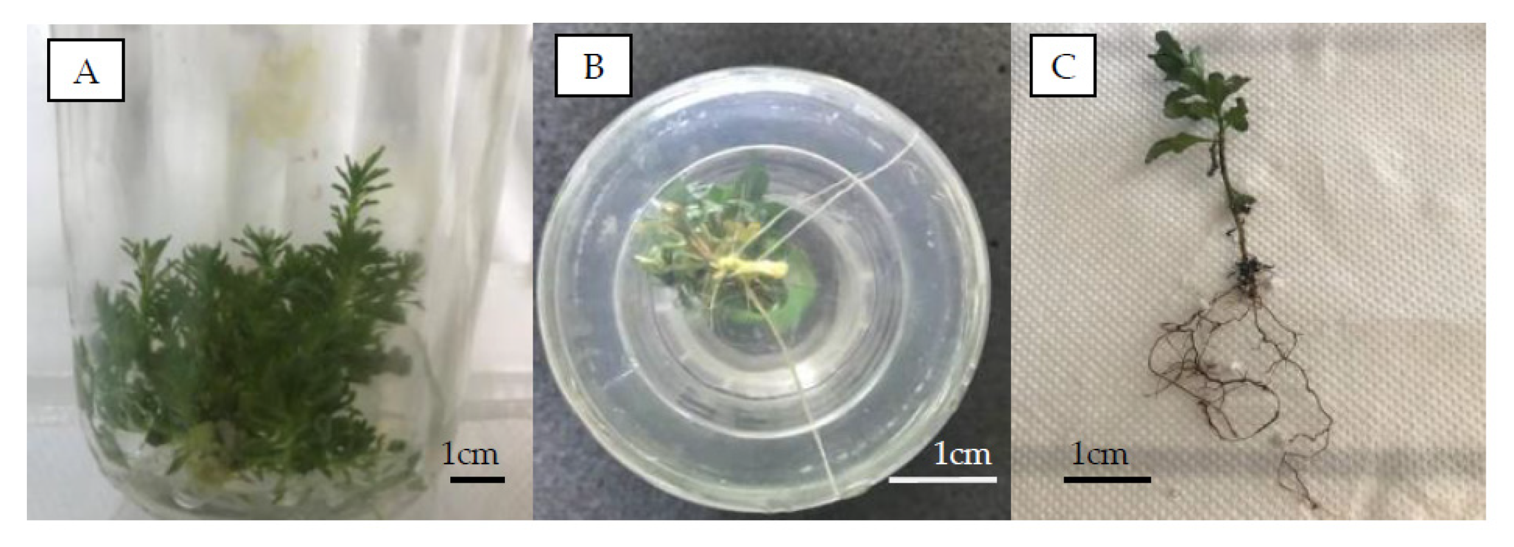
3.4. In Vitro Shoot Rooting and Plantlet Acclimatization

Both the nutrient medium and the IBA application, as well as their interaction, significantly influenced the rooting (%) of P. spinosa. Additionally, the number and length of roots produced were significantly affected by the application of IBA and its combination with the nutrient medium (Table 2). The rooting percentages were significantly higher in modified rooting MS nutrient medium than Pear Medium 1 at the IBA concentrations of 2.46 μM (44.0 vs. 20.0%) and 4.90 μM (73.0 vs. 10.0%), while at 49.00 μM IBA, both media resulted in very high percentages of rooting (79.0 and 80.0%, respectively) in comparison with the control (38.5 and 30.0%, respectively) (Table 3). Pear Medium 1 enhanced the rooting response (%) compared with modified rooting MS nutrient medium in the treatment of 24.60 μM IBA. Calluses did not develop on any shoot tested for rooting. The treatment of 4.90 μM IBA in combination with the modified rooting MS nutrient medium achieved a significantly greater number of roots (4.0 roots per shoot), whereas the same result (4.0 roots per shoot) was achieved with 24.60 μM of IBA in Pear Medium 1 (Table 3, Figure 8B). In the control treatments, more roots were produced in Pear Medium 1 than in the modified rooting MS nutrient medium (6.0 vs. 1.6 roots per shoot). In the modified rooting MS nutrient medium, the longest roots were obtained in the control and 49.00 μM IBA treatments, while in Pear Medium 1, the longest roots were obtained at 2.46 μM of IBA (Table 3). From the results obtained herein, it appeared that, in both nutrient media, the use of 49.00 μM IBA resulted in high rooting rates (79.0–80.0%) and a sufficient number of roots (2.4–2.7 roots per shoot), while the modified rooting MS nutrient medium with 49.00 μM IBA additionally provided longer roots compared with Pear Medium 1 (Table 3).
According to the literature, so far, this is the first report on P. spinosa rooting under the conditions described in this study. The process of quickly dipping the bases of the shoots into IBA solutions has long been investigated for rooting various species, and is used mainly to eliminate callus formation and facilitate the subsequent acclimatization and survival of plantlets ex vitro. Specifically, in four different Pyrus species and Cydonia oblonga, Dolcet-Sanjuan et al. [10], among other rooting processes, investigated a 15-s quick dip into 10 mM IBA, where, among the tested species, P. amygdaliformis reached 73% rooting, a satisfactory rooting percentage, as well as a sufficient number and length of roots. Yeo and Reed [43] also applied a 15-s dip into 10 mM NAA or IBA solutions to the shoots of three Pyrus rootstocks. They found that only P. communis could reach a relatively high rooting percentage (85.6%) and number of roots (8.4 roots per shoot) in IBA, whereas the 15-s dip into NAA solution seemed to be more successful for all three Pyrus rootstocks examined. In P. elaeagrifolia Pall., Aygun and Dumanoglu [44] applied a 10-s dip in IBA to in vitro-derived shoots and reportedly achieved 54.2% rooting in vitro and 55.0% rooting ex vitro. In a previous study, by working with the initial juvenile material of P. spinosa, we achieved 83.3% rooting in vitro, with a well-developed rooting system, by applying not the quick dip process, but a more complicated and laborious procedure including two stages, the induction and development of roots [2]. However, in this case, a lot of calluses were formed along with the roots at the base of the shoots. Similarly, Dolcet-Sanjuan et al. [10], for the same species, achieved 90% rooting in vitro, also reporting the development of a large number of calluses at the basal part of the shoots.
Rooted plantlets of P. spinosa were satisfactorily acclimatized (Figure 8C) after five weeks (87.5% survival) (data not shown). Dolcet-Sanjuan et al. [10], for the same species, did not provide results on plantlet acclimatization. In another report, the acclimatization of plantlets derived from the initial juvenile material of P. spinosa reached a survival rate of 87.5% [2]. Other researchers have also reported successful acclimatization with various survival rates in a number of Pyrus species, such as 50% up to 83–90% for P. communis [45] and [8] or [43], respectively, 73% for P. pyrifolia [46] and 95% for P. syrica [32]. In a more recent study, Dimitrova et al. [47] reported, for P. communis, a faster and better in vitro rooting and acclimatization protocol by using a commercial plant biostimulator of natural origin named Charkor.

4. Conclusions

Two wild almond-leaved pear genotypes (AA2 and AA3) out of thirty-two from four different locations in Greece were selected due to their rich profiles of total phenolics and antioxidants for the first time in the present work. Additionally, this research revealed a strong positive correlation between the TPC and the AC in the leaves of the studied P. spinosa trees. For the two outstanding genotypes, a simple, functional and effective micropropagation protocol was developed. The key features of the protocol included the use of Pear Medium 1 supplemented with 5 μΜ BAP for the multiplication stage, a quick dip of excised microshoots into a solution of 49 μΜ IBA and then rooting in a modified rooting MS nutrient medium and the acclimatization of young plantlets in perlite under environmentally controlled conditions. This protocol provides flexibility to use and preserve the selected plant material of P. spinosa, either for mass production or maintenance for further investigation regarding its content in valuable natural substances

Author Contributions

Conceptualization, S.A., P.T., M.T. and A.S.; methodology, S.A., P.T., A.S., S.H., S.K. and M.T.; investigation, S.A., I.N.-O. and P.T.; resources, S.A., M.T., A.S. and P.T.; software, S.A., M.T., S.K. and P.T.; validation, S.A., P.T., M.T., A.S. and I.N.-O.; formal analysis, S.A. and P.T.; data curation, S.A. and P.T.; writing—original draft preparation, P.T., S.A.,S.K. and A.E.; writing—review and editing, A.E., P.T., S.A. and S.K.; visualization, S.A., P.T. and A.E.; supervision, P.T., S.H., S.K. and A.S.; project administration, P.T.; funding acquisition, M.T., A.S. and P.T. All authors have read and agreed to the published version of the manuscript.

Funding

Part of this research was funded by the Hellenic Foundation for Research and Innovation (HFRI) and the General Secretariat for Research and Technology (GSRT) under the HFRI PhD Fellowship Grant (GA. 90733/12-9-2017).

Institutional Review Board Statement

Not applicable.

Informed Consent Statement

This research included the collection of selected plant material (cuttings) from a common species with a widespread range namely Pyrus spinosa. This Greek native plant species is not protected by the Greek Presidential Decree 67/1981 or any other national legal instrument and is not covered by the Convention on the Trade in Endangered Species of Wild Fauna and Flora. The accessed plant individuals were wild-growing in Greek public lands according to the documentation made for the selected plant material (GPS coordinates and the locations presented in Table 1, date of collection: 20–23 June 2021), and the collections were performed without causing any harm to the standing plant individuals or wild populations, complying with institutional, national, and international research guidelines such as the Convention on Biological Diversity and its provisions as incorporated in the Greek legislation.

Data Availability Statement

Not applicable.

Conflicts of Interest

The authors declare no conflict of interest.

References

  1. Zamani, A.; Attar, F.; Maroofi, H. A synopsis of the genus Pyrus (Rosaceae) in Iran. Nord. J. Bot. 2012, 30, 310–332. [Google Scholar] [CrossRef]
  2. Tsoulpha, P.; Alexandri, S.; Tsaktsira, M. Critical factors for an efficient micropropagation protocol for Pyrus spinosa. J. Appl. Hortic. 2018, 20, 190–195. [Google Scholar] [CrossRef]
  3. Tzanakis, E.; Kalogeropoulos, T.; Tzimas, S.; Chatzilazarou, A.; Katsoyannos, E. Phenols and antioxidant activity of apple, quince, pomegranate, bitter orange and almond-leaved pear methanolic extracts. e-J. Sci. Tech. 2006, 1, 16–28. [Google Scholar]
  4. Manolaraki, F.; Sotiraki, S.; Stefanakis, A.; Skampardonis, V.; Volani, M.; Hoste, H. Anthelmintic activity of some Mediterranean browse plants against parasitic nematodes. Parasitology 2010, 137, 685–696. [Google Scholar] [CrossRef] [PubMed]
  5. Mihajilov-Krstev, T.; Zlatković, B.; Stankov-Jovanović, V.; Ilić, M.; Mitić, V.; Stojanović, G. Antioxidant and antimicrobial activities of almond-leafed pear (Pyrus spinosa Forssk.) fruits. Oxid. Commun. 2013, 36, 1079–1089. [Google Scholar]
  6. Kundaković, T.; Ćirić, A.; Stanojković, T.; Soković, M.; Kovačević, N. Cytotoxicity and antimicrobial activity of Pyrus pyraster Burgsd. and Pyrus spinosa Forssk. (Rosaceae). Afr. J. Microbiol. Res. 2014, 8, 511–518. [Google Scholar]
  7. Fadi, M.S.; Hartmann, H.T. Isolation, purification and characterization of an endogenous root-promoting factor obtained from basal section of pear hardwood cuttings. Plant Physiol. 1967, 42, 541–549. [Google Scholar]
  8. Al-Maarri, K.; Arnaud, Y.; Miginiac, E. Micropropagation of Pyrus communis cultivar ‘Passe Crassane’ seedlings and cultivar ‘Williams’: Factors affecting root formation in vitro and ex vitro. Scien. Hortic. 1994, 58, 207–214. [Google Scholar] [CrossRef]
  9. Singh, S.K.; Ojha, R.K.; Prasad, K.K. Rooting capacity of cutting of pear (Pyrus communis L.). Environ. Ecol. 2009, 27, 1990–1992. [Google Scholar]
  10. Dolcet-Sanjuan, R.; Mok, D.W.S.; Mok, M.C. Micropropagation of Pyrus and Cydonia and their responses to Fe-limiting conditions. Plant Cell Tiss. Org. Cult. 1990, 21, 191–199. [Google Scholar] [CrossRef]
  11. Pandey, K.B.; Rizvi, S.I. Plant polyphenols as dietary antioxidants in human health and disease. Oxid. Med. Cell. Long. 2009, 9, 270–278. [Google Scholar] [CrossRef] [Green Version]
  12. Sanches-Silva, A.; Testai, L.; Nabavi, S.F.; Battino, M.; Devi, K.P.; Tejada, S.; Sureda, A.; Xu, S.; Yousefi, B.; Majidinia, M.; et al. Therapeutic potential of polyphenols in cardiovascular diseases: Regulation of mTOR signaling pathway. Pharmacol. Res. 2020, 152, 104626. [Google Scholar] [CrossRef]
  13. Singh, S.; Sk, M.F.; Sonawane, A.; Kar, P.; Sadhukhan, S.; Sk, M.F.; Sonawane, A.; Kar, P.; Sadhukhan, S. Plant-derived natural polyphenols as potential antiviral drugs against SARS-CoV-2 via RNA-dependent RNA polymerase (RdRp) inhibition: An in-silico analysis. J. Biomol. Struct. Dyn. 2020, 28, 6249–6264. [Google Scholar] [CrossRef]
  14. Singleton, V.L.; Rossi, J.A. Colorimetry of total phenolics with phosphomolybdic-phosphotungstic acid reagents. Amer. J. Enol. Vitic. 1965, 16, 144–158. [Google Scholar]
  15. Brand-Williams, W.; Cuvelier, M.E.; Berset, C.L.W.T. Use of a free radical method to evaluate antioxidant activity. LWT-Food Sci. Techn. 1995, 28, 25–30. [Google Scholar] [CrossRef]
  16. Murashige, T.; Skoog, F. A revised medium for rapid growth and bioassays with tobacco tissue cultures. Physiol. Plant. 1962, 15, 473–497. [Google Scholar] [CrossRef]
  17. Reed, B.M.; DeNoma, J.; Wada, S.; Postman, J. Micropropagation of pear (Pyrus sp.). In Protocols for Micropropagation of Selected Economically-Important Horticultural Plants; Lambardi, M., Ozudogru, E., Jain, S., Eds.; Humana Press: Totowa, NJ, USA, 2013; pp. 3–18. [Google Scholar]
  18. Lloyd, G.; McCown, B. Commercially-feasible micropropagation of mountain laurel, Kalmia latifolia, by use of shoot tip culture. Proc. Intl. Plant Prop. Soc. 1980, 30, 421–427. [Google Scholar]
  19. Driver, J.; Kuniyuki, A.H. In vitro propagation of Paradox walnut rootstock. HortScience 1984, 19, 507–509. [Google Scholar] [CrossRef]
  20. Ratner, B. The correlation coefficient: Its values range between +1/−1, or do they? J. Target. Meas. Anal. Mark. 2009, 17, 139–142. [Google Scholar] [CrossRef] [Green Version]
  21. He, J.; Yin, T.; Chen, Y.; Cai, L.; Tai, Z.; Li, Z.; Liu, C.; Wang, Y.; Ding, Z. Phenolic compounds and antioxidant activities of edible flowers of Pyrus pashia. J. Funct. Foods 2015, 17, 371–379. [Google Scholar] [CrossRef]
  22. Grygorieva, O.; Kucharska, A.Z.; Piórecki, N.; Klymenko, S.; Vergun, O.; Brindza, J. Antioxidant activities and phenolic compounds in fruits of various genotypes of American persimmon (Diospyros virginiana L.). Acta Sci. Pol. Techn. Aliment. 2018, 17, 117–124. [Google Scholar]
  23. Chena, G.L.; Chen, S.G.; Zhao, Y.Y.; Luo, C.X.; Li, J.; Gao, Y.Q. Totalphenolic contents of 33 fruits and their antioxidant capacities before and after in vitro digestion. Indust. Crops. Prod. 2014, 57, 150–157. [Google Scholar] [CrossRef]
  24. Ekin, H.N.; Gokbulut, A.; Aydin, Z.U.; Donmez, A.A.; Orhan, I.E. Insight into anticholinesterase and antioxidant potential of thirty-four Rosaceae samples and phenolic characterization of the active extracts by HPLC. Industr. Crops Prod. 2016, 91, 104–113. [Google Scholar] [CrossRef]
  25. Zahid, K.; Ahmed, M.; Khan, F. Comparative evaluation of total phenolics, total flavonoids content and antiradical activity in six selected species of family Rosaceae using spectroscopic method. Amer. J. Biomed. Sci. Res. 2019, 3, 352–357. [Google Scholar] [CrossRef]
  26. Yang, S.W.; Lee, H.; Song, J.M.; Choi, S.E.; Cheong, E.J. Analysis of total phenolic, flavonoid contents, and antioxidant capacity extract from leaves of selected accessions of two wild pear species, Pyrus pyrifolia and P. ussuriensis. J. For. Environ. Sci. 2021, 37, 226–234. [Google Scholar] [CrossRef]
  27. Pandey, N.; Pant, J. Determination of physicochemical and pharmacological screening of leaves and flowers part of Pyrus pashia. Intl. J. Herb. Med. 2020, 8, 28–32. [Google Scholar]
  28. Kadota, M.; Niimi, Y. Effects of cytokinin types and their concentrations on shoot proliferation and hyperhydricity in in vitro pear cultivar shoots. Plant Cell Tiss. Org. Cult. 2003, 72, 261–265. [Google Scholar] [CrossRef]
  29. Poudyal, Β.Κ.; Zhang, Y.; Du, G. Adventitious shoot regeneration from the leaves of some pear varieties (Pyrus spp.) grown in vitro. Front. Agr. China 2008, 2, 82–92. [Google Scholar] [CrossRef]
  30. Liu, J.; Zhang, X.; Poudyal, B.K.; Zhang, Y.; Jiao, Z.; Qi, J. Adventitious shoot regeneration from the leaves of in vitro grown ‘Zhongli 1’ pear (Pyrus spp.). Front. Agr. China 2009, 3, 60–66. [Google Scholar] [CrossRef]
  31. Βerardi, G.; Infante, R.; Neri, D. Micropropagation of Pyrus calleryana Dcn. from seedlings. Scien. Hortic. 1993, 53, 157–165. [Google Scholar] [CrossRef]
  32. Shibli, R.A.; Ajlouni, M.M.; Jaradat, A.; Aljanabi, S.; Shatnawi, M. Micropropagation in wild pear (Pyrus syrica). Scien. Hortic. 1997, 68, 237–242. [Google Scholar] [CrossRef]
  33. Caboni, E.; Tonelli, M.G.; Lauri, P.; D’Angeli, S.; Damiano, C. In vitro shoot regeneration from leaves of wild pear. Plant Cell Tiss. Org. Cult. 1999, 59, 1–7. [Google Scholar] [CrossRef]
  34. Wada, S.; Niedz, R.P.; DeNoma, J.; Reed, B.M. Mesos components (CaCl2, MgSO4, KH2PO4) are critical for improving pear micropropagation. In Vitro Cell. Dev. Biol.-Plant 2013, 49, 356–365. [Google Scholar] [CrossRef]
  35. Poothong, S.; Reed, B.M. Modeling the effects of mineral nutrition for improving growth and development of micropropagated red raspberries. Scien. Hortic. 2014, 165, 132–141. [Google Scholar] [CrossRef]
  36. Bell, R.L.; Srinivasan, C.; Lomberk, D. Effect of nutrient media on axillary shoot proliferation and preconditioning for adventitious shoot regeneration of pears. In Vitro Cell. Dev. Biol.-Plant 2009, 45, 708–714. [Google Scholar] [CrossRef]
  37. Quoirin, M.; Lepoivre, P. Improved media for in vitro culture of Prunus sp. Acta Hortic. 1977, 78, 437–442. [Google Scholar] [CrossRef]
  38. Cozza, R.; Turco, D.; Bati, C.B.; Bitonti, M.B. Influence of growth medium on mineral composition and leaf histology in micropropagated plantlets of Olea europaea. Plant Cell Tiss. Org. Cult. 1997, 51, 215–223. [Google Scholar] [CrossRef]
  39. De Monteiro, A.C.B.; Higashi, E.N.; Gonçalves, A.N.; Rodriguez, A.P.M. A novel approach for the definition of the inorganic medium components for micropropagation of yellow passionfruit (Passiflora edulis Sims. f. flavicarpa Deg.). In Vitro Cell. Dev. Biol. Plant 2000, 36, 527–531. [Google Scholar] [CrossRef]
  40. Gribble, K.; Conroy, J.P.; Holford, P.; Milham, P.J. In vitro uptake of minerals by Gypsophila paniculata and hybrid eucalypts, and relevance to media mineral formulation. Aust. J. Bot. 2002, 50, 713–723. [Google Scholar] [CrossRef]
  41. Nas, M.N.; Read, P.E. A hypothesis for the development of a defined tissue culture medium of higher plants and micropropagation of hazelnuts. Scien. Hortic. 2004, 101, 189–200. [Google Scholar] [CrossRef]
  42. Aranda-Peres, A.N.; Peres, L.E.P.; Higashi, E.N.; Martinelli, A.P. Adjustment of mineral elements in the culture medium for the micropropagation of three Vriesea bromeliads from the Brazilian Atlantic Forest: The importance of calcium. HortScience 2009, 44, 106–112. [Google Scholar] [CrossRef]
  43. Yeo, D.; Reed, B. Micropropagation of three Pyrus rootstocks. HortScience 1995, 30, 620–623. [Google Scholar] [CrossRef] [Green Version]
  44. Aygun, A.; Dumanoglu, H. In vitro shoot proliferation and in vitro and ex vitro root formation of Pyrus elaeagrifolia Pallas. Front. Plant Sci. 2015, 6, 225. [Google Scholar] [CrossRef] [PubMed]
  45. Shen, X.-S.; Mullins, M.G. Propagation in vitro of pear, Pyrus communis L., cultivars ‘William’s Bon Chrétien’, ‘Packham’s Triumph’ and ‘Beurré Bosc’. Scien. Hortic. 1984, 23, 51–57. [Google Scholar] [CrossRef]
  46. Bhojwani, S.S.; Mullins, K.; Cohen, D. In vitro propagation of Pyrus pyrifolia. Scien. Hortic. 1984, 24, 247–254. [Google Scholar] [CrossRef]
  47. Dimitrova, N.; Nacheva, L.; Berova, M. Optimisation of rooting and acclimatization of Pyrus communis L. with the biostimulator Charkor. Silva 2019, 20, 47–56. [Google Scholar]
Figure 1. Total phenols contents (TPCs) in the fresh leaves of the 32 Pyrus spinosa wild-growing genotypes from 4 different locations in Greece (red: Agia Anastasia, blue: Chalkidiki, green: Lagadas and yellow: Pieria). Means ± SD sharing the same letter among 32 genotypes are statistically insignificant and dissimilar letters on bars indicate their statistical differences (Duncan’s multiple range test at p ≤ 0.05 (n = 3)).
Sustainability 15 04511 g001
Figure 2. Antioxidant capacity (AC) of the fresh leaves of the 32 Pyrus spinosa wild-growing genotypes from 4 different locations in Greece (red: Agia Anastasia, blue: Chalkidiki, green: Lagadas and yellow: Pieria). Means ± SD sharing the same letter among 32 genotypes are statistically insignificant and dissimilar letters on bars indicate their statistical differences (Duncan’s multiple range test at p ≤ 0.05 (n = 3)).
Sustainability 15 04511 g002
Figure 3. Principal component analysis according to data from the TPC and AC of P. spinosa leaves, in which the 32 genotypes are depicted. The genotypes are divided into three groups depending on the location: red circle Chalkidiki population, green circle Agia Anastasia and blue circle Pieria and Lagadas populations.
Sustainability 15 04511 g003
Figure 4. Relationship between the TPC and AC found in the fresh leaves of the 32 Pyrus spinosa wild-sourced genotypes from 4 different locations in Greece.
Sustainability 15 04511 g004
Figure 5. Effect of different 6-Benzyloaminopurine (BAP) concentrations (0, 5, 10 and 20 μM) on the number and length of shoots formed on Pyrus spinosa (genotypes AA2 and AA3) explants. Means ± SD sharing the same letter are statistically insignificant and dissimilar letters on bars indicate their statistical differences, among the means of the number of roots and between the means of the length of the roots (Duncan’s multiple range test at p ≤ 0.05).
Sustainability 15 04511 g005
Figure 6. Effect of different nutrient media (modified MS, Pear Medium 1 and 2, Woody Plant Medium and Driver and Kuniyuki walnut medium) on the number and length of shoots produced on Pyrus spinosa (genotypes AA2 and AA3) explants. Means ± SD sharing the same letter are statistically insignificant and dissimilar letters on bars indicate their statistical differences among the means of the number of roots and between the means of the length of the roots (Duncan’s multiple range test at p ≤ 0.05).
Sustainability 15 04511 g006
Figure 7. Effect of different levels of PPFD (10 μmol·m−2·s−1 for one week followed by 35 μmol·m−2·s−1 for the next four weeks, 10, 35 and 100 μmol·m−2·s−1 for five weeks) on the number and length of shoots produced on Pyrus spinosa (genotypes AA2 and AA3) explants. Means ± SD sharing the same letter are statistically insignificant and dissimilar letters on bars indicate their statistical differences among the means of the number of roots and between the means of the length of the roots (Duncan’s multiple range test at p ≤ 0.05).
Sustainability 15 04511 g007
Figure 8. (A) Shoot proliferation of a P. spinosa culture maintained under 10 μmol·m−2·s−1 for the first week followed by exposure to 35 μmol·m−2·s−1 for another four weeks, (B) microshoot of P. spinosa rooted in modified rooting MS nutrient medium after a quick dip of its base into a solution of 4.90 μM IBA and (C) bare-root plantlet of P. spinosa after five weeks of acclimatization.
Sustainability 15 04511 g008
Table 1. Geographical coordinates of the selected P. spinosa genotypes from four different locations of Greece.
LocationGenotype CodeLatitude NLongitude E
Agia AnastasiaAA140.4745249°23.1694133°
AA240.4743708°23.1695997°
AA340.4743795°23.1697057°
AA440.4737353°23.1718045°
AA540.4780138°23.1882478°
AA640.4779023°23.1879031°
AA740.4802906°23.1907855°
AA840.4805263°23.1905726°
ChalkidikiCh140.4604397°23.3776510°
Ch240.4604397°23.3776510°
Ch340.4597619°23.3768460°
Ch440.4598438°23.3775906°
Ch540.4598609°23.3773378°
Ch640.4568795°23.4084984°
Ch740.4573448°23.4074041°
Ch840.4628896°23.3385812°
LagadasL140.7540624°23.1392016°
L240.7536982°23.1393273°
L340.7567682°23.1404019°
L440.7567329°23.1409849°
L540.7566468°23.1413470°
L640.7551287°23.1477250°
L740.7579819°23.1565756°
L840.7591659°23.1594043°
PieriaP140.3047421°23.4914045°
P240.3047421°23.4912045°
P340.3031678°23.4919882°
P440.3031678°23.4919882°
P540.3057794°23.4882214°
P640.3058068°23.4902324°
P740.3070443°23.4839155°
P840.3064999°23.4832791°
Table 2. Significance of factors (medium and IBA concentration) and their interaction, estimated by multifactor ANOVA on the rooting response (%) and the number and length of roots, on in vitro-derived shoots of Pyrus spinosa (genotypes AA2 and AA3).
FactorsRooting (%)Number of RootsLength of Roots (cm)
dfFSign.dfFSign.dfFSign.
Medium1527.3680.000 ***11.4320.233 NS10.0000.986 NS
IBA conc.4736.2690.000 ***45.1480.001 ***46.5840.000 ***
Medium x IBA conc.4448.2470.000 ***420.9630.000 ***416.5260.000 ***
Rooting percentage: R squared = 0.998 (adjusted R squared = 0.996), p ≤ 0.05. Number of roots: R squared = 0.400 (adjusted R squared = 0.363), p ≤ 0.05. Length of roots: R squared = 0.409 (adjusted R squared = 0.372), p ≤ 0.05. NS = non-significant, *** p ≤ 0.001, df = degree of freedom, F = ratio of two variances, Sign. = significance.
Table 3. Effect of different rooting media and IBA concentrations on the rooting response and the number and length of roots formed on in vitro derived shoots of Pyrus spinosa (genotypes AA2 and AA3).
Rooting (%)Number of RootsLength of Roots (cm)
IBA (μM)Modified Rooting MSPear Medium 1Modified Rooting MSPear Medium 1Modified Rooting MSPear Medium 1
0.0038.5 ± 7.7 b1A230.0 ± 7.2 bcA1.6 ± 0.2 cB6.3 ± 0.7 aA4.5 ± 0.2 aA2.9 ± 0.7 bB
2.4644.0 ± 7.8 bA20.0 ± 6.3 cdB3.0 ± 0.4 bA1.0 ± 0.0 dB1.3 ± 0.0 dB4.2 ± 1.0 aA
4.9073.0 ± 7.0 aA10.0 ± 4.7 dB4.0 ± 0.5 aA1.0 ± 0.0 dB1.8 ± 0.1 cA1.5 ± 0.0 dB
24.6022.0 ± 6.5 cB40.0 ± 7.7 bA1.5 ± 0.2 cB4.0 ± 0.4 bA1.6 ± 0.1 cB2.7 ± 0.2 bA
49.0079.0 ± 6.5 aA80.0 ± 6.3 aA2.7 ± 0.3 bA2.4 ± 0.2 cA3.9 ± 0.3 bA1.8 ± 0.2 cB
1,2 Means ± SD of rooting percentages, number and length of roots sharing the same lowercase letter within a column and the same capital letter within a row are not significantly different according to the Bonferroni test at p ≤ 0.05.
Disclaimer/Publisher’s Note: The statements, opinions and data contained in all publications are solely those of the individual author(s) and contributor(s) and not of MDPI and/or the editor(s). MDPI and/or the editor(s) disclaim responsibility for any injury to people or property resulting from any ideas, methods, instructions or products referred to in the content.

