Article

Emerging Technologies for the Production of In Vitro Raised Quality Rich Swertia chirayita by Using LED Lights

Department of Biotechnology and Bioinformatics, Jaypee University of Information Technology, Waknaghat, Solan 173234, India
*
Author to whom correspondence should be addressed.
Sustainability 2023, 15(2), 1714; https://doi.org/10.3390/su15021714
Submission received: 13 December 2022 / Revised: 11 January 2023 / Accepted: 12 January 2023 / Published: 16 January 2023
(This article belongs to the Special Issue Sustainable Smart Cities and Societies Using Emerging Technologies)

Abstract

:
The major bioactive compounds in S. chirayita are amarogentin (most bitter compound) and mangiferin, which contribute to its medicinal value due to its antidiabetic, anticancer, antimicrobial and antimalarial properties. In this study, we developed a light emitting diode (LED)–based culture setup as an alternative to the existing white fluorescent lamps (WFL) used as a light source in the tissue culture conditions of the plants. The in-vitro raised plants of S. chirayita cultivated under LED lights showed a higher accumulation of shoot biomass and secondary metabolites as compared with plants growing under WFL. In the LED lights experiment, red LED accounted forthe maximum biomass accumulation (3.56 ± 0.04 g L−1), and blue LED accounted for the accumulated maximum content of amarogentin (8.025 ± 0.04 µg mg−1 DW), total phenolics (22.33 ± 1.05 mg GA g−1 DW), total flavonoids (29 ± 1.03 mg QE g−1 DW) and DPPH radical scavenging activity (50.40 ± 0.16%) in comparison with other light conditions. From the findings, we propose LED lightning as a more sustainable, eco-friendly and reliable source for the enormous production of quality rich secondary metabolites in shoot cultures of S. chirayita than the traditionally used fluorescent lights.

Graphical Abstract

1. Introduction

S. chirayita is an endangered herb in the family Gentianaceae, comprised of annual and perennial herbs, that is an erect plant and reaches up to 1.5 m in height. It is native to the Himalaya region and grows at an elevation of 1200–3000 m between Kashmir and Bhutan. It is a traditional Ayurvedic herb of Nepal, China and India [1,2] and is collected from the wild population for medicinal purposes. Almost the whole plant is used for therapeutic purposes because of its analgesic, antiviral, antimalarial, anti-inflammatory, antidiabetic and anticancer properties [3]. Numerous compounds, including phenolics, flavonoids, and the important biomarker compounds amarogentin (secoiridoid glucoside) and mangiferin (xanthone C-glucoside), which contribute to the herb’s therapeutic capabilities, are responsible for the extensive biological activities of S. chirayita [4,5]. Amarogentin possesses antidiabetic and anticancer properties, whereas mangiferin possesses anti-HIV, antidiabetic, antiparkinson and anticancerous properties [6,7,8,9,10]. Based on their high medicinal properties, amarogentin and mangiferin have become the major phytochemicals for biological experimentation. The demand for S. chirayita has increased tremendously because of its rich pharmacological values [11]. Yet, accessibility of such a valuable herb is limited because of human interferences in their natural habitat; a low seed viability rate, as their growth period is 2–3 years and reproduction occurs through seeds produced by the plant; and other environmental changes [12]. Such challenges put the status of S. chirayita under the category of “critically endangered” by the IUCN [4], and it is on a list of 32 focused medicinal plants of India by the NMPB, Govt. of India http://www.nmpb.nic.in (accessed on 10 August 2022). As per the World Health Organization (WHO), the present demand of S. chirayita to be used as an herbal raw material is ≈ US $14 billion/year and will reach up to ≈ US $5 trillion by the year 2050 [13,14].
The primary goal of the herb industry is to produce a sustainable and high-yield amount of secondary metabolites. In recent years, many methods, including micropropagation, synthetic seed technology, and elicitor’s treatments, have been employed for the synthesis of phytoconstituents [15]. Use of elicitor treatments such as methyl jasmonate, salicylic acid and sodium nitroprusside was reported in tissue cultures of S. chirayita to enhance the production of secondary metabolites [16].The implementation of these technologies on a wide scale is difficult because technology accessibility is constrained and social acceptance of gene-edited plants is low. Herb growers are constantly looking towards more dependable and environmentally friendly technologies to boost the supply of secondary metabolites without using chemicals or genetic engineering.
Elicitation is the best biotechnological strategy for enhancing the production of bioactive compounds in plant cell cultures. Light as an abiotic elicitor inspires the synthesis as well as the development of important bioactive compounds [17]. Most of the breeders are utilizing traditional fluorescent lamps for indoor cultivation, but these sources are not efficient enough due to high temperature operations, high energy consumption and unequal spectral distribution for the growth of plants [18,19,20]. As an alternative to these lights, LEDs can improve the efficacy of biomarker compounds by altering the plants’ primary and secondary metabolism and light fluency, and they provide an accurate wavelength for plant growth [21]. Additionally, the use of LEDs provides many advantages, such as cost-effectiveness, a sustainable supply of energy, and a lifelong source of light with less heat generation as compared with the conventional one [22]. Several reports have discussed the impact of various LEDs on the enhancement of bioactive compounds in medicinal plants such as Rhodiola imbricate and Panax ginseng, but none of these studies was conducted on in vitro raised plants of S. chirayita. [17,23].
Therefore, the present goal of this study was to accelerate the shoot biomass and antioxidant activity and enhance production of key secondary metabolites in shoot cultures of S. chirayita under LED lights.

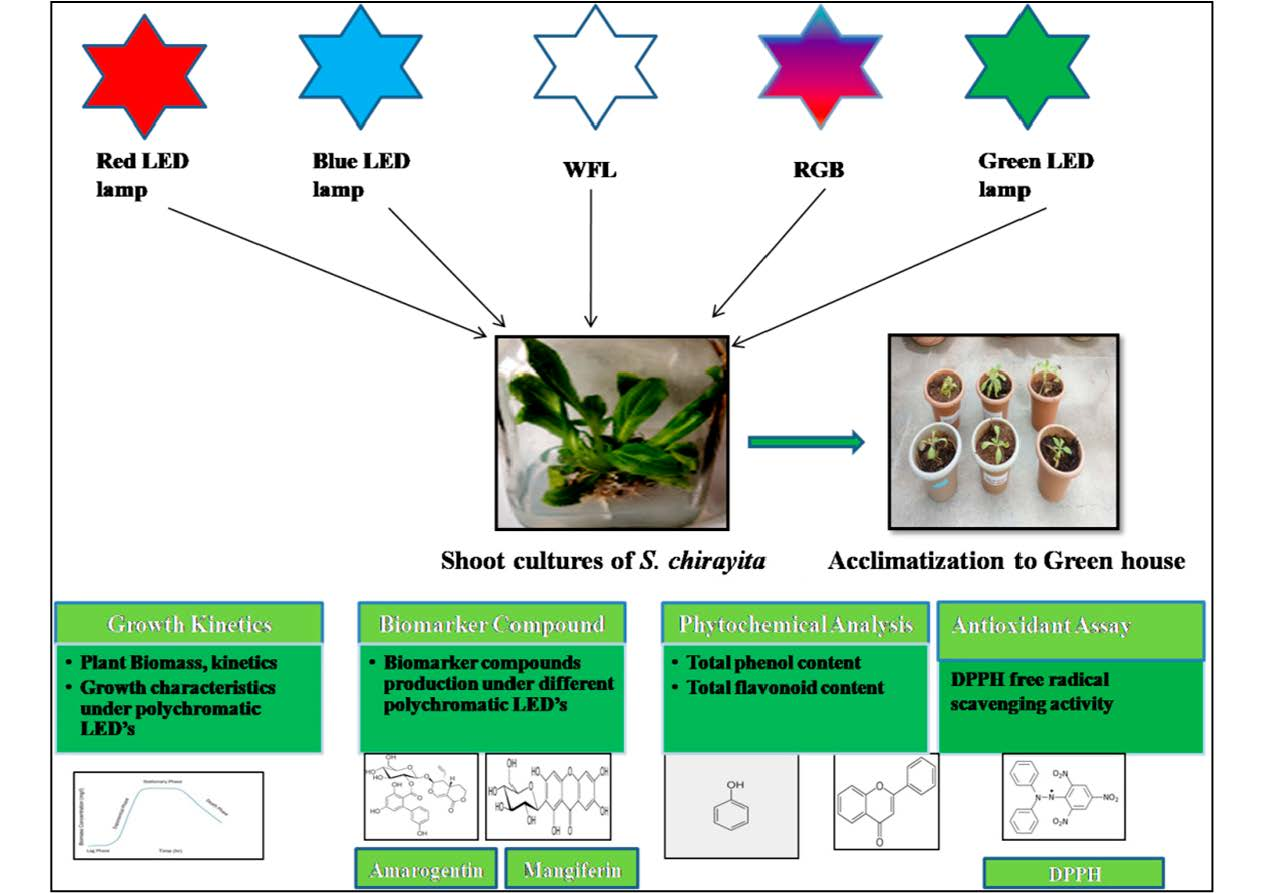
2. Material and Methods

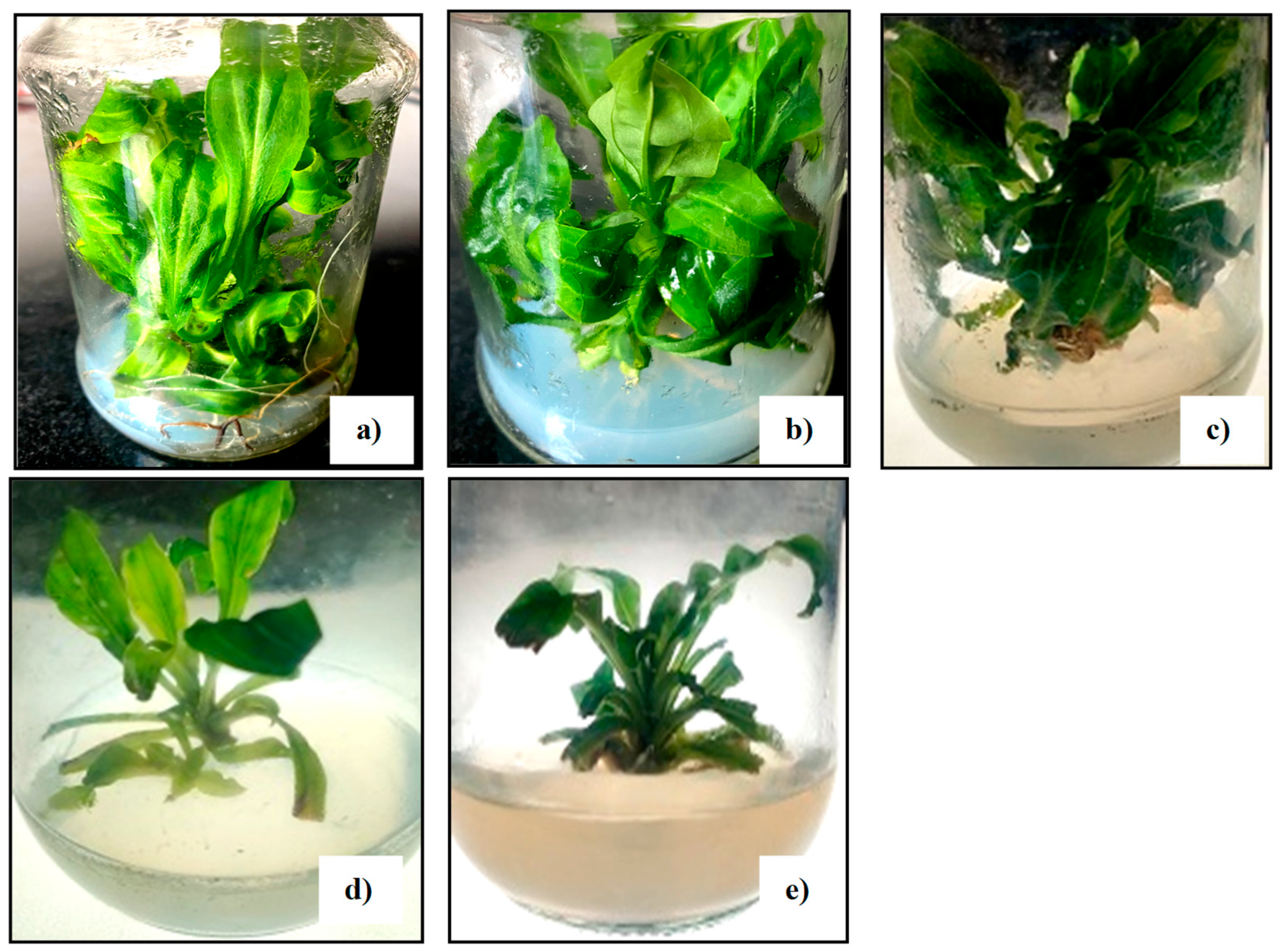
2.1. Selection of Plant Material and Establishment of In Vitro Shoots under LED Lighting

Plants of S. chirayita were collected at the Himalayan Forest Research Institute, H.P. India (20 76′ N, 67 12′ E). Further care for the plants was provided in a greenhouse at JUIT in Waknaghat, Solan, India (1400 m altitude), with controlled lighting (1300–4700 W m−2), humidity (74%), and a photoperiod of 14 h of day and 10 h of light. Shoots of S. chirayita were maintained in the plant tissue culture laboratory JUIT, Waknaghat, India. Shoots were grown in MS media [24] provided with different growth hormone concentrations such as indole-3-butyric acid (IBA) 3 mg L−1 and kinetin (KN) 1 mg L−1. After a 30-day interval, plants were routinely subcultured.
For maintaining the shoot cultures under LED lamps, in vitro grown shoot apices were used as inoculums for carrying out further experimentation. Freshly growing shoot cultures of S. chirayita (≈0.5 gm weight) were transferred to MS media (50 mL in glass jars) provided with indole-3-butyric acid (IBA) 3mg L−1 and kinetin (KN) 1mg L−1, 0.8% agar-agar, and 3% w/v sucrose, and pH was maintained at 5.6 with controlled conditions (white fluorescent light (WFL) at 3000 μmol m−2 s −1 intensity), 15 ± 1 °C temperature, and humidity (≈74%) with 16 h day and 8 h light photoperiod under aseptic conditions. Following shoots of tissue cultured plants were used for elicitation experiment by LED lamps.

2.2. Light Setup with Growth Conditions

For elicitation of medicinal compounds, various colored 12-watt LED lamps were used, which were bought from Amazon www.amazon.com (accessed on 10 August 2022) through online mode with light colors of green, red, blue (Empire brand, Tejas brand) and WFL as control. In our experimentation we used a two-part light-emitting setup. The first part was an easily removable electric circuit for LEDs, and the second consisted of a direct supply of current that was used as control. Plants of S.chirayita were randomly assigned to each LED treatment in the plant tissue culture lab at 15 °C ± 1 with a photoperiod of 16 h of light and 8 h of dark. The LED lineup conditions were as follows:
(a)
(Control: white fluorescent light at 15 °C ± 1 with 3000 μmol m−2 s −1 intensity (16 h of light and 8 h of dark photoperiod).
(b)
Red: 100% red LED at~660 nm wavelength with 20 nm of bandwidth at ½ peak height.
(c)
Blue: 100% blue LED at ~460 nm wavelength with 20 nm of bandwidth at ½ peak height.
(d)
Green: 100% green LED at ~550 nm wavelength with 20 nm of bandwidth at ½ peak height.
(e)
RGB: 40% red, 40% green and 20% blue polychromatic LEDs at 15°C± 1 inside plant tissue culture room.
After 30 days of cultivation conditions, growth metrics, total phenolics, total flavonoids, total antioxidants, and acclimation were examined by taking the plant from the culture bottles under aseptic conditions. However, the time periods for biomarker compound (amarogentin and mangiferin) accumulation in S. chirayita were analyzed at different time intervals for 30 days (days 0, 5, 15, 21, 25 and 30).

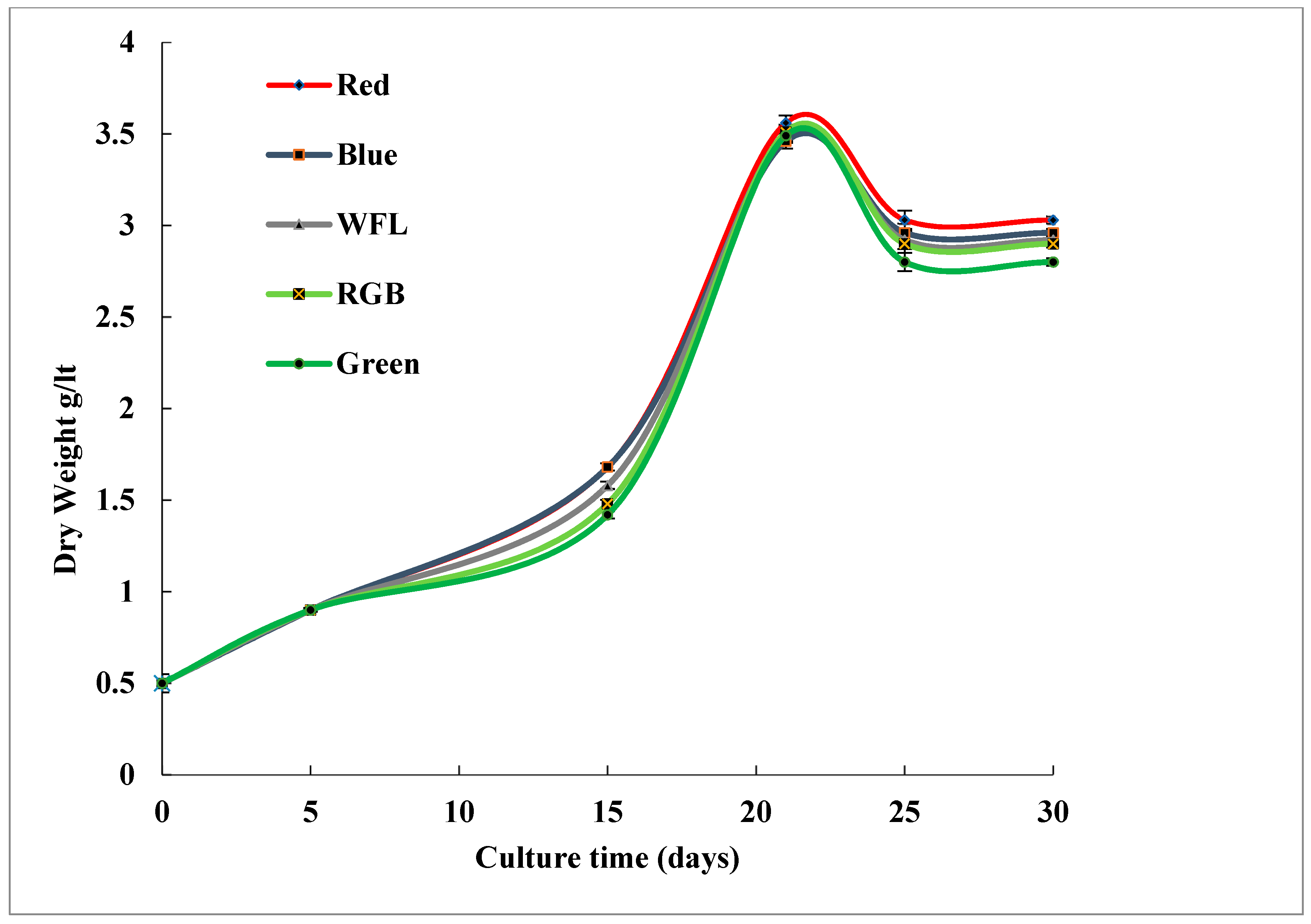
2.3. Growth Determination of Shoot Cultures

To determine the fresh weight (FW) of the shoots, they were removed from the culture medium, excess water was removed by pressing them on WhatmanTM filter paper (GE Healthcare, UK Limited), and they were weighed (Citizon, cg-203) under aseptic conditions. Simultaneously, shoots were oven dried at 45 °C (220/230 V) and finally weighed for the dry weight (DW) as expressed in (g L−1) of the medium used. Subsequently, on day 30, the growth index (GI) of shoot cultures was calculated as reported by Ketchum et al. 1995 [25],
where
GI = Wf − Wi/Wi
Wf, Final dry weight of shoots on day 30
Wi, Initial dry weight on first day
On day 30, various growth parameters were examined (plant biomass, number and length of shoots and roots (cm)) (Table 1).

2.4. Phytochemical Screening of Plant Extracts

2.4.1. Preparation of Plant Extract

The completely developed shoots were removed, dried, and ground into a fine powder using liquid nitrogen before being suspended in 100 mL of 80% methanol for an overnight period. The following day, samples were sonicated for 10 min at 30% amplitude with 2 s pulse (SONICS, Vibra CellTM). After sonication, extracts were introduced to centrifugation at 10,000 rpm for 15 min at 4 °C (Eppendr of, 5804 R), and supernatant was filtered with syringe filters (0.22 µm) PVDF. Remaining extract was kept at 4 °C until further analysis.

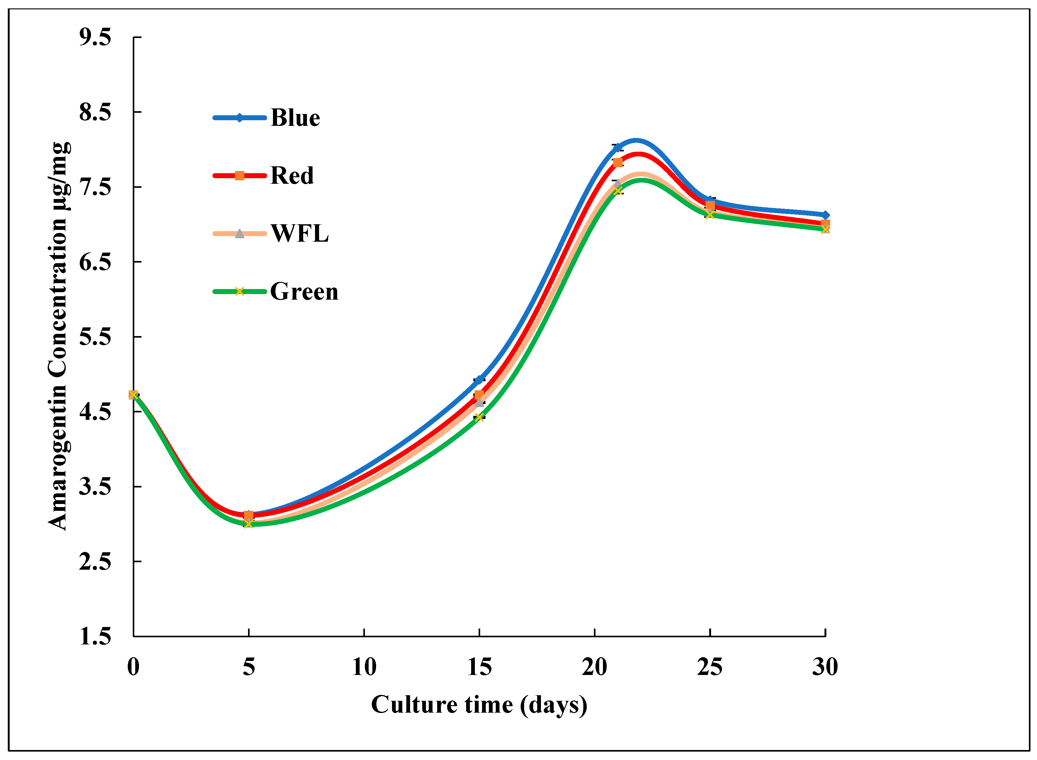
2.4.2. Quantification of Bioactive Compounds through Reverse-Phase HPLC

Shoot cultures grown under various LED lamps were subjected to reverse-phase HPLC (Agilent 11,200 series) along with an HPLC pump using C18 (5 µm) Waters column and Photodiode Array Detector (Waters 2996) to estimate the amounts of amarogentin and mangiferin. The filtrate was 10× diluted and inoculated in the column. Solvent A, a 0.1% TFA trifluoroacetic acid (Merck), and Solvent B, a solvent system, are employed (70:30 acetonitrile: water, Merck: MiiliQ). At a flow rate of 1.0 mL/min, the column was eluted in isocratic mode and the amarogentin and mangiferin were identified at 270 nm. The column temperature was 25 °C, and the cycle lasted 30 min. Quantification of bioactive compounds (amarogentin and mangiferin) in the plant extracts was evaluated by measuring their retention time with the authentic standards obtained from Chromadex, Inc, India. Results were expressed as µg/mg DW.

2.5. Determination of Total Phenolics and Total Flavonoid Content

2.5.1. Total Phenolic Content

Total phenolic content of plant extracts of S. chirayita grown under different LED lamps was determined by using the protocol of Kim et al. 2003 [26] with some modifications. According to this method, 0.1 mL of plant extract was mixed with 0.4 mL of distilled water, and then 0.15 mL of Folin–Coicalteu reagent was added. After proper mixing, it was incubated at room temperature for 5 min. Afterwards, 0.5 mL of 20% Na2CO3 was added. After mixing, the mixture was incubated in a dark room for 1 h. Absorbance was measured at 750 nm using a UV-visible spectrophotometer. For plotting the calibration curve, gallic acid was used (100–500 μg mL−1).The total amount of phenolics was estimated using the calibration curve equation (y = 0.003x − 0.046, r2 = 0.996), and it was represented as mg gallic acid (mg GA g−1).

2.5.2. Total Flavonoid Content

Total flavonoid content of plant extracts of S. chirayita grown under different LED lamps was determined by using the method of Ebrahimzadeh et al. 2008 [27], with some modifications. Briefly, 0.1 mL of methanolic plant extract was mixed with 0.4ml of methanol, and then 0.1 mL of 10% AlCl2 was added. Afterwards, 0.1mL of 1M sodium acetate was added to make up the final volume up to 4 mL with distilled water. Then it was incubated at room temperature for 30 min. Absorbance was taken at 415nm using a UV-visible spectrophotometer. For plotting the calibration curve, quercetin (100–500 μg mL−1, Sigma-aldrich, India) was used as standard. Flavonoid content was calculated using the following calibration curve equation (y = 0.001x − 0.019, r2 = 0.993), and the total amount of flavonoids was represented as mg quercetin (mg QE g−1).

2.6. Determination of DPPH Free Radical Scavenging Activity

The antioxidant activity of shoot culture extracts of S. chirayita was determined by DPPH free radical scavenging assay under different LED lamps by using the method of Yesmin et al. 2008 [28] with some changes. After this, 50 μL of plant extract was used with 3mL of methanolic DPPH (0.004%) solution added to it. Then it was stored in the dark for 30 min at room temperature. Absorbance was taken at 517 nm using a UV-visible spectrophotometer. The free RSA of different extracts incubated at different polychromatic LED lamps were compared with BHT (Sigma-Aldrich) as standard. DPPH free radical scavenging activity (%RSA) was determined as follows:
%RSA= {(Ab. of control − Ab. of Sample)/Ab. of control} × 100
where Ab. is absorbance.

2.7. Acclimatization and Transplantation of Light Treated Plants of S. chirayita

Well-grown shoots of S. chirayita incubated under different LED lamps were taken out from the culture conditions and settled at room temperature for approximately 2 h (20 bottles from each setup). Shoots were removed from the vessels and washed with running tap water to clear agar and finally rinsed with 0.5% Bavistin. These fully-grown shoots were then shifted to the containers having a mixture of sand, soil, perlite, vermiculite and cocopeat in the ratio of 1:1:1:1:1 v/v, covered with plastic bags for 3 weeks and incubated under plant tissue culture lab with different LED lamps (16 h of light and 8 h of darkness photoperiod). For 3 weeks these plants were regularly checked, and then the plastic was removed and the plants were shifted to big pots in the greenhouse area of JUIT, Waknaghat, India.

2.8. Statistical Analysis

Each experiment was performed three times, and the results were provided as mean standard deviation (standard deviation).To check the differences among group means, one-way ANOVA was used with the Duncan multiple range test. Values at (p ≤ 0.05) were considered significant. MS Office (Window Version 10, USA Microsoft India Devolpment Center (IDC), Hydrabad) was used for generating figures.

3. Result

3.1. Impact of LED Lighting on Growth

The present study revealed the impact of different LED lamps on growth in shoot cultures of S. chirayita. Similar growth kinetics in shoot cultures grown under various LED lamps are depicted in Figure 1. With a relevant variety of phases, the growth curve of shoot cultures incubated under various LED lamps showed a sigmoidal pattern for growth(lag phase: days 0–5, exponential phase: days 6–15, linear phase: days 16–21, deceleration phase: days 22–25 and stationary phase: days 25–30). Shoot cultures grown under different polychromatic LED lamps exhibited some variations in their morphology (Figure 2). Among various LED lamps, shoot cultures incubated under red LED showed maximum biomass accumulation (3.03 ± 0.01 g/lt DW) on day 21 of incubation. On day 30 of incubation, the maximum increase in the length of shoot and roots (6.13 ± 0.91 cm, 3.09 ± 1.33 cm, respectively) was observed in the red LED. An increase in the number of shoots and roots (5.51 ± 0.82, 1.94 ± 1.18, respectively) was also seen in shoot cultures incubated under red LED as compared with other LED lamps (Table 1). Growth index (GI) displayed the maximum accumulation in the red LED (5.06 ± 0.01) as compared with others and the control WFL (4.84 ± 0.01). However, the minimum accumulation was observed in shoots grown under green LED (4.60 ± 0.03) on day 30 of culture (Figure 3).

3.2. Impact of LED Lighting on Bioactive Compound Production

In the present work, the effect of LED lighting on the synthesis of active compounds in S. chirayita shoot cultures was investigated. Reverse-phase HPLC analysis of shoot cultures incubated under various LED lamps in the current investigation revealed substantial variation in the amount of amarogentin, whereas mangiferin was absent in all shoot cultures, with the exception of WFL utilised as a control (Figure 4). Interestingly, shoot cultures grown under RGB light did not show any signs of amarogentin. Shoot cultures expanded under various LED lights revealed comparable production kinetics (Figure 4). In shoot cultures grown under various polychromatic LED lamps, the production of amarogentin showed an exponential surge in the production curve for bioactive synthesis from days 5 through 21, followed by a steady decline until day 30.Among the various LED lamps, shoot cultures incubated under blue LED showed the maximum amount of amarogentin (8.035 ± 0.04 µg mg−1 DW) on day 21 of culture.
Upon completion at 30 days, the concentration of amarogentin in shoots culture incubated under various LED lamps ranged from 6.935 to 7.125 µg mg−1 DW. The maximum amount of amarogentin (7.125 ± 0.01 µg mg−1 DW) was quantified in shoot cultures incubated under blue LED in comparison with other lights, such as control WFL (6.945 ± 0.01 µg mg−1 DW). The minimum accumulation of amarogentin (6.935 ± 0.01 µg mg−1 DW) was quantified in shoot cultures incubated under green LED (Figure 4).

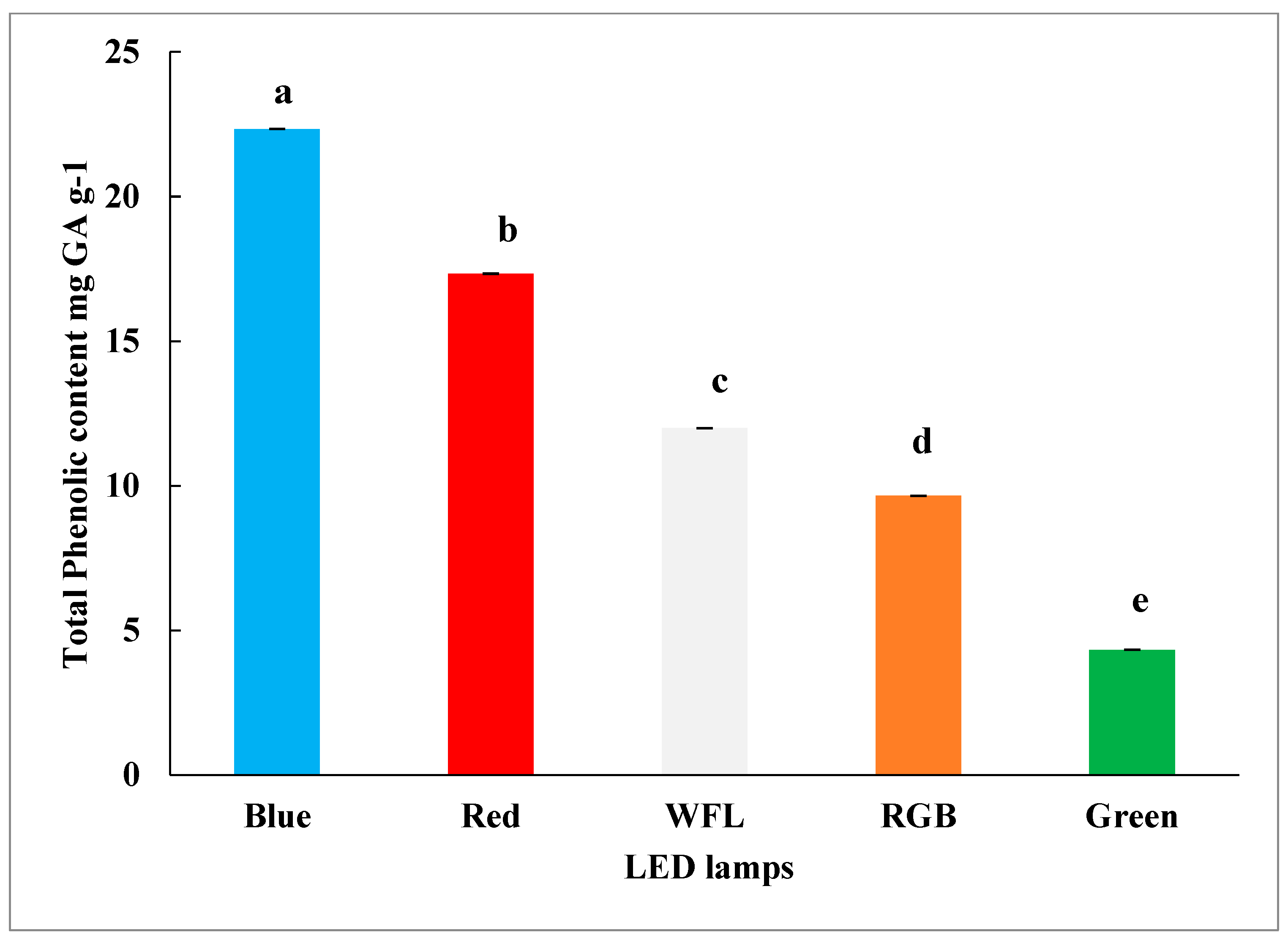
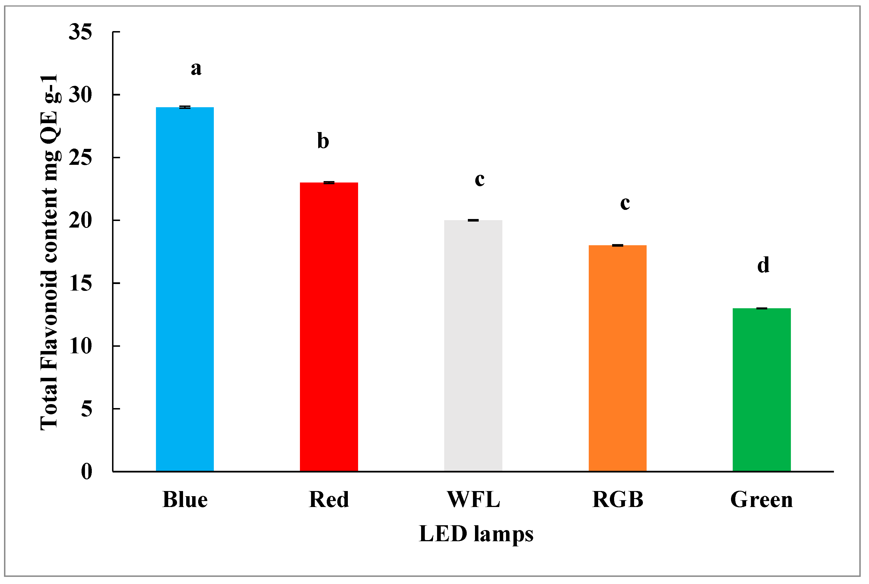
3.3. Impact of LED Lighting on Phenolic and Flavonoid Content

The current research examined how LED lighting affected S. chirayita shoot culture production of total phenolic and total flavonoid content. In the current investigation, total phenolic content in shoot cultures incubated under various LED lamps showed appreciable changes. The total phenolic amounts observed in shoot cultures varied from 4.33 to 22.33 mg GA g−1 DW of plants (Figure 5). Shoot cultures incubated under blue LED showed the highest content of total phenolics (22.33 ± 1.05 mg GA g−1 DW), in comparison with other LEDs and WFL as control (12 ± 0.06 mg GA/g DW) (Figure 5). The minimum accumulation of phenolics (4.33 ± 0.03 mg GA g−1 DW) was observed in shoot cultures incubated under green LED. Shoot cultures incubated under different LED lamps showed significant variations in the total flavonoid amount also. Total flavonoid content in shoot cultures varied from 29 to 13 mg QE/g DW of plants (Figure 6). Shoot cultures incubated under blue LED showed the highest amount of total phenolics (29 ± 1.03 mg QE g−1 DW), in comparison with other LEDs and WFL as control (20 ± 0.05 mg QE g−1 DW). The minimum accumulation of phenolics (13 ± 0.03 mg GA g−1 DW) was observed in shoot cultures incubated at green LED.

3.4. Impact of LED Lighting on DPPH Activity

As reported in the current study, shoot cultures incubated under LED lighting showed significant variations in the DPPH free radical scavenging assay. The RSA% in shoot cultures varied from 28.45 to 50.40% (Table 2). Compared with other LEDs, shoot cultures incubated under blue LEDs showed the highest RSA (50.40 0.15%), and WFL was used as control (39.02 ± 0.11%). The minimum RSA (28.45 ± 0.19%) was observed in shoot cultures incubated under green LED. Our findings are similar to those of Manivanman et al. 2015 [29], who observed the same impact of blue LED on RSA in cell cultures of Rehmannia glutinosa.

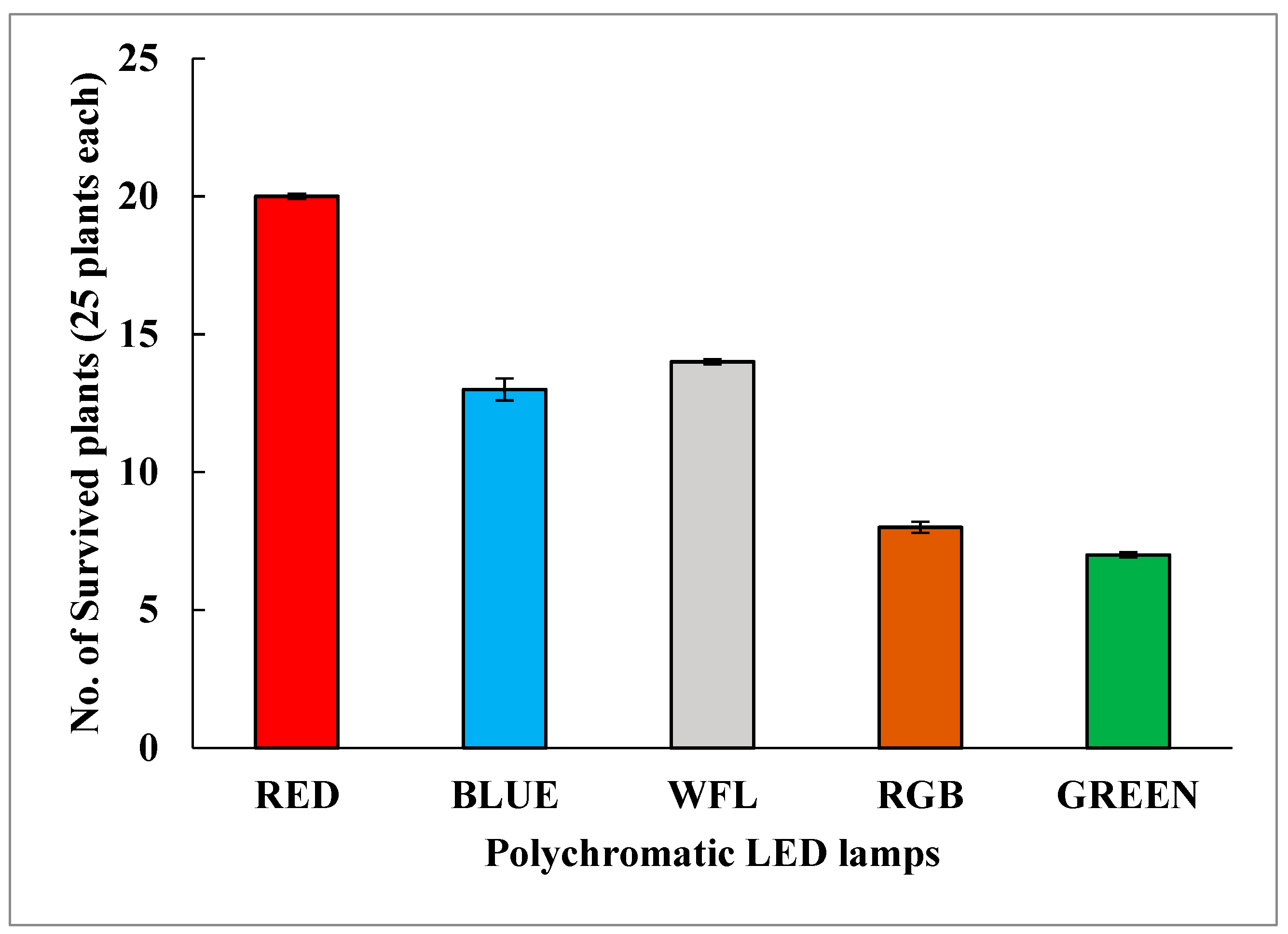
3.5. Acclimatization to Outer Environment

After successful quantification of the metabolites, the rest of the in vitro shoots of S. chirayita grown under different LED lamps were transferred to the greenhouse area. These plantlets were successfully acclimatized, with red LED shoots surviving with an 80% success rate in comparison with WFL used as control (Figure 7).

4. Discussion

Plants contain a dynamic group of photoreceptors called phytochromes (red and far red light) and cryptochromes (blue light), which help them in responding to varying photo-environmental conditions [30].The cause for enhancement in the accumulation of biomass can be credited to the considerable rise in the level of Pfr, which might have increased the growth-related enzymes in the shoot cultures [31]. In previous studies it was reported by Yu et al. 2016 [32] that red LED enhanced the photosynthetic and photochemical efficiency in Camptotheca acuminata seedlings. Our results show that different light sources have varied effects on physiological and growth patterns in cell cultures, and that these responses can vary depending on the species [33,34,35,36]. Therefore, these results demonstrated the remarkable finding that red LED increased the accumulation of biomass in S. chirayita shoot cultures. Such findings are favorable, because the red LED–incubated shoot cultures of S. chirayita can impart a sustainable supply of pharmacologically important biomarkers in an environment-friendly mode.
Amarogentin and mangiferin exhibit a wide range of bioactive properties, such as antidiabetic, anticancerous, antimalarial and antipyretic [9,10]. Previous reports have validated the impact of different light qualities on the production of bioactive compounds under various plant species’ cell culture conditions [36,37,38,39,40,41]. Our time-based investigation demonstrated that the synthesis of amarogentin was totally growth dependent in cell culture conditions. As per our knowledge, this is the first study of the impact of blue LED on the increased accumulation of amarogentin in shoot cultures of S. chirayita. The considerable conversion of the Pr to Pfr could be the cause of this [40]. Similar results were reported by [41,42] on the production of chlorogenic acid, cyanidin and jaceosidin in Populus cell cultures, when incubated under blue light. Kumar et al. 2013 [43] reported that S. chirayita plants elicited with media containing Agrobacterium rhizogenes accumulated a high amount of marker compounds as compared with other elicitors, such as salicylic acid, vanadyl sulphate, methyl jasmonate, Hoagland solution and yeast extract. Our results add to the already existing reports, which claim that the quality of different lights helps in the production of bioactive compounds [44].
The current study shows that the shoot cultures of S. chirayita, incubated under blue LED, could start out as a substitute medium for the sustainable supply of these therapeutically bioactive compounds (amarogentin). Shoot cultures of S. chirayita incubated under blue LED could aid further experimentation on discerning the detailed mechanism for enhanced production of amarogentin. In the present report, we have concluded that blue LED could increase the production of amarogentin in shoot cultures of S. chirayita, which can be scaled up by using biological tools for the enhanced production of amarogentin.
Phenolic compounds are ubiquitously present in plants and are of enormous importance due to their various biological defenses as antiproliferative agents, antioxidants, antimutagens, antiatherogenics and protectors against cardio disorders [45]. The main class of phenolic compounds that contribute to the scavenging of oxygen free radicals are flavonoids [46,47,48]. Phenolic compounds serve a diverse array of biological properties as well as health benefits [49,50,51,52]. Different types of light have been employed in earlier investigations to produce phenolics and flavonoids in tissue cultures [52,53], and numerous reports have examined the impact of polychromatic LED lights on the production of bioactive compounds as species-specific in plants [54].
The current investigation highlights the fact that shoot cultures grown under blue LED increased the production of total phenolics and total flavonoids. Our findings are consistent with those of Kapoor et al. 2018 [17], who noted comparable outcomes in Rhodiola imbricata callus cultures with regard to the generation of phenolics and flavonoids under blue light. Similar findings were observed in Stevia rebaudiana callus cultures incubated under blue light [55,56]. The enhanced accumulation of flavonoids and phenolics in blue LED–incubated shoot cultures of S. chirayita might be considered a protective response of shoot cultures against the oxidative stress caused by blue LED–induced reactive oxygen species [57]. Variation in the amount of phenolics and flavonoids in shoot cultures of S. chirayita contributed to the different light treatments and changes in the genetic expression involved during secondary metabolism machinery [52]. Our findings, along with the previous reports, revealed that different light qualities selectively enhance the production of bioactive compounds [58,59]. The current study suggests that elicitation with blue LED can serve as an alternative way for the enhanced production of phenolics and flavonoid compounds as high bioactive properties. This revealed that blue LED in cell cultures helps in the production of pharmaceutically important compounds. Our findings revealed that blue LED could increase the production of phenolics and flavonoid compounds in the shoot cultures of S. chirayita. Such compounds with high medicinal properties are of utmost priority in the pharmaceutical world.
The DPPH test is an easy method used for assessing the antioxidant capacity of different plant extracts. Its sensitivity allows it to identify active ingredients at low quantities [60,61].The present study revealed that shoot cultures incubated under blue LED displayed a higher accumulation of antioxidants, which may suggest that the higher production of antioxidants in shoot cultures protects against oxidative stress caused by reactive oxygen species [57,62]. As a result, the shoot cultures of S. chirayita produced under blue LEDs may be used as a substitute for currently available antioxidants, which are highly valued in the food and pharmaceutical industries.

5. Conclusions

The extreme endangered status of this herb warranted the development of a production platform for enriched contents of major chemical components. Among various abiotic and biotic elicitors, the application of LED lights is one of the remarkable strategies for enriching the production of secondary metabolites in tissue raised endangered medicinal herbs. The red LED as a source of elicitation provides a promising treatment for enhancement of biomass accumulation, whereas blue LED helps in the enhanced production of amarogentin, phenolics, flavonoids and antioxidants in shoot cultures of S. chirayita, in comparison with traditionally used fluorescent lamps. These important secondary metabolites play a major role as therapeutics in the pharmaceutical industry. In addition, the extracts of shoot cultures may be tested for other biological activities, such asanticancerous, antibacterial and antidiabetic, and this study could be utilized for up-scaling the bioactive compounds production on a commercial scale as well.

Author Contributions

Conceptualization, H.S. and R.G.; methodology, R.G.; validation, H.S. and R.G.; investigation, R.G.; writing—original draft preparation, R.G.; writing—review and editing, H.S. and R.G. All authors have read and agreed to the published version of the manuscript.

Funding

This research received no external funding.

Institutional Review Board Statement

Not applicable.

Informed Consent Statement

Not applicable.

Data Availability Statement

Data sharing is not applicable.

Acknowledgments

The authors are grateful to the JUIT, Waknaghat for giving the laboratory requisite to complete this experimentation and Satyam and Rhythem for helping us in this work.

Conflicts of Interest

The authors declare no conflict of interest.

References

  1. Clarke, C.B. Verbenaceae. In The Flora of British India, 4th ed.; Hooker, J.D., Ed.; L. Reeve and Co.: London, UK, 1885; pp. 560–604. [Google Scholar]
  2. Bentley, R.; Trimen, H. Medicinal Plants, Being Descriptions, with Original Figures, of the Principal Plants Employed in Medicine, and an Account of the Characters, Properties, and Uses of Their Parts and Products of Medicinal Value; J. & A. Churchill: London, UK, 1880. [Google Scholar]
  3. Bhatt, A.; Rawal, R.S.; Dhar, U. Ecological features of a critically rare medicinal plant, S. chirayita, in Himalaya. Plant Spec. Biol. 2006, 21, 49–152. [Google Scholar] [CrossRef]
  4. Joshi, P.; Dhawan, V. S. chirayita—An overview. Curr. Sci. 2005, 89, 635–640. [Google Scholar]
  5. Phoboo, S.; Pinto, M.D.S.; Barbosa, A.C.L.; Sarkar, D.; Bhowmik, P.C.; Jha, P.K. Phenolic-linked biochemical rationale for the anti-diabetic properties of S. chirayita (Roxb. ex Flem.). Phytother. Res. 2013, 27, 227–235. [Google Scholar] [CrossRef] [PubMed]
  6. Vaidya, H.; Goyal, R.K.; Cheema, S.K. Anti-diabetic activity of S.marin is due to an active metabolite, gentianine, that upregulates PPAR-c gene expression in 3T3-L1 cells. Phytother. Res. 2013, 27, 624–627. [Google Scholar] [CrossRef] [PubMed]
  7. Saha, P.; Mandal, S.; Das, A.; Das, S. Amarogentin can reduce hyper proliferation by down regulation of Cox-II and upregulation of apoptosis in mouse skin carcinogenesis model. Cancer Lett. 2006, 244, 252–259. [Google Scholar] [CrossRef]
  8. Pal, D.; Sur, S.; Mandal, S.; Das, A.; Roy, A.; Das, A. Prevention of liver carcinogenesis by amarogentin through modulation of G1/S cell cycle check point and induction of apoptosis. Carcinogenesis 2012, 33, 2424–2431. [Google Scholar] [CrossRef]
  9. Pardo-Andreu, G.L.; Paim, B.A.; Castilho, B.A.; Velho, R.F.; Delgado, J.A.; Vercesi, R. Mangifera indica L. extract (Vimang R) and its main poly phenol mangiferin prevent mitochondrial oxidative stress in atherosclerosis-pronehypercholesterolemic mouse. Pharmacol. Res. 2008, 57, 332–338. [Google Scholar] [CrossRef] [PubMed]
  10. Kavitha, M.; Nataraj, J.; Essa, M.M.; Memon, M.M.; Manivasagam, M.A. Mangiferin attenuates MPTP induced dopaminergicneurode generation and improves motor impairment, redox balance and Bcl-2/Bax expression in experimental Parkinson’s disease mice. Chem. Biol. Interact. 2013, 206, 239–247. [Google Scholar] [CrossRef]
  11. Tabassum, S.; Mahmood, S.; Hanif, J.; Hina, M.; Uzair, B. An Overview of Medicinal Importance of S. chirayita. Int. J. Appl. Sci. Technol. 2012, 2, 298–304. [Google Scholar]
  12. Pradhan, B.K.; Badola, H.K. S. chirayita, a threatened High-value Medicinal Herb: Microhabitats and conservation challenges in Sikkim Himalaya, India. Mt. Res. Dev. 2015, 35, 374–381. [Google Scholar] [CrossRef]
  13. Sharma, A. Global Medicinal Plants Demand May Touch $5 Trillion by 2050; Indian Express: Chennai, India, 2004. [Google Scholar]
  14. Joshi, K.; Chavan, P.; Warude, D.; Patwardhan, B. Molecular markers in herbal drug technology. Curr. Sci. 2004, 87, 159–165. [Google Scholar]
  15. Goraya, G.S.; Jishtu, V.; Rawat, G.S.; Ved, D.K. Wild Medicinal Plants of Himachal Pradesh: An Assessment of Their Conservation Status and Management Prioritisation; Himachal Pradesh Forest Department: Shimla, India, 2013. [Google Scholar]
  16. Ganesan, M.; Narsingh, V.; Manju, S.; Karuna, S.; Suchitra, B.; Birendra, K.; Laiq, R. Elicitation enhances swerchirin and 1,2,5,6-tetrahydroxyxanthone production in hairy root cultures of S. chirayita (Roxb.) H. Karst. Ind. Crop. Prod. 2022, 177, 114488. [Google Scholar]
  17. Kapoor, S.; Raghuvanshi, R.; Bhardwaj, P.; Sood, H.; Saxena, S.; Chaurasia, O.P. Influence of light quality on growth, secondary metabolites production and antioxidant activity in callus cultures of Rhodiola imbricate Edgew. J. Photochem. Photobiol. 2018, 183, 258–265. [Google Scholar] [CrossRef] [PubMed]
  18. Darko, É.; Heydarizadeh, P.; Schoefs, B.; Sabzalian, M.R. Photosynthesis under artificial light: The shift in primary and secondary metabolism. Philos. Trans. R. Soc. 2014, 369, 20130243. [Google Scholar] [CrossRef] [PubMed]
  19. Morrow, R.C. LED lighting in horticulture. HortScience 2008, 43, 1947–1950. [Google Scholar] [CrossRef]
  20. Schettini, E. Lighting equipment for a crop growing system in microgravity conditions for space mission. Acta Hortic. 2005, 1, 217–224. [Google Scholar] [CrossRef]
  21. Singh, D.; Basu, C.; Meinhardt-Wollweber, M.; Roth, B. LEDs for energy efficient greenhouse lighting. Renew. Sustain. Energy Rev. 2015, 49, 139–147. [Google Scholar] [CrossRef]
  22. Massa, G.D.; Kim, H.; Mitchell, C.A. Plant productivity in response to LED lighting. HortScience 2008, 43, 1951–1956. [Google Scholar] [CrossRef]
  23. Yu, K.W.; Murthy, H.N.; Hahn, E.J.; Paek, K.Y. Ginsenoside production by hairy root cultures of Panax ginseng: Influence of temperature and light quality. Biochem. Eng. J. 2005, 23, 53–56. [Google Scholar] [CrossRef]
  24. Murashige, T.; Skoog, F. A revised medium for rapid growth and bio assays with tobacco tissue cultures. Physiol. Plant. 1962, 15, 473–497. [Google Scholar] [CrossRef]
  25. Ketchum, R.E.B.; Gibson, D.M.; Gallo, L.G. Media optimization for maximum biomass production in cell cultures of pacific yew. Plant Cell Tissue Organ. Cult. 1995, 42, 185–193. [Google Scholar] [CrossRef]
  26. Kim, D.O.; Jeong, S.W.; Lee, C.Y. Antioxidant capacity of phenolic phytochemicals from various cultivars of plums. Food Chem. 2003, 81, 321–326. [Google Scholar] [CrossRef]
  27. Ebrahimzadeh, M.A.; Pourmorad, F.; Bekhradnia, A.R. Chelating activity, phenol and flavonoid content of some medicinal plants from Iran. Afr. J. Biotechnol. 2008, 7, 3188–3192. [Google Scholar]
  28. Yesmin, M.N.; Uddin, S.N.; Mubassara, S.; Akond, M.A. Antioxidant and Antibacterial Activities of Calotropis procera Linn. Am. J. Agric. Environ. Sci. 2008, 4, 550–553. [Google Scholar]
  29. Manivannan, A.; Soundararajan, P.; Halimah, N.; Ko, C.H.; Jeong, B.R. Blue LED light enhances growth, phytochemical contents, and antioxidant enzyme activities of Rehmannia glutinosa cultured in vitro. Hortic. Environ. Biotechnol. 2015, 56, 105–113. [Google Scholar] [CrossRef]
  30. Halliday, K.J.; Fankhauser, C. Phytochrome-hormonal signalling networks. New Phytol. 2003, 157, 449–463. [Google Scholar] [CrossRef] [PubMed]
  31. Wang, Y.; Zhang, H.; Zhao, B.; Yuan, X. Improved growth of Artemisia annua L. hairy roots and artemisinin production under red light conditions. Biotechnol. Lett. 2001, 23, 1971–1973. [Google Scholar] [CrossRef]
  32. Yu, W.; Liu, Y.; Song, L.; Jacobs, D.F.; Du, X.; Ying, Y.; Shao, Q.; Wu, J. Effect of Differential Light Quality on Morphology, Photosynthesis, and Antioxidant Enzyme Activity in Camptotheca acuminata Seedlings. J. Plant Growth Regul. 2016, 36, 148–160. [Google Scholar] [CrossRef]
  33. Holopainen, J.K.; Kivimäenpää, M.; Julkunen-Tiitto, R. New Light for Phytochemicals. Trends Biotechnol. 2018, 36, 7–10. [Google Scholar] [CrossRef]
  34. Hunter, D.C.; Burritt, D.J. Light quality influences adventitious shoot production from cotyledon explants of lettuce (Lactuca sativa L.). In Vitr. Cell. Dev. Biol. Plant. 2004, 40, 215–220. [Google Scholar] [CrossRef]
  35. Poudel, P.R.; Kataoka, I.; Mochioka, R. Effect of red- and blue-light-emitting diodes on growth and morphogenesis of grapes. Plant Cell Tissue Organ. Cult. 2008, 92, 147–153. [Google Scholar] [CrossRef]
  36. Tariq, U.; Ali, M.; Abbasi, B.H. Morphogenic and biochemical variations under different spectral lights in callus cultures of Artemisia absinthium L. J. Photochem. Photobiol. B Biol. 2014, 130, 264–271. [Google Scholar] [CrossRef] [PubMed]
  37. Kawka, B.; Kwiecień, I.; Ekiert, H. Influence of Culture Medium Composition and Light Conditions on the Accumulation of Bioactive Compounds in Shoot Cultures of Scutellaria lateriflora L. (American Skullcap) Grown In Vitro. Appl. Biochem. Biotechnol. 2017, 183, 1414–1425. [Google Scholar] [CrossRef] [PubMed]
  38. Pedroso, R.C.N.; Branquinho, N.A.A.; Hara, A.C.B.A.M.; Costa, A.C.; Silva, F.G.; Pimenta, L.P.; Silva, M.L.A.; Cunha, W.R.; Pauletti, P.M.; Januario, A.H. Impact of light quality on flavonoid production and growth of Hyptis marrubioides seedlings cultivated in vitro. Braz. J. Pharmacogn. 2017, 27, 466–470. [Google Scholar] [CrossRef]
  39. Lefsrud, M.L.; Kopsell, D.A.; Sams, C.E. Irradiance from distinct wavelength light-emitting diodes affect secondary metabolites in kale. HortScience 2008, 43, 2243–2244. [Google Scholar] [CrossRef]
  40. Ouyang, J.; Wang, X.; Zhao, B.; Wang, Y. Light intensity and spectral quality influencing the callus growth of Cistanche deserticola and biosynthesis of phenylethanoid glycosides. Plant Sci. 2003, 165, 657–661. [Google Scholar] [CrossRef]
  41. Zhao, D.; Xing, J.; Li, M.; Lu, D.; Zhao, Q. Optimization of growth and jaceosidin production in callus and cell suspension cultures of Saussurea medusa. Plant Cell Tissue Organ. Cult. 2001, 67, 227–234. [Google Scholar] [CrossRef]
  42. Matsumoto, T.; Nishida, K.; Noguchi, M.; Tamaki, E. Some factors affecting the anthocyanin formation by populus cells in suspension culture. Agric. Biol. Chem. 1973, 37, 561–567. [Google Scholar] [CrossRef]
  43. Kumar, V.; Chauhan, R.; Sood, H. In Vitro Production and Efficient Quantification of Major Phyto pharmaceuticalsin an Endangered Medicinal Herb, S. chirata. Int. J. Biotechnol. Bioeng. Res. 2013, 4, 495–506. [Google Scholar]
  44. Yousefzadi, M.; Sharifi, M.; Behmanesh, M.; Ghasempour, A.; Moyano, E.; Palazon, J. The effect of light on gene expression and podophyllotoxin biosynthesis in Linum album cell culture. Plant Physiol. Biochem. 2012, 56, 41–46. [Google Scholar] [CrossRef]
  45. Shahidi, F.; Ambigaipalan, P. Phenolics and polyphenolics in foods, beverages and spices: Antioxidant activity and health effects—A review. J. Funct. Foods 2015, 18, 820–897. [Google Scholar] [CrossRef]
  46. Bors, W.; Michel, C.; Saran, M. Flavonoid antioxidants: Rate constants for reactions with oxygen radicals. Methods Enzymol. 1994, 234, 420–429. [Google Scholar]
  47. Croteau, R.; Kutchan, T.M.; Lewis, N.G. Natural Products (Secondary Metabolites). In Biochemistry & Molecular Biology of Plants; Buchanan, B., Gruissem, W., Jones, R., Eds.; American Society of Plant Physiologists: Rockville, MD, USA, 2000; pp. 1250–1318. [Google Scholar]
  48. Grace, S.C. Phenolics as Antioxidants. In Antioxidants and Reactive Oxygen Species in Plants; Smirnoff, N., Ed.; Blackwell Publishing Ltd.: Hoboken, NJ, USA, 2005; pp. 141–168. [Google Scholar]
  49. Balasundram, N.; Sundram, K.; Samman, S. Phenolic compounds in plants and agri-industrial by-products: Antioxidant activity, occurrence, and potential uses. Food Chem. 2006, 99, 191–203. [Google Scholar] [CrossRef]
  50. Crozier, A.; Jaganath, I.B.; Clifford, M.N. Dietary phenolics: Chemistry, bioavailability and effects on health. Nat. Prod. Rep. 2009, 26, 1001–1043. [Google Scholar] [CrossRef]
  51. Halliwell, B.; Rafter, J.; Jenner, A. Health promotion by flavonoids, tocopherols, tocotrienols, and other phenols: Direct or indirect effects? Antioxidant or not? Am. J. Clin. Nutr. 2005, 81, 268S–276S. [Google Scholar] [CrossRef]
  52. Liu, Z.; Qi, J.L.; Chen, L.; Zhang, M.S.; Wang, X.Q.; Pang, Y.J.; Yang, Y.H. Effect of light on gene expression and shikonin formation in cultured Onosma paniculatum cells. Plant Cell Tissue Organ. Cult. 2006, 84, 39–46. [Google Scholar] [CrossRef]
  53. Yáñez, J.A.; Remsberg, C.M.; Takemoto, J.K.; Vega-Villa, K.R.; Andrews, P.K.; Sayre, C.L.; Martinez, S.E.; Davies, N.M. Polyphenols and Flavonoids: An Overview. In Flavonoid Pharmacokinetics: Methods of Analysis, Preclinical and Clinical Pharmacokinetics, Safety, and Toxicology; John Wiley & Sons, Inc.: Hoboken, NJ, USA, 2012; pp. 1–69. [Google Scholar]
  54. Taulavuori, K.; Hyöky, V.; Oksanen, J.; Taulavuori, E.; Julkunen-Tiitto, R. Species specific differences in synthesis of flavonoids and phenolic acids under increasing periods of enhanced blue light. Environ. Exp. Bot. 2016, 121, 145–150. [Google Scholar] [CrossRef]
  55. Ahmad, N.; Rab, A.; Ahmad, N. Light-induced biochemical variations in secondary metabolite production and antioxidant activity in callus cultures of Stevia rebaudiana (Bert). J. Photochem. Photobiol. B Biol. 2016, 154, 51–56. [Google Scholar] [CrossRef] [PubMed]
  56. Gupta, S.D.; Karmakar, A. Machine vision based evaluation of impact of light emitting diodes (LEDs) on shoot regeneration and the effect of spectral quality on phenolic content and antioxidant capacity in S. chirata. J. Photochem. Photobiol. B Biol. 2017, 174, 162–172. [Google Scholar] [CrossRef]
  57. Consentino, L.; Lambert, S.; Martino, C.; Jourdan, N.; Bouchet, P.E.; Witczak, J.; Castello, P.; El-Esawi, M.; Corbineau, F.; d’Harlingue, A.; et al. Blue-light dependent reactive oxygen species formation by Arabidopsis cryptochrome may define a novel evolutionarily conserved signaling mechanism. New Phytol. 2015, 206, 1450–1462. [Google Scholar] [CrossRef]
  58. Castillon, A.; Shen, H.; Huq, E. Phytochrome Interacting Factors: Central players in phytochrome-mediated light signaling networks. Trends Plant Sci. 2007, 12, 514–521. [Google Scholar] [CrossRef] [PubMed]
  59. Szopa, A.; Ekiert, H. The importance of applied light quality on the production of lignans and phenolic acids in Schisandra chinensis (Turcz.) Baill. cultures in vitro. Plant Cell Tissue Organ. Cult. 2016, 127, 115–121. [Google Scholar] [CrossRef]
  60. Kedare, S.B.; Singh, R.P. Genesis and development of DPPH method of antioxidant assay. J. Food Sci. Technol. 2011, 48, 412–422. [Google Scholar] [CrossRef] [PubMed]
  61. Kumar, V.; Van Staden, J. A Review of Swertia chirayita (Gentianaceae) as a Traditional Medicinal Plant. Front. Pharmacol. 2016, 12, 308. [Google Scholar] [CrossRef]
  62. Chen, Y.; Huang, B.; He, J.; Han, L.; Zhan, Y.; Wang, Y. In vitro and in vivo antioxidant effects of the ethanolic extract of Swertia chirayita. J. Ethnopharmacol. 2011, 136, 309–315. [Google Scholar] [CrossRef]
Figure 1. Time period for accumulation of biomass in shoot cultures of S. chirayita incubated under LED lighting. Experiment was conducted three times, and data were recorded as mean ± SD.
Sustainability 15 01714 g001
Figure 2. In vitro shoot cultures of S. chirayita incubated under LED lighting: (a) Red LED: Green and well developed shoots, (b) Blue LED: Green and compact shoots, (c) WFL: Green and compact shoots,(d) RGB: Less green and less compact shoots,(e) Green LED: Green and less compact shoots, small in size.
Sustainability 15 01714 g002
Figure 3. Impact of LED lighting on GI of shoot cultures in S. chirayita after 30 days of culture. Experiment was conducted three times, and data were recorded as mean ± SD, with a representing the highest, b representing the mild, c and d representing the least.
Sustainability 15 01714 g003
Figure 4. Time period for amarogentin production in shoot cultures of S. chirayita incubated under LED lighting. Experiment was conducted three times, and data were recorded as mean ± SD.
Sustainability 15 01714 g004
Figure 5. Impact of LED lighting on total phenolic content in shoot cultures of S. chirayita after 30 days. Experiment was conducted three times, and data were recorded as mean ± SD, with a representing the highest; b representing the mild; c, d and e representing the least.
Sustainability 15 01714 g005
Figure 6. Impact of LED lighting on total flavonoid content in shoot cultures of S. chirayita after 30 days. Experiment was conducted three times, and data were recorded as mean ± SD, with a representing the highest, b representing the mild, c and d representing the least.
Sustainability 15 01714 g006
Figure 7. Acclimatization of plants incubated under LED lighting.
Sustainability 15 01714 g007
Table 1. Growth characteristics reported under LED lighting in S. chirayita.
LED LampShoot Length (cm)Root Length (cm)No. of Shoots/ExplantsNo. of Roots/
Shoot
Red LED6.13 ± 0.91 a3.09 ± 1.33 a5.51 ± 0.82 a1.94 ± 1.18 a
Blue LED4.14 ± 0.61 b2.71 ± 1.34 ba4.91 ± 0.90 a1.70 ± 0.83 a
RGB LED3.70 ± 0.35 cb1.18 ± 0.65 ba2.61 ± 0.25 b1.42 ± 0.72 a
Green LED
WFL (Control)
2.61 ± 0.37 cb
3.96 ± 0.33 cb
0.00 ± 0.00 b
2.11 ± 1.12 ba
2.19 ± 0.37 b
3.91 ± 0.93 a
0.00 ± 0.00 a
1.59 ± 0.54 a
Experiment was conducted three times and data were recorded as mean ± SD within a column followed by the same letters are not significantly different at p < 0.05 according to Duncan multiple range test, with a representing the highest, b representing the mild and c representing the least.
Table 2. Impact of LED lighting on antioxidant activity in shoot cultures of S. chirayita.
Antioxidant Activity
Light Quality%RSA
Blue50.40 ± 0.15 a
Red43.08 ± 0.06 b
WFL39.02 ± 0.11 c
RGB35.77 ± 0.05 c
Green28.45 ± 0.19 d
Standard
BHT67.47 ± 0.05 e
Experiment was conducted three times, and data were recorded as mean ± SD within a column followed by the same letters are not significantly different at p< 0.05 according to the Duncan multiple range test, with a representing the highest, b representing the mild, c, d and e representing the least.
Disclaimer/Publisher’s Note: The statements, opinions and data contained in all publications are solely those of the individual author(s) and contributor(s) and not of MDPI and/or the editor(s). MDPI and/or the editor(s) disclaim responsibility for any injury to people or property resulting from any ideas, methods, instructions or products referred to in the content.

