Article

The Effect of Banana Rhizosphere Chemotaxis and Chemoattractants on Bacillus velezensis LG14-3 Root Colonization and Suppression of Banana Fusarium Wilt Disease

1
State Key Laboratory Breeding Base of Green Pesticide and Agricultural Bioengineering, Key Laboratory of Green Pesticide and Agricultural Bioengineering, Ministry of Education, Guizhou University, Guiyang 550025, China
2
Environment and Plant Protection Institute, Chinese Academy of Tropical Agricultural Sciences, No. 4, West Xueyuan Road, Haikou 571101, China
*
Author to whom correspondence should be addressed.
Sustainability 2023, 15(1), 351; https://doi.org/10.3390/su15010351
Submission received: 28 November 2022 / Revised: 20 December 2022 / Accepted: 20 December 2022 / Published: 26 December 2022

Abstract

:
Fusarium oxysporum f. sp. cubense (Foc) causes banana Fusarium wilt disease, which is a destructive soil-borne disease. Many plants can recruit rhizosphere microorganisms using their root exudates, thereby shaping the rhizosphere microbiome to resist pathogen infection. Therefore, this study was conducted to explore the role of root exudates in the process of biocontrol strain colonization and resistance to pathogens. In this study, the banana root exudates used as chemoattractants were obtained by hydroponics. Bacillus velezensis strain LG14-3 was isolated from the infected area of the root system of banana and showed significant chemotaxis to banana root exudates and strong inhibition of Fusarium oxysporum f. sp. cubense. Further analysis found that LG14-3 showed chemotaxis toward the components of banana root exudates, such as citric acid, succinic acid, glycine, D-galactose and D-maltose, and glycine and citric acid, which resulted in more significant chemotaxis of LG14-3. Moreover, banana root exudates enhanced the swarming motility and biofilm formation of LG14-3. Pot experiments showed that glycine and citric acid enhanced the colonization ability of Bacillus velezensis LG14-3 in the banana rhizosphere and reduced the disease severity index of banana fusarium wilt. Glycine and citric acid enhanced the growth-promoting ability of LG14-3 under pathogen stress. Our results showed that the addition of chemotactic substances enhanced the biocontrol potential of Bacillus velezensis LG14-3 to prevent banana Fusarium wilt.

1. Introduction

Banana Fusarium wilt, whose pathogen is Fusarium oxysporum f. sp. cubense (Foc), is one of the most devastating diseases of banana, which limits the development of the global banana industry [1]. Four physiological races of the pathogen have been identified [2]. Race 4 (Foc4) is the most harmful and infects nearly all banana varieties [1,2]. In recent years, much progress has been made in discovering antagonistic microorganisms to prevent and control banana wilt, but only a few strains have been commercialized. The control effect of most strains on Fusarium wilt of banana and other hosts is unstable and unsatisfactory, and performance in field conditions is often very impoverished compared to that in laboratory conditions [3].
Root exudates are the main cause of antagonistic bacterial colonization in the rhizosphere. Root exudates play a major role in the interaction between plants and plant growth-promoting rhizobacteria (PGPR) and between plants and pathogens [4]. As signal molecules, root exudates can directly mediate plant–microbe interactions, plant–plant interactions, and microbe–microbe interactions in the rhizosphere [5]. Root exudates are organic materials secreted by plant roots in the rhizosphere. These organic compounds can often be separated into two classes: low-molecular-weight compounds, which include amino acids, organic acids, sugars, phenolics and an array of secondary metabolites, and high-molecular-weight compounds, including mucilage and proteins [6]. The chemotaxis of rhizosphere bacteria to root exudates determines their effect on colonization in the rhizosphere. Chemotaxis is the process by which bacteria travel to higher concentrations of attractants or lower concentrations of repellents [7]. This property allows bacteria to receive beneficial stimulation and escape harmful stimulation [8]. Both Escherichia coli and Bacillus subtilis use a modified two-component system involving the CheA histidine kinase and CheY response regulator for transmitting signals from the receptors to the flagellar motors [9,10], triggering changes in motion. Most motile bacteria can sense and respond to low concentrations of organic compounds in the surrounding environment by chemotaxis [11].
Motile bacteria are attracted by a wide variety of chemical stimuli, including sugars, amino acids, organic acids, inorganic phosphate, and aromatic compounds [12]. In recent years, an increasing number of researchers have begun to pay attention to the interaction of root exudates and their components with bacteria in vitro. Biofilm formation and swarming motility are also important for bacterial colonization in plant roots. Bacteria achieve chemotaxis through movement. Biofilm and bacterial chemotaxis are recognized as part of the colonization mechanisms [13], and biofilms can protect bacteria from external stress. Malic acid and fumaric acid in banana root exudates attract NJN-6 and promote biofilm formation by activating relevant genes [4]. Organic acids and sugars enhance the biofilm formation of Pseudomonas sp. and Pseudomonas monteilii [14].
Bacillus spp. are common soil microorganisms that can colonize the rhizosphere and benefit plants [15]. Many Bacillus species have also been shown to have chemotactic effects on root exudates. Banana rhizosphere bacterium B. subtilis N11 showed significant chemotaxis toward banana root exudates [16]. Bacillus cereus YL6 showed chemotaxis to malate, oxalic acid, citric acid, lactic acid, and succinic acid [17]. Root exudates promote the Bacillus colonization effect on the plant rhizosphere and thus suppress pathogens. The phenolic acids of banana root exudates may have the potential to enhance root colonization and pathogen suppression abilities of Bacillus amyloliquefaciens NJN-6 [4].
Biological antagonistic bacteria are an important control measure for banana Fusarium wilt disease. The biological control of Bacillus on banana Fusarium wilt disease has been studied [18,19,20,21]. Endophytic Bacillus subtilis TR21 improved banana plant resistance to Fusarium oxysporum f. sp. cubense and promotes root growth [22]. Bacillus siamensis had strong Fusarium wilt control and growth-promoting effects on banana [23]. The combinations of Bacillus and different amendments are used to control banana Fusarium wilt disease. Biocontrol bacterium B. velezensis HN03 combined with wormcast could induce plant resistance to Fusarium wilt and suppress disease [24]. The combination of B. velezensis, Bacillus subtilis and Penicillium sp. could suppress banana disease [25].
Chemotaxis is considered an important characteristic of a successful antagonistic bacterium candidate for soil-borne pathogens [26,27]. The antagonistic microorganisms move toward plant rhizosphere chemoattractants and successfully colonize the rhizosphere to exert more effective biocontrol against pathogens [15,17,26,28]. Therefore, it is very important to optimize the selection of antagonistic bacteria sources with the chemotactic potential to act as biocontrols against soil-borne disease [26]. For example, strong chemoattractant (maize root exudates) of Bacillus amyloliquefaciens OR2-30 induced its colonization in maize roots to inhibit Fusarium graminearum infection [15]. However, little is known regarding how an antagonistic microorganism affects the suppression mechanisms of banana Fusarium wilt disease when combined with different chemoattractants.
Therefore, the present study focuses on the role of root exudates in biocontrol strain colonization in roots and resistance to pathogens. Therefore, in this study, Bacillus velezensis LG14-3 was screened based on its significant chemotactic behavior toward banana root exudates, and the effect of chemotaxis on LG14-3 biocontrol-related traits was investigated. Finally, pot experiments were conducted to assess the ability of LG14-3 to colonize banana roots and its biocontrol potential against banana wilt disease.

2. Materials and Methods

2.1. Plant Materials, Bacterial and Fungal Strains, and Culture Conditions

Tissue-cultured seedlings of Brazilian banana (Musa acuminata Cavendish cv. Brail). Fusarium oxysporum f. sp. cubense race 4 (Foc4) strain B2 (BioProject No.: PRJNA174275) and other fungal strains were cultured at 28 °C on Potato Dextrose Agar (PDA)medium (200 g/L potato, 20 g/L glucose). Bacterial strains were cultured in a Luria Bertani (LB) medium at 37 °C (10 g/L tryptone, 5 g/L yeast extract, 10 g/L NaCl, 15 g/L agar, pH 7.0). The biocontrol strains were tetracycline-resistant (400 μg/mL). All strains utilized in this study were obtained from the Environment and Plant Protection Institute, Chinese Academy of Tropical Agricultural Sciences.

2.2. Collection of Banana Root Exudates

Healthy banana seedlings were rinsed 3 times using ultrapure water. Each seedling was transferred into 100 mL deionized water in a 100 mL conical flask wrapped in tin foil. The water was changed every day. Seedlings were cultivated to 5–6 leaves and washed with sterile water 3 times. After washing, 50 mL deionized water was added to the conical flask. Seedlings were cultivated for 72 h at room temperature, and the liquid (the root exudates) was collected. A total of 500 mL of root exudates were frozen in a −70 °C refrigerator and dried with a freeze-dried dryer at low temperature until the powder was obtained. The powder was dissolved using 25 mL ultrapure water (20RE), sterilized using a 0.22 µm water phase microtool-filter membrane, and stored at −70 °C. Different concentrations of root exudates were prepared: (1) 20RE; (2) 10RE (diluted 2-fold 20RE); (3) RE (diluted 20-fold 20RE).

2.3. Isolation and Purification of Bacteria

Samples were collected from healthy banana roots in the field of the epidemic area in Lingao County of Hainan Province, China. A total of 0.1 g of banana root system was washed briefly using sterile water and ground with a mortar in 2 mL of sterile water. The suspension was heated in a water bath at 80 °C for 10 min to isolate Bacillus. The suspension was diluted, inoculated on LB medium and incubated in a constant temperature incubator at 37 °C for 12 h. Bacterial colonies with different morphologies and colors were picked and passaged, and the pure cultures obtained were stored in 30% glycerol in a −80 °C refrigerator for later experiments.

2.4. Chemotaxis Assay

Banana root secretions were used for chemotaxis analysis. Seven sugars (D-maltose, D-galactose, D-xylan, D-alginate, sucrose, D-fructose and glucose), four amino acids (L-tryptophan, histidine, glycine and D-glutamic acid) and five organic acids (succinic acid, fumaric acid, malonic acid, citric acid and L-malic acid), which are common components of banana root secretions, were also used for chemotaxis assay. The sample to be analyzed was dissolved in chemotactic buffer to a concentration of 10 mM and then sterilized by filtration.
The chemotaxis assay is based on the “drop” assay [29] with minor modifications. Briefly, cells were cultured overnight in 150 mL of LB medium. The culture was centrifuged to remove the supernatant and suspended using 100 mL chemotactic buffer (K2HPO4·3H2O 13.9 g/L, KH2PO4 5.3 g/L, EDTA 0.0067 g/L, pH 7.0). A 25 mL solution of 1% hydroxypropylmethylcellulose was added to the cell suspension. Then, a 15 mL bacterial suspension was transferred to a Petri dish. A total of 20 μL of 20RE or each substance at a concentration of 10 mM was tested. The plates were incubated at room temperature for 0.5 to 2 h and then observed for the appearance of transparent circles.
A quantitative capillary assay was performed [17,30]. Bacterial strains were cultured in LB medium until the absorbance value of the solution at wavelength 600 nm (OD600) reached 1.0, and then, the suspensions were centrifuged and resuspended in chemotaxis buffer. Then, 100 μL of the cell suspension was transferred to a 200-μL pipette tip. Instead of a capillary, a 1-mL syringe was used for the quantification assay, and 100 μL of the sample to be tested or chemotaxis buffer was aspirated separately. Then, the syringe needle was placed into the bacterial suspension in the pipette to bring the two solutions into full contact. After two hours of incubation at room temperature, the solution in the syringe was diluted with chemotaxis buffer and coated on LB plates and the number of colonies on the plates was counted after 12 h. The assay was tested 3 times.
R e l a t i v e   c h e m o t a x i s   i n d e x   ( R C I ) = t h e   b a c t e r i a l   n u m b e r   t o w a r d t h e   s a m p l e   t e s t e d   ÷ b a c t e r i a l   n u m b e r   t o w a r d   c h e m o t a x i s   b u f f e r
A value of RCI less than or equal to 1 indicates no chemotaxis, while a value of RCI greater than 1 indicates chemotaxis. Additionally, a value of RCI greater than or equal to 2 indicates a significant chemotactic response.

2.5. Identification of the Strain

The physiological and biochemistry characteristics of the bacterium were identified [31], such as Gram staining, endospore staining and methyl red (MR), catalase, citrate utilization, starch hydrolysis, nitrate reduction, protease production and cellulase production tests.
Molecular identification was performed. The genomic DNA of bacteria was extracted using an Ezup Column Bacteria Genomic DNA Purification Kit (Sangon Biotech, Shanghai, China). The 16S rRNA gene and gyrB gene were amplified by PCR. For the amplification of the 16S rRNA gene fragment, the 27F/1492R primer set was used. For the amplification of the gyrB gene fragment, the UP-1/UP-2r primer set was used. The 50 μL reaction mixture consisted of 2 μL DNA template, 2 μL 27F/UP-1, 2 µL 1492R/UP-2r, 25 μL PCR mix (0.05 units μL−1 Taq DNA polymerase; 4 mM MgCl2, and 0.4 mM dNTPs), and 19 μL nuclease-free water. The reaction conditions of the 16S rRNA gene fragment were 95 °C for 5 min, followed by 35 cycles of 95 °C for 30 s, 60 °C for 30 s and 72 °C for 2 min, with a final extension step at 72 °C for 10 min. The reaction conditions of the gyrB gene fragment were 95 °C for 5 min, followed by 35 cycles of 95 °C for 30 s, 55 °C for 30 s and 72 °C for 2 min, with a final extension step at 72℃ for 10 min. The amplification products were detected by horizontal electrophoresis through 1% agarose gels. PCR amplification products were sequenced at Hainan Nanshan Biotechnology Company (Haikou, China). Sequences were BLAST searched against the NCBI database. The reference 16S and gyrB nucleotide sequences were obtained from GenBank. The 16S and gyrB sequences of each strain were aligned using MEGA-X, and characters were weighted equally. Then, the two single-sequence datasets were concatenated and aligned using ClustalX (1.7). The neighbor-joining method with 1000 bootstrap replicates was used to build a phylogenetic tree.

2.6. Plant Growth-Promoting Properties of the Strain

The production of auxin was tested [32]. Briefly, bacteria were cultured in a liquid LB medium supplemented with 5 mM L-tryptophan. After 5 days of incubation at 37 °C with shaking at 180 rpm, the bacterial cultures were centrifuged at 6000× g for 10 min, and the supernatant was mixed (1:1, v/v) with Salkowski reagent (12 g of FeCl3 per liter in 7.9 M H2SO4) and incubated in the dark at room temperature for 30 min. The mixture turned red.
The production of siderophores was tested [33,34]. The isolate was cultured in LB liquid medium for 14 h. Bacterial liquid was inoculated into Chrome azurol S (CAS) plates for 2–3 days at 37 °C; the strain producing an orange iron chelating circle was further tested. The isolate was inoculated into LB culture medium at 37 °C for 2 days. After incubation, the cultures were centrifuged for 10 min at 10,000× g; the same volume of supernatant was mixed with CAS detection solution and reacted for 20 min. The absorbance was determined at 630 nm (i.e., As). The same volume of LB medium was mixed with CAS detection solution and reacted for 20 min as a control, and the absorbance was determined at 630 nm, which is the absorbance of Ar. The amount of iron carrier was calculated according to the formula (As − Ar)/Ar × 100%.
Nitrogen fixation was tested. One hundred microliters of bacterial suspension was inoculated in Ashby’s nitrogen-free medium (mannitol 10.0 g/L, KH2PO4 0.2 g/L, MgSO4·7H2O 0.2 g/L, NaCl 0.2 g/L, CaSO4·2H2O 0.2 g/L, CaCO3 5.0 g/L, pH 7.0–7.2). The bacteria were cultured at 28 ° C for 7 days and observed for single colony production.

2.7. Effect of Chemotaxis on the Growth of the Strain In Vitro

The bacterial cells were cultured overnight in LB medium and then adjusted to an OD600 of 1.0, resuspended 3 times with minimal medium without a carbon source ((NH4)2SO4 2.0 g/L, NaH2PO4·H2O 0.5 g/L, K2HPO4 0.5 g/L, MgSO4·7H2O 0.2 g/L, CaCl2·H2O 0.1 g/L), and then added at 1% to minimal medium containing different concentrations of chemoattractant as the sole carbon source. Cultures were cultured at 37 °C with 180 rpm shaking for 48 h, and finally, the OD600 was determined at different time points for each treatment.

2.8. Swarming Motility Assay

The effect of root exudates on the swarming ability of strains was measured as follows [15]. The bacterial cells were cultured overnight in an LB medium and then adjusted to an OD600 of 1.0. After centrifugation of the cultures, the cells were resuspended in sterile water (control) and different concentrations of root exudates (RE, 10RE and 20RE). Ten microliters of different bacterial suspensions were cultured in LB medium with 0.7% agar and incubated at 37 °C for 12 h. Strain LG11-2 was used for comparison. The assay was repeated three times.

2.9. The Effect of Chemotaxis on Bacterial Biofilm Formation

Four kinds of biofilm mediums were prepared separately: Msgg medium (control); Msgg medium and root exudates (final concentration of root exudates: 20RE); Msgg medium and citric acid (final concentration of citric acid: 0.25 mM); and Msgg medium and glycine (final concentration of glycine: 0.25 mM). The bacterial cells were cultured overnight in LB medium and then resuspended in Msgg medium and adjusted to an OD600 of 1.0. Then, 20 μL of bacterial suspension was added to a 24-well plate, and each well with 2 mL of biofilm medium, and the plate was incubated for 72 h at 30 °C in a constant temperature incubator. For quantitative analysis of biofilm formation [15], the cultures were carefully removed from wells. The remaining cells were gently washed with PBS buffer three times and fixed with 1 mL methanol for 15 min. Then, 1% crystal violet solution was added to each well for 5 min, and the excess cells were rinsed off with sterilized water. Finally, 33% glacial acetic acid was added to extract the dye, and the absorbance of the solution was measured at 570 nm. The assay was repeated three times.

2.10. Colonization Assay

Bacterial cells were cultured in LB medium at 37 °C with 180 rpm shaking for 48 h. The cells were resuspended in sterile water, and the concentration of the bacterial suspension was adjusted to 5 × 10⁷ colony-forming units/mL (cfu/mL). Forty milliliters of bacterial suspension was added to each seedling, and 6 seedlings were included in each treatment. The banana roots were taken on the 5th, 10th, and 15th days. Banana seedlings were pulled out of the soil. Banana plant roots were washed with sterile water for 3 min, soaked in 75% ethanol by volume for 1 min, rinsed with sterile water 3–5 times and ground with a sterile mortar. The ground solution was spread in tetracycline-LB solid medium, and colonies on the medium for 12 h were checked. The assay was repeated three times.
Three kinds of solutions were prepared as follows: 0.25 mM citric acid; 0.25 mM glycine; sterile water (CK). The banana roots were rinsed three times using sterile water, immersed in a solution for 1 min, placed in a conical flask (bacterial suspension 5 × 10⁷ cfu/mL) and incubated at room temperature for 72 h. Six seedlings were included in each treatment. Banana plant roots were taken, washed briefly with sterile water, ground into homogenate, coated in tetracycline-LB solid plate. After being cultured at 37 °C for 12 h, and the colonies of LG14-3 were checked. The experiment was repeated three times.

2.11. Antagonistic Assays

For antifungal bioassays, the 5 mm pathogenic fungi cake was inoculated in the center of the PDA medium. A total of 5 µL of fresh bacterial culture (OD600 = 1.0) was inoculated evenly at 3 cm from the center of the Petri dishes in all four directions; the Petri dishes were incubated at 28 °C, and the inhibition rate was calculated after 7 days. An equivalent volume of sterile water was used as a control in place of bacterial culture. T h e   i n h i b i t i o n   r a t e   ( % ) = ( D i a m e t e r   o f   t r a n s p a r e n t   c i r c l e ) ÷ D i a m e t e r   o f   c o l o n y × 100 % . The experiment was repeated three times.
The inhibition effect of strain LG14-3 metabolite on Foc was evaluated. The bacterial cells were cultured in the LB medium at 37 °C with 180 rpm shaking for 3 days and centrifuged to obtain the supernatant. Then, 100 μL fresh culture of Foc4 (107 cfu/mL) was evenly distributed into Petri dishes, and the holes were punched evenly on the PDA medium. An equal amount of filtered and debacterized supernatant was added to the holes, and the transparent circle around the holes was observed by incubation at 28 °C for 2–3 days. The experiment was repeated 3 times.
For the antibacterial bioassay, pathogenic bacteria were evenly distributed in LB medium. Five microliters of the tested culture was inoculated on Petri dishes and incubated at 37 °C for 3–5 days. The diameters of antibacterial zones were measured, and the inhibition rate was calculated. I n h i b i t i o n   r a t e   ( % ) = ( d i a m e t e r   o f   a n t i b a c t e r i a l   z o n e d i a m e t e r   o f   c o l o n y ) ÷ d i a m e t e r   o f   a n t i b a c t e r i a l   z o n e × 100 % . The experiment was repeated 3 times.

2.12. Biocontrol Experiment

The strain was cultured in LB liquid medium for 48 h, centrifuged to remove the supernatant and resuspended in sterile water, and the concentration of the bacterial solution was adjusted to 5 × 10⁷ cfu/mL. Foc4 was cultured on PDA liquid medium for 5 days, centrifuged to remove the supernatant and resuspended in sterile water, and the concentration of the suspension was adjusted to 1 × 10⁶ (conidia/mL). Four experimental treatments were carried out as follows: CK1 treatment group plants were treated with Foc; CK2 treatment group plants were treated with LG14-3 and Foc; T1 treatment group plants were treated with glycine, LG14-3 and Foc; and T2 treatment group plants were treated with citric acid, LG14-3 and Foc. First, T1 treatment group plants were soaked in 0.25 mM glycine solution for 1 min. T2 treatment group plants were soaked in 0.25 mM citric acid solution for 1 min. CK1 and CK2 treatment group plants were soaked in sterile water for 1 min. Then, plants were planted in sterilized soil. A 100 mL bacterial suspension (5 × 10⁷ cfu/mL) was added around the root of each seedling in the T1, T2 and CK2 treatment groups, and 100 mL sterile water was added around banana roots in the CK1 treatment group. Seven days later, 100 mL of Foc4 suspension was added to each pot of banana seedlings. The growth status of banana seedlings was observed daily. After 60 days, disease incidence was counted, the disease severity index was calculated [35], and plant height (distance from the ground to the tip of the uppermost spreading leaf), pseudostem girth, and pseudostem height (distance from the ground to the intersection of the petioles of the top two leaves) were measured.
Disease   severity   index = ( number   of   disease   plants   at   all   levels × representative   value   of   the   level ) ÷ total   number   of   plants × representative   value   of   the   highest   level × 100

2.13. Extraction and Determination of Chlorophyll Content

A 0.1 g mass of freshly washed banana leaves was placed into a 10 mL mixture of acetone and anhydrous ethanol (1:1, v/v), soaked for 48 h in the dark at room temperature. The absorbance of the extracted solution was measured at 645 nm and 663 nm with a mixture of acetone and anhydrous ethanol as the reference. The contents of chlorophyll a and chlorophyll b were calculated by Amon’s formula [36]: C a   ( mg / g ) = ( 12.7 × A 663 2.69 × A 645 × V / ( 1000   W ) ; C b   ( mg / g ) = ( 22.7 × A 645 4.68 × A 663 ) × V / ( 1000   W ) ; C   ( mg / g ) = C a + C b . Absorbance values at 645 nm and 663 nm are represented by A645 and A663, respectively. Ca and Cb are the contents of chlorophyll a and chlorophyll b, respectively. V is the volume of the extraction solution, and W is the mass of the leaf sample.

2.14. Statistical Analysis

The data were subjected to one-way analysis of variance (ANOVA) and Tukey’s test using IBM SPSS Statistics 26. Figures were generated using GraphPad Prism 8.

3. Results

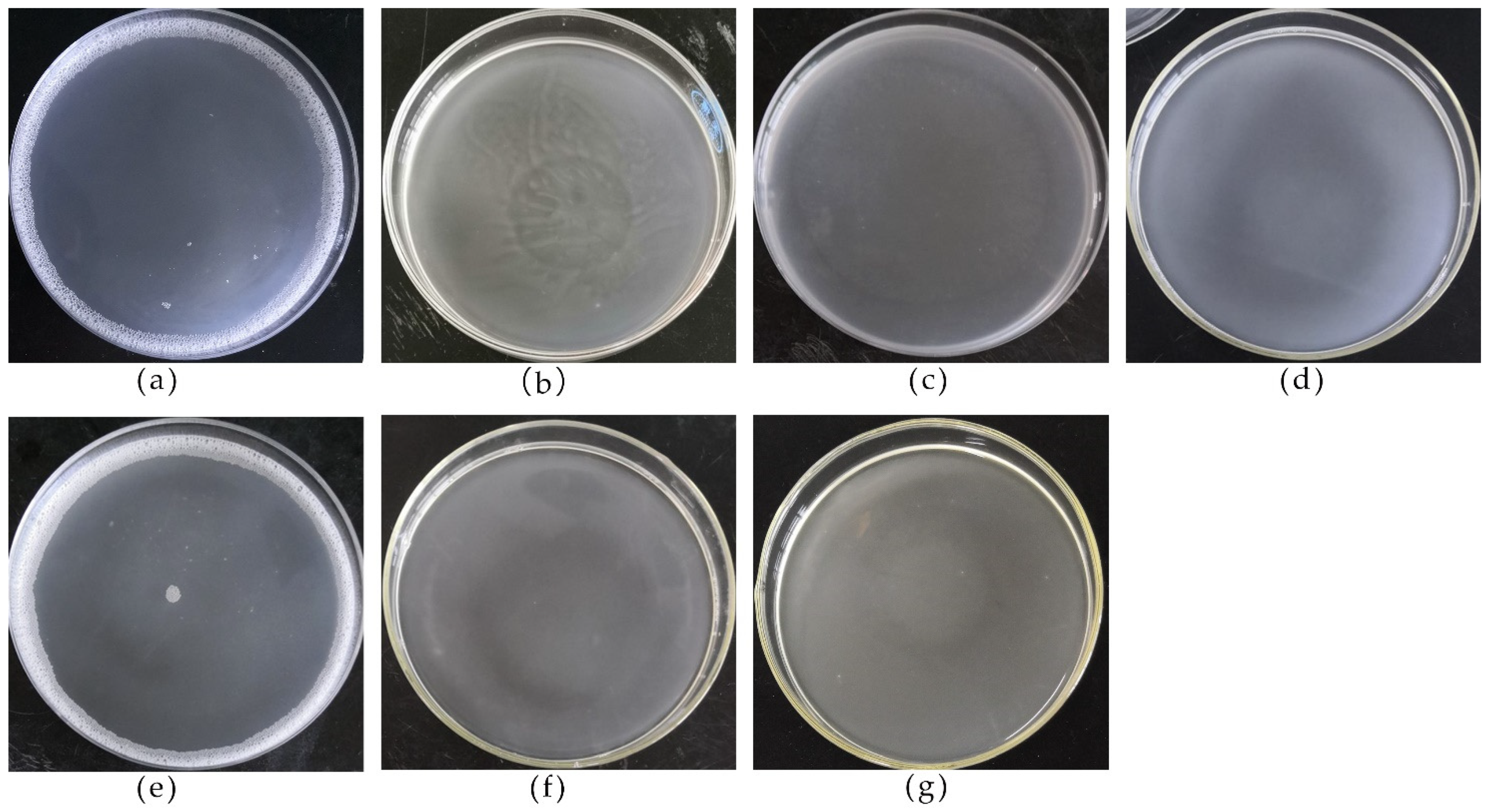
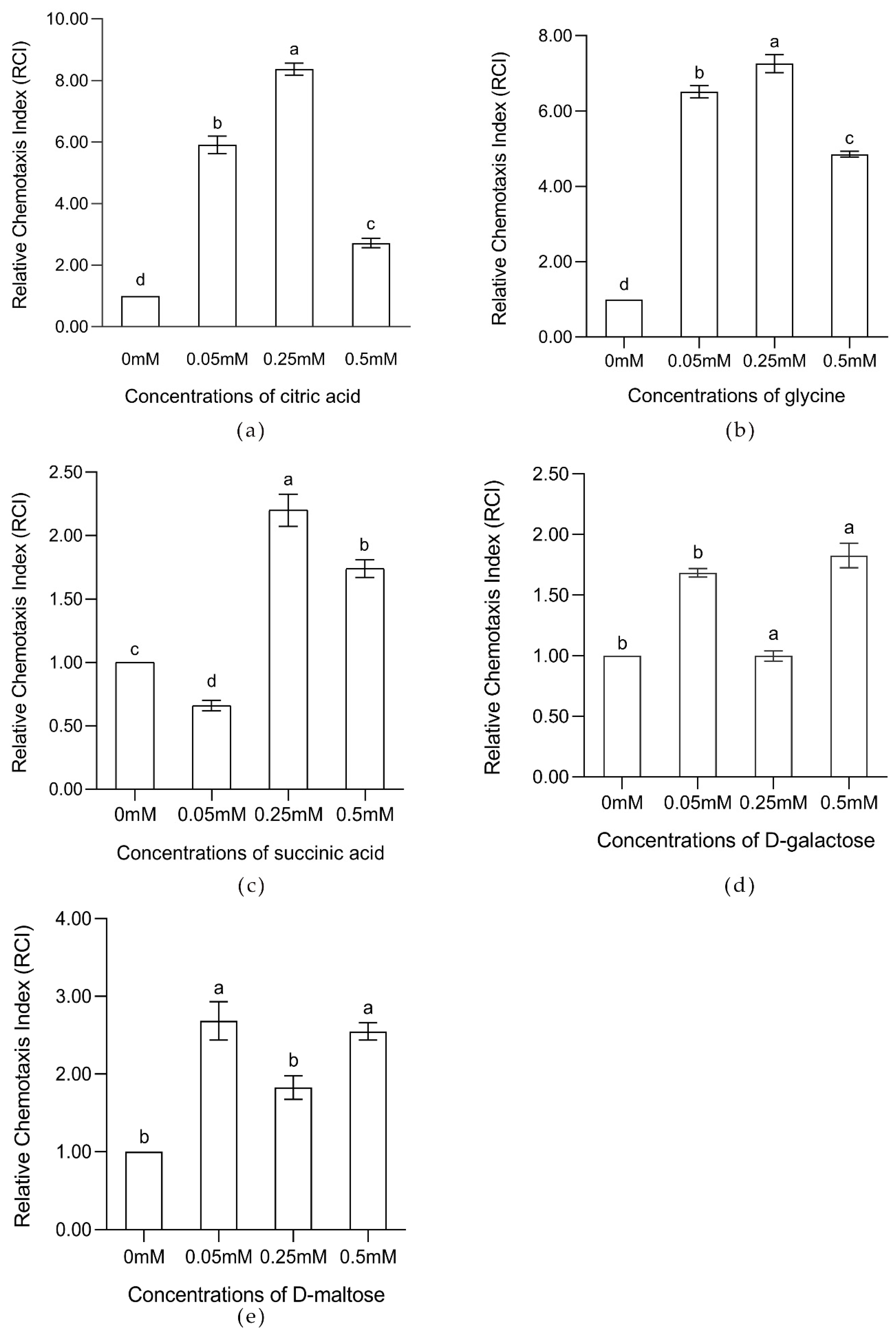
3.1. LG14-3 Exhibited Prominent Chemotactic Behavior toward Banana Root Exudates, Glycine, and Citric Acid

A chemotaxis assay was used to screen 11 bacterial strains from the banana rhizosphere that have chemotaxis to root exudates (Table 1). LG14-3 showed significant chemotaxis to banana root exudates (Figure 1b), whose relative chemotactic index (RCI) was 3.62.
The chemotaxis of LG14-3 to components of banana root exudates was further evaluated. The “Drop” assay showed that LG14-3 had chemotaxis to five compounds (10 mM), including glycine, D-galactose, D-maltose, citric acid, and succinic acid (Figure 1c–g). Quantitative capillary assays showed that glycine and citric acid had particularly marked effects (RCI ≥ 2) on LG14-3 chemotaxis at various tested concentrations. The strongest chemotactic response was for citric acid (0.25 mM), with an RCI value of 8.37. The chemotactic response of LG14-3 to glycine, citric acid, and succinic acid followed an inverted “U” shape, with a maximum attractive concentration of 0.25 mM. The chemotactic response toward D-maltose and D-galactose was biphasic, with attractive concentration maxima of 0.05 mM (Figure 2).

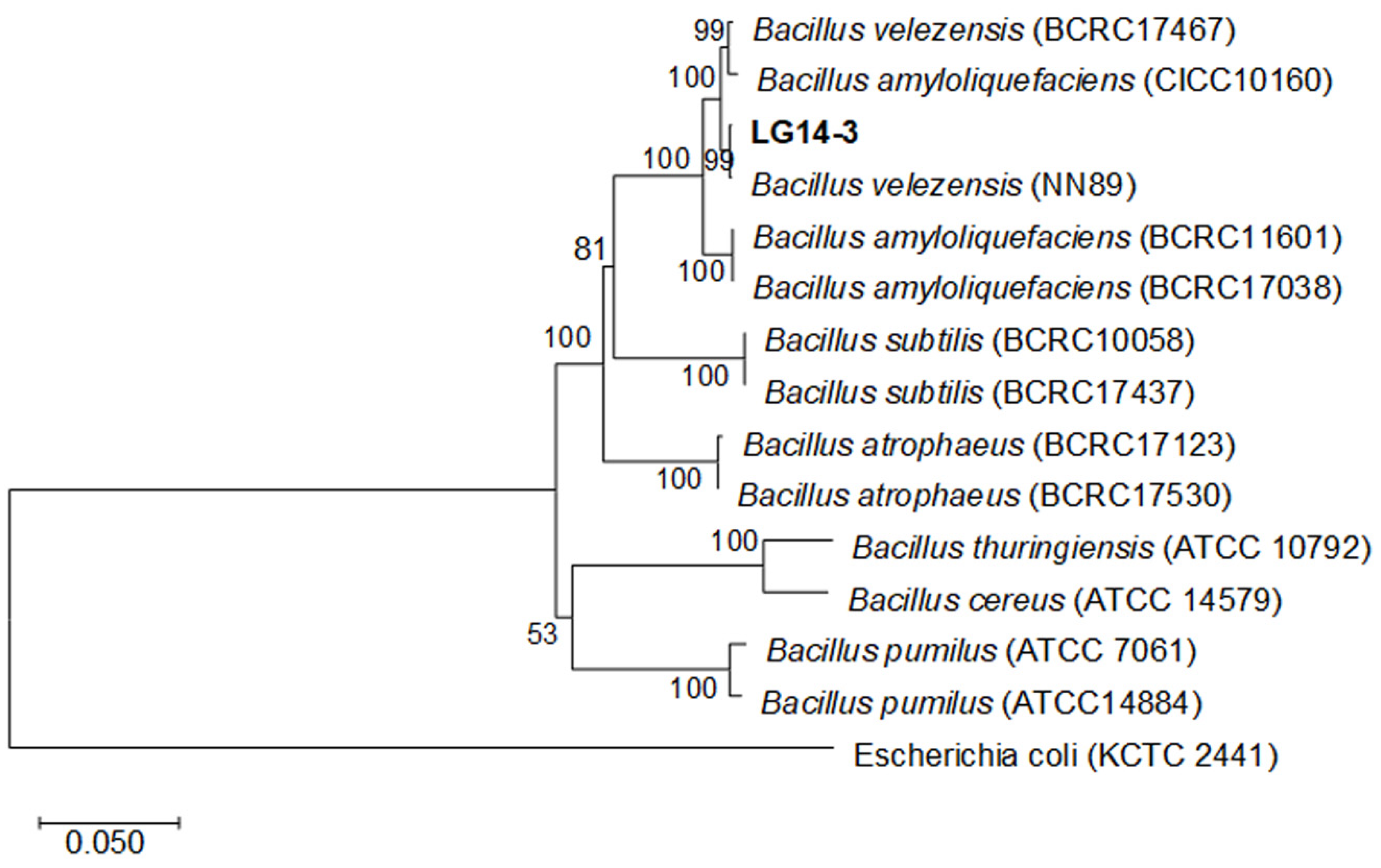
3.2. Morphological and Molecular Identification and Plant Growth-Promotion Research of LG14-3

The strain was cultured on LB agar medium at 37 °C for 12 h and produced round and creamy white colonies with dry wrinkles on the surface and a rounded depression in the middle. LG14-3 is a Gram-positive bacterium that is able to produce endospores. LG14-3 can decompose hydrogen peroxide and liquefied gelatin, utilize citrate, reduce nitrate, and produce cellulase, protease and amylase (Table 2).
For Bacillus, it is not possible to rely on 16S sequences alone to identify strains due to the high degree of sequence similarity between species [37]. Therefore, the phylogenetic tree was established using the combination of 16S and gyrB genes. The phylogenetic tree showed that strain LG14-3 and Bacillus velezensis NN89 clustered in the same large branch with 99% bootstrap (Figure 3). Based on morphological, physiological and biochemical characteristics, and phylogenetic analyses, LG14-3 was identified as Bacillus velezensis.
LG14-3 can produce indoleacetic acid (IAA) and siderophores, and the synthesis rate of siderophores is 37%. LG14-3 produced elastic and sticky, oil-drop-like single colonies on Ashby’s nitrogen-free medium, indicating that LG14-3 has nitrogen fixation (Table 2).

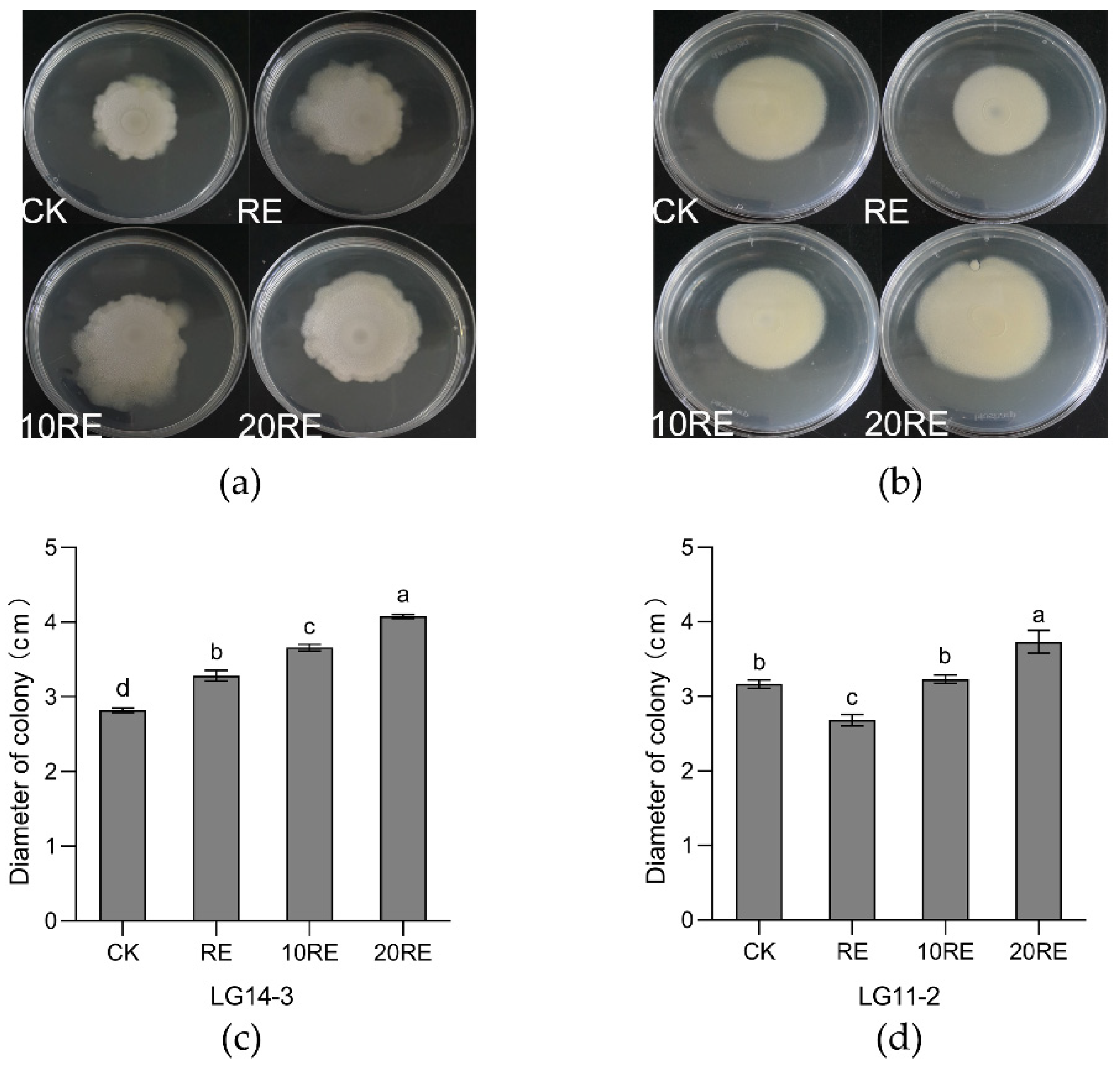
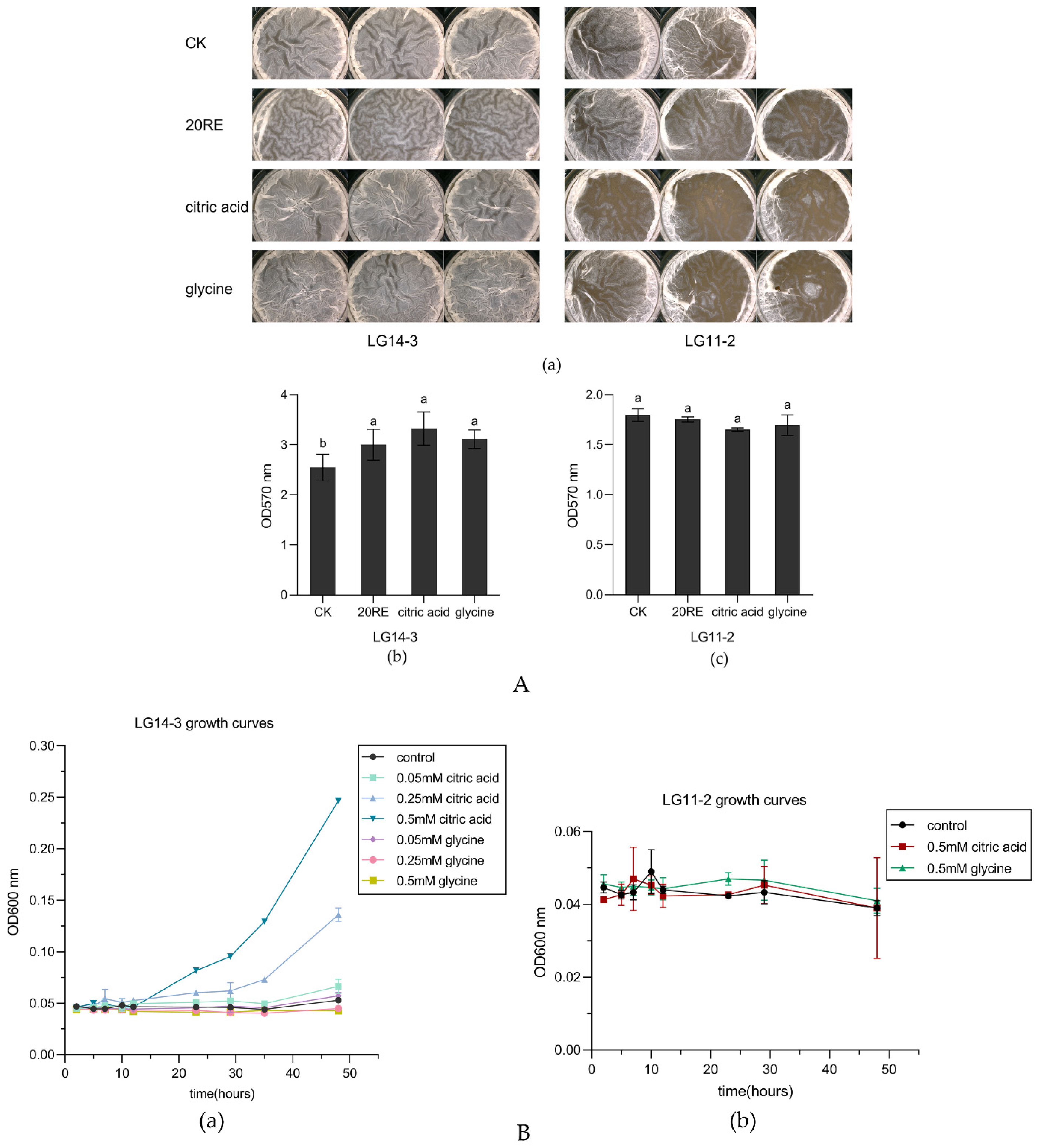
3.3. Banana Root Exudates Promoted the Swarming Ability of LG14-3

The swarming analysis showed that compared with the control (CK, sterile water), strain LG14-3 showed stronger swarming ability under root exudates, and the higher the concentration of root exudates was, the larger the area of swarming (Figure 4a,c). However, compared with the control (sterile water), 20RE could enhance the swarming ability of LG11-2, while RE and 10RE could not, and RE inhibited the swarming ability of LG11-2 (Figure 4b,d). The results showed that root exudates enhanced the swarming ability of LG14-3.

3.4. Effects of Citric Acid and Glycine on LG14-3 Growth and Biofilm Formation

Compared to the control (0 mM), the concentrations of 0.05 mM, 0.25 mM, and 0.5 mM citric acid supported cell growth up to 0.066, 0.136, and 0.247 of OD600 values, respectively. However, glycine at concentrations of 0.05 mM, 0.25 mM, and 0.5 mM did not support cell growth in the basic medium, and the OD600 values were close to or smaller than the control (Figure 5B(a)). Glycine and citric acid at 0.25 mM did not support the growth of LG11-2 (Figure 5B(b)).
Biofilm formation in plant roots indicates the successful colonization of PGPR [4]. Studies have shown that biofilms can protect rhizosphere bacteria against pathogens and are conducive to enhancing rhizosphere bacteria to obtain nutrients [13,14]. LG14-3 showed the most significant chemotactic response to 0.25 mM citric acid and 0.25 mM glycine, so the concentration used for those two compounds was 0.25 Mm; In contrast, LG11-2 had no chemotactic response to 0.25 mM citric acid or glycine. The results showed that the root exudates (20RE), 0.25 mM citric acid, and 0.25 mM glycine enhanced the biofilm formation of LG14-3. The treatment groups of 0.25 mM glycine and 0.25 mM citric acid had more obvious folds and thicker biofilms than the control and root exudates treatments. Crystal violet quantitative analysis showed that the absorbances of biofilms formed by adding chemoattractants to Msgg medium were higher than the control at 570 nm. Among them, the biofilm formed in the citric acid treatment group had the highest value at OD570 nm (Figure 5A(a,b)).
The biofilm morphology of LG11-2 showed that the biofilms formed in the treatment groups treated with glycine and citric acid were thinner. Crystal violet quantitative analysis of biofilms showed that root exudates, glycine, and citric acid did not promote the biofilm formation of LG11-2. The root exudates did not affect biofilm formation, while glycine and citric acid inhibited biofilm formation (Figure 5A(a,c)).

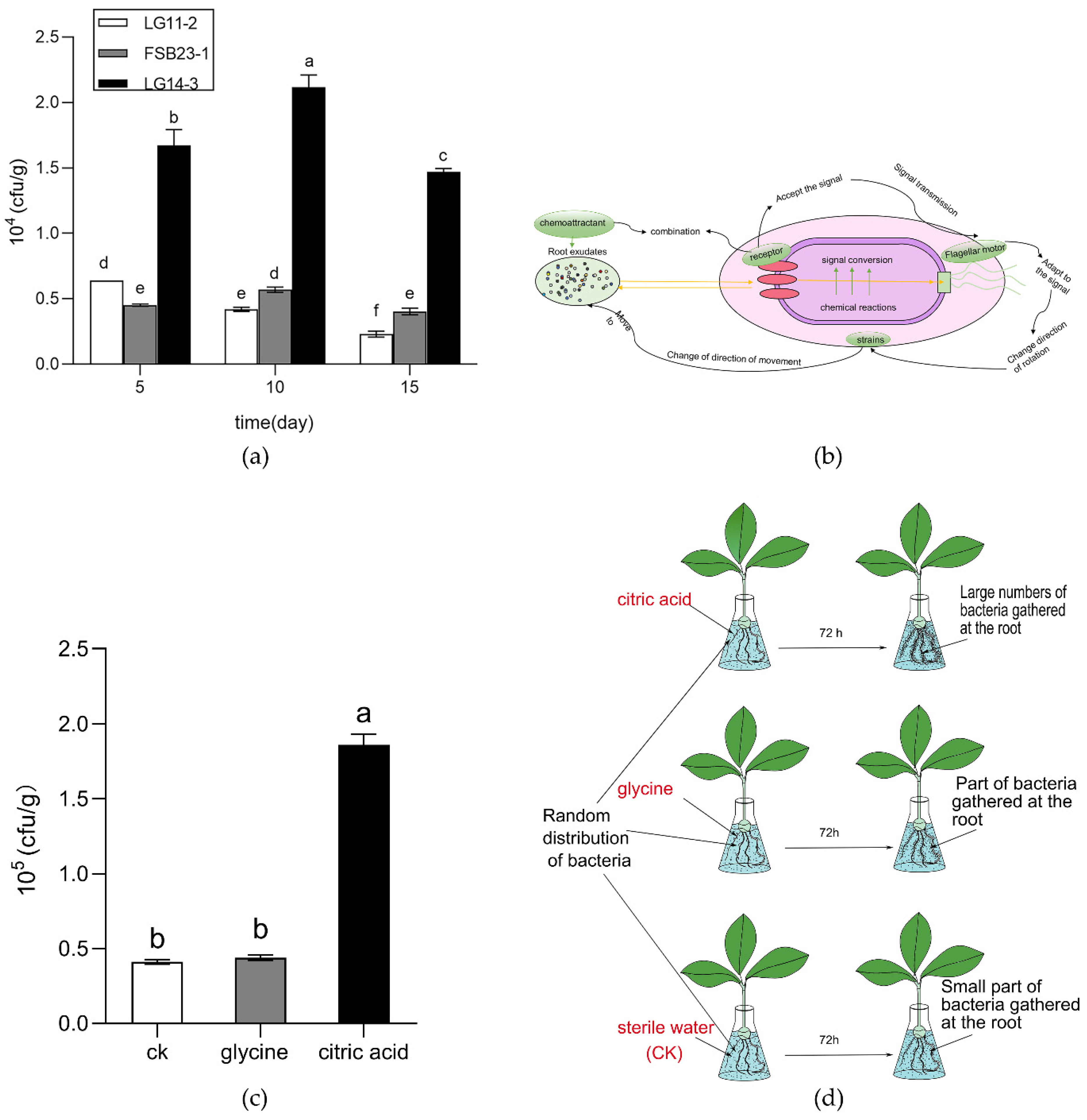
3.5. LG14-3 Has Colonization Advantages in Banana Roots, and Citric Acid can Promote Its Colonization Ability in the Banana Rhizosphere

The antagonistic activities of Bacillus velezensis LG11-2 and Bacillus velezensis FSB23-1 against Foc4 were similar to that of LG14-3 in vitro, but LG11-2 and FSB23-1 had no significant chemotactic response to banana root exudates. The colonization number of LG14-3 in banana roots reached 2.12 × 10⁴ cfu/g on the 10th day. Compared to LG11-2 and FSB23-1, the colonization amount of LG14-3 in the root was 2.6 and 3.7 times greater on the 5th day, 5.03 and 3.72 times greater on the 10th day and 6.41 and 3.63 times greater on the 15th day, respectively (Figure 6a).
The results of hydroponic experiments showed that the addition of exogenous glycine and citric acid promoted the colonization of strain LG14-3. The citric acid (0.25 mM) significantly promoted colonization by strain LG14-3 (Figure 6c).

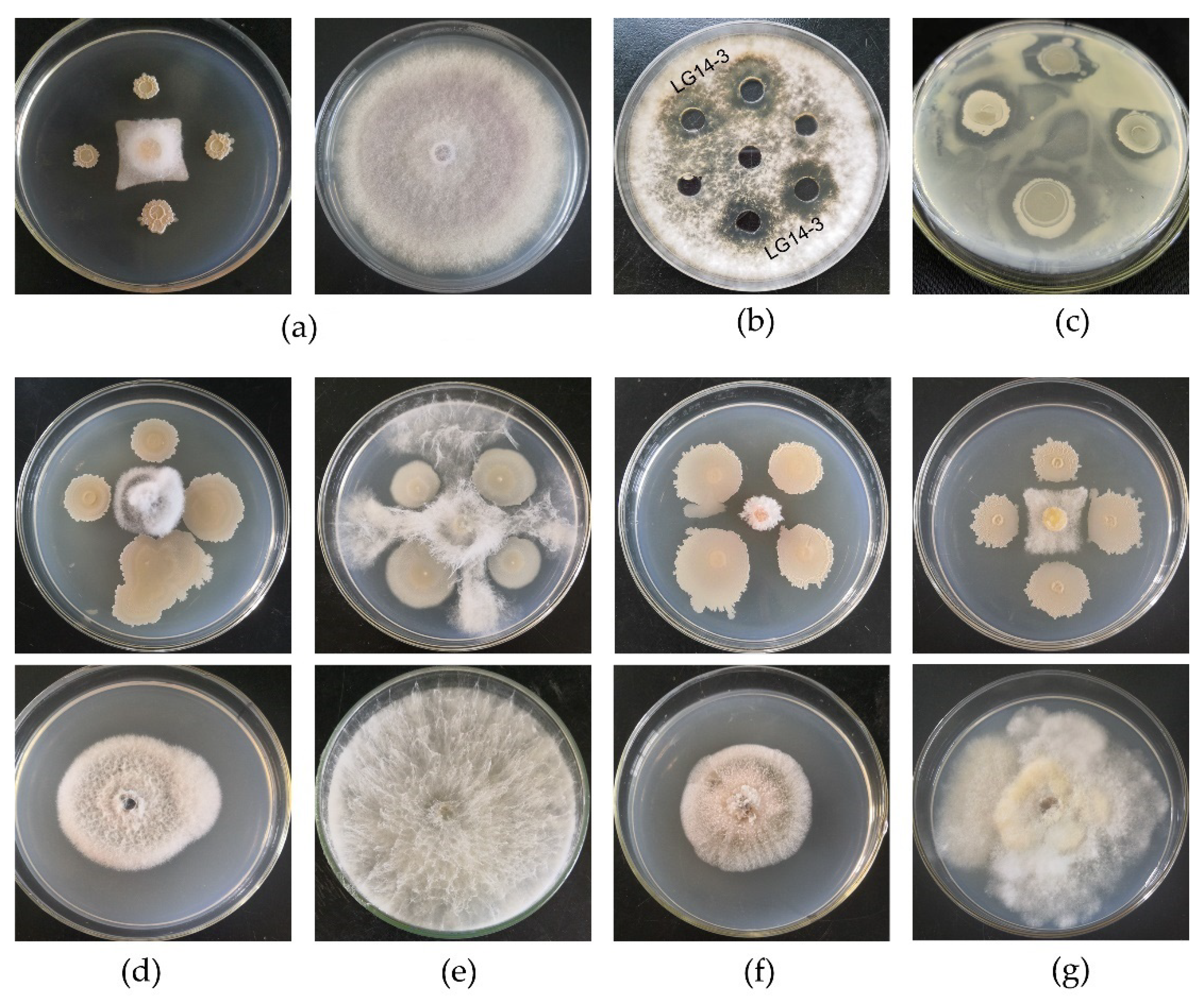
3.6. The LG14-3 Strain Exhibited Broad-Spectrum Antagonistic Activity

The LG14-3 strain has broad-spectrum antagonistic activity against Xanthomonas campestris pv. mangiferaeindicae, Pestalotiopis, Diaporthe, Gliomastix murorum, and Foc4 (Figure 7a). LG14-3 had a 78% inhibitory rate against Foc4 (Table 3). The inhibition rates of LG14-3 on Xanthomonas campestris pv. mangiferaeindicae, Pestalotiopsis, Diaporthe, Lasiodiplodia, and Gliomastix murorum, were 80.99%, 76.47%, 63.81%, 75.19%, and 76.6%, respectively (Table 3).

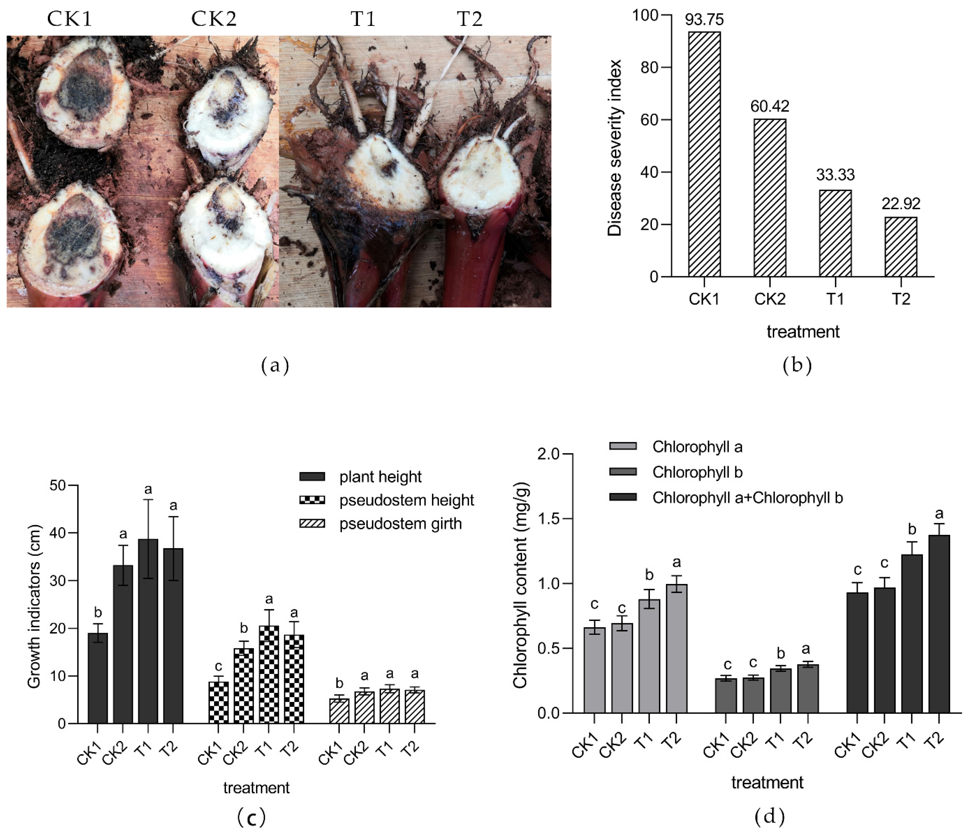
3.7. Biological Control Experiment

Compared with the CK1 (Foc) group, the blackened area of banana seedling corms in the CK2 (LG14-3 + Foc), T1 (glycine + LG14-3 + Foc), and T2 (citric acid + LG14-3 + Foc) groups was significantly reduced, and the T2 treatment group had the smallest blackened area of corms (Figure 8a) and the lowest disease severity index (22.92) (Figure 8b). The disease severity indices of CK1, CK2, and T1 were 93.75, 60.42, and 33.33, respectively. The measured data of plant height, pseudostem height, pseudostem girth, and chlorophyll content of banana seedlings of each group showed that LG14-3 promoted plant growth and increased chlorophyll content, and the exogenous substances (glycine and citric acid) enhanced the growth-promoting ability of LG14-3 on the plants (Figure 8c,d).
Overall, LG14-3 reduced the degree of banana wilt disease, and LG14-3 promoted the growth of plants under pathogenic stress (Table 4).

4. Discussion

Microorganisms that grow in the rhizosphere are ideal as biocontrol agents [38]. In this study, Bacillus velezensis LG14-3, which has a significant chemotactic effect on banana root exudates, was isolated from the healthy rhizosphere of banana in the banana Fusarium wilt disease epidemic area. Auxin (indole-3-acetic acid (IAA), which can promote plant growth, was the first hormone identified in plants [39]. The research by Patten et al. showed that bacterial IAA stimulated the development of the host plant root system [40]. For a long time, the competition of siderophores produced by bacteria for iron has been considered one of the important mechanisms by which biocontrol bacteria antagonize plant pathogens. Bacillus subtilis CAS15, which produces a siderophore, can effectively control Fusarium wilt and has the ability to promote growth [38]. Pseudomonas aeruginosa A7 produces siderophores combined with insoluble iron ions, thereby inhibiting the growth of plant pathogens and helping to control plant diseases [41]. Nitrogen (N) is an essential element for microbial and plant life [42]. Biological nitrogen fixation is the process by which nitrogen-fixing microorganisms reduce atmospheric nitrogen to ammonia available to bacteria and plants [43]. Our research results showed that LG14-3 has the ability to produce IAA and iron carriers and to perform nitrogen fixation, so it is a biocontrol strain with good potential.
Chemotaxis and biofilm formation determine the strain colonization in plant roots [13]. Malate and citric acid are the main chemoattractants of the tomato rhizosphere bacterium WCS365 [29]. Pseudomonas B4 experienced chemotaxis to biphenyls and benzoates and used them as carbon sources [11] 1-Aminocyclopropane-1-carboxylarboxylate is a strong chemoattractant for the plant-beneficial rhizobacterium Pseudomonas putida UW4 [44]. In this study, LG14-3 showed chemotaxis to the root exudate of banana and its common components (citric acid, glycine, succinate, D-galactose, and D-maltose). In previous studies, organic acids and sugars have been reported to induce and enhance the formation of bacterial biofilms in the rhizosphere [14]. In this study, glycine (0.25 mM) and citric acid (0.25 mM) enhanced the biofilm formation of LG14-3. Banana root exudates were also found to improve the swarming motility of LG14-3. Similarly, research by Tan et al. showed that five organic acids significantly increased the swarming ability of T-5 [13].
Several studies have shown that root exudates and organic acids can promote the colonization of Bacillus in roots [13,17,45,46]. As with the above results, LG14-3 has a better colonization advantage within banana roots than the other two biocontrol strains. Moreover, the addition of exogenous glycine or citric acid can increase the colonization of LG14-3 in the banana rhizosphere. Some carbon metabolites in plant root exudates can serve as food and as an energy source for rhizosphere microorganisms [5,47,48]. Similarly, citric acid supports the growth of LG14-3.
Bacillus velezensis LG14-3 showed broad-spectrum antagonistic activity against Xanthomonas campestris pv. mangiferaeindicae, Pestalotiopis, Diaporthe, Gliomastix murorum, and Fusarium oxysporum f. sp. cubense. The in vitro inhibition rate against Foc4 was more than 70%, and its metabolites also had antagonistic activity. Biocontrol using antagonistic microorganisms is a safer alternative to reduce the use of chemicals in agriculture, and it is considered a promising approach for the management of soil-borne diseases [49]. A large number of bacterial and fungal strains, as well as viruses, nematodes, and insects, have been employed as biological control agents (BCAs) in the management of soil-borne crop pathogens for decades [50], including Bacillus, Pantoea, Streptomyces, Trichoderma, Clonostachys, Pseudomonas, Burkholderia, and certain yeasts [51]. Among them, Bacillus is one of the most popular because it can form heat-resistant and dry-resistant endophytic spores [52], which are easy to store and apply [53]. Bacillus spp., especially Bacillus subtilis, Bacillus cereus, and Bacillus amyloliquefaciens, are effective for the control of plant soil-borne diseases and foliar and postharvest disease [38]. Both Bacillus amyloliquefaciens HSB1 and FZB42 were found to inhibit fungal mycelial growth in vitro and in plants and to promote the growth of wolfberry seedlings [49]. Rhizosphere Bacillus Y6 and F7 showed strong antagonistic activity against Fusarium oxysporum under laboratory and greenhouse conditions, and their lipopeptides effectively reduced the incidence of banana Fusarium wilt [53].
The results of this study demonstrated that Bacillus velezensis LG14-3 was chemotactic to banana root exudates and that citric acid and glycine in root exudates enhanced the abilities of LG14-3 root colonization and pathogen inhibition. Sustaining chlorophyll content in plants upon pathogen invasion is vital, as it will permit plant cells to continue photosynthesis [54]. Moreover, pot experiments showed that LG14-3 was efficient in increasing the chlorophyll a and chlorophyll b contents of bananas, which may be one of the reasons for the increase in plant height and weight after the LG14-3 application. In conclusion, these results demonstrate the biocontrol potential of Bacillus velezensis LG14-3 and provide data for exploring the role of root exudates in the interaction between biocontrol Bacillus, plants and soil-borne pathogens.

5. Conclusions

In this study, LG14-3 was isolated from the healthy rhizosphere of banana in the banana Fusarium wilt disease epidemic area and identified as Bacillus velezensis by morphological and molecular biology. Our research results also showed that LG14-3 has the ability to produce IAA and iron carriers and perform nitrogen fixation. A chemotaxis assay showed that LG14-3 had significant chemotactic effects on banana root exudates, glycine and citric acid. Swarming motility assays and biofilm formation assays showed that root exudates, glycine (0.25 mM) and citric acid (0.25 mM) enhanced the biofilm formation and swarming ability of LG14-3. Bacillus velezensis LG14-3 showed broad-spectrum antagonistic activity against Xanthomonas campestris pv. mangiferaeindicae, Pestalotiopis, Diaporthe, Gliomastix murorum and Fusarium oxysporum f. sp. cubense. Colonization assays and biocontrol experiments showed that citric acid and glycine in root exudates enhanced the abilities of LG14-3 root colonization and pathogen inhibition.
In summary, banana root exudates as chemoattractants were used to screen antagonistic microorganisms, and the colonization of Bacillus velezensis LG14-3 was promoted in banana roots through the dual induction of strong Chemoattractants (citric acid and glycine) and root exudates. Bacillus velezensis LG14-3 exerted their effective biocontrol effect, and thus targeted the control of banana wilt. Therefore, the use of antagonistic microorganisms and chemoattractants to control banana Fusarium wilt disease against soil-borne pathogens is a preferable approach. This study provides a new idea and method to optimize the selection of antagonistic microorganisms to control plant diseases.

Author Contributions

Conceptualization, J.H. and L.Y. (Lihua Yang); methodology, J.H., L.Y. (Lihua Yang) and L.G.; software, L.Y. (Lihua Yang); validation, L.Y. (Lihua Yang); formal analysis, J.H.; investigation, J.H., L.Y. (Lihua Yang), J.W. and C.L.; resources, J.H. and L.Y. (Laying Yang); data curation, L.Y. (Lihua Yang); writing—original draft preparation, L.Y. (Lihua Yang); writing—review and editing, J.H., Y.Z., L.G. and L.Y. (Laying Yang); visualization, L.Y. (Lihua Yang); supervision, J.H.; project administration, J.H.; funding acquisition, J.H. and J.W. All authors have read and agreed to the published version of the manuscript.

Funding

The research was supported by National key R & D program of China (No. 2017YFD0200602), Hainan Province Major Science and Technology Special Fund (No. ZDKJ2021016), Hainan Provincial Natural Science Foundation of China (321RC618), Hainan Province Agricultural Germplasm Resources Protection Project (QNJC (2022) No. 29) and Central Public-interest Scientific Institution Basal Research Fund (NO. 1630042022010).

Institutional Review Board Statement

Not applicable.

Informed Consent Statement

Not applicable.

Data Availability Statement

Not applicable.

Acknowledgments

We are grateful to Yunlong Xu and Xiyu Jiang for preparing some experiment materials.

Conflicts of Interest

The authors declare no conflict of interest. The funders had no role in the design of the study; in the collection, analyses, or interpretation of data; in the writing of the manuscript; or in the decision to publish the results.

References

  1. Ploetz, R.C. Fusarium wilt of banana is caused by several pathogens referred to as fusarium oxysporum f. sp. cubense. Phytopathology 2006, 96, 653–656. [Google Scholar] [CrossRef] [PubMed] [Green Version]
  2. Bubici, G.; Kaushal, M.; Prigigallo, M.I.; Gomez-Lama Cabanas, C.; Mercado-Blanco, J. Biological control agents against fusarium wilt of banana. Front. Microbiol. 2019, 10, 616. [Google Scholar] [CrossRef] [PubMed] [Green Version]
  3. Gao, X.; Li, T.; Liu, W.; Zhang, Y.; Shang, D.; Gao, Y.; Qi, Y.; Qiu, L. Enhancing the 1-aminocyclopropane-1-carboxylate Metabolic rate of pseudomonas sp. UW4 intensifies chemotactic rhizocompetence. Microorganisms 2020, 8, 71. [Google Scholar] [CrossRef] [PubMed] [Green Version]
  4. Yuan, J.; Zhang, N.; Huang, Q.W.; Raza, W.; Li, R.; Vivanco, J.M.; Shen, Q.R. Organic acids from root exudates of banana help root colonization of PGPR strain bacillus amyloliquefaciens NJN-6. Sci. Rep. 2015, 5, 1–8. [Google Scholar] [CrossRef] [PubMed] [Green Version]
  5. Olanrewaju, O.S.; Ayangbenro, A.S.; Glick, B.R.; Babalola, O.O. Plant health: Feedback effect of root exudates-rhizobiome interactions. Appl. Microbiol. Biotechnol. 2019, 103, 1155–1166. [Google Scholar] [CrossRef] [PubMed] [Green Version]
  6. Badri, D.V.; Vivanco, J.M. Regulation and function of root exudates. Plant Cell Environ. 2009, 32, 666–681. [Google Scholar] [CrossRef] [PubMed]
  7. Szurmant, H.; Muff, T.J.; Ordal, G.W. Bacillus subtilis CheC and FliY are members of a novel class of CheY-P-hydrolyzing proteins in the chemotactic signal transduction cascade. J. Biol. Chem. 2004, 279, 21787–21792. [Google Scholar] [CrossRef] [Green Version]
  8. Aroney, S.T.N.; Poole, P.S.; Sanchez-Canizares, C. Rhizobial chemotaxis and motility systems at work in the soil. Front. Plant Sci. 2021, 12, 725338. [Google Scholar] [CrossRef]
  9. Garrity, L.F.; Ordal, G.W. Activation of the CheA kinase by asparagine in bacillus subtilis chemotaxis. Microbiology 1997, 143, 2945–2951. [Google Scholar] [CrossRef] [Green Version]
  10. Hess, J.F.; Oosawa, K.; Kaplan, N.; Simon, M.I. Phosphorylation of three proteins in the signaling pathway of bacterial chemotaxis. Cell 1988, 53, 79–87. [Google Scholar] [CrossRef]
  11. Gordillo, F.; Chavez, F.P.; Jerez, C.A. Motility and chemotaxis of pseudomonas sp. B4 towards polychlorobiphenyls and chlorobenzoates. FEMS Microbiol. Ecol. 2007, 60, 322–328. [Google Scholar] [CrossRef] [PubMed]
  12. Kim, H.E.; Shitashiro, M.; Kuroda, A.; Takiguchi, N.; Kato, J. Ethylene chemotaxis in pseudomonas aeruginosa and other pseudomonas species. Microbes Environ. 2007, 22, 186–189. [Google Scholar] [CrossRef] [Green Version]
  13. Tan, S.Y.; Yang, C.L.; Mei, X.L.; Shen, S.Y.; Raza, W.; Shen, Q.R.; Xu, Y.C. The effect of organic acids from tomato root exudates on rhizosphere colonization of bacillus amyloliquefaciens T-5. Appl. Soil Ecol. 2013, 64, 15–22. [Google Scholar] [CrossRef]
  14. Panichikkal, J.; Edayileveetil Krishnankutty, R. Rhizobacterial biofilm and plant growth promoting trait enhancement by organic acids and sugars. Biofouling 2020, 36, 990–999. [Google Scholar] [CrossRef]
  15. Xie, S.; Jiang, L.; Wu, Q.; Wan, W.; Gan, Y.; Zhao, L.; Wen, J. Maize Root Exudates Recruit Bacillus amyloliquefaciens OR2–30 to Inhibit Fusarium graminearum Infection. Phytopathology 2022, 112, 1886–1893. [Google Scholar] [CrossRef]
  16. Zhang, N.; Wang, D.; Liu, Y.; Li, S.; Shen, Q.; Zhang, R. Effects of different plant root exudates and their organic acid components on chemotaxis, biofilm formation and colonization by beneficial rhizosphere-associated bacterial strains. Plant Soil 2013, 374, 689–700. [Google Scholar] [CrossRef]
  17. Wang, X.; Xie, H.; Ku, Y.; Yang, X.; Chen, Y.; Yang, N.; Mei, X.; Cao, C. Chemotaxis of Bacillus cereus YL6 and its colonization of Chinese cabbage seedlings. Plant Soil 2019, 447, 413–430. [Google Scholar] [CrossRef]
  18. Shen, Z.; Wang, B.; Lv, N.; Sun, Y.; Jiang, X.; Li, R.; Ruan, Y.; Shen, Q. Effect of the combination of bio-organic fertiliser with Bacillus amyloliquefaciens NJN-6 on the control of banana Fusarium wilt disease, crop production and banana rhizosphere culturable microflora. Biocontrol Sci. Technol. 2015, 25, 716–731. [Google Scholar] [CrossRef]
  19. Proboningrum, A.; Widono, S. Effectivity and compatibility of Azotobacter and Bacillus for biological control agents of fusarium wilt on banana seedlings. In Proceedings of the International Conference on Sustainable Agriculture for Rural Development (ICSARD), Purwokerto, Indonesia, 23–24 October 2019. [Google Scholar]
  20. Wang, J.; Zhao, Y.; Ruan, Y. Effects of bio-organic fertilizers produced by four bacillus amyloliquefaciens strains on banana fusarium wilt disease. Compost. Sci. Util. 2015, 23, 185–198. [Google Scholar] [CrossRef]
  21. Zhang, N.; Wu, K.; He, X.; Li, S.-Q.; Zhang, Z.-H.; Shen, B.; Yang, X.-M.; Zhang, R.-F.; Huang, Q.-W.; Shen, Q.-R. A new bioorganic fertilizer can effectively control banana wilt by strong colonization with Bacillus subtilis N11. Plant Soil 2011, 344, 87–97. [Google Scholar] [CrossRef]
  22. Sun, Y.; Huang, B.; Cheng, P.; Li, C.; Chen, Y.; Li, Y.; Zheng, L.; Xing, J.; Dong, Z.; Yu, G. Endophytic Bacillus subtilis TR21 improves banana plant resistance to fusarium oxysporum f. sp. cubense and promotes root growth by upregulating the jasmonate and brassinosteroid biosynthesis pathways. Phytopathology 2022, 112, 219–231. [Google Scholar] [CrossRef] [PubMed]
  23. Shen, N.; Li, S.; Li, S.; Zhang, H.; Jiang, M. The siderophore-producing bacterium, Bacillus siamensis Gxun-6, has an antifungal activity against Fusarium oxysporum and promotes the growth of banana. Egypt. J. Biol. Pest Control. 2022, 32, 34. [Google Scholar] [CrossRef]
  24. Wu, X.; Shan, Y.; Li, Y.; Li, Q.; Wu, C. The Soil Nutrient Environment Determines the Strategy by Which Bacillus velezensis HN03 Suppresses Fusarium wilt in Banana Plants. Front. Plant Sci. 2020, 11, 599904. [Google Scholar] [CrossRef] [PubMed]
  25. Win, T.T.; Bo, B.; Malec, P.; Fu, P. The effect of a consortium of Penicillium sp. and Bacillus spp. in suppressing banana fungal diseases caused by Fusarium sp. and Alternaria sp. J. Appl. Microbiol. 2021, 131, 1890–1908. [Google Scholar] [CrossRef]
  26. Ma, L.; Wang, W.Q.; Shi, R.; Zhang, X.M.; Li, X.; Yang, Y.S.; Mo, M.H. Effects of organic acids on the chemotaxis profiles and biocontrol traits of antagonistic bacterial endophytes against root-rot disease in Panax notoginseng. Antonie Van Leeuwenhoek 2021, 114, 1771–1789. [Google Scholar] [CrossRef]
  27. Raina, J.B.; Fernandez, V.; Lambert, B.; Stocker, R.; Seymour, J.R. The role of microbial motility and chemotaxis in symbiosis. Nat. Rev. Microbiol. 2019, 17, 284–294. [Google Scholar] [CrossRef]
  28. Yuan, J.; Wu, Y.; Zhao, M.; Wen, T.; Huang, Q.; Shen, Q. Effect of phenolic acids from banana root exudates on root colonization and pathogen suppressive properties of Bacillus amyloliquefaciens NJN-6. Biol. Control 2018, 125, 131–137. [Google Scholar] [CrossRef]
  29. Weert, S.D.; Vermeiren, H.; Mulders, I.H.M.; Kuiper, I.; Lugtenberg, B.J.J. Flagella-driven chemotaxis towards exudate components is an important trait for tomato root colonization by pseudomonas fluorescens. Mol. Plant-Microbe Interact. MPMI 2002, 15, 1173. [Google Scholar] [CrossRef] [Green Version]
  30. Mazumder, R.; Phelps, T.J.; Krieg, N.R.; Benoit, R.E. Determining chemotactic responses by two subsurface microaerophiles using a simplified capillary assay method. J. Microbiol. Meth. 1999, 37, 255–263. [Google Scholar] [CrossRef]
  31. Fang, Z.D. Research Methodology of Plant Diseases; China Agriculture Press: Beijing, China, 1998. [Google Scholar]
  32. He, D.; Singh, S.K.; Peng, L.; Kaushal, R.; Vilchez, J.I.; Shao, C.; Wu, X.; Zheng, S.; Morcillo, R.J.L.; Pare, P.W.; et al. Flavonoid-attracted Aeromonas sp. from the Arabidopsis root microbiome enhances plant dehydration resistance. ISME J. 2022, 16, 2622–2632. [Google Scholar] [CrossRef]
  33. Wang, S.; Wang, J.; Zhou, Y.; Huang, Y.; Tang, X. Prospecting the plant growth–promoting activities of endophytic bacteria Franconibacter sp. YSD YN2 isolated from Cyperus esculentus L. var. sativus leaves. Ann. Microbiol. 2022, 72, 1. [Google Scholar]
  34. Schwyn, B.; Neilands, J.B. Universal chemical assay for the detection and determination of siderophores. Anal. Biochem. 1987, 160, 47–56. [Google Scholar] [CrossRef] [PubMed]
  35. Mohamed, A.A.; Mak, C.; Liew, K.W.; Ho, Y.W. Early evaluation of banana plants at nursery stage for fusarium wilt tolerance. In Proceedings of the International Workshop on the Banana Fusarium Wilt Disease, Pahang, Malaysia, 18–20 October 1999. [Google Scholar]
  36. Arnon, D.I. Copper enzymes in isolated chloroplasts.Polyphenoloxidases in Beta vulgaris. Plant Physiol. 1949, 24, 1. [Google Scholar] [CrossRef] [Green Version]
  37. Xu, D.; Cote, J.C. Phylogenetic relationships between Bacillus species and related genera inferred from comparison of 3′ end 16S rDNA and 5′ end 16S–23S ITS nucleotide sequences. Int. J. Syst. Evol. Microbiol. 2003, 53, 695–704. [Google Scholar] [CrossRef] [PubMed] [Green Version]
  38. Yu, X.; Ai, C.; Li, X.; Zhou, G. The siderophore-producing bacterium, Bacillus subtilis CAS15, has a biocontrol effect on Fusarium wilt and promotes the growth of pepper. Eur. J. Soil Biol. 2011, 47, 138–145. [Google Scholar] [CrossRef]
  39. Shao, J.; Xu, Z.; Zhang, N.; Shen, Q.; Zhang, R. Contribution of indole-3-acetic acid in the plant growth promotion by the rhizospheric strain Bacillus amyloliquefaciens SQR9. Biol. Fertil. Soils 2015, 51, 321–330. [Google Scholar] [CrossRef]
  40. Patten, C.L.; Glick, B.R. Role of Pseudomonas putida indoleacetic acid in development of the host plant root system. Appl. Environ. Microbiol. 2002, 68, 3795–3801. [Google Scholar] [CrossRef] [Green Version]
  41. Mozumder, A.B.; Chanda, K.; Chorei, R.; Prasad, H.K. An Evaluation of Aluminum Tolerant Pseudomonas aeruginosa A7 for In Vivo Suppression of Fusarium Wilt of Chickpea Caused by Fusarium oxysporum f. sp. ciceris and Growth Promotion of Chickpea. Microorganisms 2022, 10, 368. [Google Scholar] [CrossRef]
  42. Wolińska, A.; Kuźniar, A.; Zielenkiewicz, U.; Banach, A.; Izak, D.; Stępniewska, Z.; Błaszczyk, M. Metagenomic analysis of some potential nitrogen-fixing bacteria in arable soils at different formation processes. Microb. Ecol. 2017, 73, 162–176. [Google Scholar] [CrossRef] [Green Version]
  43. Simon, Z.; Mtei, K.; Gessesse, A.; Ndakidemi, P.A. Isolation and Characterization of Nitrogen Fixing Rhizobia from Cultivated and Uncultivated Soils of Northern Tanzania. Am. J. Plant Sci. 2014, 5, 4050–4067. [Google Scholar] [CrossRef] [Green Version]
  44. Li, T.; Zhang, J.; Shen, C.; Li, H.; Qiu, L. 1-Aminocyclopropane-1-Carboxylate: A Novel and Strong Chemoattractant for the Plant Beneficial Rhizobacterium Pseudomonas putida UW4. Mol. Plant-Microbe Interact. 2019, 32, 750–759. [Google Scholar] [CrossRef] [PubMed]
  45. Liu, Y.; Chen, L.; Wu, G.; Feng, H.; Zhang, G.; Shen, Q.; Zhang, R. Identification of Root-Secreted Compounds Involved in the Communication Between Cucumber, the Beneficial Bacillus amyloliquefaciens, and the Soil-Borne Pathogen Fusarium oxysporum. Mol. Plant-Microbe Interact. 2017, 30, 53–62. [Google Scholar] [CrossRef] [Green Version]
  46. Zhang, H.; Chen, Q.F.; Shang, N.; Li, N.; Niu, Q.H.; Hong, Q.; Huang, X. The enhanced mechanisms of Hansschlegelia zhihuaiae S113 degrading bensulfuron-methyl in maize rhizosphere by three organic acids in root exudates. Ecotoxicol. Environ. Saf. 2021, 223, 112622. [Google Scholar] [CrossRef] [PubMed]
  47. Mendes, R.; Kruijt, M.; de Bruijn, I.; Dekkers, E.; van der Voort, M.; Schneider, J.H.; Piceno, Y.M.; DeSantis, T.Z.; Andersen, G.L.; Bakker, P.A.; et al. Deciphering the rhizosphere microbiome for disease-suppressive bacteria. Science 2011, 332, 1097–1100. [Google Scholar] [CrossRef] [PubMed]
  48. Liu, X.; Zhang, K.; Liu, Y.; Xie, Z.; Zhang, C. Oxalic Acid From Sesbania rostrata Seed Exudates Mediates the Chemotactic Response of Azorhizobium caulinodans ORS571 Using Multiple Strategies. Front. Microbiol. 2019, 10, 2727. [Google Scholar] [CrossRef] [Green Version]
  49. Uwaremwe, C.; Yue, L.; Wang, Y.; Tian, Y.; Zhao, X.; Liu, Y.; Zhou, Q.; Zhang, Y.; Wang, R. An Endophytic Strain of Bacillus amyloliquefaciens Suppresses Fusarium oxysporum Infection of Chinese Wolfberry by Altering Its Rhizosphere Bacterial Community. Front. Microbiol. 2021, 12, 782523. [Google Scholar] [CrossRef]
  50. Niu, B.; Wang, W.; Yuan, Z.; Sederoff, R.R.; Sederoff, H.; Chiang, V.L.; Borriss, R. Microbial Interactions Within Multiple-Strain Biological Control Agents Impact Soil-Borne Plant Disease. Front. Microbiol. 2020, 11, 585404. [Google Scholar] [CrossRef]
  51. Lahlali, R.; Ezrari, S.; Radouane, N.; Kenfaoui, J.; Esmaeel, Q.; El Hamss, H.; Belabess, Z.; Barka, E.A. Biological Control of Plant Pathogens: A Global Perspective. Microorganisms 2022, 10, 596. [Google Scholar] [CrossRef]
  52. Emmert, E.A.; Handelsman, J. Biocontrol of plant disease: A (gram-) positive perspective. FEMS Microbiol. Lett. 1999, 171, 1–9. [Google Scholar] [CrossRef]
  53. Cao, Y.; Pi, H.; Chandrangsu, P.; Li, Y.; Wang, Y.; Zhou, H.; Xiong, H.; Helmann, J.D.; Cai, Y. Antagonism of Two Plant-Growth Promoting Bacillus velezensis Isolates Against Ralstonia solanacearum and Fusarium oxysporum. Sci. Rep. 2018, 8, 4360. [Google Scholar] [CrossRef]
  54. Id, S.M.; Shahzad, A.N.; Qureshi, M.K. Acuities into tolerance mechanisms via different bioassay during Brassicaceae-Alternaria brassicicola interaction and its impact on yield. PLoS ONE 2020, 15, e0242545. [Google Scholar]
Figure 1. The “drop” assay was conducted. Drops added to the center of the plate contained, (a) chemotactic buffer; (b) root exudates; (c) citric acid; (d) glycine; (e) succinic acid; (f) D-maltose; (g) D-galactose.
Sustainability 15 00351 g001
Figure 2. Chemotactic responses of LG14-3 to different chemotaxis at final concentrations of 0.05 mM, 0.25 mM, 0.5 mM by capillary assay. The chemotaxis buffer was supplemented with (a) citric acid; (b) glycine; (c) succinic acid; (d) D-galactose; and (e) D-maltose. Bars indicate standards errors from the means of three replicates. Letters above the columns represent significant difference for each treatment according to Duncan’s multiple range test (p < 0.05).
Sustainability 15 00351 g002
Figure 3. Phylogenetic tree based on 16S and gyrB genes.
Sustainability 15 00351 g003
Figure 4. The effects of root exudates on the swarming motility. 5 μL Bacillus suspensions (107 cfu/mL) mixed with different concentrations of banana root exudates (RE,10RE, and 20RE) or sterilized water (CK) were inoculated in the LB medium with 0.7% agar. After incubating at 37 °C for 12 h, the diameters of colonies were measured. (a,c), LG14-3; (b,d), LG11-2. Bars indicate standard errors from the means of three replicates. Letters above the columns represent significant difference for each treatment according to Duncan’s multiple range test (p < 0.05).
Sustainability 15 00351 g004
Figure 5. (A) Influence of chemoattractant on the biofilm formation. (a) biofilm phenotype of B. velezensis LG14-3 and LG11-2 in biofilm medium; (b,c), crystal violet staining was used to quantify the biofilm. Bars indicate standard errors from the means of three replicates. Letters above the columns represent significant differences for each treatment according to Duncan’s multiple range test (p < 0.05); (B) Growth curves of B. velezensis LG14-3 and LG11-2 in minimal medium containing citric acid and glycine at the concentration indicated in the text as a carbon source. Bars indicate standard errors from the means of three replicates.
Sustainability 15 00351 g005
Figure 6. Colonization assay. (a) Effect on the colonization of B. velezensis LG14-3, LG11-2, and FSB23-1 in the banana root. (b) Schematic diagram of chemotaxis of strains to root exudates. (c,d) The effect of glycine and citric acid on the colonization of LG14-3 in the rhizosphere of banana. (a,c) Bars indicate standard errors from the means of three replicates. Letters above the columns represent significant difference for each treatment according to Duncan’s multiple range test (p < 0.05).
Sustainability 15 00351 g006
Figure 7. The antagonistic effect of strain LG14-3. Pathogens, (a), (b), Foc4, ((b), the antagonistic effect of metabolites of LG14-3); (c) Xanthomonas citri pv. mangiferaeindicae; (d) Diaporthe; (e) Pestalotiopsis; (f) Lasiodiplodia; (g) Gliomastix murorum.
Sustainability 15 00351 g007
Figure 8. The effects of LG14-3 strain on banana Fusarium wilt disease. (a) the lesion of corms of CK1 (Foc), CK2 (LG14-3 + Foc), T1 (LG14-3 + glycine + Foc) and T2 (LG14-3 + citric acid +Foc); (b) disease severity index of banana Fusarium wilt disease, letters above the columns represent disease severity index; (c) after the 60 days of Foc4 inoculated, the plant height, pseudostem girth, and pseudostem height were measured. (d) after the 60 days of Foc4 inoculated, chlorophyll content was measured. (c,d) bars indicate standard errors from the means. Letters above the columns represent significant difference for each treatment according to Duncan’s multiple range test (p < 0.05).
Sustainability 15 00351 g008
Table 1. Relative chemotaxis index (RCI) to root exudates in strains.
Bacillus StrainsRelative Chemotaxis Index (RCI)
Bacillus velezensis strain LG11-21.93 ± 0.02
Bacillus velezensis strain YH-61.25 ± 0.11
Bacillus velezensis strain C31.91 ± 0.05
Bacillus aerophilus strain YC-61.56 ± 0.02
Bacillus aerophilus strain CM131.71 ± 0.08
Bacillus aerophilus strain HZ-B1.09 ± 0.02
Bacillus aerophilus strain JS-51.36 ± 0.07
Bacillus velezensis strain X2-11.2 ± 0.05
Bacillus megaterium strain Y1R31.44 ± 0.04
Bacillus velezensis strain LG14-33.62 ± 0.09
Bacillus velezensis strain A31.58 ± 0.06
Note: Values are the means ± standard errors.
Table 2. Morphological and Biochemical characterizations of LG14-3.
Morphological CharactersMorphological CharactersCarbon Source
ShapeRoundGram staining+Sorbitol+
ColorCreamy whiteMethyl red test+D-maltose+
Gram staining+Catalase test+Mannitol+
Endospore production+Nitrate reduction+Sucrose+
Citrate test+D-galactose+
Starch hydrolysis+D-xylan+
protease production+Glucose+
Cellulase production+Glucose+
Nitrogen fixation+
Phosphate solubilization
Iron carrier production+
Indoleacetic acid production+
Note: +, Positive; −, Negative.
Table 3. Inhibition rates of LG14-3 on pathogens.
PathogensInhibition Rates (%)
Foc478.00 ± 0.94
Xanthomonas campestris pv. mangiferaeindicae80.99 ± 4.55
Diaporthe63.81 ± 1.90
Lasiodiplodia75.19 ± 1.70
Gliomastix murorum76.60 ± 2.13
Pestalotiopsis76.47 ± 2.35
Note: Values are the means ± standard errors.
Table 4. Effects of chemoattractant on Bacillus velezensis LG14-3 suppressing of banana Fusarium wilt disease.
CK1CK2T1T2
Disease severity index 93.7560.4233.0023.00
Growth indicators (cm)Plant height19.04 ± 1.93 b33.21 ± 4.17 a38.78 ± 8.25 a36.75 ± 6.68 a
Pseudostem height8.86 ± 1.11 c15.84 ± 1.48 b20.61 ± 3.32 a18.70 ± 2.73 a
Pseudostem girth5.28 ± 0.76 b6.79 ± 0.75 a7.35 ± 0.83 a7.09 ± 0.62 a
Chlorophyll content (mg/g)Chlorophyll a0.66 ± 0.05 c0.70 ± 0.06 c0.88 ± 0.07 b1.00 ± 0.06 a
Chlorophyll b0.27 ± 0.02 c0.28 ± 0.02 c0.35 ± 0.02 b0.38 ± 0.02 a
Chlorophyll a + Chlorophyll b0.93 ± 0.08 c0.97 ± 0.08 c1.23 ± 0.09 b1.37 ± 0.09 a
Note: Values are the means ± standard errors. Letters represent significant differences for each treatment according to Duncan’s multiple range test (p < 0.05).
Disclaimer/Publisher’s Note: The statements, opinions and data contained in all publications are solely those of the individual author(s) and contributor(s) and not of MDPI and/or the editor(s). MDPI and/or the editor(s) disclaim responsibility for any injury to people or property resulting from any ideas, methods, instructions or products referred to in the content.

