
A Novel Machine-Learning-Based Hybrid CNN Model for Tumor Identification in Medical Image Processing

 , , ,
, , ,  ,
, 
Abstract
:1. Introduction
2. Related Research
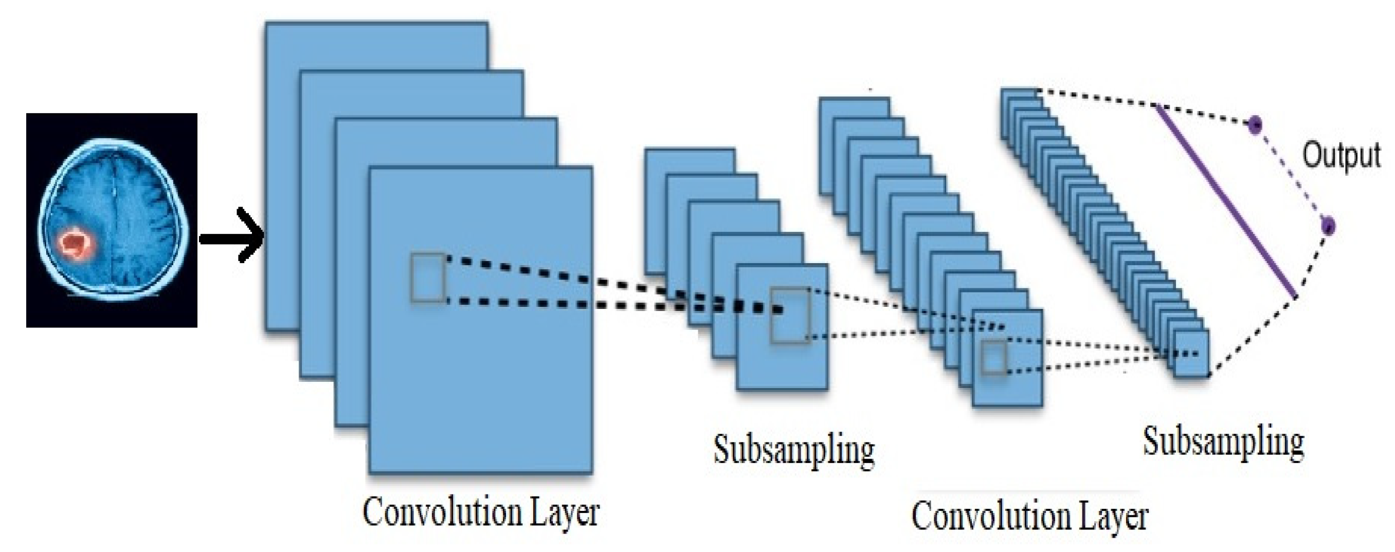
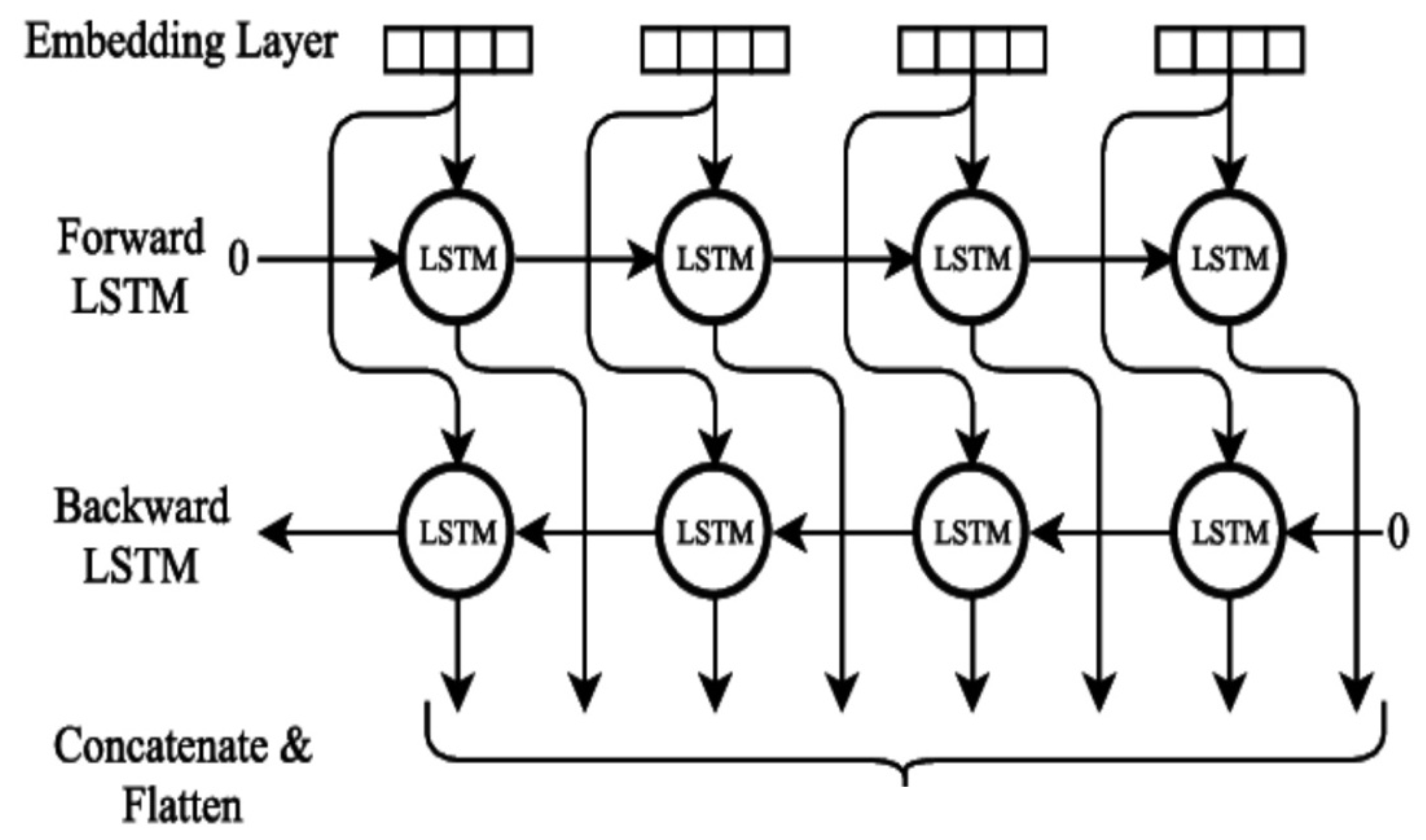
3. Joint-Extraction Method of Oncology Medical Events
3.1. Taks Analysis
3.2. Design Methods
4. Datasets and Result Evaluation
Experimental Results
- Not using the RoBERT pre-training language model, but using randomly initialized token-embedding representation;
- Not using any external resources;
- No rules are used to clean the dataset, character replacement, and other preprocessing operations.
- For the transfer learning ability of the evaluation method, the CCKS2020 dataset, the data distribution of the training set, and the test set are fairly different;
- The algorithm can significantly expand the number and types of medical record texts labelled, which are critical to improving model performance;
- The pseudo-labeled samples generated by this algorithm are random and may not necessarily match the actual scene. Therefore, adding too much pseudo-labelled data will make the model correct. Sexuality produces a certain amount of interference, which affects model performance improvement.
5. Conclusions
Author Contributions
Funding
Institutional Review Board Statement
Informed Consent Statement
Data Availability Statement
Acknowledgments
Conflicts of Interest
References
- Hasegawa, R. Automatic Detection and Segmentation of Liver Tumors in Multi-phase CT Images by Phase Attention Mask R-CNN. In Proceedings of the 2021 IEEE International Conference on Consumer Electronics (ICCE), Las Vegas, NV, USA, 10–12 January 2021; pp. 1–5. [Google Scholar] [CrossRef]
- Archa, S.P.; Kumar, C.S. Segmentation of Brain Tumor in MRI Images Using CNN with Edge Detection. In Proceedings of the 2018 International Conference on Emerging Trends and Innovations in Engineering and Technological Research (ICETIETR), Ernakulam, India, 11–13 July 2018; pp. 1–4. [Google Scholar] [CrossRef]
- Prakash, R.M.; Kumari, R.S.S. Classification of MR Brain Images for Detection of Tumor with Transfer Learning from Pre-trained CNN Models. In Proceedings of the 2019 International Conference on Wireless Communications Signal Processing and Networking (WiSPNET), Chennai, India, 21–23 March 2019; pp. 508–511. [Google Scholar] [CrossRef]
- Hasegawa, R.; Iwamoto, Y.; Lin, L.; Hu, H.; Chen, Y. Automatic Segmentation of Liver Tumor in Multiphase CT Images by Mask R-CNN. In Proceedings of the 2nd Global Conference on Life Sciences and Technologies (LifeTech), Kyoto, Japan, 10–12 March 2020; pp. 231–234. [Google Scholar] [CrossRef]
- Someswararao, C.; Shankar, R.S.; Appaji, S.V.; Gupta, V. Brain Tumor Detection Model from MR Images using Convolutional Neural Network. In Proceedings of the International Conference on System, Computation, Automation and Networking (ICSCAN), Pondicherry, India, 3–4 July 2020; pp. 1–4. [Google Scholar] [CrossRef]
- Yasir, M.; Hayat, U.; Rahman, A.U.; Riaz, R. Classification and Detection of Glioblastoma Tumor from MRI Images. In Proceedings of the International Bhurban Conference on Applied Sciences and Technologies (IBCAST), Islamabad, Pakistan, 12–16 January 2021; pp. 322–327. [Google Scholar] [CrossRef]
- Somasundaram, S.; Gobinath, R. Current Trends on Deep Learning Models for Brain Tumor Segmentation and Detection—A Review. In Proceedings of the International Conference on Machine Learning, Big Data, Cloud and Parallel Computing (COMITCon), Faridabad, India, 14–16 February 2019; pp. 217–221. [Google Scholar] [CrossRef]
- Raut, G.; Raut, A.; Bhagade, J.; Gavhane, S. Deep Learning Approach for Brain Tumor Detection and Segmentation. In Proceedings of the International Conference on Convergence to Digital World—Quo Vadis (ICCDW), Mumbai, India, 18–20 February 2020; pp. 1–5. [Google Scholar] [CrossRef]
- Mao, Y.; Yin, Z.; Schober, J.M. Iteratively training classifiers for circulating tumor cell detection. In Proceedings of the IEEE 12th International Symposium on Biomedical Imaging (ISBI), Brooklyn, NY, USA, 16–19 April 2015; pp. 190–194. [Google Scholar] [CrossRef]
- Kashif, M.N.; Ahmed Raza, S.E.; Sirinukunwattana, K.; Arif, M.; Rajpoot, N. Handcrafted features with convolutional neural networks for detection of tumor cells in histology images. In Proceedings of the IEEE 13th International Symposium on Biomedical Imaging (ISBI), Prague, Czech Republic, 13–16 April 2016; pp. 1029–1032. [Google Scholar] [CrossRef]
- Moutushi, N.-E.-J.; Tara, K. Comparison among Supervised Classifiers for Classification of Brain Tumor. In Proceedings of the 2020 IEEE Region 10 Symposium (TENSYMP), Dhaka, Bangladesh, 5–7 June 2020; pp. 304–307. [Google Scholar] [CrossRef]
- Shelke, S.M.; Mohod, S.W. Automated Segmentation and Detection of Brain Tumor from MRI. In Proceedings of the International Conference on Advances in Computing, Communications and Informatics (ICACCI), Bangalore, India, 19–22 September 2018; pp. 2120–2126. [Google Scholar] [CrossRef]
- Abdullah, A.A.; Chize, B.S.; Nishio, Y. Implementation of an improved cellular neural network algorithm for brain tumor detection. In Proceedings of the International Conference on Biomedical Engineering (ICoBE), Penang, Malaysia, 27–28 February 2012; pp. 611–615. [Google Scholar]
- Hemanth, G.; Janardhan, M.; Sujihelen, L. Design and Implementing Brain Tumor Detection Using Machine Learning Approach. In Proceedings of the 3rd International Conference on Trends in Electronics and Informatics (ICOEI), Tirunelveli, India, 23–25 April 2019; pp. 1289–1294. [Google Scholar] [CrossRef]
- Gayanga, T.D.L.; Pathirana, G.P.S.N.; Sandanayake, T.C. Detecting and Capturing the Intensity of a Brain Tumor using MRI Images. In Proceedings of the 4th International Conference on Information Technology Research (ICITR), Moratuwa, Sri Lanka, 10–13 December 2019; pp. 1–6. [Google Scholar] [CrossRef]
- Choudhury, C.L.; Mahanty, C.; Kumar, R.; Mishra, B.K. Brain Tumor Detection and Classification Using Convolutional Neural Network and Deep Neural Network. In Proceedings of the International Conference on Computer Science, Engineering and Applications (ICCSEA), Gunupur, India, 13–14 March 2020; pp. 1–4. [Google Scholar] [CrossRef]
- Jemimma, T.A.; Vetharaj, Y.J. Watershed Algorithm based DAPP features for Brain Tumor Segmentation and Classification. In Proceedings of the International Conference on Smart Systems and Inventive Technology (ICSSIT), Tirunelveli, India, 13–14 December 2018; pp. 155–158. [Google Scholar] [CrossRef]
- Siar, M.; Teshnehlab, M. Brain Tumor Detection Using Deep Neural Network and Machine Learning Algorithm. In Proceedings of the 9th International Conference on Computer and Knowledge Engineering (ICCKE), Mashhad, Iran, 24–25 October 2019; pp. 363–368. [Google Scholar] [CrossRef]
- Ezhilarasi, R.; Varalakshmi, P. Tumor Detection in the Brain using Faster R-CNN. In Proceedings of the 2nd International Conference on I-SMAC (IoT in Social, Mobile, Analytics and Cloud) (I-SMAC) I-SMAC (IoT in Social, Mobile, Analytics and Cloud) (I-SMAC), Palladam, India, 30–31 August 2018; pp. 388–392. [Google Scholar] [CrossRef]
- Vinoth, R.; Venkatesh, C. Segmentation and Detection of Tumor in MRI images Using CNN and SVM Classification. In Proceedings of the Conference on Emerging Devices and Smart Systems (ICEDSS), Tiruchengode, India, 2–3 March 2018; pp. 21–25. [Google Scholar] [CrossRef]
- Nair, R.; Soni, M.; Bajpai, B.; Dhiman, G.; Sagayam, K.M. Predicting the Death Rate Around the World Due to COVID-19 Using Regression Analysis. Int. J. Swarm Intell. Res. 2022, 13, 1–13. [Google Scholar] [CrossRef]
- Sharma, S.; Gupta, S.; Gupta, D.; Juneja, S.; Singal, G.; Dhiman, G.; Kautish, S. Recognition of Gurmukhi Handwritten City Names Using Deep Learning and Cloud Computing. Sci. Program. 2022, 2022, 5945117. [Google Scholar] [CrossRef]
- Zeidabadi, F.A.; Doumari, S.A.; Dehghani, M.; Montazeri, Z.; Trojovsky, P.; Dhiman, G. MLA: A New Mutated Leader Algorithm for Solving Optimization Problems. Comput. Mater. Contin. 2022, 70, 5631–5649. [Google Scholar] [CrossRef]
- Zeidabadi, F.A.; Doumari, S.A.; Dehghani, M.; Montazeri, Z.; Trojovsky, P.; Dhiman, G. AMBO: All Members-Based Optimizer for Solving Optimization Problems. Comput. Mater. Contin. 2022, 70, 2905–2921. [Google Scholar] [CrossRef]
- Alharbi, Y.; Alferaidi, A.; Yadav, K.; Dhiman, G.; Kautish, S. Denial-of-Service Attack Detection over IPv6 Network Based on KNN Algorithm. Wireless Commun. Mob. Comput. 2021, 2021, 8000869. [Google Scholar] [CrossRef]
- Balakrishnan, A.; Kadiyala, R.; Dhiman, G.; Ashok, G.; Kautish, S.; Yadav, K.; Maruthi Nagendra Prasad, J. A Personalized Eccentric Cyber-Physical System Architecture for Smart Healthcare. Secur. Commun. Netw. 2021, 2021, 1747077. [Google Scholar] [CrossRef]
- Juneja, S.; Juneja, A.; Dhiman, G.; Jain, S.; Dhankhar, A.; Kautish, S. Computer Vision-Enabled Character Recognition of Hand Gestures for Patients with Hearing and Speaking Disability. Mob. Inform. Syst. 2021, 2021, 4912486. [Google Scholar] [CrossRef]
- Balakrishnan, A.; Ramana, K.; Dhiman, G.; Ashok, G.; Bhaskar, V.; Sharma, A.; Gaba, G.S.; Masud, M.; Al-Amri, J.F. Multimedia Concepts on Object Detection and Recognition with F1 Car Simulation Using Convolutional Layers. Wireless Commun. Mob. Comput. 2021, 2021, 5543720. [Google Scholar] [CrossRef]
- Das, S.R.; Sahoo, A.K.; Dhiman, G.; Singh, K.K.; Singh, A. Photo voltaic integrated multilevel inverter based hybrid filter using spotted hyena optimizer. Comput. Electr. Eng. 2021, 96, 107510. [Google Scholar] [CrossRef]
- Dhiman, G.; Kaur, G.; Haq, M.A.; Shabaz, M. Requirements for the Optimal Design for the Metasystematic Sustainability of Digital Double-Form Systems. Math. Probl. Eng. 2021, 2021, 2423750. [Google Scholar] [CrossRef]
- Juneja, S.; Dhiman, G.; Kautish, S.; Viriyasitavat, W.; Yadav, K. A Perspective Roadmap for IoMT-Based Early Detection and Care of the Neural Disorder, Dementia. J. Healthc. Eng. 2021, 2021, 6712424. [Google Scholar] [CrossRef] [PubMed]
- Hu, Y.; Sharma, A.; Dhiman, G.; Shabaz, M. The Identification Nanoparticle Sensor Using Back Propagation Neural Network Optimized by Genetic Algorithm. J. Sens. 2021, 2021, 6712424. [Google Scholar] [CrossRef]
- Uppal, M.; Gupta, D.; Juneja, S.; Dhiman, G.; Kautish, S. Cloud-Based Fault Prediction Using IoT in Office Automation for Improvisation of Health of Employees. J. Healthc. Eng. 2021, 2021, 8106467. [Google Scholar] [CrossRef]
- Kansal, L.; Gaba, G.S.; Sharma, A.; Dhiman, G.; Baz, M.; Masud, M. Performance Analysis of WOFDM-WiMAX Integrating Diverse Wavelets for 5G Applications. Wirel. Commun. Mob. Comput. 2021, 2021, 5835806. [Google Scholar] [CrossRef]
- Vaishnav, P.K.; Sharma, S.; Sharma, P. Analytical review analysis for screening COVID-19 disease. Int. J. Modern Res. 2021, 1, 22–29. [Google Scholar]
- Chatterjee, I. Artificial intelligence and patentability: Review and discussions. Int. J. Modern Res. 2021, 1, 15–21. [Google Scholar]
- Kumar, R.; Dhiman, G. A comparative study of fuzzy optimization through fuzzy number. Int. J. Modern Res. 2021, 1, 1–14. [Google Scholar]
- Iqbal, M.S.; Ahmad, I.; Bin, L.; Khan, S.; Rodrigues, J.J.P.C. Deep learning recognition of diseased and normal cell representation. Trans. Emerg. Telecommun. Technol. 2020, 32, e4017. [Google Scholar] [CrossRef]
- Iqbal, M.S.; Luo, B.; Mehmood, R.; Alrige, M.A.; Alharbey, R. Mitochondrial Organelle Movement Classification (Fission and Fusion) via Convolutional Neural Network Approach. IEEE Access 2019, 7, 86570–86577. [Google Scholar] [CrossRef]
- Iqbal, M.S.; El-Ashram, S.; Hussain, S.; Khan, T.; Huang, S.; Mehmood, R.; Luo, B. Efficient cell classification of mitochondrial images by using deep learning. J. Opt. 2019, 48, 113–122. [Google Scholar] [CrossRef]
- Iqbal, M.S.; Ahmad, I.; Khan, T.; Khan, S.; Ahmad, M.; Wang, L. Recent Advances of Deep Learning in Biology. In Deep Learning for Unmanned Systems; Koubaa, A., Azar, A.T., Eds.; Studies in Computational Intelligence; Springer: Cham, Switzerland, 2021; Volume 984. [Google Scholar] [CrossRef]


| Training | Testing | ||
|---|---|---|---|
| Lung | 62.67 | Liver | 28.72 |
| Milk | 20.81 | Intestine | 13.18 |
| Intestine | 4.0 | Stomach | 12.16 |
| Kidney | 2.38 | Lung | 8.11 |
| Liver | 1.92 | Pancreas | 7.43 |
| Esophagus | 1.13 | Uterus | 5.41 |
| Other | 7.09 | Other | 24.99 |
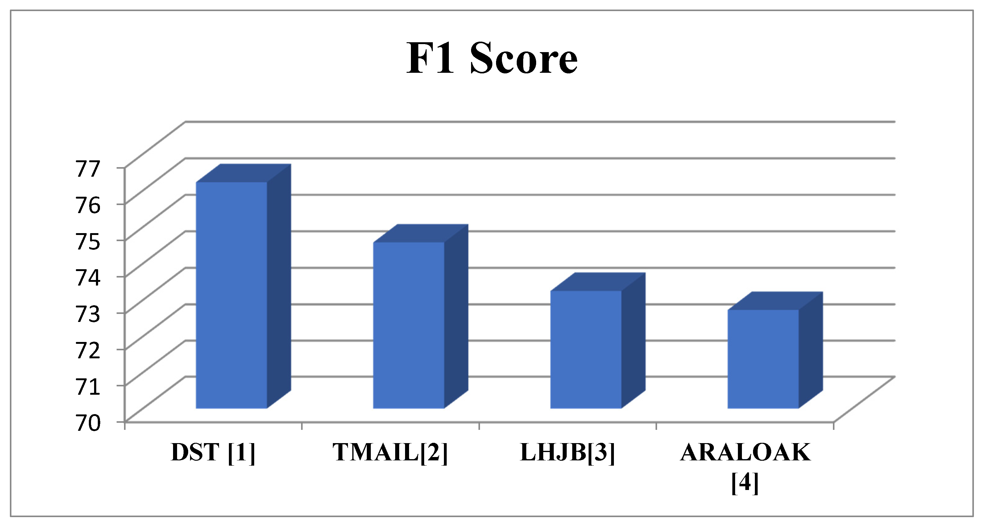
| Team | F1 Score |
|---|---|
| DST [1] | 76.23 |
| TMAIL [2] | 74.58 |
| LHJB [3] | 73.25 |
| ARALOAK [4] | 72.73 |
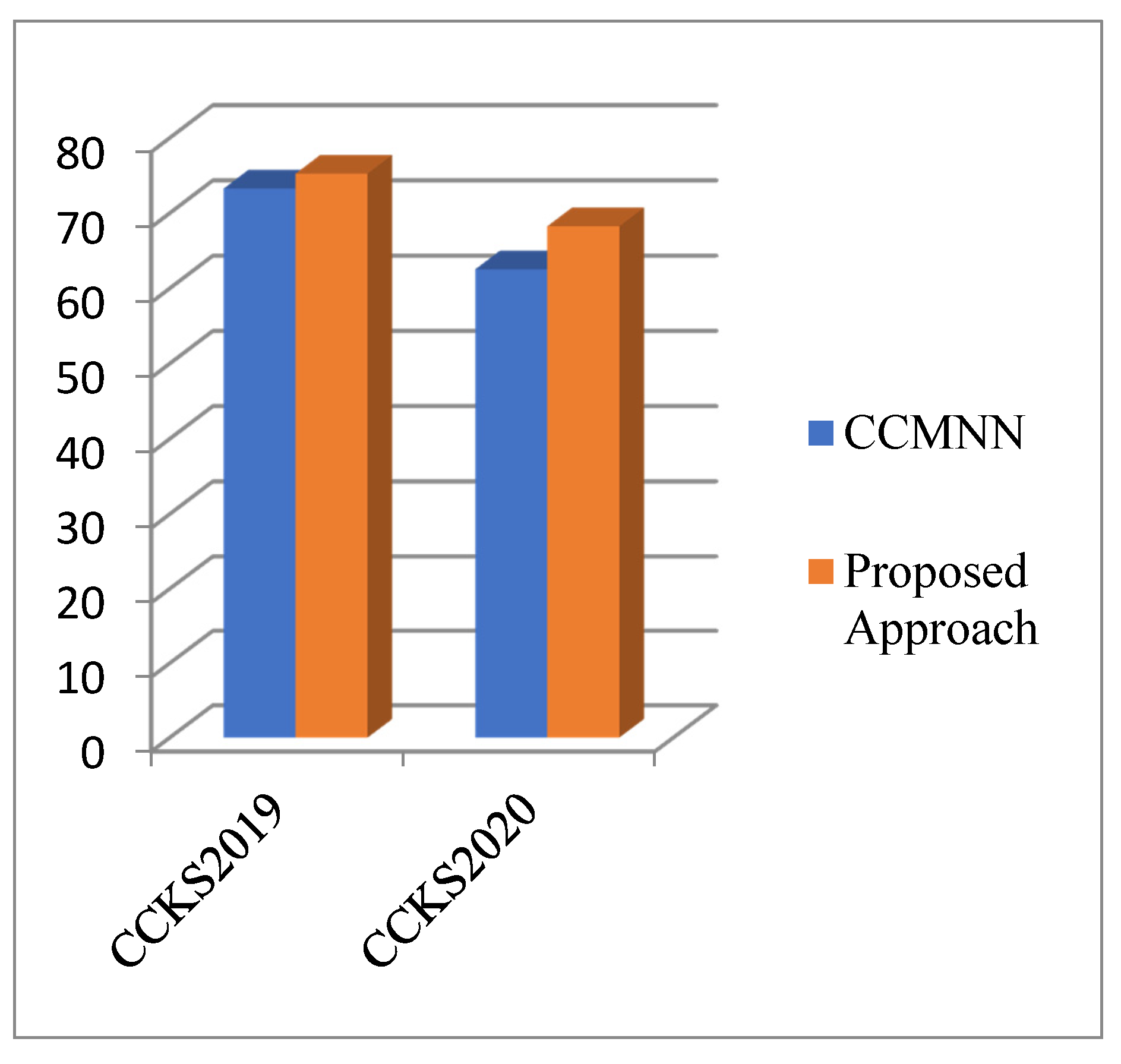
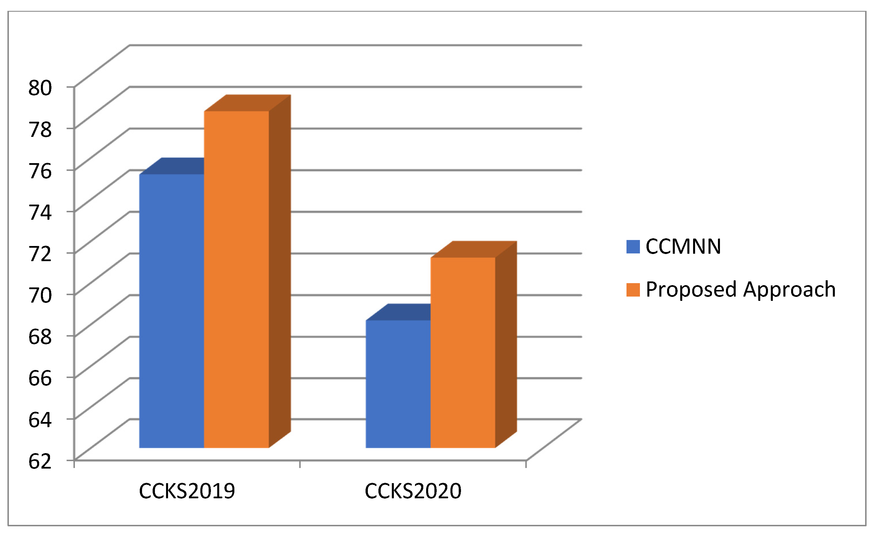
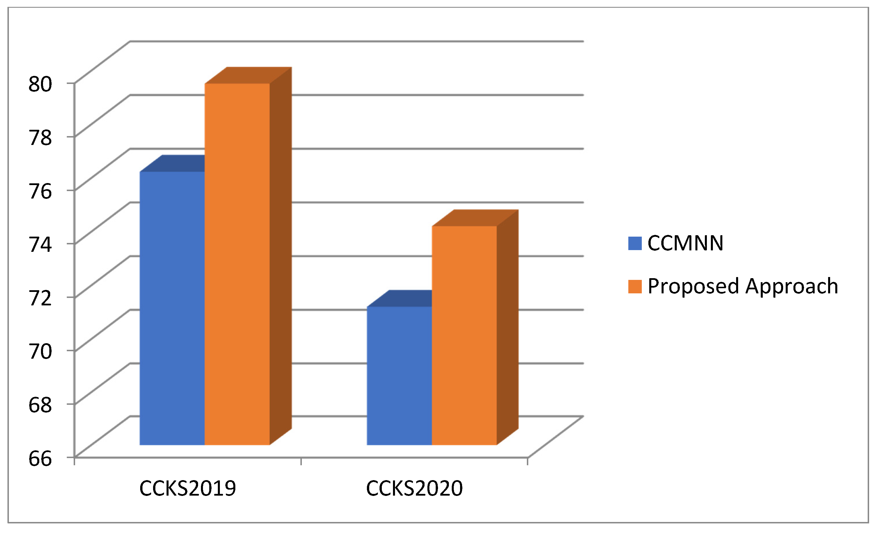
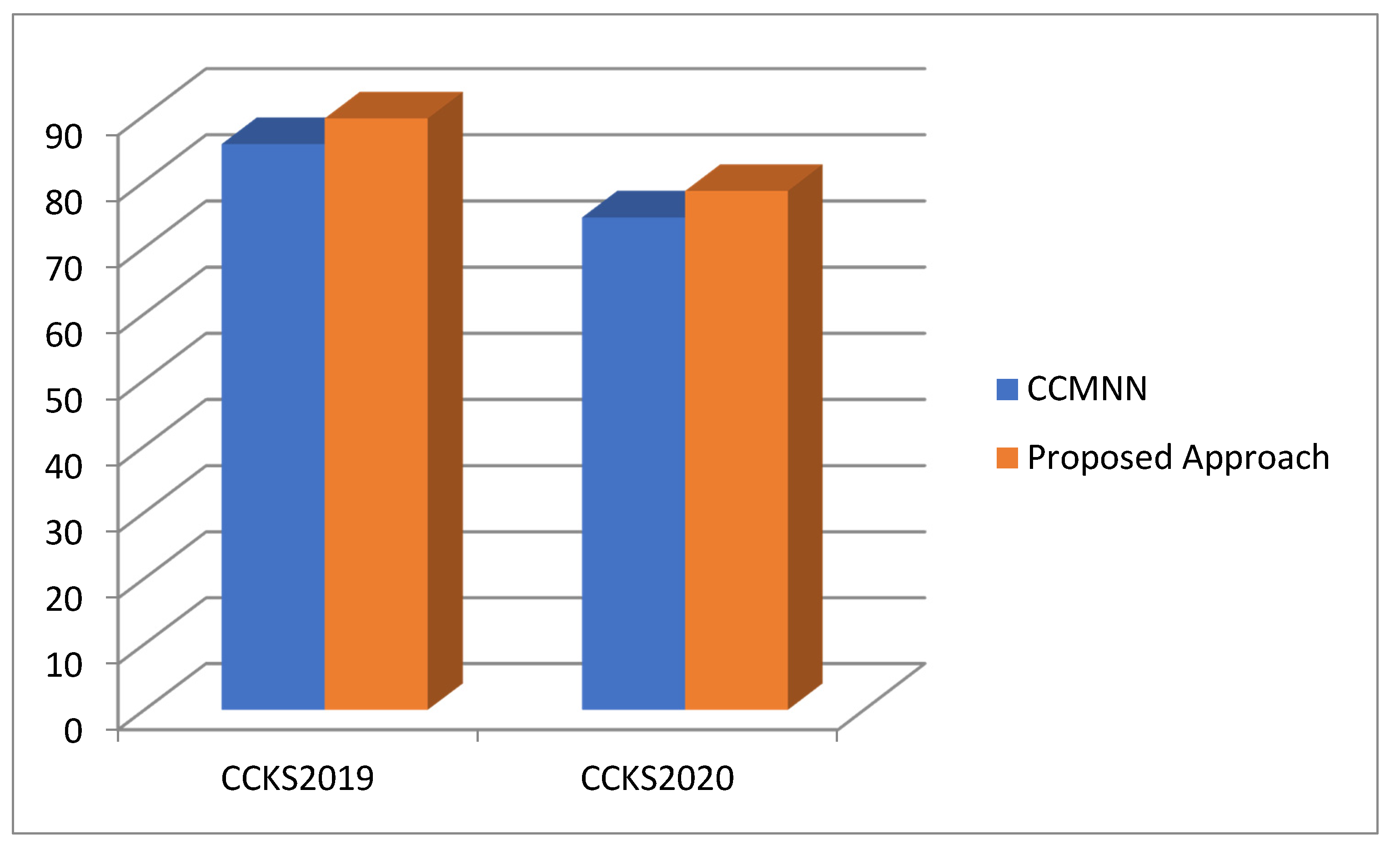
| CCKS2020 | CCKS2020 | |||||||
|---|---|---|---|---|---|---|---|---|
| P | R | F1 Score | Accuracy | P | R | F1 Score | Accuracy | |
| CCMNN | 73.26 | 75.21 | 76.24 | 85.65 | 62.54 | 68.2 | 71.21 | 74.56 |
| Proposed Approach | 75.25 | 78.24 | 79.52 | 89.52 | 68.25 | 71.21 | 74.21 | 78.56 |
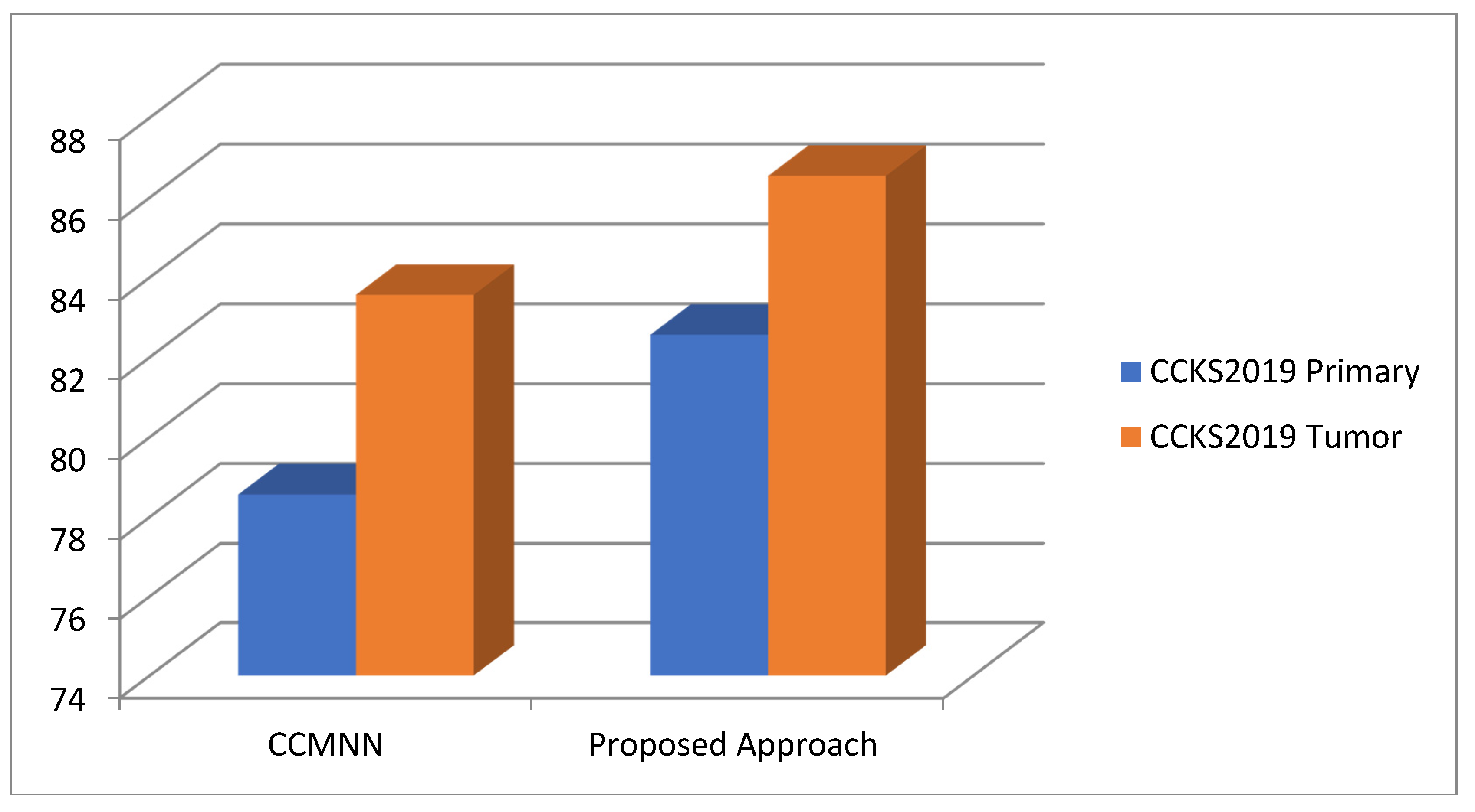
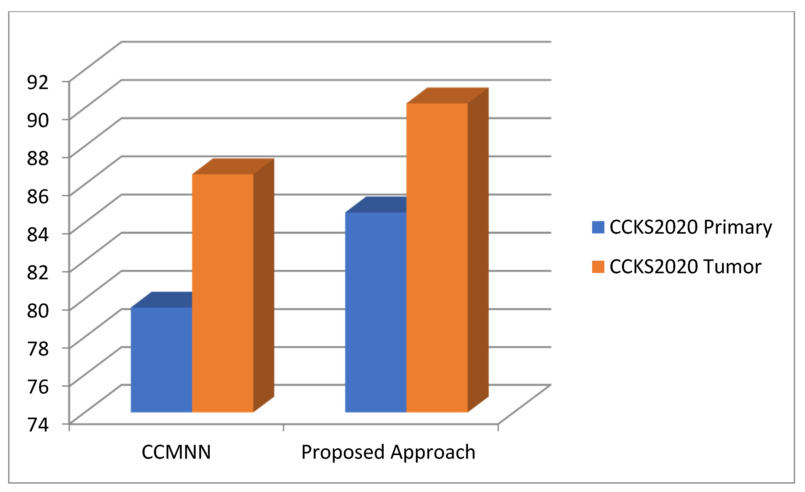
| CCKS2019 | CCKS2020 | |||
|---|---|---|---|---|
| Primary | Tumor | Primary | Tumor | |
| CCMNN | 78.56 | 83.56 | 79.54 | 86.52 |
| Proposed Approach | 82.56 | 86.54 | 84.52 | 90.23 |
Publisher’s Note: MDPI stays neutral with regard to jurisdictional claims in published maps and institutional affiliations. |
© 2022 by the authors. Licensee MDPI, Basel, Switzerland. This article is an open access article distributed under the terms and conditions of the Creative Commons Attribution (CC BY) license (https://creativecommons.org/licenses/by/4.0/).
Share and Cite
Dhiman, G.; Juneja, S.; Viriyasitavat, W.; Mohafez, H.; Hadizadeh, M.; Islam, M.A.; El Bayoumy, I.; Gulati, K. A Novel Machine-Learning-Based Hybrid CNN Model for Tumor Identification in Medical Image Processing. Sustainability 2022, 14, 1447. https://doi.org/10.3390/su14031447
Dhiman G, Juneja S, Viriyasitavat W, Mohafez H, Hadizadeh M, Islam MA, El Bayoumy I, Gulati K. A Novel Machine-Learning-Based Hybrid CNN Model for Tumor Identification in Medical Image Processing. Sustainability. 2022; 14(3):1447. https://doi.org/10.3390/su14031447
Chicago/Turabian StyleDhiman, Gaurav, Sapna Juneja, Wattana Viriyasitavat, Hamidreza Mohafez, Maryam Hadizadeh, Mohammad Aminul Islam, Ibrahim El Bayoumy, and Kamal Gulati. 2022. "A Novel Machine-Learning-Based Hybrid CNN Model for Tumor Identification in Medical Image Processing" Sustainability 14, no. 3: 1447. https://doi.org/10.3390/su14031447