Article

Sustainable Production of Biodiesel Using UV Mutagenesis as a Strategy to Enhance the Lipid Productivity in R. mucilaginosa

by
Joseph Antony Sundarsingh Tensingh
and
Vijayalakshmi Shankar
*
School of Bioscience and Technology, CO2 Research and Green Technologies Centre, VIT University, Vellore 632014, India
*
Author to whom correspondence should be addressed.
Sustainability 2022, 14(15), 9079; https://doi.org/10.3390/su14159079
Submission received: 13 May 2022 / Revised: 28 June 2022 / Accepted: 5 July 2022 / Published: 25 July 2022
(This article belongs to the Special Issue Biological Treatment Technologies of Domestic Waste)

Abstract

:
The future of petroleum-based fuel is biodiesel. Biodiesel is an eco-friendly fuel that can be used in any diesel engine without any alterations. Researchers have focused on biodiesel that can be produced from microbial lipids extracted from high lipid-yielding microbes. In this study, microbial cultures were screened for high lipid-yielding capabilities and mutated using UV radiation at three different time intervals of 30, 75, and 90 min. The Nile red fluorescence method was used to analyze high lipid-yielding microbes. An outstanding increase in biomass and lipid productivity was noted when the microbes were exposed to UV for 30 min. For example, an M30-8 UV-mutated strain produced a lipid yield of 68.5%. The lipids produced from the wild and mutated strains were analyzed using GCMS and FTIR spectrophotometric analysis. Then, the lipids extracted from both wild VS3 and UV-mutated M30-8 strains were transesterified using a base catalyst and the produced biodiesel was analyzed using ASTM standards. The aim and objective of the research was to mutate high lipid-yielding microbes by using UV radiation and produce biodiesel from the lipids extracted from both wild and UV-mutated strains.

1. Introduction

Oleaginous micro-organisms have the characteristic ability to produce and accumulate lipids, which constitute around 20% of their dry cell weight [1]. The lipids extracted from microbes are also known as single-cell oils and these lipids can replace the lipids extracted from the waste of animals and plants [2]. The accumulation of lipids by oleaginous micro-organisms is fully based on their genetic profile and differs significantly both according to species and among different strains of the same species [3]. The lipids are mainly composed of triacylglycerols and stearyl esters. Triacylglycerol is composed of three main fatty acid chains in which glycerol plays a major role and it can be used in the production of biodiesel [4]. Biodiesel is a major example of triacylglycerol-based biofuels that require less amount of feedstock, and the cost of biodiesel production is less compared with that of fossil fuels. In this study, we observed that oleaginous microalgae, yeasts, and fungi are the furthermost micro-organisms used in lipid biotechnology research [5]. With regards to lipid-producing micro-organisms, oleaginous microalgae can potentially accumulate a lipid content of around 70%, as a yield of their dry cell weight [6], Whereas the production of lipids from microalgae differs between species and strains and mainly depends on their cultural conditions. Similarly, oleaginous yeasts are unicellular micro-organisms that are also capable of producing excellent sources of lipids. Oleaginous yeast species include Yarrowia, Candida, Rhodotorula, Rhodosporidium, Cryptococcus, Trichosporon, and Lipomyces, and can produce a lipid yield of up to 80% in their biomass under stress conditions, while cultured in a wide range of carbon sources [7].
A mutation is a process by which a change of gene sequence occurs in an organism. Mutations can differ in various forms, from a change in single base pair to deletion, duplications, inversions, and insertions in mega base pair [8]. Alterations in the nucleotide sequence of a small portion of a genome and phenotypic results can differ from the specific site and location of the mutation [9]. Mutations can result from misplacement of a nucleotide sequence in DNA replication or by inducing an organism via exposure to mutagens. Mutagens are of three different types, comprising physical, chemical, and biological [10].
Generally, mutagens cause damage to DNA directly, and that can affect the results during DNA replication and cause errors, whereas severely damaged DNA can stop replicating and cause cell death [11]. PSOS operon plays a major role when the DNA is damaged. The response of a cell to DNA damage causes cell cycle arrest and stimulates mutagenesis [12]. The rec A gene stimulates the SOS response by identifying single-stranded DNA and activating mutagenic DNA polymerases. Physical mutation can be followed by exposing the specific micro-organism to radiation or UV light [13]. The radioactive waves produced from UV radiation damage DNA by producing covalent linkages between adjacent pyrimidine bases. The pyrimidine dimer produced from the covalent linkage is not capable of fixing in the double-strand structure of DNA, causing replication and translation to become inhibited [14]. Typically, the formation of dimer results in a deletion of base pairs, causing mutation to occur, whereas other types of radiation can have different effects on mutation depending on their wavelength and the intensity of radiation, but mainly mutations occur in the process of insertion and deletions of DNA base pairs [15].
According to a recent survey, replacement of diesel fuel in agricultural engines will be more effective for the farmers by utilizing biodiesel with an appropriate blend [16]. Transesterification plays a major role in the reduction of high viscous oil to produce low viscous biodiesel [17]. Therefore, extensive usage of biodiesel can reduce more pollutants, such as carbon dioxide, nitrogen oxide, and hydrocarbons [18].
This study mainly focuses on biodiesel production using micro-organisms of both wild and mutated strains. Mutation of microbes was performed using UV radiation. The mutated microbes were then screened for radioactive fluorescence units, in order to analyze the mutation rate. The mutated microbes were cultured and lipids were extracted from both wild and mutated microbes. The lipids extracted from the microbes were transesterified using a base catalyst to produce biodiesel. The quality of biodiesel was then assessed using ASTM standards.

2. Materials and Methods

2.1. Isolation and Screening of Microbes

The soil sample was collected from an oil-contaminated site near the VVD coconut oil factory in Tuticorin, Tamil Nadu, India. The soil sample was then serially diluted aseptically using double-distilled water under a laminar airflow chamber for serial dilution and pour plate method. The diluted sample of different concentrations varied between 10−9, 10−8, 10−7, 10−6, 10−5, 10−4, 10−3, 10−2, and 10−1. An amount of 1 mL of stock solution was diluted aseptically in 9 mL of double-distilled water and mixed well using a micropipette. Each 1 mL of the sample was taken from a previously mixed solution. These samples were then cultured in YEPD broth (10 g/L of yeast extract; 20 g/L of peptone; 20 g/L of dextrose in 1000 mL of double-distilled water) and NA (3 g of beef extract; 5 g of peptone; 0.5 g of sodium chloride; 20 g of agar mixed with 1000 mL of double-distilled water) media [19]. Around 10 colonies of bacteria and 10 colonies of fungi were selected from 10−7 and 10−6 dilutions from the oil-contaminated soil sample. A few micro-organisms showed fast growth compared to other isolated micro-organisms and produced high lipid content. The isolated micro-organisms were screened for high lipid content by staining the micro-organisms using Nile red staining [20]. By using Nile red fluorescence staining method, the amount of lipids determined via the relative fluorescence unit of the cell suspension was recorded at 540 nm [21,22,23].

2.2. Physical Mutation of Microbes Using UV Radiation Method

The isolated and screened micro-organisms were mutated using the UV radiation process. Around 5 mL of cell culture was taken aseptically using a micropipette from the broth at the exponential phase. Then it was transferred into an open Petri plate and directly exposed to UV irradiation (Germicidal lamp, UVC 30 W, Philips, Amsterdam, The Netherlands) at a 25 cm distance for 30, 70, and 90 min [24]. After mutation, the mutants were placed in a dark room for 24 h, to avoid recovery of cells from light interaction. The mutated cells were then kept under continuous light incubation for 2–3 weeks. After incubation, single colonies appeared on the agar plate. The colonies developed on the agar plate were selected and transferred aseptically into a sterile 96-wells plate. Using VarioskanTM LUX multimode microplate reader (Thermo Fisher Scientific, Waltham, MA, USA), the density of cells was measured in each culture plate [25].

2.3. Molecular Sequencing

The molecular sequencing method was used to identify the isolated micro-organism. The genomic DNA was isolated from the micro-organism using Qiagen DNeasy Kit. Using standard PCR reaction, the fragments were amplified using the primers LR7 and 5.8 SR. After amplification, the DNA fragments were purified using a gene O-spin purification kit and directly sequenced to identify the species of the micro-organism. The sequenced genome was submitted to GenBank [26].

2.4. Culture and Growth Conditions of Selected Strains

The selected microbe was inoculated in a 500 mL conical flask containing 250 mL of YEPD broth medium containing 1% yeast extract, 2% peptone, and 2% glucose in double-distilled water. The culture was kept under a rotary shaker at 170 rpm for 24 h and maintained the temperature at 30 °C. Around 1 mL of culture was suspended from the broth and pelletized using a centrifuge at 13,000 rpm for 6 min. The pelletized cells were weighed under a vacuum weighing machine to estimate the production of biomass cultured in the broth [27].

2.5. Analysis of Cell Dry Weight

By using the gravimetrical analysis method, the production of biomass from the culture was analyzed by weighing the cell’s dry weight. Around 10 mL of broth culture from the exponential phase was transferred into a 15 mL centrifuge tube and centrifuged at 10,000 rpm for 10 min [28]. After centrifugation, the supernatant was discarded and the pellet was collected and dried in a hot air oven at 65 °C for 1 day. The dried pellet was then weighed in a vacuum weighing machine [29,30].

2.6. Extraction of Lipids

In this process of lipid extraction, Bligh and Dyer’s method was followed. This is a common method for total lipid extraction [31]. In this method, the centrifuged biomass is dried and mixed with CH3OH:CHCl3:H2O at a ratio of 2:1:0.8. (v/v/v) and vortexed for 15 min. An amount of 0.85% of KCl is then added to the solution to make up a final ratio of 2:2:1.8 (v/v/v) [32]. The mixture was again vortexed for 15 min. After vertexing, the solvent was gently placed on the stand to remove cell debris. Then the lower lipid phase was collected and evaluated for total lipid content [33].

2.7. Quantification of Lipids

Lipid content was quantified using a glass Petri plate. Initially, the weight of the plate was measured in a vacuum weighing machine, then the extracted lipids were added to the Petri plate. The plates were then placed at 45 °C in a hot air oven for 1 h to make sure that all the solvents except the lipids were vaporized from the plates. After drying, the plates were kept at room temperature and weighed as the final value [34]. The differences between the initial weight and the final weight were used to determine the concentration of lipids. The percentage was obtained by calculating the initial and final weight of lipids to the total weight of the sample as mentioned in the formula,
Lipid % = Initial   weight   of   lipid     Final   weight   of   lipid Total   weight   of   sample   ×   100

2.8. Gas Chromatography and Mass Spectroscopy

Around 200 μL of extracted lipids was transferred into a round bottom flask and mixed with 4 mL of 0.5 M potassium hydroxide solution via shaking. The flask containing the stock solution was attached to the reflux condenser gently and kept in a boiling water bath for 15 min via periodic shaking. After boiling, 1.6 mL of methanolic hydrochloric acid was added into the flask and the solution was boiled again for 25 min by fixing the reflux condenser gently. After boiling, the reflux condenser was firmly removed from the flask and the solution cooled at room temperature. Subsequently, 8 mL of double-distilled water was added and the contents mixed slowly by shaking. Then, w6 mL of n-Hexane was added to the solution and mixed continuously for 2 min to extract the fatty esters present in the solution [35]. This process was repeated 3 times to extract the whole fatty acid methyl esters as the sample for GCMS. An Agilent 6890 gas chromatograph was used to analyze and identify the fat content present in the extracted lipids. The gas chromatograph is equipped with a 2 mm direct injector liner and an EC-5 column of 250 μ I.D., 0.25 μ film thickness. It also consists of a split injection which is used for loading the sample at the ratio of 10:1. It was automated to start when the oven temperature reached 35 °C and then held for 2 min, before rising at a rate of 20 °C per minute up to 300 °C and held for a further 5 min. The flow rate of helium carrier gas was constantly programmed as 2 mL/min. AJEOL GCmateII bench top double-focusing magnetic sector mass spectrometer operating in electron ionization (EI) mode with TSS-2000 software was used for all analyses [36].

2.9. Fourier Transformed Infrared Spectroscopy

In this study, an FTIR spectrometer was used to identify the quality of the catalyst using a functional group in the range of cm−1. The catalysts which were produced were mixed with KBr separately to form tablets and then attached to the FTIR sample plate. For an individual and composite, a spectrum was collected at 400–4000 [37].

2.10. Transesterification

Biodiesel was produced by converting the lipids extracted from the microbes by using the transesterification process. The catalyst was prepared by adding 4 gm of sodium hydroxide pellets to 100 mL of methanol. Then 1 mL of lipids extracted from the microbe was suspended in a conical flask containing 10 mL of freshly prepared catalyst in the ratio of 1:10 lipids to catalyst [38]. The conical flask containing the mixture solvents was placed on the thermomagnetic stirrer for 6 h at 70 °C and the rotation speed of the magnetic stirrer was maintained at 450 rpm [39,40]. After this process, the solvent mixture was transferred into a separating funnel and kept overnight without disturbing the solvents [32]. After 24 h of separation, the solvents were found to be in two layers: the top layer was biodiesel and the bottom layer was methanol [41]. Then the methanol was removed gently from the separating funnel and the biodiesel was collected [42,43]. The collected biodiesel was then kept in a hot air oven for a few minutes to remove the residual methanol present in it. The properties of the biodiesel were then analyzed using ASTM standards [44,45].

2.11. Biodiesel Properties

The properties of biodiesel were analyzed to determine the ability of the fuel [46]. The main properties of biodiesel are determined using specific analyses such as density (which is determined by ASTM D4052 [47]), specific gravity, and kinematic viscosity (which is determined by the flow of a fluid based on gravity). The volume of fuel is placed on the capillary tube viscometer to analyze the time taken for the fuel to flow under gravity, measured thermostatically in a boiling water bath at 40 °C [48,49]. Flash point and fire point are determined by ASTM D93, pour point is determined by ASTM D2500, calorific value is determined by the amount of heat liberated during the process of combustion, and the acid value is determined by free fatty acid titration and cetane number [50,51]. These methods were performed in triplicate and the repeated values were noted and compared with ASTM standards [52,53].

3. Results and Discussion

3.1. Screening of High Lipid-Yielding Microbe

The soil sample was collected from an oil-contaminated site near the VVD coconut oil factory in Tuticorin, Tamil Nadu, India. The samples were cultured in an agar medium using the serial dilution method and around 10 colonies of bacteria and fungi were screened for high lipid-yielding microbes using Nile red staining, out of which one strain (BS2) showed a high lipid-yielding profile, as represented in Table 1, and it was selected for the UV-mutation process.

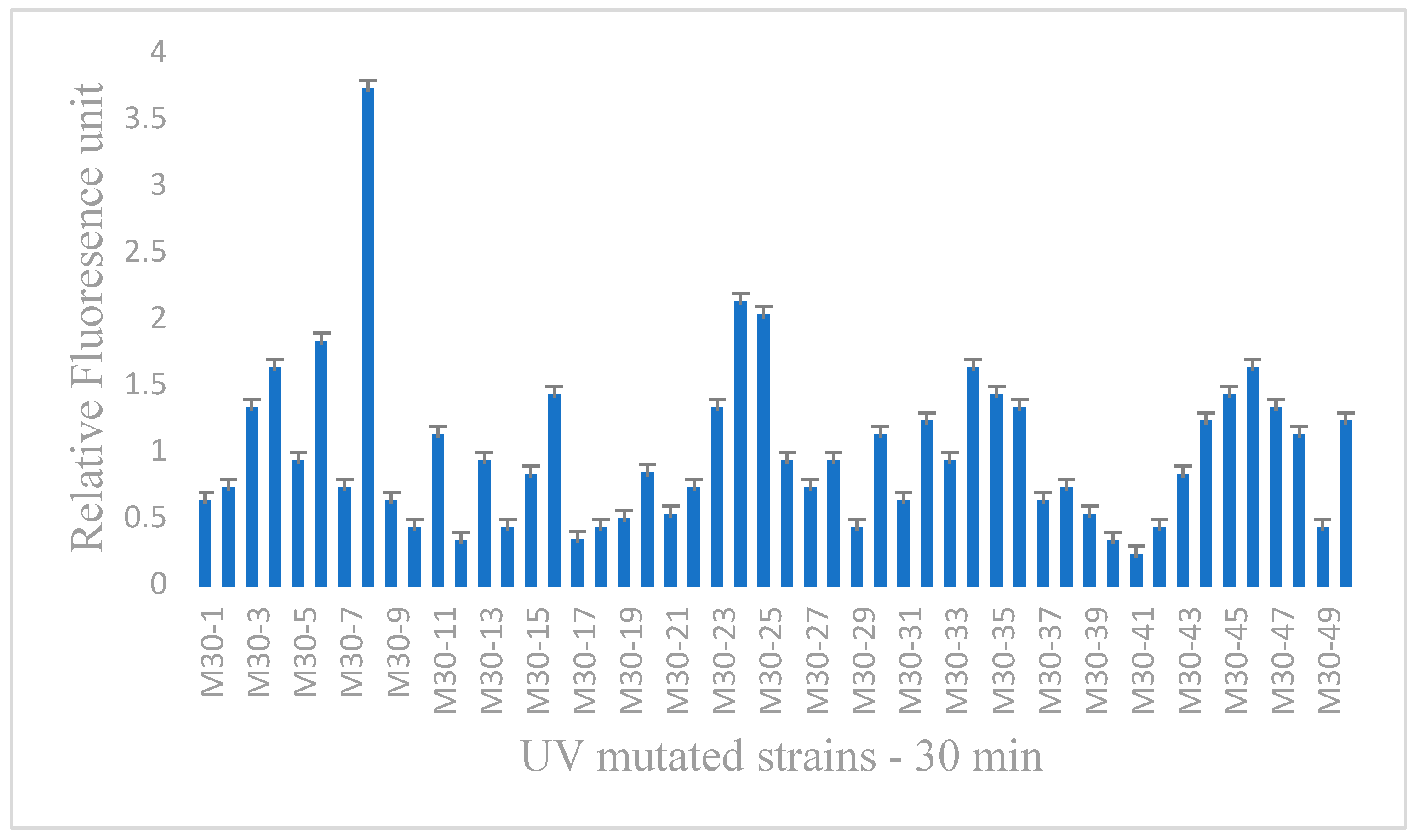
3.2. Screening and Selection of UV-Mutated Strains

The isolated micro-organism was subjected to UV radiation at a distance of 25 cm for different time intervals of 30, 75, and 90 min, respectively, and each plate contained 50 colonies. The colonies were mixed with Nile red fluorescence stain to analyze high lipid-producing strains using a relative fluorescence unit. In this analysis, mutant strains of different periods showed different lipid profiles, out of which three mutant strains were selected from three time intervals, as represented in Table 2. The M30-8 strain displayed 3.7 RFU, as shown in Figure 1, the M75-24 strain displayed 1.8 RFU, as shown in Figure 2, and the M90-14 strain displayed 0.9 RFU, as shown in Figure 3. These colonies were selected as high lipid-yielding colonies from different time intervals of UV radiation and showed a high mutation rate. The mutated strains were sequenced to analyze the homology difference with wild strain VS3. Similarly, Rike Rachmayati et al. (2020) [14] have screened high lipid-yielding microalgae by using Nile red fluorescence staining to analyze wild and UV-mutated strains, while Chen et al. (2009) [27] have applied the Nile red staining method to identify the lipid content present in oleaginous micro-organisms, and Halim et al. (2015) [26] have shown that Nile red stain emits fluorescence when it interacts with lipids present in the microbial bodies. Hence, Nile red staining is used to screen high lipid-yielding microbes from the UV-mutated strain.

3.3. Molecular Identification of Wild and Mutated Strains

The genomic DNA was isolated from wild strain VS3 and UV-mutated strains were amplified using polymerize chain reaction. The DNA isolated from wild strain VS3 and UV-mutated strains were sequenced and submitted to GenBank and the accession number of wild strain VS3 and UV-mutated strains were received. The isolated micro-organism wild strain VS3 was found to be Rhodotorula mucilaginosa and the accession number was OL635991. The UV-mutated strain of three different time intervals represented as M30-8, M75-24, and M90-14 (as shown in Table 3) received accession numbers of OL658823, OL658824, and OL658825, respectively. The distance tree was analyzed by using the accession number of wild and UV-mutated strains in BLAST, as shown in Figure 4.

3.4. Production of Biomass and Lipids from Wild and UV-Mutated Strain

The wild strain Rhodotorula mucilaginosa VS3 and the UV-mutated strains M30-8, M75-24, and M90-14 were inoculated and cultured in YEPD broth media. The culture was maintained in a rotary shaker at 28 °C and 150 rpm. Around 1 mL of culture was taken every 12 h in the exponential phase of the culture from each broth aseptically and the biomass was calculated gravimetrically by using cell dry weight analysis. The lipids extracted from the biomass using the Bligh and Dyer extraction method were also analyzed. A gradual increase in the growth of biomass is represented in Figure 5, and the production of lipids is analyzed and represented in Figure 6. The wild strain VS3 has recorded a maximum of 18.7 g/L per day, and the UV-mutated strain M30-8 has produced 62.1 g/L per day; M75-24 has produced 53.2 g/L per day and M90-14 has produced 48.5 g/L per day, as represented in Table 4. The M30-8 strain produced a high amount of biomass from a broth culture. The percentage of lipids produced from the biomass of the wild strain and the mutant strain was analyzed and the amount of lipids extracted from the wild strain was noted as 41.3%, whereas the mutant strains of M30-8, M75-24, and M90-18 produced 68.5%, 56.3%, and48.6% of lipids, respectively, at the exponential phase of the culture. Similarly, to our study Rike Rachmayati et al. (2020) [14] also found a gradual increase in the growth of biomass in UV-mutated Chlorella sp., compared to the growth of biomass in the wild strain, and Liu et al. (2015) [28] have shown high lipid content in UV mutagenized microalgal species compared to the lipid content produced by wild strain. Therefore, UV mutation shows an increase in ACC concentration that enhances lipids. In our study, M30-8 strain has produced a high amount of biomass and lipids, and the lipids produced from the M30-8 strain have been analyzed using GCMS and FTIR for biodiesel production.

3.5. GCMS Analysis of Lipids Extracted from VS3 and M30-8 Strain

The fatty acid composition of lipids extracted from both wild strain VS3 and UV-mutated M30-8 strain of Rhodotorula mucilaginosa was analyzed using gas chromatography and mass spectroscopy. In this study, the fatty acid composition of the wild strain consists of tetra decanoic acid, pentadecanoic acid, n-hexadecanoic acid, and hexadecanoic acid, which is confirmed by the peak produced at different retention times (RT) in the spectroscopy, as shown in Figure 7. The UV-mutated strain M30-8 consists of tetra decanoic acid, pentadecanoic acid, n-hexadecanoic acid, hexadecanoic acid, 9-octadecanoic acid, and Oleic acid is confirmed by the peak produced at different retention times which is almost similar to the wild strain as shown in Figure 8. Similar compounds of fatty acids have been identified in both wild and mutated strains, as represented in Table 5. Similarly, in Kanakdande et al. (2021) [22], the presence of decanoic acid, hexadecenoic acid, and tetradecanoic acid is used to obtain biodiesel.

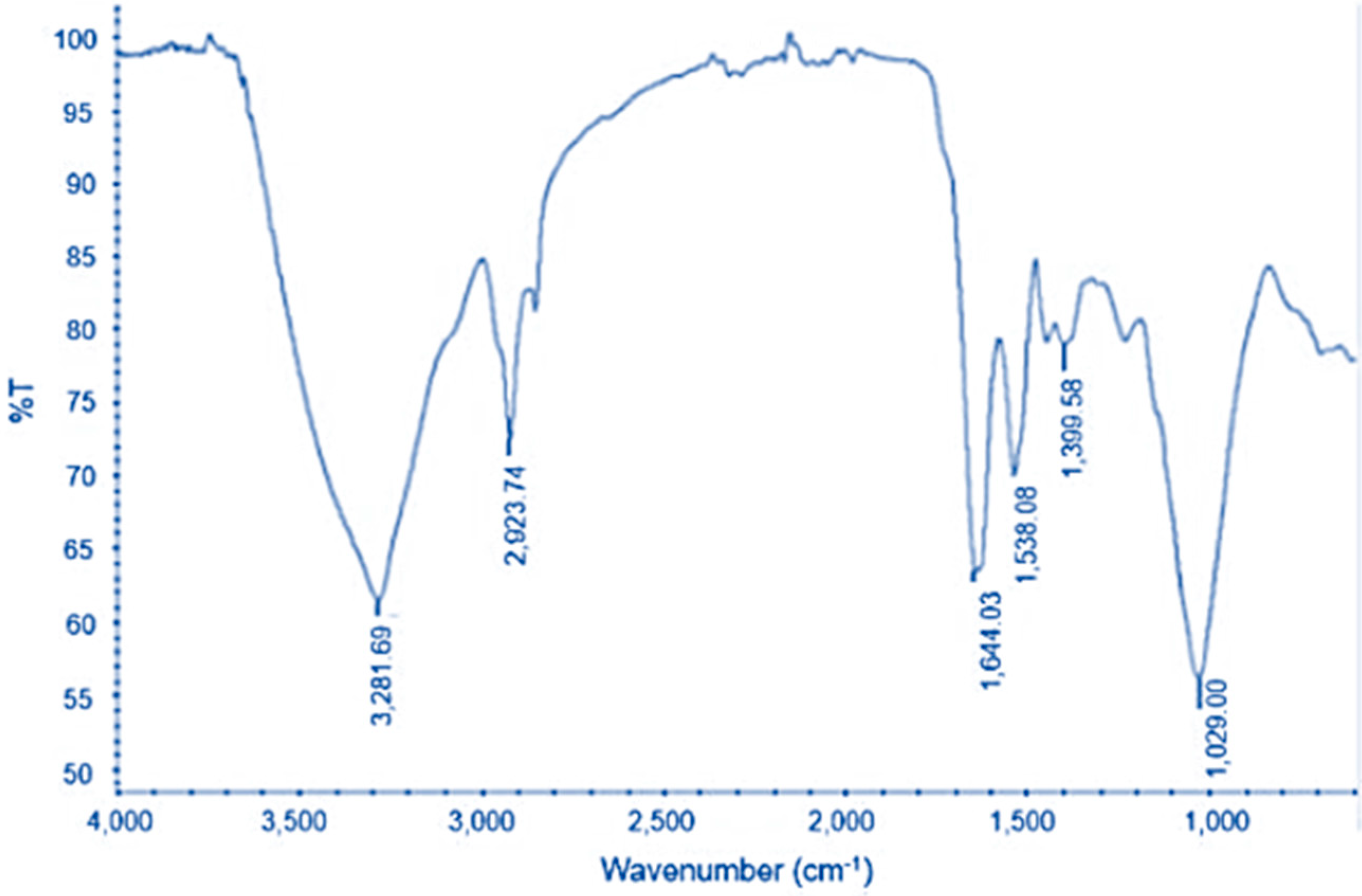
3.6. FTIR Analysis of Lipids Extracted from VS3 and M30-8 Strain

The IR bands of lipids extracted from the wild strain of R. mucilaginosa consist of wave numbers at 1636.45 which represent the presence of C=O acid stretching, while the wave numbers produced at 1028.01 indicate C=O acid bending. Therefore, the presence of the carboxylic acid functional group was justified through FTIR, as shown in Figure 9. The lipids produced from the mutant strain also show the IR regions consisting of a visible peak at 1644.03, indicating the presence of C=O acid stretching, while the wave number produced at 1029.00 represents C=O acid bending, confirming the presence of the carboxylic acid functional group, as shown in Figure 10. The peaks of the FTIR spectrum indicate and confirm the presence of fatty acid methyl esters which are used to produce biodiesel. Similarly, Kanakdande et al. (2021) [22] have mutated Bacillus amyloliquefaciens using UV radiation, and the lipids extracted from mutated species were observed at the peak at 1712 cm−1 showing that the presence of C=O stretching and the peak of 1256 cm−1 was due to the presence of C-O stretching, which is used for biodiesel production.

3.7. Biodiesel Properties of VS3 and M30-8 Strain

Both the wild strain VS3 and the UV-mutated strain M30-8 of R. mucilaginosa produced biodiesel within the limits of ASTM standards. The density of biodiesel produced from R. mucilaginosa wild strain VS3 was recorded as 860 kg/m3, whereas UV-mutated strain M30-8 was recorded as 865 kg/m3. The specific gravity of biodiesel was recorded as 0.86 in the VS3 strain and 0.88 in the M30-8 U-V-mutated strain. The kinematic viscosity of biodiesel was reported as 5.2 mm2/s in the VS3 strain and 5.5 mm2/s in the M30-8 strain, as per ASTM D445 standard. The flashpoint of wild strain VS3 was 160 °C, whereas in the M30-8 UV-mutated strain of R. mucilaginosa it was 155 °C. The fire point of wild strain VS3 and the UV-mutated strain M30-8 was 165 °C and 167 °C, as per ASTM D93 standards. The pour point of wild strain VS3 and UV-mutated strain M30-8 was recorded as −2–8 °C and −2–6 °C, as per ASTM D2500 standards. The calorific value of wild and mutated strains resulted in 37,500 kJ/kg in VS3 and 37,650 kJ/kg in the M30-8 strain. The acid value of biodiesel produced from wild and UV-mutated strains was tested using the free fatty acid titration method, which resulted in 0.1 mg/KOH and 0.3 mg/KOH in the M30-8 strain. The cetane number was recorded as 56 min in both wild strains VS3 and the UV-mutated strain M30-8, as represented in Table 6. Similarly, Elangovan T et al. (2016) [52] analyzed the properties of biodiesel from various oil resources using ASTM standards, which were almost similar to our study. A comparison of properties of oil extracted from jatropha, peppermint, and pine oil is shown in the table.

4. Conclusions

This study mainly concentrated on biodiesel production using high lipid-yielding microbes. High lipid-yielding microbes were screened using Nile red fluorescence stain. The screened microbe was subjected to UV radiation for three different t of 30, 75, and 90 min and the growth of biomass and lipid content in the mutated strains was observed. The M30-8 strain achieved a high growth rate of 62.1 mg/L biomass and a lipid content of 68.5% compared to other M75-24 and M90-14 UV-mutated strains. Thus, the M30-8 strain was selected along with the wild strain VS3 of Rhodotorula mucilaginosa for further gas chromatography-mass spectroscopy and Fourier transmission infrared analysis. The presence of tetra decanoic acid, hexadecenoic acid, pentadecanoic acid, and octadecanoic acid were analyzed using mass spectroscopy, which is useful in the production of biodiesel. The lipid extracted from wild and UV-mutated strains were mixed with sodium hydroxide methanol solution and processed for transesterification to produce biodiesel. The produced biodiesel was analyzed according to ASTM standards.

Author Contributions

Investigation, J.A.S.T.; Supervision, V.S.; Writing—original draft, J.A.S.T.; Writing—review & editing, V.S. All authors have read and agreed to the published version of the manuscript.

Funding

This research received no external funding.

Institutional Review Board Statement

Not applicable.

Informed Consent Statement

Not applicable.

Conflicts of Interest

The authors declare no conflict of interest.

References

  1. Ahmad, A.L.; Yasin, N.H.M.; Derek, C.J.C.; Lim, J.K. Microalgae as a sustainable energy source for biodiesel production: A review. Renew. Sustain. Energy Rev. 2011, 15, 584–593. [Google Scholar] [CrossRef]
  2. Balat, M. Potential alternatives to edible oils for biodiesel production—A review of current work. Energy Convers. Manag. 2011, 52, 1479–1492. [Google Scholar] [CrossRef]
  3. Chen, J.; Li, J.; Dong, W.; Zhang, X.; Tyagi, R.D.; Drogul, P.; Surampalli, R.Y. The potential of microalgae in biodiesel production. Renew. Sustain. Energy Rev. 2018, 90, 336–346. [Google Scholar] [CrossRef]
  4. Chisti, Y. Biodiesel from microalgae. Biotechnol. Adv. 2007, 25, 294–306. [Google Scholar] [CrossRef]
  5. Knothe, G. Biodiesel, and renewable diesel: A comparison. Prog. Energy Combust. Sci. 2010, 36, 364–373. [Google Scholar] [CrossRef]
  6. Aloklah, B.; Alhajali, A.; Yaziji, S. Identification of some yeasts by fatty acid profiles. Pol. J. Microbiol. 2014, 63, 467–472. [Google Scholar] [CrossRef]
  7. Rawat, I.; Kumar, R.R.; Mutanda, T.; Bux, F. Biodiesel from microalgae: A critical evaluation from laboratory to large scale production. Appl. Energy 2013, 103, 444–467. [Google Scholar] [CrossRef]
  8. Mata, T.M.; Martins, A.A.; Caetano, N.S. Microalgae for biodiesel production and other applications: A review. Renew. Sustain. Energy Rev. 2010, 14, 217–232. [Google Scholar] [CrossRef] [Green Version]
  9. Ramadhas, A.S.; Jayaraj, S.; Muraleedharan, C. Biodiesel Production from High FFA Rubber Seed Oil. Fuel 2005, 84, 335–340. [Google Scholar] [CrossRef]
  10. Demirbas, A. Progress, and recent trends in biodiesel fuels. Energy Convers. Manag. 2009, 50, 14–34. [Google Scholar] [CrossRef]
  11. Kodym, A.; Afza, R. Physical and Chemical Mutagenesis. Methods Mol. Biol. 2003, 236, 189–203. [Google Scholar]
  12. Sharma, K.K.; Schuhmann, H.; Schenk, P.M. High lipid induction in microalgae for biodiesel production. Energy J. 2012, 5, 1532–1553. [Google Scholar] [CrossRef] [Green Version]
  13. Sivaramakrishnan, R.; Incharoensakdi, A. Enhancement of lipid production in Scenedesmus sp. by UV mutagenesis and hydrogen peroxide treatment. Bioresour. Technol. 2017, 235, 366–370. [Google Scholar] [CrossRef]
  14. Rachmayati, R.; Agustriana, E.; Rahman, D.Y. UV Mutagenesis as a Strategy to Enhance Growth and Lipid Productivity of Chlorella sp. 042. J. Trop. Biodivers. Biotechnol. 2020, 5, 218–227. [Google Scholar] [CrossRef]
  15. Kumar, A.K. UV mutagenesis treatment for improved production of endoglucanase and β-glucosidase from newly isolated thermotolerant actinomycetes, Streptomyces griseoaurantiacus. Bioresour. Bioprocess. 2015, 2, 22. [Google Scholar] [CrossRef] [Green Version]
  16. Paneerselvam, P.; Venkadesan, G.; Panithasan, M.S.; Alaganathan, G.; Wierzbicki, S.; Mikulski, M. Evaluating the Influence of Cetane Improver Additives on the Outcomes of a Diesel Engine Characteristics Fueled with Peppermint Oil Diesel Blend. Energies 2021, 14, 2786. [Google Scholar] [CrossRef]
  17. Panithasan, M.S.; Gopalakichenin, D.; Venkadesan, G.; Malairajan, M. Evaluating the working characters of a diesel engine fueled with biodiesel blends added with rice husk Nano particles. Energy Sources Part A Recovery Util. Environ. Eff. 2020. [Google Scholar] [CrossRef]
  18. Panithasan, M.S.; Gopalakichenin, D.; Venkadesan, G. Assessing the combined outcome of rice husk nano additive and water injection method on the performance, emission and combustion characters of the low viscous pine oil in a diesel engine. SAE Stud./Young Prof. Tech. Pap. Compet. 2019. [Google Scholar] [CrossRef]
  19. Yao, R.; Li, M.; Deng, S.; Hu, H.; Wang, H.; Li, F. Mutagenesis of Trichoderma viride by ultraviolet and plasma. Plasma Sci. Technol. 2012, 14, 353–356. [Google Scholar] [CrossRef]
  20. Choi, T.-O.; Kim, K.-H.; Kim, G.-D.; Choi, T.-J.; Jeon, Y.J. The Evaluation of UV-induced Mutation of the Microalgae, Chlorella vulgaris in Mass Production Systems. J. Life Sci. 2017, 27, 1137–1144. [Google Scholar]
  21. Banerjee, C.; Dubey, K.K.; Shukla, P. Metabolic engineering of microalgal based biofuel production: Prospects and challenges. Front. Microbiol. 2016, 7, 432. [Google Scholar] [CrossRef] [Green Version]
  22. Kanakdande, A.P.; Khobragade, C.N.; Mane, R.S. Ultraviolet-induced random mutagenesis in Bacillus amyloliquefaciens (MF 510169) for improving biodiesel production. Fuel 2021, 304, 121380. [Google Scholar] [CrossRef]
  23. Huang, G.-H.; Chen, G.; Chen, F. Rapid screening method for lipid production in alga based on Nile red fluorescence. Biomass Bioenergy 2009, 33, 1386–1392. [Google Scholar] [CrossRef]
  24. Pick, U.; Rachutin-Zalogin, T. Kinetic anomalies in the interactions of Nile red with microalgae. J. Microbiol. Methods 2012, 88, 189–196. [Google Scholar] [CrossRef]
  25. Uprety, B.K.; Dalli, S.S.; Rakshit, S.K. Bioconversion of crude glycerol to microbial lipid using a robust oleaginous yeast Rhodosporidiumtoruloides ATCC 10788 capable of growing in the presence of impurities. Energy Convers. Manag. 2017, 1, 117–128. [Google Scholar] [CrossRef]
  26. Halim, R.; Webley, P.A. Nile Red Staining for Oil Determination in Microalgal Cells: A New Insight through Statistical Modelling. Int. J. Chem. Eng. 2015, 2015, 695061. [Google Scholar] [CrossRef]
  27. Chen, W.; Zhang, C.; Song, L.; Sommerfeld, M.; Hu, Q. A high throughput Nile red method for quantitative measurement of neutral lipids in microalgae. J. Microbiol. Methods. 2009, 77, 41–47. [Google Scholar] [CrossRef]
  28. Liu, S.; Zhao, Y.; Liu, L.; Ao, X.; Ma, L.; Wu, M. Improving cell growth and lipid accumulation in green microalgae Chlorella sp. via UV irradiation. Appl. Biochem. Biotechnol. 2015, 175, 3507–3518. [Google Scholar] [CrossRef]
  29. Ramadhani, A.P.; Prashantyo, M.H.; Soedarmodjo, T.P.; Widjaja, A. The Effect of UV-B Mutation on Biodiesel from Microalgae Botryococcusbraunii using Esterification, Transesterification, and Combination of Esterification Transesterification. AIP Conf. Proc. 2020, 2217, 030021. [Google Scholar]
  30. Cheirsilp, B.; Kitcha, S.; Torpee, S. Co-culture of an oleaginous yeast Rhodotorulaglutinis and a microalga Chlorella vulgaris for biomass and lipid production using pure and crude glycerol as a sole carbon source. Ann. Microbiol. 2012, 62, 987–993. [Google Scholar] [CrossRef]
  31. Bligh, E.G.; Dyer, W.J. A rapid method of total lipid extraction and purification. Can. J. Biochem. Physiol. 1959, 37, 911–917. [Google Scholar] [CrossRef]
  32. Sheng, J.; Vannela, R.; Rittmann, B.E. Evaluation of methods to extract and quantify lipids from Synechocystis PCC 6803. Bioresour. Technol. 2011, 102, 1697–1703. [Google Scholar] [CrossRef]
  33. Folch, J.; Lees, M.; Sloane Stanley, G.H. A simple method for the isolation and purification of total lipids from animal tissues. J. Biol. Chem. 1957, 226, 497–509. [Google Scholar] [CrossRef]
  34. Fernandez, C.M.; Ramos, M.J.; Perez, A.; Rodriguez, J.F. Production of biodiesel from winery waste: Extraction refining and transesterification of grape seed oil. Bioresour. Technol. 2010, 101, 7019–7024. [Google Scholar] [CrossRef]
  35. Bradley, D.; Michael, R.W.; Alex, T.M.; Robert, M.W.; Michael, D.M.; Daniel, J.D.; Dye, D.J.; Bugbee, B.; Byard, D. Wood, and Lance, C. Seefeldt. Biodiesel from Microalgae, Yeast, and Bacteria: Engine Performance and Exhaust Emissions. Energy Fuels 2013, 27, 220–228. [Google Scholar]
  36. Chen, W.; Sommerfeld, M.; Hu, Q. Microwave-assisted Nile red method for in-vivo quantification of neutral lipids in microalgae. Bioresour. Technol. 2011, 102, 135–141. [Google Scholar] [CrossRef]
  37. Kuan, I.-C.; Kao, W.-C.; Chen, C.-L.; Yu, C.-Y. Microbial Biodiesel Production by Direct Transesterification of Rhodotorulaglutinis Biomass. Energy J. 2018, 11, 1036. [Google Scholar]
  38. Shapaval, V.; Brandenburg, J.; Blomqvist, J.; Tafntseva, V.; Passoth, V.; Sandgren, M.; Kohler, A. Biochemical profiling, prediction of total lipid content and fatty acid profile in oleaginous yeasts by FTIR spectroscopy. Biotechnol. Biofuels 2019, 12, 140. [Google Scholar] [CrossRef] [Green Version]
  39. Yousuf, A.; Khan, M.R.; Islam, M.A.; Wahid, Z.A.; Pirozzi, D. Technical difficulties, and solutions of direct transesterification process of microbial oil for biodiesel synthesis. Biotechnol. Lett. 2017, 39, 13–23. [Google Scholar] [CrossRef] [Green Version]
  40. Hoekmana, S.K.; Brocha, A.; Robbins, C.; Ceniceros, E.; Natarajan, M. Review of biodiesel composition, properties, and specifications. Renew. Sustain. Energy Rev. 2012, 16, 143–169. [Google Scholar] [CrossRef]
  41. Gohain, M.; Bardhan, P.; Laskar, K.; Sarmah, S.; Mandal, M.; Bora, U.; Kalita, M.C.; Goud, V.V.; Deka, D. Rhodotorula mucilaginosa: A source of heterogeneous catalyst for biodiesel production from yeast single cell oil and waste cooking oil. Renew. Energy 2020, 160, 220–230. [Google Scholar] [CrossRef]
  42. Arora, N.; Yen, H.-W.; Philippidis, G.P. Harnessing the Power of Mutagenesis and Adaptive Laboratory Evolution for High Lipid Production by Oleaginous Microalgae and Yeasts. Sustainability 2020, 12, 5125. [Google Scholar] [CrossRef]
  43. Mohammad, B.; Najafi, H.; Pezeshki, P. Bacterial mutation; Types, mechanisms and mutant detection methods: A review. Eur. Sci. J. 2013, 4, 1857–7881. [Google Scholar]
  44. Lemmer, K.C.; Zhang, W.; Langer, S.J.; Dohnalkova, A.C.; Hu, D.; Lemke, R.A.; Piotrowski, J.S.; Orr, G.; Noguera, D.R.; Donohue, T.J. Mutations That Alter the Bacterial Cell Envelope Increase Lipid Production. mBio 2017, 8, e00513-17. [Google Scholar] [CrossRef] [Green Version]
  45. Pandey, R.K.; Rai, A.; Kundu, K.; Karmakar, R.; Roy, P.; Chowdhury, A.R. Effect of process parameters for standardization of esterification of cottonseed oil for the production of biodiesel. Int. J. Curr. Sci. 2013, 8, 74–78. [Google Scholar]
  46. Dong, T.; Knoshaug, E.P.; Pienkos, P.T.; Laurens, L.M.L. Lipid recovery from wet oleaginous microbial biomass for biofuel production: A critical review. Appl. Energy 2016, 177, 879–895. [Google Scholar] [CrossRef] [Green Version]
  47. Prabhu, A.A.; Gadela, R.; Bharali, B.; Deshavath, N.N.; Dasu, V.V. Development of high biomass and lipid yielding medium for newly isolated Rhodotorula mucilaginosa. Fuel 2019, 239, 874–885. [Google Scholar] [CrossRef]
  48. Karmakar, R.; Kundu, K.; Rajor, A. Fuel properties and emission characteristics of biodiesel produced from unused algae grown in India. Pet. Sci. 2018, 15, 385–395. [Google Scholar] [CrossRef] [Green Version]
  49. Canakci, M.; Sanli, H. Biodiesel production from various feedstocks and their effects on the fuel properties. J. Ind. Microbiol. Biotechnol. 2008, 35, 431–441. [Google Scholar] [CrossRef]
  50. Kumar, V.; Nanda, M.; Kumar, S.; Chauhan, P.K. The effects of ultraviolet radiation on growth, biomass, lipid accumulation, and biodiesel properties of microalgae. Energy Sources 2018, 40, 787–793. [Google Scholar] [CrossRef]
  51. Buasri, A.; Chaiyut, N.; Katlekha, P.; Mongkolwatee, W.; Boonrawd, S. Biodiesel production from crude palm oil with a high content of free fatty acids and fuel properties. Chiang Mai Univ. J. Nat. Sci. 2009, 8, 115–124. [Google Scholar]
  52. Elangovan, T.; Anbarasu, G. Analysis of biodiesel properties from various oil resources and develop relationships among the properties. Asian J. Inf. Technol. 2016, 7, 2658–2664. [Google Scholar]
  53. Xue, J.; Grift, T.E.; Hansen, A.C. Effect of biodiesel on engine performances and emissions. Renew. Sustain. Energy Rev. 2011, 15, 1098–1116. [Google Scholar] [CrossRef]
Figure 1. Relative fluorescence unit of 30 min UV-radiated strains.
Sustainability 14 09079 g001
Figure 2. Relative fluorescence unit of 75 min UV-radiated strains.
Sustainability 14 09079 g002
Figure 3. Relative fluorescence unit of 90 min UV-radiated strains.
Sustainability 14 09079 g003
Figure 4. Distance tree analysis of wild strain VS3 and UV-mutated M30-8, M75-24, and M90-14 strains.
Sustainability 14 09079 g004
Figure 5. A graphical representation of biomass production in wild strain VS3 and UV-mutated strains of M30-8, M75-24, and M90-14, of Rhodotorula mucilaginosa.
Sustainability 14 09079 g005
Figure 6. A graphical representation of lipid yield in wild strain VS3 and UV-mutated strains of M30-8, M75-24, and M90-14, of Rhodotorula mucilaginosa.
Sustainability 14 09079 g006
Figure 7. Gas chromatographic image of lipids extracted from a wild strain of Rhodotorula mucilaginosa.
Sustainability 14 09079 g007
Figure 8. Gas chromatographic image of lipids extracted from UV-mutated M30-8 strain of Rhodotorula mucilaginosa.
Sustainability 14 09079 g008
Figure 9. FTIR image of lipids extracted from a wild strain of Rhodotorula mucilaginosa.
Sustainability 14 09079 g009
Figure 10. FTIR image of lipids extracted from UV-mutated M30-8 strain of Rhodotorula mucilaginosa.
Sustainability 14 09079 g010
Table 1. A tabular representation of the concentration of lipids extracted from the fungal and bacterial strains isolated from the soil sample.
Bacterial ColoniesConcentration of Lipid (mg/mL)Fungal ColoniesConcentration of Lipid (mg/mL)
BS10.28FS10.27
BS20.58FS20.34
BS30.04FS30.13
BS40.39FS40.14
BS50.27FS50.12
BS60.14FS60.22
BS70.09FS70.13
BS80.21FS80.13
BS90.14FS90.4
BS100.13FS100.12
Table 2. Relative fluorescence unit of UV-mutated Rhodotorula mucilaginosa at different time intervals of 30 min, 75 min, and 90 min at 540 nm.
S.NoRelative Fluorescence Unit of UV-Mutated R. mucilaginosa of Different Time Intervals at 540 nm
M-30 StrainM-75 StrainM-90 Strain
10.61.60.9
20.71.11.1
31.30.80.7
41.60.51.2
50.91.20.4
61.80.90.6
70.70.20.3
83.71.20.8
90.61.70.1
100.411.1
111.10.91.4
120.30.60.6
130.91.41.6
140.40.10.9
150.81.30.5
161.40.80.8
170.310.220.2
180.40.70.6
190.470.60.1
200.811.10.7
210.50.30.2
220.70.40.5
231.30.91.1
242.11.80.4
2521.30.3
260.90.41
270.70.70.5
280.91.20.6
290.40.10.8
301.10.70.3
310.60.50.1
321.20.30.9
330.90.60.7
341.61.10.3
351.40.91.1
361.30.50.8
370.60.20.5
380.70.40.2
390.51.20.7
400.30.70.1
410.20.10.6
420.40.50.3
430.80.30.5
441.21.11
451.40.90.8
461.60.60.9
471.31.10.2
481.10.50.7
490.40.30.1
501.21.10.6
Table 3. The accession number for the isolated Rhodotorula mucilaginosa wild strain and UV-mutated strains of different periods.
Micro-OrganismAccession NumberSequence LengthPercentage Identity
Rhodotorula mucilaginosa VS3OL635991536100%
Rhodotorula mucilaginosa M30-08OL65882353898.52%
Rhodotorula mucilaginosa M70-24OL65882453798.88%
Rhodotorula mucilaginosa M90-14OL65882553698.70%
Table 4. A tabular representation of biomass and percentage of lipids produced from the wild strain and UV-mutated strains of Rhodotorula mucilaginosa.
Micro-OrganismProduction of Biomass (mg/L)Percentage of Lipids (%)
Rhodotorula mucilaginosa VS318.741.3
Rhodotorula mucilaginosa M30-0862.168.5
Rhodotorula mucilaginosa M70-2453.256.3
Rhodotorula mucilaginosa M90-1448.548.6
Table 5. GC-MS analysis of fatty acid compounds extracted from both wild and UV-mutated strains of Rhodotorula mucilaginosa.
Peak NoRT (Min.)Compound NameFatty Acid (%)
VS3M30-8VS3M30-8
117.5817.72Tetra decanoic acid5.0711.26
219.2919.63Pentadecanoic acid9.2413.16
319.4919.77n-hexadecenoic acid11.2415.24
422.6621.64hexadecenoic acid6.859.29
5-25.259-octadecanoic acid-13.58
6-26.84Oleic acid-6.77
Table 6. Biodiesel properties of wild strain VS3 and UV-mutated strain M30-8 of R. mucilaginosa.
S.NoPropertiesUnitsIndian StandardAmerican StandardBiodieselComparison of OilsTest Procedure
VS3M30-8JatrophaPeppermintPine
1Densitykg/m3860–900 860865897890886ASTM D4052
2Specific gravity 0.860.88
3Kinematic viscositymm2/s2.5–6.01.9–6.05.25.54.743.31.3ASTM D445
4Flashpoint°C1201501601551358253ASTM D93
5Fire point°C13016016516716016664ASTM D93
6Pour point°C −2 to 8−2 to 6−7 ASTM D2500
7Calorific valuekJ/kg38,500-37,50037,65039,60032,00041,900Demirbas, 2008
8Acid valuemg/KOH0.50 max0.80 max0.10.3 FFA Titration
9Cetane numbermin51475656541832Krisnangkura, 1986
Publisher’s Note: MDPI stays neutral with regard to jurisdictional claims in published maps and institutional affiliations.

