Article

Development of Green Methods for the Determination of Elemental Impurities in Commercial Pharmaceutical Tablets

by
Vitoria Hagemann Cauduro
1,
Alessandra Schneider Henn
1,
Rochele Sogari Picoloto
1,
Edson Irineu Muller
1,
Marcia Foster Mesko
2 and
Erico Marlon Moraes Flores
1,*
1
Departamento de Química, Universidade Federal de Santa Maria, Santa Maria 97105-900, Brazil
2
Centro de Ciências Químicas, Farmacêuticas e de Alimentos, Universidade Federal de Pelotas, Capão do Leão 96160-000, Brazil
*
Author to whom correspondence should be addressed.
Sustainability 2022, 14(1), 422; https://doi.org/10.3390/su14010422
Submission received: 15 November 2021 / Revised: 21 December 2021 / Accepted: 28 December 2021 / Published: 31 December 2021

Abstract

:
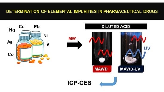
In this study, two methods based on the use of diluted acids were developed: microwave-assisted wet digestion (MAWD) and microwave-assisted ultraviolet digestion (MAWD-UV). These methods are evaluated for the digestion of oral pharmaceutical drugs and further determination of elemental impurities from classes 1 (As, Cd, Hg and Pb) and 2A (Co, Ni and V) by inductively coupled plasma optical emission spectrometry (ICP-OES). Commercial drugs for the treatment of type 2 diabetes are used. No prior comminution is performed. For MAWD, the optimized conditions were 2 mol L−1 or 3 mol L−1 HNO3, 1 mL of 50% H2O2 and a 45 min or 55 min irradiation program. For MAWD-UV, the condition using 1 mol L−1 HNO3, 1.6 mL of 50% H2O2 and a 55 min irradiation program enabled the digestion of all samples. In this way, efficient methods are proposed for the digestion of commercial pharmaceutical tablets for further determination of class 1 and 2A elemental impurities (ICH Q3D guidelines).

Graphical Abstract

1. Introduction

In recent years, an increasing interest in the determination of elemental impurities in pharmaceutical products has been observed in the literature, pushed by stricter limits introduced by several documents, such as the ICH Q3D guidelines [1] and United States Pharmacopeia (USP) chapters 232 and 233, implemented in 2018 [2,3]. Additionally, these impurities have been separated in three classes (1, 2A and 2B, and 3) by ICH Q3D guidelines, according to their toxicity and probability of occurrence in the pharmaceutical product [1]. Class 1 is composed by As, Cd, Hg and Pb, which are toxic and relatively abundant [4]. Class 2 elements are those whose toxicity is route dependent, and are subdivided into class 2A (Co, Ni and V) and class 2B (Ag, Au, Ir, Os, Pd, Pt, Rh, Ru, Se and Tl). Class 2A contains some abundant elements that should be monitored, while class 2B elements are less likely to be present in the product and should be monitored only in case of intentional addition. Lastly, elements Ba, Cr, Cu, Li, Mo, Sb and Sn form class 3, presenting low oral toxicity, but should be monitored in parenteral and inhalational routes of administration [1]. Out of these, classes 1 and 2A should be monitored through all routes of exposure, also being the most critical for the oral route. Aside from the toxicological aspect, the control of elemental impurities is also useful for the identification of possible faults in the production process and storage of pharmaceutical products [5].
When considering the continuous or prolonged use drugs, a more thorough quality control is important to avert acute and/or chronic issues that could be caused by the presence of elemental impurities [1]. This is the case for drugs used in the control of type 2 diabetes, which is a disease caused by insulin resistance in peripheral organs and pancreatic β-cell dysfunction. In 2017, approximately 8.8% of the world population was estimated to be affected by either type 1 or 2 diabetes [6,7,8,9]. Although an improvement in lifestyle and diet can help to attenuate the effects of this disease, it is also necessary to administrate continuous use oral drugs, such as metformin hydrochloride, which reduces glucose production in the liver and enhances its sensitivity to insulin [9].
In this regard, researchers have developed a variety of sample preparation methods coupled to sensitive detection techniques in order to improve detectability of elemental impurities. Plasma-based techniques, such as inductively coupled plasma optic emission spectrometry (ICP-OES) [10,11,12,13,14] and inductively coupled plasma mass spectrometry (ICP-MS) [11,13,15,16,17,18,19,20,21], are frequently used with this purpose, due to their high sensitivity, multielemental capacity and wide linear range [5,22].
Since plasma-based techniques, in their traditional assembly, require sample introduction in liquid form, a previous sample decomposition process is usually necessary when analyzing oral drugs, such as those used in the treatment of type 2 diabetes. However, it is important to mention that solutions containing high acidity, easy to ionize elements or carbon content can cause spectral interferences and/or matrix effects in these techniques, especially in ICP-MS [23,24,25,26]. Hence, it is often necessary to perform a pretreatment of the samples prior to the detection step to minimize possible interferences [5].
Several sample preparation methods have been developed aiming for elemental impurities determination in pharmaceutical ingredients, such as microwave-assisted wet digestion (MAWD) [10,12,13,18,19,27], microwave-induced combustion (MIC) [15], dissolution [16] and MAWD with single reaction chamber (MAWD-SRC) [20,28]. Out of these, MAWD is usually the method of choice, as modern microwave equipment enables precise control of temperature and pressure inside digestion vessels, as well as provide the direct heating of the samples [29]. However, concentrated oxidants and acids are usually employed in MAWD procedures, which can result in a high volume of residue, also being associated with higher blank values [30,31]. Additionally, high acidity or carbon content could lead to interferences in the determination step.
In this sense, in an attempt to minimize the drawbacks and environmental impact of MAWD, greener methods using milder conditions have been developed in the past years [30,31]. In particular, MAWD using diluted acid could be an interesting alternative for the decomposition of pharmaceutical drugs for further elemental impurities determination and has been successfully used for biological and botanical samples [30,32,33,34]. This method usually employs diluted nitric acid and a source of oxygen (H2O2 or O2 pressurization) [30,31]. The oxygen provided to the system regenerates the acid by reacting with the nitrogen oxides formed during sample decomposition, which in term makes it possible to reduce reagent waste while maintaining digestion efficiency [30,31].
In addition to MAWD using diluted acid, the microwave-assisted ultraviolet digestion (MAWD-UV) method could potentially improve the digestion efficiency of diluted acid in samples containing molecules with π bonds, such as aromatic rings [35]. In this method, the microwaves activate an UV lamp placed inside the reaction vessel. The UV radiation then acts as a catalyst for the oxidant reagents (usually HNO3 and H2O2), forming highly reactive •OH radicals, which can accelerate degradation of organic molecules [35]. This method has been applied to several matrices, such as electric and electronic polymeric waste [36], foods [37], seaweed [38,39,40], crude oil [41,42] and dyes [43].
It is important to mention as well that most published studies focus on the decomposition of active pharmaceutical ingredients (API). Only a few studies were found in the literature on the digestion of the commercial pharmaceutical drug, and even less when inorganic excipients are present in the formulation [10,12,18,28,44]. In this sense, the aim of this study is to develop sample preparation methods for oral drugs used in the treatment of type 2 diabetes and the further determination of class 1 and 2A elemental impurities. The proposed methods are developed with the aim of reducing reagent consumption and waste generation, as well as using milder digestion conditions while maintaining digestion efficiency.

2. Materials and Methods

2.1. Instrumentation

Both MAWD and MAWD-UV methods were carried out using a Multiwave 3000 sample preparation system (Anton Paar, Graz, Austria), equipped with an 8XQ80 rotor and eight 80 mL quartz vessels. For the MAWD-UV procedures, low pressure Cd electrodeless discharge UV lamps purged with Ar (part number 16847, Anton Paar), with emission in 228 nm and 326 nm, were used. These lamps were anchored inside the digestion vessels using polytetrafluoroethylene (PTFE) holders supplied by the manufacturer. The maximum operating pressure and temperature were set according to the manufacturer (80 bar and 280 °C for MAWD, or 250 °C for MAWD-UV), so as to not cause damage to the equipment.
The determination of class 1 and 2A elemental impurities, major elements and dissolved carbon in the digests was carried out by ICP-OES, using a Spectro Ciros CCD spectrometer (Spectro Analytical Instruments, Kleve, Germany) with sealed optic, in axial view. Argon 99.996% (Air Liquide S.A., São Paulo, Brazil) was used for plasma generation, nebulization and as auxiliary gas for the ICP-OES instrument. The instrumental conditions are described in Table 1. Emission lines were selected based on abundancy, sensitivity and possible interferences. It is important to mention that carbonaceous gases were removed prior to C determination by purging the digests and calibration solutions with Ar for 2 min at 0.1 L min−2.
The determination of residual acidity in the digests was carried out using an automatic titrator (Titrando 836, Metrohm, Herisau, Switzerland) equipped with a magnetic stirrer (803 Ti Stand, Metrohm), a 20 mL burette (Dosino 800, Metrohm) and a combined glass pH electrode for aqueous medium (LL Ectrode Plus, 6.0262.100, Metrohm). Weighing procedures were performed using an analytical scale with 0.0001 g of resolution and maximum load of 220 g (AY220, Shimadzu, Barueri, Brazil).

2.2. Samples, Reagents and Standards

Ultrapure water with resistivity of 18.2 MΩ cm (Millipore, Burlington, USA) was used for the preparation of solutions and reagents. The nitric acid (65%, 1.4 kg L−1, Sigma Aldrich, USA) and hydrochloric acid (37%, 1.19 kg L−1, Merck, Darmstadt, Germany) used in this study were previously distilled in a sub-boiling system (duoPUR, Milestone, Sorisole, Italy), except for decontamination procedures, for which 65% HNO3 was used. Hydrogen peroxide (50%, 1.14 kg L−1, Vetec, Rio de Janeiro, Brazil) was used in MAWD and MAWD-UV procedures.
For standard addition experiments, a stock solution with varied concentrations of the analytes was prepared from the dilution of As, Cd, Co, Hg, Ni, Pb and V monoelementar reference solutions (1000 mg L−1, Merck) in water. Calibration solutions for As, Cd, Pb and V (1 to 100 µg L−1 in 5% inversed aqua regia), and for Ca, Co, Fe, K, Mg, Na and Ni (10 to 500 µg L−1 in 5% inversed aqua regia) were prepared by sequential dilution of a stock standard solution (10 mg L−1, SCP33MS, SCP Science, Quebec, Canada). Mercury calibration solutions (1 to 100 µg L−1 in 5% inversed aqua regia) were prepared by sequential dilution of a monoelementar reference solution (1000 mg L−1, Merck). Citric acid (Vetec) was dissolved in 5% HNO3 to obtain a standard 1000 mg L−1 carbon stock solution, which was sequentially diluted to obtain the C calibration solutions (10 to 500 mg L−1). Yttrium was used as internal standard for C determination. For this, a Y monoelementar reference solution (1000 mg L−1 in 2% HNO3, Spex, Metuchen, USA) was added to both digests and calibration solutions (final concentration of 1 mg L−1). The titrant used for determination of residual acidity of the digests was prepared by dissolution of KOH (Merck) in water to obtain a 0.1 mol L−1 solution, which was standardized using potassium hydrogen phthalate (C8H5KO4, Merck).
Six types of continuous use oral drugs applied for the treatment of type 2 diabetes were purchased in local drugstores. The composition of samples is presented in Table 2. Samples were obtained as tablets and no previous comminution was performed prior to digestion. For this, one or more tablets from each type of sample were used in order to reach a sample mass of approximately 500 mg (ranging from 420 to 600 mg). The relative API content of the samples was calculated as the ratio between mean tablet mass and API dose multiplied by 100. Metformin hydrochloride (MET) and canagliflozin (CANA) samples were arbitrarily chosen for the optimization of the MAWD and MAWD-UV methods. It is important to notice that, apart from MET, all of the APIs contain aromatic groups (Figure S1, Supplementary Materials). Additionally, some of the rings are bonded to deactivating groups such as –COOH (repaglinide, REPA), –Cl (glibenclamide, GLIB) and –F (CANA and sitagliptin phosphate, SITA), which confer stability to the molecule, potentially making it more resistant to the digestion procedure.

2.3. Sample Preparation Methods

2.3.1. MAWD Method

For the MAWD procedures, 1-to-5 sample tablets (Table 2) were inserted into the digestion vessels, followed by the addition of 6 mL of digesting solution. Afterwards, the vessels were closed and inserted into the rotor, which was capped and submitted to the microwave irradiation program. After cooling, the rotor was removed from the microwave oven and pressure inside the vessels was carefully released. Digests were transferred to 50 mL polypropylene (PP) vessels and diluted to 25 mL with water. The efficiency of digestion was evaluated by the dissolved carbon content of the digests. Residual acidity and recovery of the analytes were also evaluated [41].

2.3.2. Evaluation of Digesting Solution

Nitric acid in the concentrations of 1, 2, 3, 7 and 14.4 mol L−1 was evaluated as digesting solutions. For these experiments, 1 mL of 50% H2O2 was used as auxiliary reagent (except for the 14.4 mol L−1 HNO3 condition, for which H2O2 was not added in order not to dilute the acid). It is important to mention that the solution volume and HNO3 concentration inside digestion vessels were not altered with the H2O2 addition, since the added volume was taken into account for the preparation of the diluted HNO3 solutions.

2.3.3. Evaluation of the Irradiation Program

Two irradiation programs were evaluated for the MAWD method. At first, an irradiation program adapted from a previous work [33], named program 1, was used for the digestion of MET samples (Table 2). This program consisted of three steps: (i) a 5 min ramp to 1000 W, (ii) 10 min of irradiation at 1000 W and (iii) 20 min at 0 W (cooling step). Afterwards, a longer irradiation program (program 2) was evaluated for CANA samples, consisting of: (i) a 10 min ramp to 1000 W, (ii) 15 min of irradiation at 1000 W and (iii) 20 min at 0 W (cooling step).

2.3.4. Evaluation of Simultaneous Cooling during Irradiation

Previous studies have found that simultaneous cooling during microwave irradiation can favor condensation inside the digestion vessels [33]. This could in turn reduce internal pressure and enable longer irradiation times at maximum power in systems where power is regulated by vessel temperature and pressure, such as Multiwave 3000. Additionally, dislocating the equilibrium towards the liquid phase could favor the acid regeneration reactions, enhancing digestion efficiency [33]. Hence, two ventilation levels were evaluated for MAWD procedures: an air flow of 60 m3 h−1 (FAN 1), conventionally employed in this system, and a higher air flow, of 125 m3 h−1 (FAN 2). For this evaluation, 6 mL of a 2 mol L−1 HNO3 solution containing 1 mL of 50% H2O2 was used as digestion solution, and irradiation program 1 was applied.

2.3.5. Evaluation of the Auxiliary Reagent

Hydrogen peroxide was evaluated as a source of O2 for the regeneration of diluted HNO3 in MAWD procedures [31]. For this, either 1 or 2 mL of 50% H2O2 were added to the digestion solution, which were equivalent to final concentrations of 8.3 and 16.7% in the digestion vessel, respectively. This evaluation was carried out using 2 mol L−1 HNO3 and irradiation program 1.

2.4. MAWD-UV Method

For the MAWD-UV method, 1-to-5 sample tablets (Table 2) were inserted into the quartz vessels containing the bottom PTFE holder for the UV lamp and the digesting solution. Afterwards, the UV lamp, already equipped with the top PTFE holder, was placed inside the vessels using a quartz rod. A higher digesting solution volume, of 10 mL, was used for all MAWD-UV procedures in order to maximize contact with the lamp bulb and enhance the UV radiation effects. After placement of the UV lamps, the vessels were closed and fixated in the rotor, which was capped and inserted in the microwave equipment. The irradiation program for MAWD-UV was adapted from program 2, as the manufacturer recommends heating ramps with duration of 10 to 15 min [45]. The steps of program 2 were maintained; however, the higher air flow, FAN 2, was used during the whole procedure. This was performed in order to keep irradiation at maximum power for longer, due to the intensity of the UV emission being dependent of the applied microwave power [35,41]. After the decomposition procedures, the digests were transferred to 50 mL PP vessels and diluted to 25 mL with water.

Evaluation of MAWD-UV Experimental Parameters

The digestion conditions for MAWD-UV were evaluated taking into consideration the conditions in which a complete digestion was not observed for MAWD using only diluted acid. For this, MET samples were used and 1 mol L−1 HNO3 was evaluated as digesting solution. Afterwards, the addition of 1.6 or 3.2 mL of 50% H2O2 was evaluated as auxiliary reagent. The addition of a higher H2O2 volume was carried out to obtain the same concentrations evaluated for MAWD (final concentration of 8.3 or 16.7% in the digestion vessels, respectively). The most adequate condition was chosen based on dissolved carbon and residual acidity of the digests.
A summary of all evaluated experimental conditions for both MAWD and MAWD-UV methods is shown in Figure 1.

2.5. Statistical Treatments

For statistical calculations, GraphPad InStat Software (GraphPad InStat Software Inc., San Diego, USA, Version 3.00, 1997) was used to carry out two-way ANOVA and Student’s t-test analyses at a confidence level of 95%.

3. Results and Discussion

3.1. Evaluation of Digesting Solution for MAWD

The digestion of MET samples using 14.4 mol L−1 HNO3 was carried out in order to establish reference values for dissolved carbon and residual acidity. With the 50% H2O2 volume fixed as 1 mL and using irradiation program 1, different HNO3 concentrations were evaluated. When MET digestion was carried out using 2, 3 or 14.4 mol L−1 HNO3, clear and transparent digests were obtained. However, digestion using 1 mol L−1 HNO3 was incomplete, resulting in a yellowish and cloudy digest with non-decomposed residual sample. An intermediate condition using 1.5 mol L−1 HNO3 was also evaluated. However, digestion was also incomplete when using this condition. For all procedures, the presence of an insoluble white solid, corresponding to the SiO2 excipient of the MET sample, was observed in digests.
As can be expected, high residual acidity (55 ± 2%) and low dissolved carbon content (<25 mg L−1) were observed when 14.4 mol L−1 HNO3 was used. This is due to the excess of HNO3 in the oxidation reaction. A high residual acidity was also observed when 3 mol L−1 was used (36 ± 1%), due to the added H2O2, which could regenerate the acid during the decomposition. It is important to mention that, even though residual acidity was high for this condition, in this case this is not detrimental to the detection technique, since the initial HNO3 concentration was already low (final concentration around 7.8%). Rather, it indicated the success in regenerating the acid by the reaction of H2O2 with the NO generated during digestion [30]. Additionally, the dissolved carbon content for this condition was also low, 56.0 ± 13.2 mg L−1. When 2 mol L−1 HNO3 was used, a higher dissolved carbon content (2000 ± 130 mg L−1) was observed. The residual acidity was 12 ± 2% for this condition, indicating a higher H2O2 consumption as well. The carbon concentration obtained for this condition is in accordance with the values reported in the literature for the decomposition of metformin using 2 mol L−1 HNO3 and the more extreme temperature and pressure conditions of MAWD with single reaction cell (MAWD-SRC) [28].
In a previous study [26], it was observed that up to 8 g L−1 of dissolved carbon could be present in solutions without major influences in element determination by ICP-OES. This information agrees to that observed for standard addition experiments in the evaluated conditions. Recoveries between 95 and 108% were observed for all analytes, even for the condition with higher dissolved carbon. As the carbon concentration and residual acidity did not impair the analysis, and analyte recoveries were quantitative, the condition in which 2 mol L−1 HNO3 was used was selected for the decomposition of the other samples. The residual acidity and dissolved carbon content of the digests can be observed in Figure 2. It is important to mention that particulate matter, identified as the inorganic fraction of the samples (Table 2), not decomposed by MAWD, was present in digests from CANA, MET, REPA and SITA samples.
As can be seen in Figure 2, the use of 2 mol L−1 HNO3 was adequate for all samples, apart from CANA, for which the digests presented a strong yellowish color and non-decomposed residue. Samples GLIB, REPA and SITA, which had lower relative API content (4, 2 and 19%, respectively), presented the lowest carbon contents and highest residual acidities. For the PIO sample, although its relative API content is similar to SITA (23%), the observed carbon concentration was considerably higher. This was most probably due to the absence of inorganic excipients in PIO, while SITA presented CaHPO4, talc, TiO2, Fe2O3 and FeO in its composition. Hence, the original carbon content of PIO was higher.
As MET and CANA present the highest relative API content (65 and 48%, respectively) in the tablets, it is understandable that carbon content in the digests of these samples was also higher. It is important to mention as well that the molecular structure canagliflozin is much more stable and complex than metformin, due to the aromatic rings and deactivation effect of F, hence being less susceptible to acid attack [46,47]. In this sense, additional parameters were evaluated for the digestion of CANA samples.

3.2. Evaluation of the Irradiation Program

For the decomposition of CANA samples, the irradiation program 2, which consisted of a longer ramp and permanence in the set power, was evaluated. With this program, a more gradual increase in internal pressure was expected, allowing for continuous irradiation during the ramp, as well as longer permanence in high temperature and pressure. As CANA decomposition was incomplete using 2 mol L−1 HNO3, concentrated HNO3 (14.4 mol L−1) was used for this evaluation, and H2O2 was not added. For both programs 1 and 2, residual acidity was higher than 75%, indicating an excess of acid. However, there was a reduction of 1.5 times in carbon content, from 3310 ± 160 mg L−1 for program 1 to 2230 ± 270 mg L−1 for program 2. This indicates a higher digestion efficiency when the sample was subjected to microwave irradiation for a longer period. Hence, irradiation program 2 was selected for further evaluations.

3.3. Evaluation of Simultaneous Cooling during Irradiation

The use of simultaneous cooling (FAN 2) was evaluated for both CANA and MET samples. For CANA, the experiment was performed using 14.4 mol L−1 HNO3 and irradiation program 2, while for MET, 2 mol L−1 HNO3, 1 mL of 50% H2O2 and irradiation program 1 were used. When comparing conditions using FAN 1 to those using FAN 2, a higher residual acidity was observed for both CANA (from 80 ± 3% for FAN1 to 85 ± 1% for FAN 2) and MET samples (from 12 ± 2% for FAN 1 to 15 ± 1% for FAN 2). However, this increase was not significant. These results are in agreement with the hypothesis that simultaneous cooling favors acid regeneration [33]. However, no significant differences were observed in dissolved carbon content of the digests of both CANA (Student’s t-test, p = 0.232) and MET (Student’s t-test, p = 0.984), indicating that simultaneous cooling did not enhance digestion efficiency, despite the higher residual acidity. Hence, FAN 1 was selected for further evaluations.

3.4. Evaluation of Diluted Acid and Auxiliary Reagent for CANA Decomposition

In order to avoid the high acidity and the need for sample dilution associated to the use of concentrated acid, the use of diluted HNO3 in the concentrations of 3, 7 and 10 mol L−1 was evaluated for CANA using irradiation program 2 and 2 mL of 50% H2O2. Dissolved carbon and residual acidity results for this evaluation are informed in Figure 3A.
As can be seen in Figure 3A, digestion efficiency was not impaired by the use of diluted HNO3. In fact, the opposite was observed, as there was a significant reduction (Student’s t test, p = 0.0075) in carbon content when diluted HNO3 was used in combination with 2 mL of 50% H2O2 in comparison to the digestion using concentrated acid. This can be due to both the acid regeneration and the oxidative action of the H2O2. No significant difference was observed among the conditions using diluted acid, for both dissolved carbon content (ANOVA, p = 0.255) and residual acidity (ANOVA, p = 0.195). Hence, 3 mol L−1 HNO3 was selected for further optimizations.
As residual acidity was high for the chosen condition (almost 90%), the condition using 1 mL of 50% H2O2 was also evaluated for CANA, as there was probably an excess of reagent in the system. For this evaluation, 3 mol L−1 HNO3 and irradiation program 2 were used. Dissolved carbon content and residual acidity for this evaluation are presented in Figure 3B.
With the reduction in H2O2 volume, a residual acidity lower than 50% was achieved, indicating the higher consumption of the oxygen for acid regeneration. Additionally, the lower H2O2 volume did not affect the digestion efficiency in a significant way. For this reason, the condition using 3 mol L−1 HNO3, 1 mL of 50% H2O2 and irradiation program 2 was considered adequate for the digestion of CANA samples. It is important to mention that, when the same conditions were applied using irradiation program 1, incomplete digestion of the sample was observed. Hence, the use of a longer program was of high relevance for the digestion of CANA. For the other samples, the condition using 2 mol L−1 HNO3, 1 mL of 50% H2O2 and irradiation program 1 was considered the most adequate, as there was less reagent consumption and sample preparation time was lower.

3.5. Concentration of Major Elements in the Samples

According to previous works [24,25,26], the presence of easily ionized elements, such as Na, K, Ca and Mg, in concentrations higher than 1 g L−1 could cause a decrease in plasma energy, affecting analyte ionization. This was observed particularly for Pb (220 nm), Co (230 nm) and Ni (231 nm) when 10 g L−1 of Na and Ca were added to standard solutions.
In this sense, after the most adequate conditions for MAWD digestion were selected, characterization of the digests regarding major element composition was performed (Table 3). It is important to mention that determination by ICP-OES could be performed without previous dilution, since residual acidity was low and insoluble solids were deposited at the bottom of the PP vessels.
As can be seen in Table 3, when using the optimized conditions for MAWD with diluted HNO3 and H2O2, the sum of easily ionized element concentrations was lower than 150 mg L−1 for all samples analyzed in this study. As this concentration is much lower than what is reported to be problematic in the literature, there was no need to perform further experiments in regard to interferences caused by these elements.

3.6. Evaluation of MAWD-UV

The MAWD-UV method was first evaluated for the digestion of MET samples using 1 mol L−1 HNO3 (for which the MAWD method was unsuccessful). For this procedure, 1.6 mL of 50% H2O2, 10 mL of digesting solution and irradiation program 2 with simultaneous cooling were used. It is important to mention that MAWD was also performed using 10 mL of digesting solution and irradiation program 2 with simultaneous cooling, in order to check the influence of solution volume in digestion efficiency. However, results showed no statistical difference compared to MAWD using 6 mL of digesting solution (Student’s t-test, p = 0.2435) and irradiation program 1.
With MAWD-UV, the digestion of MET samples was achieved, confirming that the use of UV radiation during the heating step enhances digestion efficiency. When comparing the addition of different volumes of 50% H2O2, no significant difference was observed in the dissolved carbon content of the digests (Student’s t-test, p = 0.058). Hence, the digestion by MAWD-UV using 1.6 mL of 50% H2O2 and 1 mol L−1 HNO3 was considered suitable and was applied for the remaining samples. Dissolved carbon content and residual acidity of the digests using the optimized conditions are expressed in Figure 4.
It is possible to observe in Figure 4 that the use of the MAWD-UV method was successful in digesting all samples using 1 mol L−1 HNO3, resulting in a relatively low carbon content. It is worthy of mention that digestion was efficient even for CANA samples, which could only be decomposed using 3 mol L−1 HNO3 when applying the MAWD method. Thus, MAWD-UV could be considered as a promising alternative to decompose more complex oral drugs.

3.7. Analytical Figures of Merit of the Proposed Methods

Standard addition experiments were performed during the development of the proposed methods, in order to evaluate the accuracy. The specified permitted daily dose (PDE) of each analyte was used to define the concentration of the standard solution, and the oral drug with the higher daily dose (2.5 g day−1, MET) was selected for the calculation of the PDE in concentration [1]. The calculation of the stock solution concentration and added volume also took the sample mass (500 mg) and final digest volume (25 mL) into consideration. Hence, 100 µL of the stock solution containing As (30 mg L−1), Cd (10 mg L−1), Co (100 mg L−1), Hg (60 mg L−1), Ni (400 mg L−1), Pb (10 mg L−1) and V (200 mg L−1) was added onto sample tablets prior to digestion procedures.
The results of standard addition experiments in optimized conditions of the proposed MAWD and MAWD-UV methods are shown in Table S1. In brief, analyte recoveries ranged between 90 and 110% for samples, for both digestion methods. It is important to notice that standard additions were performed during the determination step as well to check for matrix interferences, and eventual dilutions were in agreement with each other. Hence, it can be inferred that the carbon content, residual acidity and major element composition in the digests did not affect the accuracy of the method.
Additionally, the accuracy of the optimized conditions was evaluated using certified reference materials (CRMs). Due to the lack of certified materials with a chemical composition similar to the pharmaceutical drugs used in this study, a CRM addition was performed to the sample matrix (GLIB sample was randomly chosen for this experiment). For this procedure, the sample tablets were ground using an agate mortar and pestle. Then, 250 mg of sample was mixed with 250 mg of biological or botanical CRM and the mixture was pressed as pellets in a hydraulic press with 1 ton for 1 min. The CRMs used consisted of dogfish liver (DOLT-4, National Research Council of Canada, Canada), lobster hepatopancreas (TORT-2, National Research Council of Canada) and an aquatic plant (BCR 60, Community Bureau of Reference, Belgium).
The results obtained for this experiment, for both MAWD and MAWD-UV, are shown in Table 4. For MAWD, 500 mg of sample (250 mg of GLIB mixed with 250 mg of CRM), 2 mol L−1 HNO3, 1 mL of 50% H2O2 and irradiation program 1 were used, while for MAWD-UV, 500 mg of sample (250 mg of GLIB mixed with 250 mg of CRM) 1 mol L−1 HNO3, 1.6 mL of 50% H2O2 and irradiation program 2 with simultaneous cooling were applied.
When comparing the obtained and certified values of the CRMs, no significant difference was found, for either MAWD or MAWD-UV, for all analytes, with the exception of V in CRM BCR 60. In this case, the obtained value was significantly lower than the certified value (Student’s t-test, p = 0.0017 for MAWD and p = 0.0005 for MAWD-UV). It is important to mention, however, that the V value in this CRM is not certified, with no information regarding the associated uncertainty being available. Additionally, V values obtained for the DOLT-4 and TORT-2 CRMs were acceptable when both methods were applied. Hence, both the proposed MAWD and MAWD-UV methods were considered suitable for the digestion of oral drugs used in the treatment of type 2 diabetes for the further determination of classes 1 and 2A elemental impurities by ICP-OES. Additionally, the RSD for the measurements was equal to or lower than 10% for all analytes.
For the calculation of LOQs, 10 consecutive measurements of digestion blanks were carried out. The standard deviation of these measurements was multiplied by 10, and added to the mean blank value, taking the sample mass (approximately 500 mg) and final digest volume (25 mL) into consideration. Correlation coefficients (r) of the calibration curves were used to express linearity, with a minimum acceptable r of 0.995 being considered. The LOQs obtained for the proposed MAWD and MAWD-UV methods are shown in Table 5. Both methods presented very similar LOQs and, when considering the limits established by ICH Q3D guidelines, the obtained LOQs were at least two times lower than the PDEs for all analytes. Hence, it was possible to carry out the determination of classes 1 and 2A elemental impurities in the oral drug samples in order to meet the ICH Q3D guidelines criteria.
The digestion of the samples was also performed without the addition of standard solutions, and the concentrations of the analytes were below the LOQs of the methods. Hence, it was verified that the final products analyzed in this study were in accordance to the ICH Q3D guidelines, being virtually free of class 1 and 2A elemental impurities.
With the use of diluted HNO3 and H2O2 as digesting solution, it was possible to minimize reagent consumption, as well as the risks associated with the use of concentrated acids. Additionally, both the proposed MAWD and MAWD-UV methods were efficient in decomposing the organic matrix of the oral drug samples, without prior comminution of the tablets.
It is important to mention that it was not possible to digest all samples using the mildest conditions when employing the MAWD method. In this case, it was necessary to use a different condition for CANA samples (3 mol L−1 HNO3 and irradiation program 2). For the MAWD-UV method, on the other hand, all samples could be efficiently digested using milder conditions (1 mol L−1 HNO3). In fact, the MAWD-UV method was more efficient in the digestion of samples containing a higher relative API content, especially those containing unsaturated and aromatic functional groups. However, for simpler matrices, both MAWD and MAWD-UV methods present good digestion efficiency, hence MAWD could be used due to its lower cost of application.

4. Conclusions

With the proposed MAWD and MAWD-UV methods, it was possible to efficiently digest oral pharmaceutical drugs used in the treatment of type 2 diabetes for the further determination of class 1 and 2A elemental impurities by ICP-OES. It should be highlighted that diluted HNO3 and H2O2 were used as digesting solution for both methods, enabling low blank values, and relatively low residual acidity and dissolved carbon content, especially for MAWD-UV. The MAWD-UV method enabled the digestion of up to 600 mg of sample (Table 2), without prior comminution, using only 10 mL of 1 mol L−1 HNO3 containing 1.6 mL of 50% H2O2 as digesting solution, and a total preparation time of 55 min. For the MAWD method, on the other hand, it was possible to digest the same amount of sample using only 6 mL of 2 mol L−1 HNO3 containing 1.0 mL of 50% H2O2 as digesting solution in a total preparation time of 45 min (in the case of CANA samples, 6 mL of 3 mol L−1 HNO3 containing 1.0 mL of 50% H2O2 as digesting solution and preparation time of 55 min).
Quantitative recoveries were obtained for both methods after standard addition experiments, and the experimental values found after the digestion of botanical and biological CRMs were in agreement with certified values. Therefore, the present study enabled the digestion of oral pharmaceutical drugs using mild conditions and relatively fast procedures (< 1 h). Finally, it is important to mention that these methods were developed using diluted reagents, reducing the risks associated with concentrated acids, as well as possible interferences. Considering large scale applications and green chemistry principles, the proposed methods could also help to minimize the impact of analytical procedures used in the quality control of pharmaceutical products. This was possible by reducing reagent use and laboratory effluent generation.

Supplementary Materials

The following Supplementary Materials can be downloaded at: https://www.mdpi.com/article/10.3390/su14010422/s1: Figure S1: Molecular structure of the APIs contained in the oral drugs used in this study: (A) canagliflozin, (B) glibenclamide, (C) metformin hydrochlorate, (D) pioglitazone hydrochloride, (E) repaglinide, and (F) sitagliptin phosphate; Table S1: Analyte recovery after oral drug samples digestion by MAWD and MAWD-UV using optimized conditions (values in percentage, mean ± standard deviation, n = 3).

Author Contributions

Conceptualization, R.S.P. and E.M.M.F.; methodology and analysis, V.H.C.; writing—original draft preparation, V.H.C. and A.S.H.; writing—review and editing, M.F.M., E.I.M. and E.M.M.F.; visualization, M.F.M., E.I.M. and R.S.P.; supervision, E.M.M.F.; project administration, E.M.M.F.; funding acquisition, E.M.M.F. All authors have read and agreed to the published version of the manuscript.

Funding

This research was funded by Conselho Nacional de Desenvolvimento Científico e Tecnológico (CNPq), Instituto Nacional de Ciência e Tecnologia de Bioanalítica (INCTBio, Proc. No. 573672/2008-3).

Institutional Review Board Statement

Not applicable.

Informed Consent Statement

Not applicable.

Data Availability Statement

Not applicable.

Acknowledgments

The authors are grateful to Conselho Nacional de Desenvolvimento Científico e Tecnológico (CNPq), INCTBio and Coordenação de Aperfeiçoamento de Pessoal de Nível Superior (CAPES) for supporting this study.

Conflicts of Interest

The authors declare no conflict of interest.

References

  1. ICH Guideline for Elemental Impurities Q3D(R1). Available online: https://www.ich.org/products/guidelines/quality/quality-single/article/guideline-for-elemental-impurities-copy-1.html (accessed on 7 June 2021).
  2. Elemental impurities–procedures. In The United States Pharmacopeia, 40th ed.; United States Pharmacopeial: Rockville, MD, USA, 2017.
  3. Elemental impurities–limits. In The United States Pharmacopeia, 40th ed.; United States Pharmacopeial: Rockville, MD, USA, 2017.
  4. Peixoto, N.C.; Serafim, M.A.; Flores, E.M.M.; Bebianno, M.J.; Pereira, M.E. Metallothionein, zinc, and mercury levels in tissues of young rats exposed to zinc and subsequently to mercury. Life Sci. 2007, 81, 1264–1271. [Google Scholar] [CrossRef] [PubMed]
  5. Barin, J.S.; Mello, P.A.; Mesko, M.F.; Duarte, F.A.; Flores, E.M.M. Determination of elemental impurities in pharmaceutical products and related matrices by ICP-based methods: A review. Anal. Bioanal. Chem. 2016, 408, 4547–4566. [Google Scholar] [CrossRef] [PubMed]
  6. Chatterjee, S.; Khunti, K.; Davies, M.J. Type 2 diabetes. Lancet 2017, 389, 2239–2251. [Google Scholar] [CrossRef]
  7. Cho, N.H.; Shaw, J.E.; Karuranga, S.; Huang, Y.; Fernandes, J.D.R.; Ohlrogge, A.W.; Malanda, B. IDF Diabetes Atlas: Global estimates of diabetes prevalence for 2017 and projections for 2045. Diabetes Res. Clin. Pract. 2018, 138, 271–281. [Google Scholar] [CrossRef]
  8. Kahn, C.R. Insulin action, diabetogenes, and the cause of type II diabetes. Diabetes 1994, 43, 1066–1085. [Google Scholar] [CrossRef]
  9. Stumvoll, M.; Goldstein, B.J.; Haeften, T.W. Type 2 diabetes: Principles of pathogenesis and therapy. Lancet 2005, 365, 1333–1346. [Google Scholar] [CrossRef]
  10. Menoutis, J.; Parisi, A.; Verma, N. Study of the use of axial viewed inductively coupled plasma atomic emission spectrometry with ultrasonic nebulization for the determination of select elemental impurities in oral drug products. J. Pharm. Biomed. 2018, 152, 12–16. [Google Scholar] [CrossRef]
  11. Silva, C.S.; Pinheiro, F.C.; Amaral, C.D.B.; Nobrega, J.A. Determination of As, Cd, Hg and Pb in continuous use drugs and excipients by plasma-based techniques in compliance with the United States Pharmacopeia requirements. Spectrochim. Acta Part B At. Spectrosc. 2017, 138, 14–17. [Google Scholar] [CrossRef]
  12. Støving, C.; Jensen, H.; Gammelgaard, B.; Stürup, S. Development and validation of an ICP-OES method for quantitation of elemental impurities in tablets according to coming US pharmacopeia chapters. J. Pharm. Biomed. 2013, 84, 209–214. [Google Scholar] [CrossRef]
  13. Wollein, U.; Bauer, B.; Habernegg, R.; Schramek, N. Potential metal impurities in active pharmaceutical substances and finished medicinal products—A market surveillance study. Eur. J. Pharm. Sci. 2015, 77, 100–105. [Google Scholar] [CrossRef]
  14. Nunes, T.S.; Muller, C.C.; Balestrin, P.; Muller, A.L.H.; Mesko, M.F.; Mello, P.A.; Muller, E.I. Determination of chlorine and sulfur in high purity flexible graphite using ion chromatography (IC) and inductively coupled plasma optical emission spectrometry (ICP OES) after pyrohydrolysis sample preparation. Anal. Methods 2015, 7, 2129–2134. [Google Scholar] [CrossRef]
  15. Barin, J.S.; Tischer, B.; Picoloto, R.S.; Antes, F.G.; Silva, F.E.B.; Paula, F.R.; Flores, E.M.M. Determination of toxic elements in tricyclic active pharmaceutical ingredients by ICP-MS: A critical study of digestion methods. J. Anal. At. Spectrom. 2014, 29, 352–358. [Google Scholar] [CrossRef]
  16. Chahrour, O.; Malone, J.; Collins, M.; Salmon, V.; Greenan, C.; Bombardier, A.; Ma, Z.; Dunwoody, N. Development and validation of an ICP-MS method for the determination of elemental impurities in TP-6076 active pharmaceutical ingredient (API) according to USP. J. Pharm. Biomed. 2017, 145, 84–90. [Google Scholar] [CrossRef]
  17. Fischer, L.; Zipfel, B.; Koellensperger, G.; Kovac, J.; Bilz, S.; Kunkel, A.; Venzago, C.; Hann, S. Flow injection combined with ICP-MS for accurate high throughput analysis of elemental impurities in pharmaceutical products according to USP. J. Pharm. Biomed. 2014, 95, 121–129. [Google Scholar] [CrossRef]
  18. Gonzalez, M.H.; Silva, C.S.; Amaral, C.D.B.; Bianchi, S.R.; Oliveira, L.H.B.; Coelho, J.S.; Oliveira, A.; Nogueira, A.R.A. Determination of Elemental Impurities in Acyclovir Ointment and Raw Materials Using Microwave Acid Digestion (MW-AD) and ICP-MS. J. Braz. Chem. Soc. 2017, 28, 98–105. [Google Scholar] [CrossRef]
  19. Li, G.; Schoneker, D.; Ulman, K.L.; Sturm, J.J.; Thackery, L.M.; Kauffman, J.F. Elemental impurities in pharmaceutical excipients. J. Pharm. Sci. 2015, 104, 4197–4206. [Google Scholar] [CrossRef] [Green Version]
  20. Muller, A.L.H.; Oliveira, J.S.S.; Mello, P.A.; Muller, E.I.; Flores, E.M.M. Study and determination of elemental impurities by ICP-MS in active pharmaceutical ingredients using single reaction chamber digestion in compliance with USP requirements. Talanta 2015, 136, 161–169. [Google Scholar] [CrossRef]
  21. Barin, J.S.; Pereira, J.S.; Mello, P.A.; Knorr, C.L.; Moraes, D.P.; Mesko, M.F.; Nóbrega, J.A.; Korn, M.G.; Flores, E.M. Focused microwave-induced combustion for digestion of botanical samples and metals determination by ICP OES and ICP-MS. Talanta 2012, 94, 308–314. [Google Scholar] [CrossRef]
  22. Hill, S.J. Inductively Coupled Plasma Spectrometry and Its Applications, 2nd ed.; Blackwell Publishing: Oxford, UK, 2007; p. 446. [Google Scholar]
  23. Grindlay, G.; Mora, J.; Loos-Vollebregt, M.; Vanhaecke, F. A systematic study on the influence of carbon on the behavior of hard-to-ionize elements in inductively coupled plasma–mass spectrometry. Spectrochim. Acta Part B At. Spectrosc. 2013, 86, 42–49. [Google Scholar] [CrossRef]
  24. Stepan, M.; Musil, P.; Poussel, E.; Mermet, J.M. Matrix-induced shift effects in axially viewed inductively coupled plasma atomic emission spectrometry. Spectrochim. Acta Part B At. Spectrosc. 2001, 56, 443–453. [Google Scholar] [CrossRef]
  25. Todolí, J.L.; Gras, L.; Hernandis, V.; Mora, J. Elemental matrix effects in ICP-AES. J. Anal. At. Spectrom. 2002, 17, 142–169. [Google Scholar] [CrossRef]
  26. Wiltsche, H.; Winkler, M.; Tirk, P. Matrix effects of carbon and bromine in inductively coupled plasma optical emission spectrometry. J. Anal. At. Spectrom. 2015, 30, 2223–2234. [Google Scholar] [CrossRef] [Green Version]
  27. Tu, Q.; Wang, T.; Antonucci, V. High-efficiency sample preparation with dimethylformamide for multi-element determination in pharmaceutical materials by ICP-AES. J. Pharm. Biomed. 2010, 52, 311–315. [Google Scholar] [CrossRef]
  28. Pinheiro, F.C.; Barros, A.I.; Nobrega, J.A. Microwave-assisted sample preparation of medicines for determination of elemental impurities in compliance with United States Pharmacopeia: How simple can it be? Anal. Chim. Acta 2019, 1065, 1–11. [Google Scholar] [CrossRef]
  29. Müller, E.I.; Mesko, M.F.; Moraes, D.P.; Korn, M.d.G.A.; Flores, E.M.M. Chapter 4—Wet Digestion Using Microwave Heating. In Microwave-Assisted Sample Preparation for Trace Element Analysis; Flores, E.M.M., Ed.; Elsevier: Amsterdam, The Netherlands, 2014; pp. 99–142. [Google Scholar]
  30. Bizzi, C.A.; Flores, E.L.M.; Nobrega, J.A.; Oliveira, J.S.S.; Schmidt, L.; Mortari, S.R. Evaluation of a digestion procedure based on the use of diluted nitric acid solutions and H2O2 for the multielement determination of whole milk powder and bovine liver by ICP-based techniques. J. Anal. At. Spectrom. 2014, 29, 332–338. [Google Scholar] [CrossRef]
  31. Bizzi, C.A.; Nóbrega, J.A.; Barin, J.S. Chapter 6—Diluted Acids in Microwave-Assisted Wet Digestion. In Microwave-Assisted Sample Preparation for Trace Element Analysis; Flores, E.M.M., Ed.; Elsevier: Amsterdam, The Netherlands, 2014; pp. 179–204. [Google Scholar]
  32. Bizzi, C.A.; Barin, J.S.; Muller, E.I.; Schmidt, L.; Nobrega, J.A.; Flores, E.M.M. Evaluation of oxygen pressurized microwave-assisted digestion of botanical materials using diluted nitric acid. Talanta 2011, 83, 1324–1328. [Google Scholar] [CrossRef]
  33. Bizzi, C.A.; Nobrega, J.A.; Barin, J.S.; Oliveira, J.S.S.; Schmidt, L.; Mello, P.A.; Flores, E.M.M. Effect of simultaneous cooling on microwave-assisted wet digestion of biological samples with diluted nitric acid and O2 pressure. Anal. Chim. Acta 2014, 837, 16–22. [Google Scholar] [CrossRef]
  34. Pardinho, R.B.; Dalla Vecchia, P.; Mendes, A.L.G.; Bizzi, C.A.; Mello, P.A.; Duarte, F.A.; Flores, E.M.M. Determination of toxic elements in yerba mate by ICP-MS after diluted acid digestion under O2 pressure. Food Chem. 2018, 263, 37–41. [Google Scholar] [CrossRef]
  35. Pereira, J.S.F.; Wiltsche, H.; Knapp, G. Chapter 7—Microwave-Assisted Ultraviolet Digestion. In Microwave-Assisted Sample Preparation for Trace Element Analysis; Flores, E.M.M., Ed.; Elsevier: Amsterdam, The Netherlands, 2014; pp. 205–229. [Google Scholar]
  36. Iop, G.D.; Krzyzaniak, S.R.; Silva, J.S.; Flores, E.M.M.; Costa, A.B.; Mello, P.A. Feasibility of microwave-assisted ultraviolet digestion of polymeric waste electrical and electronic equipment for the determination of bromine and metals (Cd, Cr, Hg, Pb and Sb) by ICP-MS. J. Anal. At. Spectrom. 2017, 32, 1789–1797. [Google Scholar] [CrossRef]
  37. Hartwig, C.A.; Pereira, R.M.; Rondan, F.S.; Cruz, S.M.; Duarte, F.A.; Flores, E.M.M.; Mesko, M.F. The synergic effect of microwave and ultraviolet radiation for chocolate digestion and further determination of As, Cd, Ni and Pb by ICP-MS. J. Anal. At. Spectrom. 2016, 31, 523–530. [Google Scholar] [CrossRef]
  38. Picoloto, R.S.; Pereira, R.M.; Costa, V.C.; Hartwig, C.A.; Pereira, C.M.P.; Colepicolo, P.; Duarte, F.A.; Mesko, M.F. Investigating essential and toxic elements in Antarctic macroalgae using a green analytical method. J. Appl. Phycol. 2017, 29, 741–749. [Google Scholar] [CrossRef]
  39. Mesko, M.F.; Picoloto, R.S.; Ferreira, L.R.; Costa, V.C.; Pereira, C.M.P.; Colepicolo, P.; Muller, E.I.; Flores, E.M.M. Ultraviolet radiation combined with microwave-assisted wet digestion of Antarctic seaweeds for further determination of toxic elements by ICP-MS. J. Anal. At. Spectrom. 2015, 30, 260–266. [Google Scholar] [CrossRef]
  40. Soares, B.M.; Vieira, A.A.; Lemões, J.S.; Santos, C.M.; Mesko, M.F.; Primel, E.G.; Montes D’Oca, M.G.; Duarte, F.A. Investigation of major and trace element distribution in the extraction-transesterification process of fatty acid methyl esters from microalgae Chlorella sp. Bioresour. Technol. 2012, 110, 730–734. [Google Scholar] [CrossRef] [PubMed] [Green Version]
  41. Pereira, J.S.; Picoloto, R.S.; Pereira, L.S.; Guimaraes, R.C.; Guarnieri, R.A.; Flores, E.M.M. High-efficiency microwave-assisted digestion combined to in situ ultraviolet radiation for the determination of rare earth elements by ultrasonic nebulization ICPMS in crude oils. Anal. Chem. 2013, 85, 11034–11040. [Google Scholar] [CrossRef]
  42. Souza, J.P.; Barela, P.S.; Kellermann, K.; Santos, M.F.P.; Moraes, D.P.; Pereira, J.S.F. Microwave-assisted ultraviolet digestion: An efficient method for the digestion of produced water from crude oil extraction and further metal determination. J. Anal. At. Spectrom. 2017, 32, 2439–2446. [Google Scholar] [CrossRef]
  43. Zhang, X.; Wang, Y.; Li, G.; Qu, J. Oxidative decomposition of azo dye C.I. Acid Orange 7 (AO7) under microwave electrodeless lamp irradiation in the presence of H2O2. J. Hazard. Mater. 2006, 134, 183–189. [Google Scholar] [CrossRef]
  44. Nam, K.H.; Isensee, R.; Infantino, G.; Putyera, K.; Wang, X. Microwave-induced combustion for ICP-MS: A generic approach to trace elemental analyses of pharmaceutical products. Spectroscopy 2011, 26, 2–7. [Google Scholar]
  45. Microwave-Assisted UV Digestion (MUV) Instruction Sheet D19IB018EN-A. Multiwave 3000 Microwave Sample Preparation System; Software Version v1.27-Synt; Anton Paar GmbH: Graz, Austria, 2003; Available online: https://www.anton-paar.com/corp-en/services-support/document-finder/application-reports/microwave-assisted-uv-digestion-of-food-related-liquids/ (accessed on 10 June 2021).
  46. Würfels, M.; Jackwerth, E.; Stoeppler, M. Residues from biological materials after pressure decomposition with nitric acid: Part 1. Carbon conversion during sample decomposition. Anal. Chim. Acta 1989, 226, 1–16. [Google Scholar] [CrossRef]
  47. Würfels, M.; Jackwerth, E.; Stoeppler, M. Residues from biological materials after pressure decomposition with nitric acid: Part 2. Identification of the reaction products. Anal. Chim. Acta 1989, 226, 17–30. [Google Scholar] [CrossRef]
Figure 1. Flowchart of the evaluated parameters for MAWD and MAWD-UV optimization.
Sustainability 14 00422 g001
Figure 2. Dissolved carbon content (bars) and residual acidity (line) of digests after MAWD digestion. Decomposition of approximately 500 mg of oral drug samples was performed using 2 mol L−1 HNO3 as digesting solution, 1 mL of 50% H2O2 as auxiliary reagent and irradiation program 1. Bars represent mean values with their respective standard deviation (n = 3). Lighter colored bar represents incomplete digestion.
Sustainability 14 00422 g002
Figure 3. Dissolved carbon content (bars) and residual acidity (line) of digests after decomposition of approximately 500 mg of CANA by MAWD applying (A) different HNO3 concentrations as digesting solution, using 2 mL of 50% H2O2 as auxiliary reagent and irradiation program 2; and (B) different H2O2 volumes, using 3 mol L−1 HNO3 and irradiation program 2. Blue bars represent mean values with their respective standard deviation (n = 3). Gray colored bar represents digestion using concentrated acid and no auxiliary reagent.
Sustainability 14 00422 g003
Figure 4. Dissolved carbon content (bars) and residual acidity (line) of sample digests after MAWD-UV decomposition. Sample mass of approximately 500 mg of oral drug samples, 1 mol L−1 HNO3 as digesting solution, 1.6 mL of 50% H2O2 as auxiliary reagent and irradiation program 2 with simultaneous cooling. Bars represent mean values with their respective standard deviation (n = 3).
Sustainability 14 00422 g004
Table 1. Instrumental conditions for class 1 and 2A elemental impurities, major elements and dissolved carbon determination by ICP-OES.
ParameterICP-OES
RF power (W)1400
Plasma flow rate (L min−1)12.0
Auxiliary gas flow rate (L min−1)1.0
Nebulizer gas flow rate (L min−1)1.00
Spray chamberDouble path, Scott type
NebulizerCross-flow
Analytes Emission line, nm
As189.042 (I)
Ca396.847 (II)
Cd214.438 (II)
Co230.786 (II)
Fe259.941 (II)
Hg194.227 (II)
K766.491 (I)
Mg279.553 (II)
Na589.592 (I)
Ni231.604 (II)
Pb220.353 (II)
V292.402 (II)
C193.030 (I)
Y371.029 (II)
I: Atomic emission line; II: Ionic emission line.
Table 2. Composition of the drugs used in this study.
Sample IDAPIAPI Dose (mg)Mean Tablet Mass (mg)N *Pharmaceutical ClassExcipients
METMetformin
hydrochlorate
5006001BiguanideCornstarch, copolymer of poly(vinyl alcohol) and macrogol, SiO2, povidone, magnesium stearate, sodium starch glycolate, macrogol
GLIBGlibenclamide51254SulfonylureaLactose monohydrate, povidone, crospovidone, magnesium stearate
SITASitagliptin phosphate502102DPP-4 inhibitorMicrocrystalline cellulose, CaHPO4, croscarmellose sodium, sodium starch glycolate, magnesium stearate, sodium stearyl fumarate, poly(vinyl alcohol), macrogol, talc, TiO2, Fe2O3, FeO
PIOPioglitazone hydrochloride451803ThiazolidinedioneLactose monohydrate, croscarmellose sodium, sodium starch glycolate, hyprolosis, magnesium stearate, H2O
CANACanagliflozin1002102SGLT2 inhibitorMicrocrystalline cellulose, anhydrous lactose, croscarmellose sodium, hyprolosis, magnesium stearate, poly(vinyl alcohol), TiO2, macrogol, talc, FeO
REPARepaglinide21005MeglitinideMicrocrystalline cellulose, CaPO4·2H2O, CaCO3, cornstarch, povidone, crospovidone, sodium lauryl sulfate, magnesium stearate, Fe2O3
DPP-4: Dipeptidyl peptidase-4 enzyme; SGLT2: sodium-glucose-2 co-transporter, API: Active pharmaceutical ingredient, * Number of tablets used to reach approximately 500 mg for the digestion procedures.
Table 3. Major element concentration in samples after digestion using the optimized MAWD conditions (values are expressed in µg g−1, mean ± standard deviation, n = 3).
SampleElement
CaFeKMgNa
CANA a27.7 ± 2.0142 ± 126.1 ± 1.0574 ± 22964 ± 24
GLIB b96.2 ± 3.33.95 ± 0.9084.0 ± 2.2304 ± 6<83.9
MET b20.5 ± 0.82.25 ± 0.2910.1 ± 1.01376 ± 401856 ± 140
PIO b17.8 ± 0.83.50 ± 0.2959.1 ± 0.8191 ± 135129 ± 267
REPA b<10.8101 ± 483.4 ± 0.81407 ± 824865 ± 113
SITA b<10.8242 ± 527.9 ± 1.41139 ± 753012 ± 35
a: MAWD: 500 mg of sample, 3 mol L−1 HNO3, 1 mL of 50% H2O2 and irradiation program 2. b: MAWD: 500 mg of sample, 2 mol L−1 HNO3, 1 mL of 50% H2O2 and irradiation program 1.
Table 4. Results obtained for classes 1 and 2A elemental impurities (As, Cd, Co, Hg, Ni, Pb and V) after digestion of botanical and biological CRMs mixed with GLIB matrix by the proposed MAWD and MAWD-UV methods (values in µg g−1, mean ± standard deviation, n = 3).
AnalyteCRM BCR 60CRM DOLT-4CRM TORT-2
Obtained ValueCertified Value Obtained ValueCertified Value Obtained ValueCertified Value
MAWD
As6.87 ± 0.198.00 *9.71 ± 0.539.66 ± 0.6222.0 ± 0.521.6 ± 1.8
Cd2.19 ± 0.022.20 ± 0.1024.5 ± 0.224.3 ± 0.826.7 ± 0.326.7 ± 0.6
Co3.97 ± 0.094.00 *<0.640.250 *<0.640.510 ± 0.090
Hg<0.770.340 ± 0.0402.62 ± 0.062.58 ± 0.22<0.770.270 ± 0.060
Ni40.1 ± 2.140.0 *0.992 ± 0.0730.970 ± 0.1102.53 ± 0.062.50 ± 0.19
Pb63.5 ± 1.963.8 ± 3.2<1.220.160 ± 0.023<1.220.350 ± 0.130
V4.75 ± 0.096.00 *0.575 ± 0.0390.600 *1.66 ± 0.031.64 ± 0.19
MAWD-UV
As7.08 ± 0.198.00 *9.44 ± 0.579.66 ± 0.6221.0 ± 0.521.6 ± 1.8
Cd2.21 ± 0.032.20 ± 0.1025.7 ± 0.324.3 ± 0.826.4 ± 0.426.7 ± 0.6
Co3.91 ± 0.074.00 *<0.480.250 *0.507 ± 0.0140.510 ± 0.090
Hg<0.650.340 ± 0.0402.41 ± 0.072.58 ± 0.22<0.650.270 ± 0.060
Ni38.7 ± 1.140.0 *1.06 ± 0.060.970 ± 0.1102.49 ± 0.092.50 ± 0.19
Pb64.2 ± 2.563.8 ± 3.2<1.090.160 ± 0.023<1.090.350 ± 0.130
V4.25 ± 0.076.00 *0.549 ± 0.0530.600 *1.76 ± 0.051.64 ± 0.19
* Informed value.
Table 5. Limits of quantification obtained for the proposed MAWD and MAWD-UV methods (values in µg g−1).
AnalyteMAWDMAWD-UV
As1.571.80
Cd0.060.07
Co0.640.48
Hg0.770.65
Ni0.500.64
Pb1.221.09
V0.100.18
Publisher’s Note: MDPI stays neutral with regard to jurisdictional claims in published maps and institutional affiliations.