Article

Green Technology Approach for Reinforcement of Calcium Chloride Cured Sodium Alginate Films by Isolated Bacteria from Palm Oil Mill Effluent (POME)

by
Briant Kang Xian Ho
1,
Baharin Azahari
2,
Mohd Firdaus Bin Yhaya
3,
Amir Talebi
1,
Charles Wai Chun Ng
4,
Husnul Azan Tajarudin
4,* and
Norli Ismail
1,*
1
School of Industrial Technology, Environmental Division, Universiti Sains Malaysia, Penang 11800, Malaysia
2
School of Industrial Technology, Bioresource, Paper and Coatings Division, Universiti Sains Malaysia, Penang 11800, Malaysia
3
School of Dental Sciences, Universiti Sains Malaysia, Kelantan 16150, Malaysia
4
School of Industrial Technology, Bioprocess Technology Division, Universiti Sains Malaysia, Penang 11800, Malaysia
*
Authors to whom correspondence should be addressed.
Sustainability 2020, 12(22), 9468; https://doi.org/10.3390/su12229468
Submission received: 17 September 2020 / Revised: 5 October 2020 / Accepted: 7 October 2020 / Published: 13 November 2020

Abstract

:
The suitability of bacteria application as fillers to reinforce calcium chloride cured sodium alginate film was investigated through the determination of the physical, morphological and mechanical properties of composite films. There were six species of bacteria isolated from palm oil mill effluent sample. The bacteria sample selected for filler reinforcement has a sub-micron diameter of 0.83 ± 0.13 µm. The growth curve of selected bacteria revealed that four days of broth culture produced the maximum bacteria mass. The composite films were produced with reinforcement of 0.1 g, 0.2 g, 0.3 g and 0.4 g of bacteria respectively. Overall, the increment of bacteria mass resulted in the production of yellowish composite films with improved morphological, physical and mechanical properties. The results revealed that the composite films reinforced with 0.3 g and 0.4 g of bacteria appeared to have less curling on the surface of the film. The water absorption properties of the films were initially 140.74% and remained constant at an approximate of 200% after the reinforcement. The tensile strength properties showed a total increment of approximately 22.70% (from 36.10 ± 1.94 MPa to 44.29 ± 0.60 MPa). Based on the results, bacteria fillers were not able to enhance the elongation properties because only about 0.6% of overall increment was observed which was considered insignificant. It was concluded that the bacteria biomass has the potential to be used as fillers to reinforce calcium chloride cured sodium alginate film.

1. Introduction

The rapid development of modern science and technology, which focuses on industrial evolution, economy and environmental protection, had led the researchers to a new material designing era in which high-performance composite materials were introduced to replace or improve most of the other materials [1].
The composite material is a type of complex with the multi-phase and multi-component system, consisting of the matrix material, reinforced material, and composed of matrix phase, reinforcement phase and interphase [2]. The composite materials are usually made by combining two species in which one of the materials is the matrix or binder, and another material is fillers. Within the composite, the matrix and filler are separated into two distinct phases, as they do not dissolve into each other.
Alginate has the potential to be utilized as a source of edible or biodegradable films, due to its excellent film-forming property [3]. Even though edible films prepared from hydrocolloids (such as alginate) formed highly transparent films with high mechanical strength [4], the former showed poor water resistance due to their hydrophilicity [5]. According to Kester and Fennema [6], the films also had a poor water vapor barrier property.
To overcome the disadvantages, the water solubility of sodium alginate films can be eliminated by replacing the hydrophilic groups of the alginate with polyvalent metal ions [7]. According to Pavlath et al. [8], water resistance of the alginate film can be improved by using calcium ions from CaCl2 to crosslink the film. There are two methods for the crosslinking process. The first method is carried out by immersing the alginate film in CaCl2 solution while the second method is employed by mixing of CaCl2 with alginate during film preparation [8]. Rhim [7] demonstrated that the first method was better as far as water resistance was concerned.
Bacteria, well known for its diminutive size, typically a few microns in length, were among the first life forms and the simplest organisms to evolve on earth. Bacteria are the most abundant and ubiquitous forms of life on Earth which play a vital role both in productivity and in cycling the substances essential to all other life forms [9]. Our environment has a lot of beneficial microbial bacteria which have potential to generate sustainable materials and food [10]
The advancement of research regarding filler production also stimulates the discovery of small-sized fillers. Researchers nowadays are focusing on the invention and application of small-sized fillers in the composite material due to their significant impact on a wide range of properties of the composite product even at low filler content [11,12,13]. The sub-micron dimension is greater than nano-scale but smaller than micro-scale. Li [10] explained that sub-micron fillers can significantly affect different types of properties such as mechanical, barrier, and thermal properties of the composite material.
In some instances, fillers were used to fill up the voids and spaces in the material while reinforcing the material by increasing the interfacial bonding between filler and matrix [14]. Various kinds of fillers have been used for selective modification of properties, and all of them fit well with required application. Currently, most of the fillers used in composite materials intended for engineering application are inorganic. They are not biodegradable and difficult to be decomposed even after an extended period of time [2]. The production of the inorganic fillers is highly dependent on the different kinds of chemical methods and processes [15]. These methods are not just costly, but along the way are causing environmental pollution and associated safety and health issues due to the materials, chemicals and scientific approaches employed.
The size of filler plays an essential role in producing a high-quality composite film. Understandably, the nano-filler is preferable but in fact, the resulting nano-composites are difficult to process and do not value as much in an industrial context [16]. Thus, bacteria of nearly sub-micron size might be an alternative option for composite film reinforcement. The source of raw materials for small-sized filler production which is renewable and abundantly available is required to ensure the quantity and quality of filler produced [11]. From this point of view, bacteria are a suitable source as they can be found anywhere and are cultured in enormous quantity.
This research aimed to employ the calcium chloride cured sodium alginate film as the matrix material while using the bacteria as reinforcing fillers. The suitability of bacteria as fillers to reinforce calcium chloride cured sodium alginate film was investigated through the determination of the morphological and mechanical properties of films reinforced with a different mass of bacteria. Composite films with and without the reinforcement of bacteria were analyzed in order to determine whether the addition of bacteria affected the morphological, physical and mechanical properties including water absorption properties (%), tensile strength (MPa), and percentage elongation (%) of the film.

2. Materials and Methods

2.1. Isolation and Preparation of Bacteria

The isolation of bacteria samples were mainly based on BENSON Manual for Microbiological Applications [17] and Laboratory Exercises in Microbiology [18]. Bacteria were isolated from Palm oil mill effluent (POME). Palm oil mill effluent (POME) with a total volume of 300 mL was collected from the collecting pond at the effluent treatment plant of a palm oil mill within 24 h from the discharging time of POME into the effluent treatment plant.
Nutrient agar (Sigma Aldrich) culture media were prepared and poured into empty Petri dishes. The agar plates were then kept in an incubator for 24 h to determine the possibility of contamination of the plates. A series of serial dilution was carried out for POME sample before spread plate culture. Overall, 10 dilutions in a series were plated to ensure that results could be obtained in countable range. The plates with 30 to 300 colony-forming units were selected for streak plate culture. A quadrant streak was performed to isolate a single colony of bacteria on nutrient agar. The single colony of bacteria was then sub-streaked on another agar plate to ensure that the colony only contain a single species of bacteria. All the steps were repeated and replicated for three times to obtain an average and viable result.
The morphological characteristics of colony formed were defined in terms of size, color, opacity, forms and elevations. Each type of bacteria was prepared on a microscope slide by using heat smear technique. Gram staining was carried out to classify and distinguish bacterial species in the Gram-positive or Gram-negative group. The morphological and physical characteristics (type, structure and diameter) of Gram-stained bacteria sample were determined by using an optical microscope. From the microscopic images, the type of bacteria with sub-micron size was selected to be applied as reinforcing filler.

2.2. Preparation of Mass Volume of Bacteria

Nutrient broth culture media were prepared in conical flasks. The liquid broths were kept in an incubator for 24 h to determine the possibility of contamination of the broths. One loop of selected bacteria sample was transferred from agar plate into a broth media. The broth media containing bacteria sample was incubated for 24 h before the media was transferred into the remaining fresh broth media to grow the bacteria in large quantity. During the incubation period, 7 broth media were selected to grow the bacteria from 1 day to 7 days respectively to determine the optimum incubation period and media to produce the highest rate of bacterial biomass. These broth media were then autoclaved (Model ES-215, Tomy, Ltd., Japan) to inhibit the growth of bacteria at the desired period [19].
After cultivation process, broth media was then centrifuged at 10,000 rpm and 4 °C (Model 5500, Kubota Co., Japan) to separate the bacteria from liquid broth. The pellets of bacteria were collected, and the wet weight was measured.
The collected bacteria pellets were freeze-dried for 24 h at −41 °C by using a freeze dryer (Model 4.5 Liter −50 °C Benchtop, Labconco Co., USA) to remove all the moisture content. Then, the dry weight of bacteria sample was measured using balance (Model MS Semi-Micro, Mettler-Toledo International Inc.). The growth curve of bacteria was plotted according to the dry weight of bacteria, obtained from 1 day to 7 days of broth culture.

2.3. Preparation of Film

Commercial grade Na-alginate (Sigma-Aldrich) and CaCl2 pellet of brand R&M Chemicals were purchased. The film-forming solution was prepared by dissolving 4.0 g of sodium alginate powder into 300 mL of distilled water [7]. Various amounts of bacteria (0.1 g–0.4 g) were poured into the film-forming solutions. A solution without any bacteria was prepared as control. All solutions were boiled with continuous stirring until the powder was wholly dissolved and the solution became clear.
Glass molds (dimension: 16 cm (length) × 16 cm (width) × 1.5 cm (height)) were prepared and sprayed with silicon dioxide to coat the entire mold area to allow easy release of films from the mold. The film-forming solutions containing a specific amount of bacteria (0 g, 0.1 g, 0.2 g, 0.3 g, 0.4 g) were then poured into different glass molds. The bubbles formed during pouring process were sucked using a dropper. Each beaker of the solution was able to produce two pieces of the film products. Films were allowed to dry in a drying oven at 40 °C for 24 h. Eventually, the dried films were peeled off from the glass molds.
Crosslinking of the dried films was performed by using CaCl2 solution through the immersion method [7]. By using an electronic balance, 12 g of CaCl2 was measured and mixed with 600 mL of distilled water to obtain a 2% (w/v) CaCl2 solution. The dried films were then immersed in the 2% (w/v) CaCl2 solution for 2 min for the crosslinking process [20]. Then, the CaCl2 solution was discarded, and the treated films were folded between blotting papers to prevent curling of films during drying process at ambient condition.

2.4. Measurement of Film Thickness

The thickness of the film was determined based on TAPPI T411 standard [21]. The thickness of the film was measured using a precision micrometer. The pressure foot and the surface of the anvil of the precision micrometer were cleaned beforehand. The precision micrometer was calibrated before any measurement was taken to prevent errors of measurement. The thickness of each film was measured based on five randomly chosen spots. The average results were calculated and tabulated.

2.5. Preparation of Water Absorption Test

The film product was cut into three smaller films with a dimension of 30 mm × 30 mm and dried in an oven for 24 h at 105 °C. Then, the dry weight of each film was measured before it was soaked in distilled water for 24 h. The films were taken out after the water absorption process. The wet weight of each film was then measured. The water absorption properties (WAP) (%) of the films were calculated by using the following formula:
WAP = (WW − DW)/(DW) ×100%
where WW is the wet weight of film (g), and DW is the dry weight of film (g).
WAP measurements for each film were replicated three times with individually prepared films as the replicated experimental units, and each replication being the mean of three tested sampling units taken from the same film.

2.6. Preparation of Tensile Test

The films were cut into dumbbell shape by using a dumbbell-shape cutter before tensile strength analysis was carried out. The tensile strength analysis was carried out by using Universal Testing Machine (Model AI-7000M, Gotech Testing Machines, Ltd., Taiwan) based on ASTM D 882 Standard Test Method [22]. The thickness and cross-sectional area of each film were first keyed into the analysis software machine. Secondly, the film was placed in the clamping jaws and preceded with the tensile test. The speed of testing was maintained at 5 mm/min throughout the tensile test.

2.7. Preparation of Sample for Cross-Sectional Morphologies Test

The cross-sectional morphologies of the films were observed by using scanning electron microscopy (SEM) analysis. Films were dried in a vacuum oven at 70 °C for 24 h. A film sample with a dimension of 1 cm × 1 cm was prepared and mounted onto stubs, sputter-coated with gold in a turbo-pumped sputter coater (Model Q150T S, Quorum Technologies, Ltd., UK). The cross-sectional morphologies and microstructure of each film product were observed and photographed using scanning electron microscopes (Model Quanta FEG 650, Thermo Fisher Scientific, Ltd., USA).

3. Results and Discussion

3.1. Morphological Characteristics of Bacteria Colony

Six different colonies with the corresponding morphological characteristics were obtained from the plate culture as presented in Table 1.
The results showed that there were at least six species of bacteria isolated from POME sample that can be cultured and grown by using nutrient agar. Only colony No. 3 was medium in size while colonies No. 1, No. 2 and No. 6 were small in size. For colonies No. 4 and No. 5, their size was categorized as punctiform because both appeared as tiny spots. The colonies No. 4 and No. 5 with punctiform colonies were predicted to be formed by bacteria species which are very small in size. In terms of color, colony No. 3 appeared in yellow while all the other colonies were buff in color. The yellow color appearance was due to the production of pigment by bacteria [23,24] of colony No. 3 when they grow in the medium. The opacity of colony was determined based on the transparency of colony. For colonies No. 3, No. 5 and No. 6 with opaque characteristics, the colonies were not transparent. On the other hand, colonies No. 1, No. 2 and No. 4 with translucent properties looked almost transparent, but of distorted vision as looking through frosted glass. The form was referred as the shape of the colony while elevation was the cross-sectional or side view of the colony.

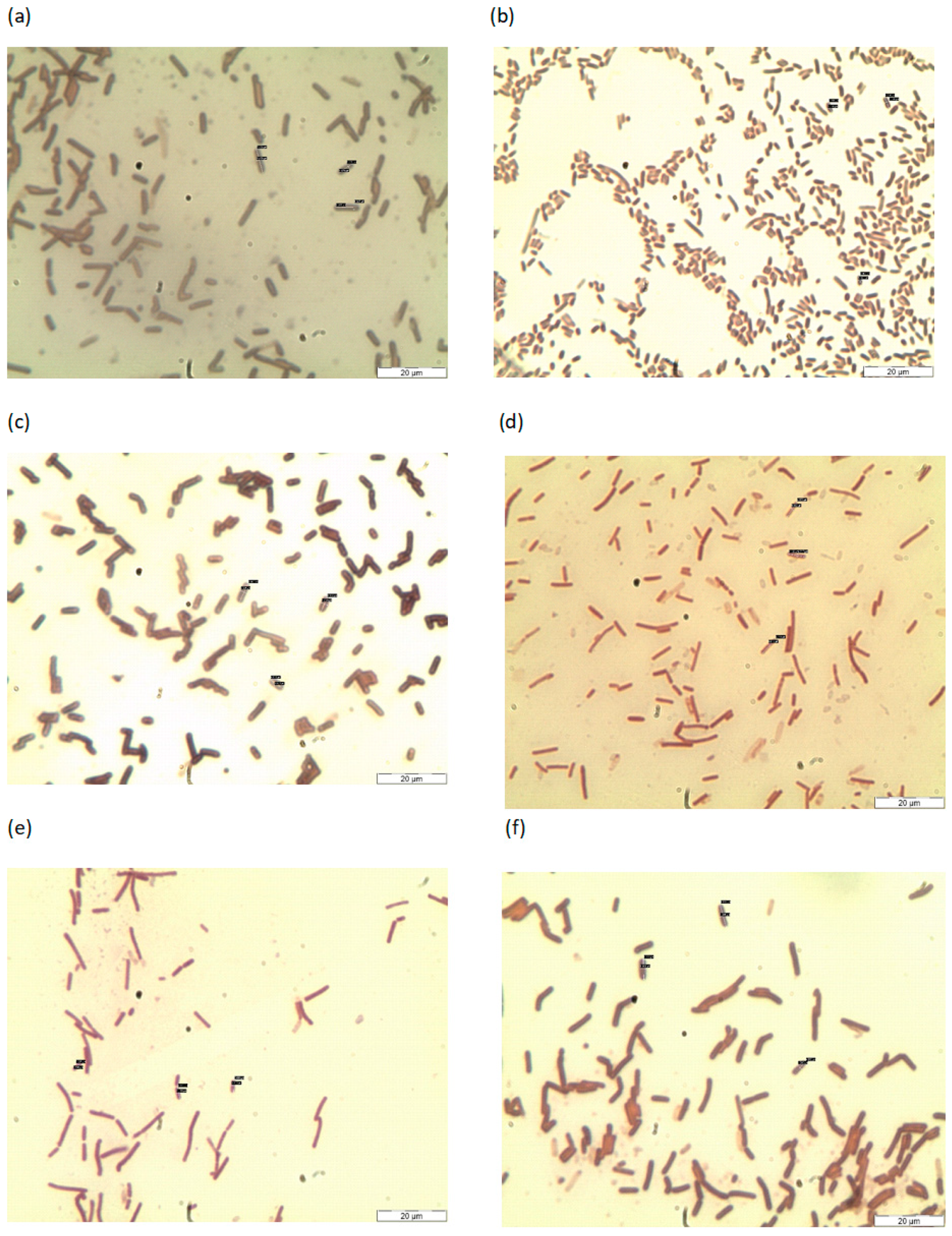
3.2. Microscopic Observation of Isolated Bacteria

The microscopic images and observation of the bacteria sample were attached in Figure 1 and Table 2 shows morphology of bacteria (Gram staining and shape). The results revealed that bacteria sample 5 showed crystal violet and Gram-positive reaction, while all the other samples showed pinkish red and Gram-negative. According to Robert et al. [18], the Gram stain reaction was based on the amount of peptidoglycan found in the cell walls of bacteria. Bacteria sample with rod-shaped structure were categorized as bacillus while only bacteria sample 2 was coccobacillus in term of the shape. Bacteria sample 2 is a type of bacterium under bacilli category but with very short rods which might be mistaken as cocci [25]. Coccobacillus was termed for bacteria sample 2 as an intermediate between bacilli (rod-shaped bacteria) and cocci (spherical bacteria). These bacteria samples under bacillus group might be endospore-forming aerobic or facultatively anaerobic and exhibited a wide range of physiological abilities [25] that allow them to live in every natural environment including POME. Only one endospore is formed per cell, and the spores are resistant to cold, heat, desiccation, radiation, and disinfectants. Results obtained showed that there was no connection between the shape of a bacterium and its reactions and colors in the Gram staining.

3.3. Selection of Bacteria

The selection was mainly based on the diameter of bacteria samples as presented in Table 3. The sub-micron fillers fulfilled the criteria where one of its phases has one, two or three dimensions in within the scale 0.1 to 1 µm [11]. Li [11] proved that sub-micron fillers would give substantial impact on a wide range of properties of the composite product even at low filler content. Based on these criteria, only bacteria sample 4 with diameter 0.83 ± 0.13 µm was chosen to be applied as filler to reinforce sodium alginate film. The other bacteria samples have a larger dimension than the required criteria. Bacteria sample 4 were bacillus in shape with Gram-negative reaction.

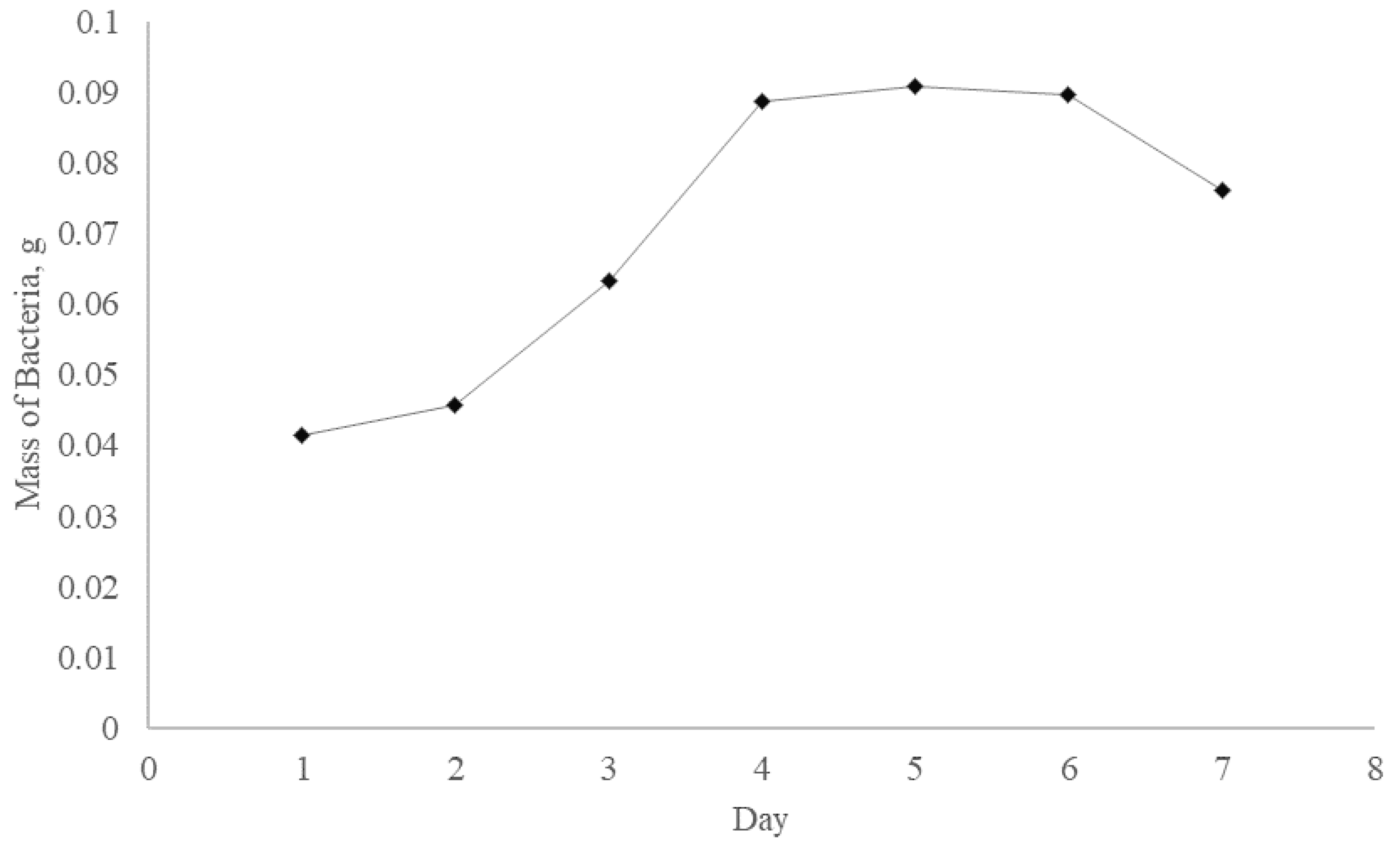
3.4. The Growth Rate of Selected Bacteria

Figure 2 shows the growth curve of selected bacteria (sample 4). Broth culture of selected bacteria showed lag phase from day one to day two. During the lag phase, bacteria did not reproduce immediately. The lag phase was the period where the individual bacteria were maturing and adapting themselves to growth conditions. However, the cells were not dormant but changed only very little as they were gathering nutrients in readiness for cell division [9]. Only little to no cell division occurred, about an increment of 0.004 g for bacteria mass during the lag phase, which can last for one hour up to several days.
The log phase occurred after day two and lasted until day four. The bacteria cells were metabolically active and doubled at a constant rate. The mass of bacteria almost doubled up which was from 0.046 g on day two to 0.089 g on day four. Zwietering et al. [26] also proved that doubling of bacteria will continue at a constant rate, so both the rate of population and the number of cell increase doubled with each consecutive period. Lag phase could not continue endlessly as the medium was soon depleted of nutrients and enriched with waste products.
The period from day four to day six showed the stationary phase where the mass of bacteria maintained almost the same which were 0.087 g, 0.091 g, and 0.090 g respectively. During stationary phase, the death rate was equaled by the growth rate because different growth-limiting factors limited the creation of new cells. These growth-limiting factors include depletion of an essential nutrient in medium and presence of toxic waste products generated from cell metabolism. Bridges et al. [27] presented evidence that mutations can occur during stationary phase.
After day six, the curve showed a death phase until day seven. The death phase was marked by the accumulation of toxic substances which resulted in the decrement of bacteria mass from 0.090 g to 0.076 g. Bacteria died as many cells underwent autolysis, and most cells were lacking surrounding nutrients and facing other injurious conditions [26]. The death phase was predicted to continue after day seven.
As a result, day four was selected to be used as the production period of bacteria sample 4 as day four was the beginning of the stationary phase with almost the maximum mass of bacteria that could be cultured. Although the culture produced the highest mass of bacteria on day five, it was not selected to prevent any occurrence of cell mutation during the constant rate period [27].

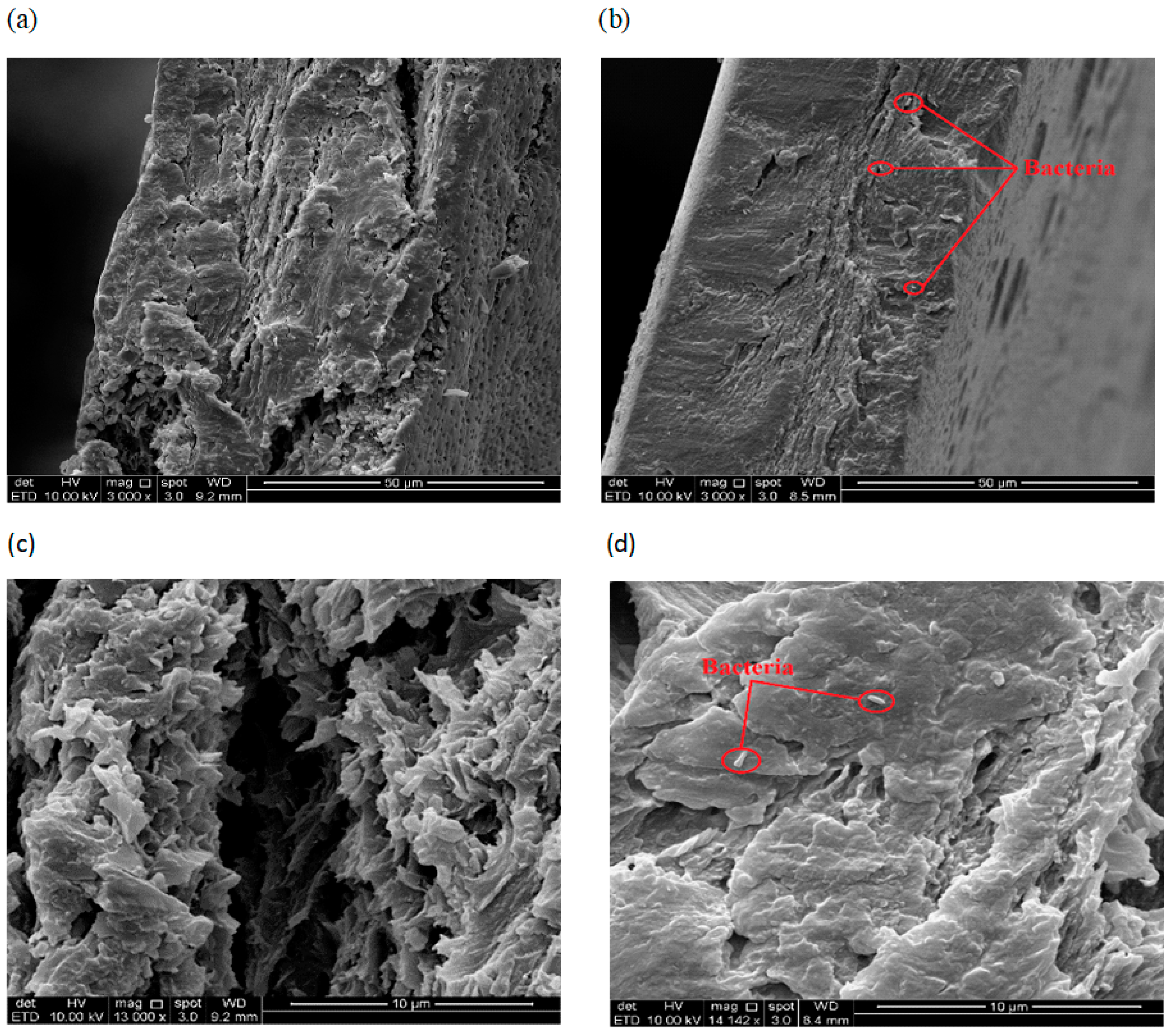
3.5. Cross-Sectional Morphology of Films

SEM micrographs, shown in Figure 3a–d revealed the comparison between the cross-sectional morphology of sodium alginate film without and with bacteria reinforcement respectively. According to Figure 1a, the film without bacteria reinforcement showed rough and uneven cross-sectional surface with visible cracking along the surface. Figure 1b demonstrated that the film which was reinforced by bacteria had become more compact with a slightly striated cross-section without noticeable cracking along the surface, typical of a stronger film with more homogenous structure.
Figure 1c,d were the SEM micrographs that presented the magnified spot of cross-sectional morphology shown in Figure 1 and Figure 2 respectively. The inside areas of both films were observed through the magnification. The results showed in Figure 3 revealed that the components were not bonded tightly and colossal gap and voids were present between components of the film without bacteria reinforcement due to the cracking surface of the film. On the other side, components in Figure 4 were bonded firmly and left little to no gap between components. The micrograph in Figure 4 also showed that the bacteria were able to reduce the voids between components and enhanced bonding of components in the film which contributed to a composite film with improved mechanical properties.

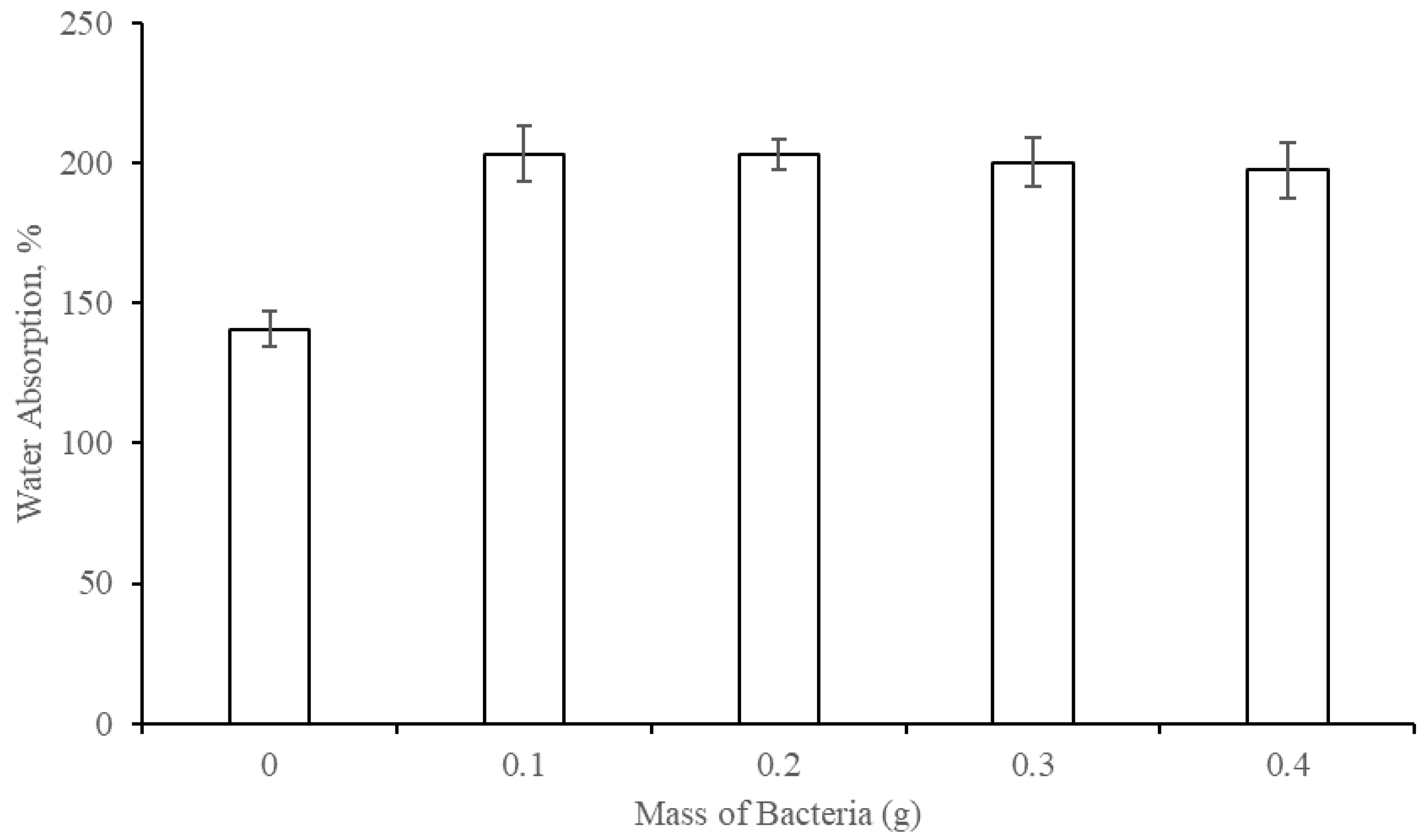
3.6. Water Absorption Properties (WAP)

Figure 4 shows the effect of the mass of bacteria reinforcing filler on the water absorption properties of the composite films. The WAP of the film increased significantly from 140.74 ± 6.42% to 203.28 ± 10.06% for films without and with 0.1 g bacteria reinforcement, respectively, due to the absorption of water molecules by bacteria cell when the films were immersed in distilled water. Bacteria sample 4 were Gram-negative bacteria that were enveloped by an outer membrane made of lipopolysaccharide and protein [28,29,30]. Both the charged or polar hydrophilic amino acid side chains in protein molecules can attract water molecules due to hydrogen bond formation. When water molecules were attracted to the outer surface of a bacteria cell, osmosis occurred as there was different concentration gradient of water molecules between the outer and inner side of a bacteria cell. Water molecules entered the dry bacteria cell of low water molecule concentration and allowed more water molecules to be trapped in bacteria cell and absorbed as a part of the composite film.
The trend for WAP of the film remains constant at about 200% from 0.1 g to 0.4 g of bacteria reinforcement. It was logical that higher mass of bacteria contributed to the higher amount of water molecules absorbed. However, the steady trend of WAP might be due to the formation of endospore or thick wax-like layers around the outside of the bacterial cell wall [31]. These external layers were functioned to protect the bacteria cell, prohibiting the water molecules from contacting and entering the bacteria cell. Another reason might be due to the saturation of water molecules inside bacteria cell as no more water molecules could be absorbed by bacteria cell through osmosis. Bacteria cells were surrounded by a rigid cell wall or termed as peptidoglycan [32]. The role of bacteria cell wall was to act as a physical barrier which physically limited the size and shape of the cell as well as preventing an excessive amount of water intake that could burst and kill the cell. Therefore, no more water molecules could be absorbed when the bacteria cells were saturated.
Size of bacteria have been proven by many previous researchers as micro-size and it has capability to seal cracks, pores or voids in the micro-size, too. Once the cracks, pores or voids are sealed, reduction in water ingress is observed. Obviously, bacterial activity or cell could seal the pores, voids and micro-cracks where other sealants are unable to work through [33].

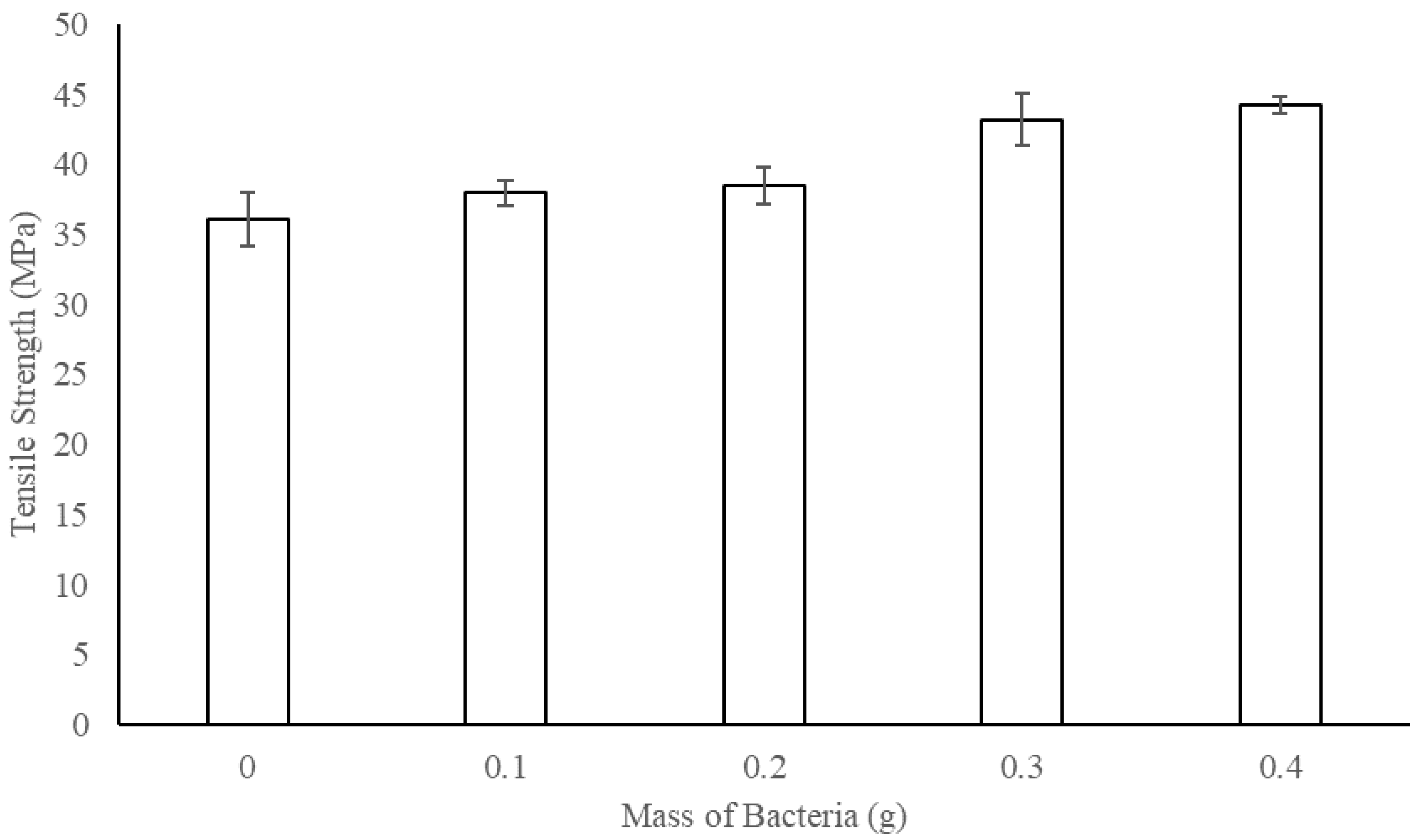
3.7. Tensile Strength (TS)

Figure 5 shows the effect of the mass of bacteria reinforcing filler on the tensile strength of the composite film. The TS of the film increased steadily from the film without bacteria reinforcement, 36.10 ± 1.94 MPa, to film with 0.4 g bacteria reinforcement, 44.29 ± 0.60 MPa. A noticeable increment of TS was observed from 0.2 g bacteria reinforcement, 38.50 ± 1.28 MPa, to 0.3 g bacteria reinforcement, 43.18 ± 1.84 MPa. The noticeable increment of TS can be explained by the presence of enough amounts of bacteria to be dispersed evenly in composite film. The results obtained showed a total increment of about 22.70% in term of TS in comparison to the film with 0.4 g of bacteria reinforcement and film without bacteria reinforcement. The enhancement of TS might be due to the hydrogen bonding which occurred between the hydroxide groups of sodium alginate components [20] and the lipopolysaccharide or protein molecules presented on the outer membrane of bacteria cells [29]. The hydroxide groups of sodium alginate were able to form hydrogen bonds with protein molecules [34,35]. The protein-alginate hydrogen bonding occurred in composite film which adjoined the bacteria and the components in the film, which strengthened the inner area of the film. The explanation was supported by the SEM micrographs shown in Figure 1, Figure 2, Figure 3 and Figure 4, as bacteria were able to reduce the voids and to close up the gap between components in the film. The higher the mass of bacteria, the bigger the surface area for interaction. Thus, more protein-alginate interaction and bonding can occur which resulted in higher TS. It is important to point out that the TS properties might be further improved through reinforcement of bacteria with mass higher than 0.4 g unless fractures occur on the films.

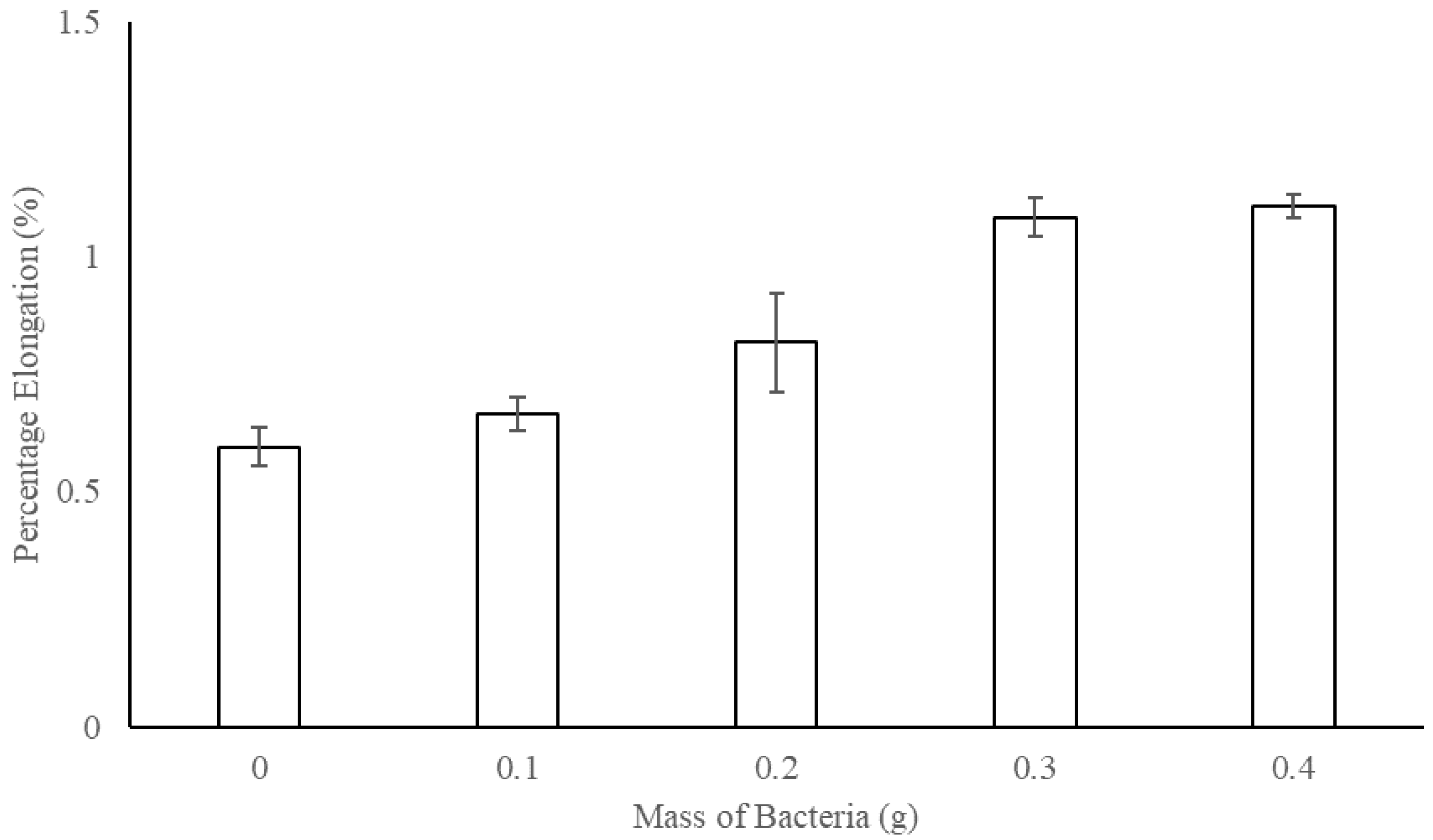
3.8. Percentage Elongation (E)

Figure 6 shows the effect of the mass of bacteria reinforcing filler on the percentage elongation of the composite film. The E of the film increased slightly from the film without bacteria reinforcement, 0.60 ± 0.04%, to the films reinforced with 0.3 g bacteria, 1.08 ± 0.04%, and remain almost constant with a further increment of 0.4 g bacteria, 1.11 ± 0.03%. The conventional trade between tensile strength and elongation is that one grows while the other decreases [36] as the film became more rigid and stronger but broke at a shorter length [36]. However, with composite materials like bacteria and sodium alginate, the composite product was not only a mixture of two bulk materials. Thus, an additional concept should be applied rather than the general rule of bulk materials. The shape of the reinforcing phase and the structure of the reinforced matrix were considered as they contributed significantly to the mechanical properties of the composite film. According to Ku et al. [37], there were some ways to increase both strength and elasticity of the composite product, mainly through crosslinking and reinforcement of stiffer matrix material with more elastic filler like long or endless fibers.
After all, bacteria filler were not concluded to have the ability to enhance the elongation properties together with the improvement of tensile strength properties for the composite films. There was only about 0.5% of overall increment observed, which was considered insignificant, and more data needs to be obtained for the E of the composite films with higher mass reinforcement of bacteria.

4. Conclusions

Bacteria sample with sub-micron diameter, 0.83 ± 0.13µm, was selected to reinforce calcium chloride cured sodium alginate film. Based on the SEM micrographs obtained for cross-sectional morphology, the surface structure of film without bacteria reinforcement was rough, uneven, and presented with cracking and voids. The water absorption properties of the film increased significantly from 140.74% to 203.28% for films without and with 0.1 g bacteria reinforcement, respectively, but remain constant at about 200% from 0.1 g to 0.4 g of bacteria reinforcement. The tensile strength properties showed a total increment of about 22.70% from the film without bacteria reinforcement, 36.10 ± 1.94 MPa, to the film reinforced with 0.4 g of bacteria, 44.29 ± 0.60 MPa. After all, bacteria filler were not concluded to have the ability to enhance the elongation properties of composite films because only about 0.5% of overall increment was observed, which was considered insignificant. This research revealed that bacteria were suitable to be used as filler to reinforce material like CaCl2 cured sodium alginate film. The results indicated that the films reinforced by using bacteria filler possessed improved morphological, physical and mechanical properties, and had the potential to be incorporated into many fields such as the innovation of water filtration membranes and alternative packaging material.

Author Contributions

B.K.X.H. carried out experiments, analysed data, and drafted the manuscript. B.A., M.F.B.Y., and N.I. conceived the study, and participated in research coordination. A.T., C.W.C.N., and H.A.T. rechecked the data, drafted and reviewed the manuscript. All authors have read and agreed to the published version of the manuscript.

Funding

This work was supported by the Ministry of Education (MOE) under Fundamental Research Grant Scheme (FRGS), grant number 203.PTEKTIND.6711701.

Acknowledgments

This research was carried out at School of Industrial Technology, Universiti Sains Malaysia, Penang, Malaysia. The authors wish to thank Azieda A.T. and Nor Azlina I. for their technical support and valuable contributions to this research.

Conflicts of Interest

The authors declare no conflict of interest.

Abbreviations

POMEpalm oil mill effluent
SEMscanning electron microscopy
CaCl2calcium chloride
WAPwater absorption properties
TStensile strength
Epercentage elongation

References

  1. Wang, R.-M.; Zheng, S.-R.; Zheng, Y.G. Polymer Matrix Composites and Technology, 1st ed.; Cambridge Woodhead Publishing Elsevier: Amsterdam, The Netherlands, 2011. [Google Scholar]
  2. Hull, D.; Clyne, T. An Introduction to Composite Materials, 2nd ed.; Cambridge University Press: Cambridge, MA, USA, 1996. [Google Scholar]
  3. Deepa, B.; Abraham, E.; Pothan, L.A.; Cordeiro, N.; Faria, M.; Thomas, S. Biodegradable nanocomposite films based on sodium alginate and cellulose nanofibrils. Materials 2016, 9, 50. [Google Scholar] [CrossRef] [PubMed] [Green Version]
  4. Moon, R.J.; Martini, A.; Nairn, J.; Simonsen, J.; Youngblood, J. Cellulose nanomaterials review: Structure, properties and nanocomposites. Chem. Soc. Rev. 2011, 40, 3941–3994. [Google Scholar] [CrossRef] [PubMed]
  5. Guilbert, S.; Gontard, N.; Cuq, B. Technology and applications of edible protective films. Packag. Technol. Sci. 1995, 8, 339–346. [Google Scholar] [CrossRef]
  6. Kester, J.J.; Fennema, O. Edible films and coatings: A review. Food Technol. 1986, 40, 47–59. [Google Scholar]
  7. Rhim, J.-W. Physical and mechanical properties of water resistant sodium alginate films. LWT-Food Sci. Technol. 2004, 37, 323–330. [Google Scholar] [CrossRef]
  8. Pavlath, A.E.; Voisin, A.; Robertson, G.H. Pectin-based biodegradable water insoluble films. In Macromolecular Symposia, 1st ed.; WILEY-VCH Verlag GmbH & Co. KGaA: Weinheim, Germany, 1999; Volume 140, pp. 107–113. [Google Scholar]
  9. Tortora, G.J.; Funke, B.R.; Case, C.L.; Johnson, T.R. Microbiology: An Introduction, 12th ed.; Benjamin Cummings: San Francisco, CA, USA, 2004. [Google Scholar]
  10. Chun Ng, C.W.; Ismail, A.F.; Zaini Makhtar, M.M.; Fikri Jamaluddin, M.N.; Tajarudin, H.A. Conversion of food waste via two-stage fermentation to controllable chicken Feed Nutrients by local isolated microorganism. Int. J. Recycl. Org. Waste Agric. 2020, 9, 33–47. [Google Scholar]
  11. Li, Y. The Development of Sub-Micro Filler Enhanced Polymer Composites; Nottingham Trent University: Nottingham, UK, 2007. [Google Scholar]
  12. Marquis, D.M.; Chivas-Joly, C.; Guillaume, É. Properties of Nanofillers in Polymer, 1st ed.; INTECH Open Access Publisher: Rijeka, Croatia, 2011. [Google Scholar]
  13. Arivalagan, K.; Ravichandran, S.; Rangasamy, K. Nanomaterials and its potential applications. Int. J. Chemtech Res. 2011, 3, 534–538. [Google Scholar]
  14. Mousavinasab, S.M. Effects of filler content on mechanical and optical properties of dental composite resin. In Metal, Ceramic and Polymeric Composites for Various Uses, 1st ed.; INTECH Open Access Publisher: Rijeka, Croatia, 2011. [Google Scholar]
  15. Abdollahi, M.; Alboofetileh, M.; Rezaei, M.; Behrooz, R. Comparing physico-mechanical and thermal properties of alginate nanocomposite films reinforced with organic and/or inorganic nanofillers. Food Hydrocoll. 2013, 32, 416–424. [Google Scholar] [CrossRef]
  16. Lau, A.K.; Bhattacharyya, D.; Ling, C.H. Nanocomposites for engineering applications. J. Nanomater. 2009, 8, 19. [Google Scholar] [CrossRef] [Green Version]
  17. Brown, A.E. Benson’s Microbiological Applications: Laboratory Manual in General Microbiology, 8th ed.; The McGraw-Hill: New York, NY, USA, 2012. [Google Scholar]
  18. Robert, A.P.; Lorraine, F.; Walter, M.; Ronald, M. Laboratory Exercises in Microbiology, 3rd ed.; John Wiley & Sons: Hoboken, NJ, USA, 2009. [Google Scholar]
  19. Trevors, J. Sterilization and inhibition of microbial activity in soil. J. Microbiol. Methods 1996, 26, 53–59. [Google Scholar] [CrossRef]
  20. Rhim, J.-W.; Kim, J.-H.; Kim, D.-H. Modification of Na-Alginate Films by CaCl2 Treatment. Korean J. Food Sci. Technol. 2003, 35, 217–221. [Google Scholar]
  21. Tappi. Thickness (Caliper) of Paper, Paperboard, and Combined Board. Standard Test Method t411 om-97; Tappi: Atlanta, GA, USA, 1997. [Google Scholar]
  22. ASTM, D. 882-88. Standard test methods for tensile properties of thin plastic sheeting. In Annual Book of ASTM Standards 8; ASTIM International: West Conshohocken, PA, USA, 1989. [Google Scholar]
  23. Baker, J.A. Light as a factor in the production of pigment by certain bacteria. J. Bacteriol. 1938, 35, 625. [Google Scholar] [CrossRef] [PubMed] [Green Version]
  24. Seleen, W.; Stark, C. Some characteristics of green-fluorescent pigment-producing bacteria. J. Bacteriol. 1943, 46, 491. [Google Scholar] [CrossRef] [Green Version]
  25. Turnbull, P.C.; Kramer, J.; Melling, J. Bacillus. In Medical Microbiology, 4th ed.; Galveston University of Texas Medical Branch: Galveston, TX, USA, 1991. [Google Scholar]
  26. Zwietering, M.; Jongenburger, I.; Rombouts, F.; Van’t Riet, K. Modeling of the bacterial growth curve. Appl. Environ. Microbiol. 1990, 56, 1875–1881. [Google Scholar] [CrossRef] [PubMed] [Green Version]
  27. Bridges, B.A.; Foster, P.L.; Timms, A.R. Effect of endogenous carotenoids on “adaptive” mutation in Escherichia coli FC40. Mutat. Res. Fundam. Mol. Mech. Mutagenesis 2001, 473, 109–119. [Google Scholar] [CrossRef]
  28. Nikaido, H.; Vaara, M. Molecular basis of bacterial outer membrane permeability. Microbiol. Rev. 1985, 49, 1. [Google Scholar] [CrossRef] [Green Version]
  29. Koebnik, R.; Locher, K.P.; Van Gelder, P. Structure and function of bacterial outer membrane proteins: Barrels in a nutshell. Mol. Microbiol. 2000, 37, 239–253. [Google Scholar] [CrossRef]
  30. Voulhoux, R.; Bos, M.P.; Geurtsen, J.; Mols, M.; Tommassen, J. Role of a highly conserved bacterial protein in outer membrane protein assembly. Science 2003, 299, 262–265. [Google Scholar] [CrossRef]
  31. Leggett, M.J.; McDonnell, G.; Denyer, S.P.; Setlow, P.; Maillard, J.Y. Bacterial spore structures and their protective role in biocide resistance. J. Appl. Microbiol. 2012, 113, 485–498. [Google Scholar] [CrossRef]
  32. Schleifer, K.H.; Kandler, O. Peptidoglycan types of bacterial cell walls and their taxonomic implications. Bacteriol. Rev. 1972, 36, 407. [Google Scholar] [CrossRef] [Green Version]
  33. Chahal, N.; Siddique, R.; Rajora, A. Influence of bacteria on the compressive strength, water absorption and rapid chloride permeability of fly ash concrete. Constr. Build. Mater. 2012, 28, 351–356. [Google Scholar] [CrossRef]
  34. Yang, L.; Guo, J.; Yu, Y.; An, Q.; Wang, L.; Li, S.; Huang, X.; Mu, S.; Qi, S. Hydrogen bonds of sodium alginate/Antarctic krill protein composite material. Carbohydr. Polym. 2016, 142, 275–281. [Google Scholar] [CrossRef] [Green Version]
  35. Mishima, Y.; Momma, K.; Hashimoto, W.; Mikami, B.; Murata, K. Crystal structure of AlgQ2, a macromolecule (alginate)-binding protein of Sphingomonas sp. A1, complexed with an alginate tetrasaccharide at 1.6-Å resolution. J. Biol. Chem. 2003, 278, 6552–6559. [Google Scholar] [CrossRef] [PubMed] [Green Version]
  36. Ku, H.; Wang, H.; Pattarachaiyakoop, N.; Trada, M. A review on the tensile properties of natural fiber reinforced polymer composites. Compos. Part. B Eng. 2011, 42, 856–873. [Google Scholar] [CrossRef] [Green Version]
  37. Hernández-Muñoz, P.; Villalobos, R.; Chiralt, A. Effect of cross-linking using aldehydes on properties of glutenin-rich films. Food Hydrocoll. 2004, 18, 403–411. [Google Scholar] [CrossRef]
Figure 1. The microscopic observation of bacteria sample: (a) Bacterial sample a; (b) Bacterial sample b; (c) Bacterial sample c; (d) Bacterial sample d; (e) Bacterial sample e; (f) Bacterial sample f.
Sustainability 12 09468 g001
Figure 2. The growth curve of bacteria sample 4.
Sustainability 12 09468 g002
Figure 3. Cross-sectional morphology and a magnified spot cross-sectional morphology for film without and with incorporated bacillus bacteria: (a) Cross-sectional morphology of the film without bacteria reinforcement; (b) Cross-sectional morphology of the film with bacteria reinforcement; (c) A magnified spot of the cross-sectional morphology of film without bacteria reinforcement; (d) A magnified spot of the cross-sectional morphology of film with bacteria reinforcement.
Sustainability 12 09468 g003
Figure 4. The effect of the mass of bacteria reinforcing filler on the water absorption properties of the composite films.
Sustainability 12 09468 g004
Figure 5. The effect of the mass of bacteria reinforcing filler on the tensile strength of the composite film.
Sustainability 12 09468 g005
Figure 6. The effect of the mass of bacteria reinforcing filler on the percentage elongation of the composite films.
Sustainability 12 09468 g006
Table 1. Morphological characteristics of bacteria colonies.
Colony No.SizeColorOpacityFormElevations
1SmallBuffTranslucentRoundRaised
2SmallBuffTranslucentRoundConvex
3MediumYellowOpaqueFilamentousHilly
4PunctiformBuffTranslucentRoundFlat
5PunctiformBuffOpaqueIrregularRaised
6SmallBuffOpaqueRoundRaised
Table 2. Gram reaction and shape of bacteria observed through an optical microscope.
Bacteria SampleGram ReactionShape
1NegativeBacillus
2NegativeCoccobacillus
3NegativeBacillus
4NegativeBacillus
5PositiveBacillus
6NegativeBacillus
Table 3. The diameter of bacteria samples.
Bacteria SampleDiameter, µm
11.62 ± 0.13
21.25 ± 0.10
32.02 ± 0.26
40.83 ± 0.13
51.30 ± 0.10
61.82 ± 0.12
Publisher’s Note: MDPI stays neutral with regard to jurisdictional claims in published maps and institutional affiliations.

