
Successful Bone Marrow Transplantation in a Patient with Acute Myeloid Leukemia Developed from Severe Congenital Neutropenia Using Modified Chemotherapy and Conditioning Regimen for Leukemia
 , ,
, ,
Abstract
:1. Introduction
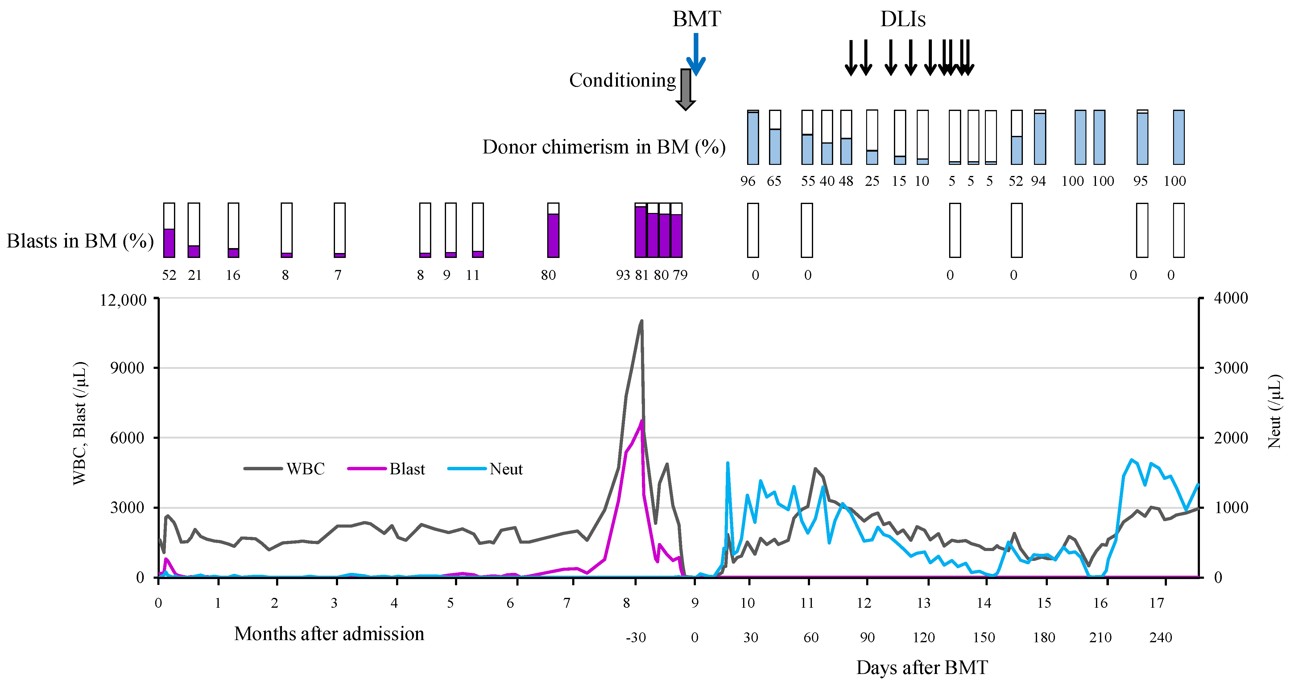
2. Case Report
3. Discussion and Conclusions
Author Contributions
Funding
Institutional Review Board Statement
Informed Consent Statement
Data Availability Statement
Acknowledgments
Conflicts of Interest
References
- Welte, K.; Zeidler, C. Severe congenital neutropenia. Hematol. Oncol. Clin. N. Am. 2009, 23, 307–320. [Google Scholar] [CrossRef] [PubMed]
- Skokowa, J.; Dale, D.C.; Touw, I.P.; Zeidler, C.; Welte, K. Severe congenital neutropenias. Nat. Rev. Dis. Primers 2017, 3, 17032. [Google Scholar] [CrossRef]
- Makaryan, V.; Zeidler, C.; Bolyard, A.A.; Skokowa, J.; Rodger, E.; Kelley, M.L.; Boxer, L.A.; Bonilla, M.A.; Newburger, P.E.; Shimamura, A.; et al. The diversity of mutations and clinical outcomes for ELANE-associated neutropenia. Curr. Opin. Hematol. 2015, 22, 3–11. [Google Scholar] [CrossRef]
- Klein, C.; Grudzien, M.; Appaswamy, G.; Germeshausen, M.; Sandrock, I.; Schäffer, A.A.; Rathinam, C.; Boztug, K.; Schwinzer, B.; Rezaei, N.; et al. HAX1 deficiency causes autosomal recessive severe congenital neutropenia (Kostmann disease). Nat. Genet. 2007, 39, 86–92. [Google Scholar] [CrossRef] [PubMed]
- Boztug, K.; Appaswamy, G.; Ashikov, A.; Schäffer, A.A.; Salzer, U.; Diestelhorst, J.; Germeshausen, M.; Brandes, G.; Lee-Gossler, J.; Noyan, F.; et al. A syndrome with congenital neutropenia and mutations in G6PC3. N. Engl. J. Med. 2009, 360, 32–43. [Google Scholar] [CrossRef] [PubMed]
- Person, R.E.; Li, F.-Q.; Duan, Z.; Benson, K.F.; Wechsler, J.; Papadaki, H.A.; Eliopoulos, G.; Kaufman, C.; Bertolone, S.J.; Nakamoto, B.; et al. Mutations in proto-oncogene GFI1 cause human neutropenia and target ELA2. Nat. Genet. 2003, 34, 308–312. [Google Scholar] [CrossRef]
- Donadieu, J.; Leblanc, T.; Bader Meunier, B.; Barkaoui, M.; Fenneteau, O.; Bertrand, Y.; Maier-Redelsperger, M.; Micheau, M.; Stephan, J.L.; Phillipe, N.; et al. Analysis of risk factors for myelodysplasias, leukemias and death from infection among patients with congenital neutropenia. Experience of the French Severe Chronic Neutropenia Study Group. Haematologica 2005, 90, 45–53. [Google Scholar]
- Rosenberg, P.S.; Alter, B.P.; Bolyard, A.A.; Bonilla, M.A.; Boxer, L.A.; Cham, B.; Fier, C.; Freedman, M.; Kannourakis, G.; Kinsey, S.; et al. The incidence of leukemia and mortality from sepsis in patients with severe congenital neutropenia receiving long-term G-CSF therapy. Blood 2006, 107, 4628–4635. [Google Scholar] [CrossRef]
- Choi, S.W.; Boxer, L.A.; Pulsipher, M.A.; Roulston, D.; Hutchinson, R.J.; Yanik, G.A.; Cooke, K.R.; Ferrara, J.L.M.; Levine, J.E. Stem cell transplantation in patients with severe congenital neutropenia with evidence of leukemic transformation. Bone Marrow Transplant. 2005, 35, 473–477. [Google Scholar] [CrossRef] [PubMed]
- Connelly, J.A.; Choi, S.W.; Levine, J.E. Hematopoietic stem cell transplantation for severe congenital neutropenia. Curr. Opin. Hematol. 2012, 19, 44–51. [Google Scholar] [CrossRef] [PubMed]
- Zeidler, C.; Welte, K.; Barak, Y.; Barriga, F.; Bolyard, A.A.; Boxer, L.; Cornu, G.; Cowan, M.J.; Dale, D.C.; Flood, T.; et al. Stem cell transplantation in patients with severe congenital neutropenia without evidence of leukemic transformation. Blood 2000, 95, 1195–1198. [Google Scholar]
- Dale, D.C.; Bonilla, M.A.; Davis, M.W.; Nakanishi, A.M.; Hammond, W.P.; Kurtzberg, J.; Wang, W.; Jakubowski, A.; Winton, E.; Lalezari, P.; et al. A randomized controlled phase III trial of recombinant human granulocyte colony-stimulating factor (filgrastim) for treatment of severe chronic neutropenia. Blood 1993, 81, 2496–2502. [Google Scholar] [CrossRef]
- Rosenberg, P.S.; Zeidler, C.; Bolyard, A.A.; Alter, B.P.; Bonilla, M.A.; Boxer, L.A.; Dror, Y.; Kinsey, S.; Link, D.C.; Newburger, P.E.; et al. Stable long-term risk of leukaemia in patients with severe congenital neutropenia maintained on G-CSF therapy. Br. J. Haematol. 2010, 150, 196–199. [Google Scholar] [CrossRef]
- Fioredda, F.; Iacobelli, S.; van Biezen, A.; Gasper, B.; Ancliff, P.; Donadieu, J.; Aliurf, M.; Peters, C.; Calvillo, M.; Matthes-Martin, S.; et al. Stem cell transplantation in severe congenital neutropenia: An analysis from the European Society for Blood and Marrow Transplantation. Blood 2015, 126, 1885–1892. [Google Scholar] [CrossRef] [PubMed]
- Rotulo, G.A.; Beaupain, B.; Rialland, F.; Paillard, C.; Nachit, O.; Galambrun, C.; Gandemer, V.; Bertrand, Y.; Neven, B.; Dore, E.; et al. HSCT may lower leukemia risk in ELANE neutropenia: A before-after study from the French Severe Congenital Neutropenia Registry. Bone Marrow Transplant. 2020, 55, 1614–1622. [Google Scholar] [CrossRef]
- Zeidler, C.; Nickel, A.; Sykora, K.W.; Welte, K. Improved outcome of stem cell transplantation for severe chronic neutropenia with or without secondary leukemia: A long-term analysis of European data for more than 25 years by the SCNIR. Blood 2013, 122, 3347. [Google Scholar] [CrossRef]
- Skokowa, J.; Steinemann, D.; Katsman-Kuipers, J.E.; Zeidler, C.; Klimenkova, O.; Klimiankou, M.; Ünalan, M.; Kandabarau, S.; Makaryan, V.; Beekman, R.; et al. Cooperativity of RUNX1 and CSF3R mutations in severe congenital neutropenia: A unique pathway in myeloid leukemogenesis. Blood 2014, 123, 2229–2237. [Google Scholar] [CrossRef] [PubMed]
- Setty, B.A.; Yeager, N.D.; Bajwa, R.P. Heterozygous M1V variant of ELA-2 gene mutation associated with G-CSF refractory severe congenital neutropenia. Pediatr. Blood Cancer 2011, 57, 514–515. [Google Scholar] [CrossRef]
- Hashem, H.; Abu-Arja, R.; Auletta, J.J.; Rangarajan, H.G.; Varga, E.; Rose, M.J.; Bajwa, R.P.S. Successful second hematopoietic cell transplantation in severe congenital neutropenia. Pediatr. Transplant. 2018, 22, e13078. [Google Scholar] [CrossRef] [PubMed]
- Rashidi, A.; Fisher, S.I. Spontaneous remission of acute myeloid leukemia. Leuk. Lymphoma 2015, 56, 1727–1734. [Google Scholar] [CrossRef]
- Pluchart, C.; Munzer, M.; Mauran, P.; Abély, M. Transient remission of childhood acute lymphoblastic and myeloid leukemia without any cytostatic treatment: 2 case reports and a review of literature. J. Pediatr. Hematol. Oncol. 2015, 37, 68–71. [Google Scholar] [CrossRef]
- Jeha, S.; Chan, K.W.; Aprikyan, A.G.; Hoots, W.K.; Culbert, S.; Zietz, H.; Dale, D.C.; Albitar, M. Spontaneous remission of granulocyte colony-stimulating factor-associated leukemia in a child with severe congenital neutropenia. Blood 2000, 96, 3647–3649. [Google Scholar] [CrossRef] [PubMed]
- Touw, I.P. Game of clones: The genomic evolution of severe congenital neutropenia. Hematol. Am. Soc. Hematol. Educ. Program 2015, 2015, 1–7. [Google Scholar] [CrossRef] [PubMed]
- Ahmed, O.G.; Lambert, E.M. Obstructive sleep apnea in a 5 month old with tonsillar hypertrophy secondary to congenital neutropenia: Case report and literature review. Int. J. Pediatr. Otorhinolaryngol. 2017, 96, 103–105. [Google Scholar] [CrossRef] [PubMed]
- Morikawa, K.; Morikawa, A.; Nakamura, M.; Miyawaki, T. Characterization of granulocyte colony-stimulating factor receptor expressed on human lymphocytes. Br. J. Haematol. 2002, 118, 296–304. [Google Scholar] [CrossRef]


| First Admission | First Discharge | Second Admission | Days after Transplantation | Normal Range | ||||
|---|---|---|---|---|---|---|---|---|
| 257 | 600 | 1290 | 1654 | |||||
| WBCs (/µL) | 1650 | 1510 | 10,810 | 2950 | 3230 | 5230 | 4920 | 3300–8600 |
| Neutrophils (%) | 0.5 | 0 | 0 | 45 | 53 | 58 | 60 | 38.5–81.5 |
| Blasts (%) | 10.5 | 4 | 60 | 0 | 0 | 0 | 0 | |
| Hb (g/dL) | 6.3 | 7.0 | 7.6 | 9.2 | 12.4 | 12.8 | 12.3 | 11.6–14.8 |
| Platelets (/µL) | 151,000 | 412,000 | 57,000 | 208,000 | 201,000 | 204,000 | 219,000 | 158,000–348,000 |
| CRP (mg/dL) | 3.45 | 1.73 | 12.0 | 0.05 | 0.07 | 0.02 | 0.01 | 0–0.14 |
| G-CSF (pg/dL) | 162 | 121 | <39.0 | |||||
Disclaimer/Publisher’s Note: The statements, opinions and data contained in all publications are solely those of the individual author(s) and contributor(s) and not of MDPI and/or the editor(s). MDPI and/or the editor(s) disclaim responsibility for any injury to people or property resulting from any ideas, methods, instructions or products referred to in the content. |
© 2024 by the authors. Licensee MDPI, Basel, Switzerland. This article is an open access article distributed under the terms and conditions of the Creative Commons Attribution (CC BY) license (https://creativecommons.org/licenses/by/4.0/).
Share and Cite
Matsumura, R.; Mochizuki, S.; Morishita, Y.; Hayakawa, H.; Karakawa, S.; Kawaguchi, H.; Okada, S.; Hyakuna, N.; Kobayashi, M. Successful Bone Marrow Transplantation in a Patient with Acute Myeloid Leukemia Developed from Severe Congenital Neutropenia Using Modified Chemotherapy and Conditioning Regimen for Leukemia. Hematol. Rep. 2024, 16, 98-105. https://doi.org/10.3390/hematolrep16010010
Matsumura R, Mochizuki S, Morishita Y, Hayakawa H, Karakawa S, Kawaguchi H, Okada S, Hyakuna N, Kobayashi M. Successful Bone Marrow Transplantation in a Patient with Acute Myeloid Leukemia Developed from Severe Congenital Neutropenia Using Modified Chemotherapy and Conditioning Regimen for Leukemia. Hematology Reports. 2024; 16(1):98-105. https://doi.org/10.3390/hematolrep16010010
Chicago/Turabian StyleMatsumura, Risa, Shinji Mochizuki, Yusuke Morishita, Hiroko Hayakawa, Shuhei Karakawa, Hiroshi Kawaguchi, Satoshi Okada, Nobuyuki Hyakuna, and Masao Kobayashi. 2024. "Successful Bone Marrow Transplantation in a Patient with Acute Myeloid Leukemia Developed from Severe Congenital Neutropenia Using Modified Chemotherapy and Conditioning Regimen for Leukemia" Hematology Reports 16, no. 1: 98-105. https://doi.org/10.3390/hematolrep16010010