Article

Somatic Embryogenesis and Plant Regeneration of Farmer-Preferred Passion Fruit Varieties Grown in Kenya

1
Department of Plant Science, Kenyatta University, Nairobi P.O. Box 43844-00100, Kenya
2
Department of Biochemistry, University of Nairobi, Nairobi P.O. Box 30197-00100, Kenya
3
Department of Biochemistry and Biotechnology, Kenyatta University, Nairobi P.O. Box 43844-00100, Kenya
*
Author to whom correspondence should be addressed.
Int. J. Plant Biol. 2023, 14(4), 1180-1189; https://doi.org/10.3390/ijpb14040086
Submission received: 30 August 2023 / Revised: 15 October 2023 / Accepted: 6 December 2023 / Published: 11 December 2023
(This article belongs to the Section Plant Reproduction)

Abstract

:
In vitro regeneration of passion fruit has great prospects for mass production of disease-free planting materials. The objective of this study was to develop an in vitro regeneration system through somatic embryogenesis for farmer-preferred genotypes grown in Kenya. Callus induction and somatic embryogenesis were carried out using leaf and immature seed explants. The explants were cultured on Murashige and Skoog medium augmented with different concentrations of 2,4-dichlorophenoxyacetic acid (2,4-D) alone and 2,4-D combined with 1.0 mg L−1 thidiazuron (TDZ). Data were recorded and subjected to analysis of variance. The highest number of somatic embryos was obtained from KPF4 genotype using MS medium supplemented with 8 mg L−1 2,4-D and 1 mg L−1 TDZ. The embryos were converted to plants on germination medium comprising of MS augmented with 0.5 mg L−1 6-benzyl amino purine (BAP). The plantlets were hardened for 4 weeks in plastic pots. The survival rate during hardening of in vitro regenerated plants was 77.8%. The present study reports a regeneration system through somatic embryogenesis for KPF4 passion fruit grown in Kenya. The in vitro regeneration system can be utilized for mass propagation and genetic improvement of KPF4 variety grown in Kenya.

1. Introduction

Passion fruit (Passiflora edulis Sims) is a high value horticultural fruit crop in the tropical and subtropical countries where it is grown for nutritional, medicinal and industrial purposes. The fruit is rich in iron, vitamin A, B2, C, non-nutritive phytochemicals and polyphenols. It is also rich in a broad spectrum of minerals such as potassium, phosphorous, calcium, iron, sodium, magnesium and sulphur [1,2]. Furthermore, the various parts of the plant contain antimicrobial, anti-inflammatory, antioxidant, and anti-sedative properties. Hence, they are used for treatment of various ailments [3].
Passion fruit (Passiflora edulis Sims) has its origin in tropical America. In Kenya, two distinct forms are largely grown in a wide range of altitudes: the purple passion fruit and the yellow passion fruit [4]. The perennial fruit has a great economic potential due to the increasing demand for fresh fruits and processed juice. However, its production is constrained by diseases and lack of disease-free planting materials.
Conventional propagation of passion fruit is predominantly carried out by seed, cutting or grafting [5,6]. However, propagation by these methods presents a number of challenges such as enhanced levels of genetic variation and low propagation efficiencies [7]. The modes of propagation also lead to build-up of fungal, bacterial and viral diseases resulting in total yield losses. Furthermore, passion fruit plants are susceptible to diseases, and the success of conventional propagation methods is considerably dependent on the donor plant’s physiological state [5]. Hence, in vitro regeneration methods offer a better alternative for mass propagation of clonal plants and disease-free plants [8].
Direct and indirect organogenesis are the most popular pathways for in vitro regeneration of several species of passion fruit [9]. They lead to production of true-to-type plants within a short period of time and the availability of superior individuals for large-scale commercial plantation [10]. Several studies have reported in vitro regeneration of passion fruit through organogenesis from various explants [7,11,12,13]. However, somatic embryogenesis offers higher propagation efficiencies and limits formation of chimeras [14]. It is a potent biotechnology tool by which a somatic plant cell is converted into an embryo for mass propagation and hence can aid in the rapid multiplication of newly released varieties [15]. Furthermore, in vitro regeneration through somatic embryogenesis offers advantages over organogenesis in exploring the possibility of spontaneously generating multiple embryos from a single cell.
Additionally, somatic embryogenesis has critical applications in genetic improvement and germplasm conservation of many species of plants [5]. Unlike organogenesis, the bipolar nature of somatic embryos also allows for direct regeneration of plantlets without having to separate shoot and root development stages. Therefore, in vitro regeneration via somatic embryogenesis has a huge potential in plant biotechnology. Several studies have reported somatic embryogenesis in passion fruit from different explants [16,17,18,19]. However, no studies have been carried out to establish a regeneration system through somatic embryogenesis for farmer-preferred varieties in Kenya. The objective of this study was to develop an efficient system for production and proliferation of somatic embryos from leaf explants and immature seeds and regeneration of plantlets of purple and KPF4 varieties of passion fruit in Kenya.

2. Materials and Methods

2.1. Source of Explants

Two farmer-preferred passion fruit genotypes, namely purple and KPF4, were acquired as ripe fruits as well as potted seedlings from Kenya Agricultural and Livestock Research Organization (KALRO) and Jomo Kenyatta University of Agriculture and Technology (JKUAT), respectively.

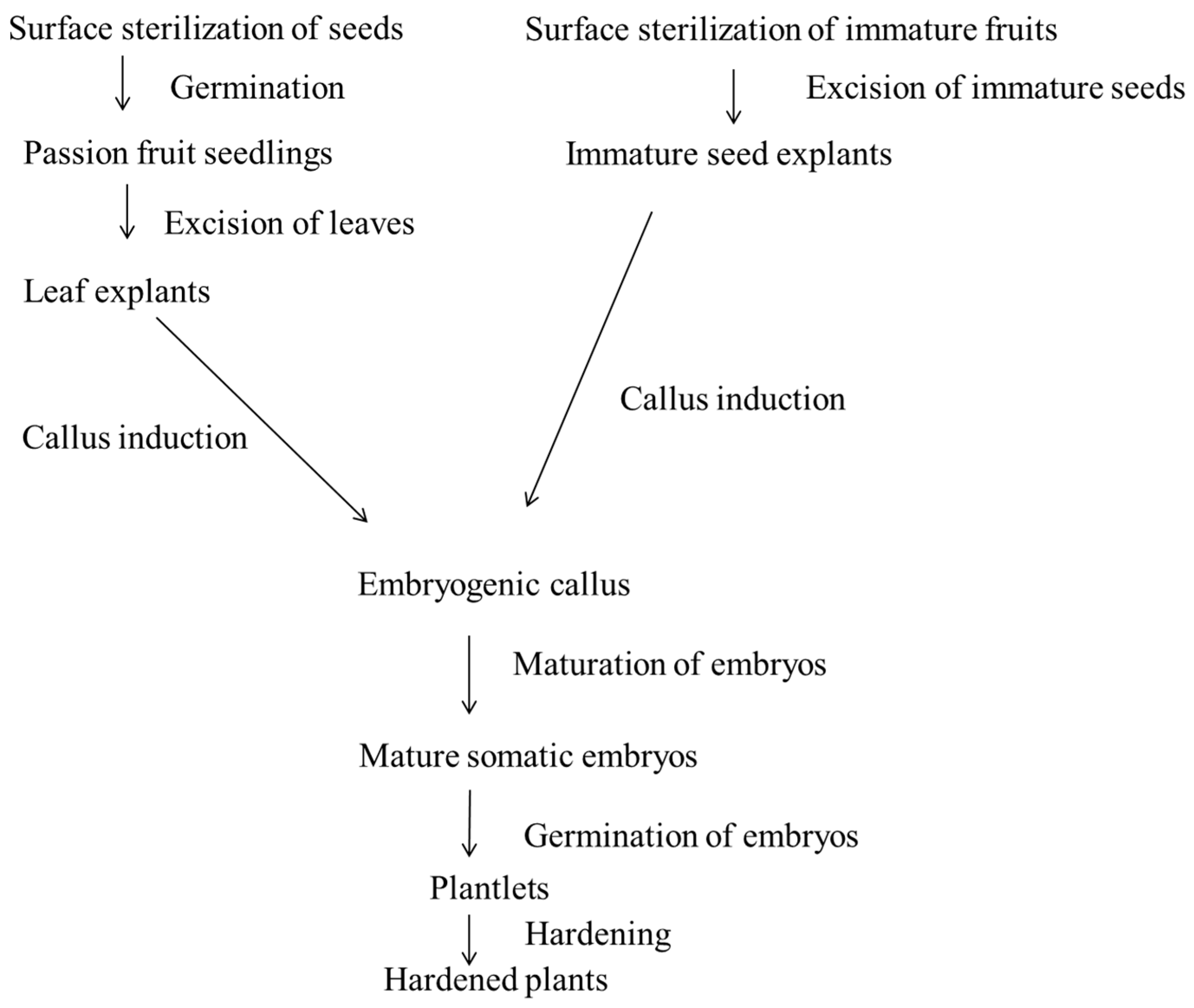
2.2. Explant Preparation

Mature seeds extracted from ripe fruits were rinsed with tap water before drying in the sun for 3 days. The seeds were surface-sterilized with 70% (v/v) ethanol for 5 min, followed by 2.5% sodium hypochlorite for 20 min and then rinsed four times in sterile distilled water. An approximately 2 mm cut was carefully made on the lateral sides of each seed before germinating them aseptically in culture vessels containing MS media [20] supplemented with sucrose (2%) and gelrite (0.24%) at pH 5.8. They were then incubated at 26 ± 2 °C. Leaves from twenty-one-day-old seedlings were excised and used as explants for somatic embryogenesis.
Immature fruits were also surface-sterilized with 70% (v/v) ethanol for 2 min, followed by 2.5% sodium hypochlorite for 10 min and then rinsed four times in sterile distilled water. The fruits were aseptically split open and immature seeds excised for induction of somatic embryos.

2.3. Somatic Embryogenesis from Leaf Disc and Immature Seed Explants

Explants from both varieties (purple and KPF4) consisting of 6 mm2 leaf discs were excised from the first two leaves of 21-day-old seedlings and used for induction of callus and somatic embryos. The excised explants were placed in Petri dishes (5 explants per Petri dish) containing MS medium supplemented with 3% sucrose and 0.8% agar and supplemented with 0.5–16.0 mg L−1 2,4-D for callus induction. Excised explants were also placed in Petri dishes (5 explants per Petri dish) containing MS medium supplemented with 3% sucrose and 0.8% agar without hormones (control). Cultures were incubated at 26 ± 2 °C with a 16 h photoperiod for 4 weeks. Experiments were set up in a completely randomized design. There were 6 replicates for each treatment and the experiments were repeated three times. The embryogenic calli and early stages of somatic embryos were transferred to different maturation treatments: MS with 6% sucrose, MS with 1% activated charcoal, MS with 0.1 or 0.2 mg L−1 abscisic acid (ABA) for 4 weeks. Maturated somatic embryos were transferred into different media containing different concentrations of growth regulators: MS without hormones and MS with BAP (0.5–3.0 mg L−1) and kinetin (0.5–3.0 mg L−1) for shoot and root induction. Data on the frequency of explants with callus and the frequency of callus with somatic embryos were recorded for analysis.
Seeds obtained from immature fruits were placed in culture bottles containing MS basal medium and B5 vitamins. The medium was supplemented with 3% sucrose, 2.4% gelrite, 200 mg L−1 glutamine and 500 mg L−1 casein hydrolysate. Immature seed explants were also cultured on MS medium supplemented with 3% sucrose and 0.8% agar in the absence of hormones (control). The medium was also augmented with 4.0–32.0 mg L−1 2,4-D and 1.0 mg L−1 TDZ. After 6 weeks, the embryogenic callus was transferred to MS basal salt and B5 vitamins containing 1% (w/v) activated charcoal or 0.1 mg L−1 ABA for maturation for 4 weeks. The somatic embryos were transferred into different media containing different concentrations of growth regulators: MS without hormones and MS with BAP (0.5–3.0 mg L−1) and kinetin (0.5–3.0 mg L−1) for plant regeneration. Data on the frequency of explants with callus and the frequency of callus with somatic embryos were recorded for analysis. A flow diagram displaying the in vitro regeneration of KPF4 genotype and purple passion fruit through somatic embryogenesis is presented in Figure 1.

2.4. Data Analysis

Data on explants forming callus, explants with embryogenic callus and callus with somatic embryos were analyzed using analysis of variance (ANOVA) with Genstat 15th edition statistical program to evaluate significant differences. The separation of means was carried out using Tukey’s honest significant difference test at 5% level.

3. Results

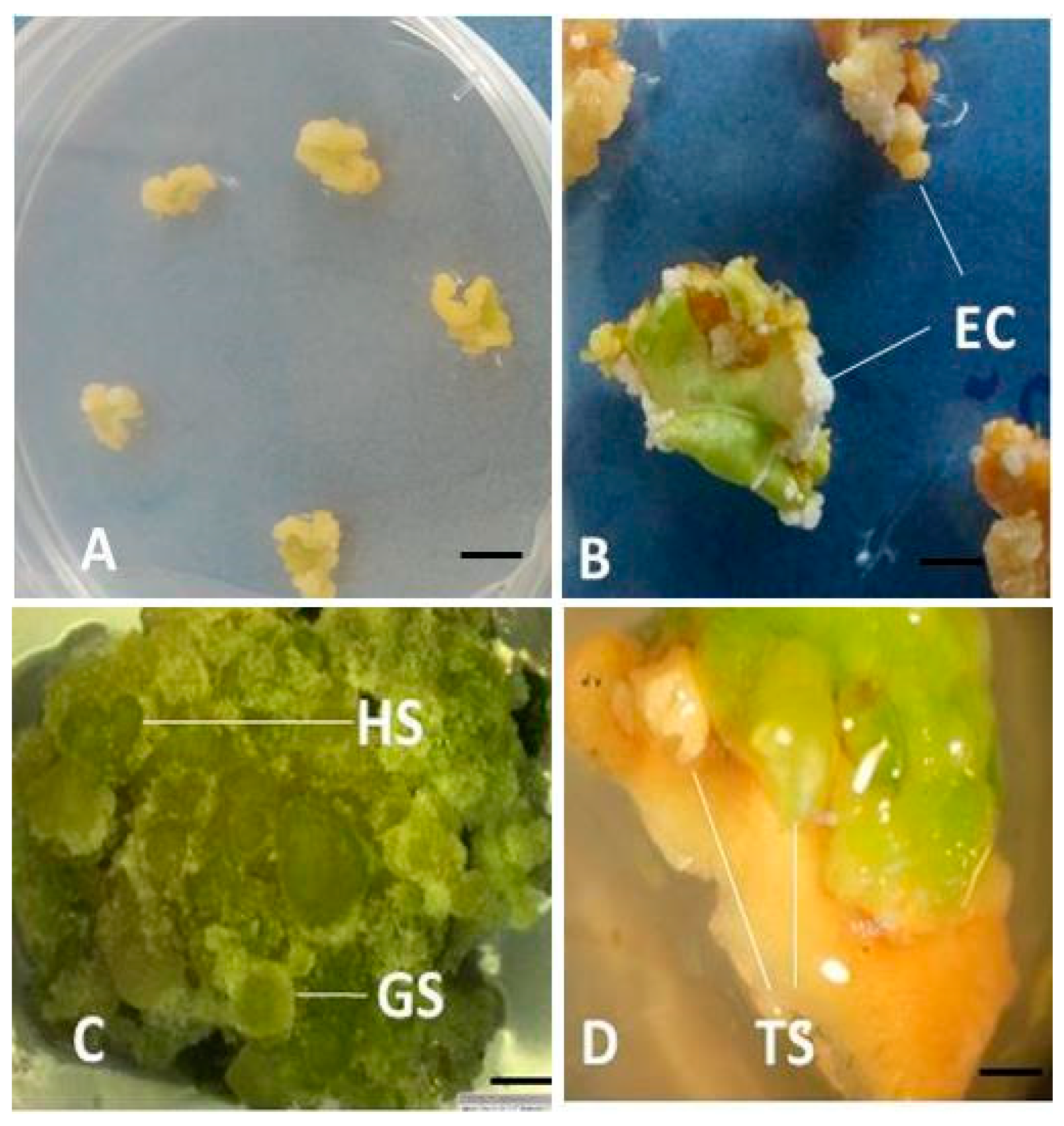
3.1. Effect of 2,4-D on Callus Induction and Somatic Embryogenesis from Leaf Explants of Two Genotypes of Passion Fruits

In hormone-free medium (control), callus was recorded on leaf explants from KPF4 genotype. The leaves displayed some chlorosis from the fourth week of culture on callus initiation medium and then turned brown. On the other hand, leaf explants from the purple variety of passion fruit did not form callus on hormone-free medium.
Explants from purple genotype of passion fruit generated loose non-embryogenic calluses on different concentrations of 2,4-D (Figure 2A). Embryos were not formed from the purple genotype of passion fruit. When KPF4 leaf explants were inoculated on MS medium supplemented with 0.5 to 16 mg L−1 2,4-D, callus initiation was visible from the cut edges from the fourth day of culture. The friable callus from KPF4 leaf explants began to show some level of organization into defined structures which appeared like the globular stages of somatic embryos in media augmented with 4 mg L−1 2,4-D (Figure 2B–D).
At lower concentrations of 2,4-D (<4.0 mg L−1), no somatic embryos were recorded in the KPF4 genotype. The number of KPF4 leaf explants with somatic embryos increased with an increase in concentration of 2,4-D up to 8 mg L−1 2,4-D which generated the highest number of explants with somatic embryos which was significantly (p ≤ 0.05) different from all other treatments except that with 10 mg L−1 (Table 1). An increase in PGR concentration from 0.5 to 6 mg L−1 2,4-D resulted in an increase in the frequencies of callus induction in the purple genotype. At concentrations above 6 mg L−1 2,4-D, a slight reduction in the frequency of callus induction was recorded. However, the reduction was not statistically significant (p > 0.05). In addition, an increase in the PGR concentration resulted in an increase in the frequencies of explants with embryogenic callus and somatic embryos of the KPF4 genotype, attaining the highest mean of 100 and 91.1, respectively, at 8 mg L−1 2,4-D (Table 1).
When embryogenic calli were transferred to hormone-free MS medium, MS supplemented with 1% activated charcoal, MS supplemented with 6% sucrose, MS supplemented with 1 mg L−1 BAP, MS supplemented with 0.1 mg L−1 ABA and MS supplemented with 0.2 mg L−1 ABA, they turned green but later turned brown from the 15th day of culture and finally died. Frequent subcultures only preserved the green coloration with no further development.

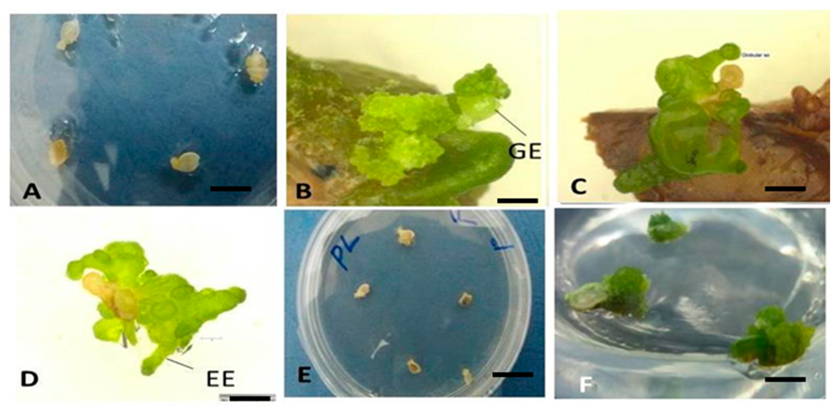
3.2. Effect of 2,4-D and TDZ on Callus Induction and Somatic Embryogenesis from Immature Seeds of KPF4 and Purple Genotypes of Passion Fruit

Immature seeds from KPF4 fruits inoculated on MS basal salts and B5 vitamins supplemented with 4 mg L−1, 8 mg L−1, 16 mg L−1 and 32 mg L−1 in combination with 1 mg L−1 TDZ yielded white friable embryogenic calli from the 5th day of culture (Figure 3A). Embryogenic calli were not formed from purple passion fruit seed explants. Most calluses appeared from the basal end of the seed while a few masses of callus appeared from the surface of the seed. The highest frequency of callus induction (95.5% for KPF4 and 93.3% for purple cultivar) was obtained from callus initiation medium augmented with 8 mg L−1 2,4-D in combination with 1 mg L−1 TDZ (Table 2). These frequencies were significantly (p ≤ 0.05) different from other treatments. Small calluses were also observed on hormone-free MS medium (control) after one week.
Callus from hormone-free callus initiation medium after 6 weeks of culture did not produce somatic embryos for both genotypes tested when they were transferred to hormone-free MS basal salts with B5 vitamins augmented with 1% activated charcoal. Similarly, no somatic embryos were observed from calli obtained from purple passion fruit on initiation and maturation medium. The calli lost the green color after 5 weeks. However, on subculturing to fresh medium with 1% activated charcoal, the green color was retained for a longer time.
On the other hand, for KPF4 genotype, the callus induced in increasing concentrations of 2,4-D formed different stages of somatic embryos after one week of transfer to MS basal salts and B5 vitamins supplemented with 1% activated charcoal (Figure 3B–D). The callus from initiation medium containing 8.0 mg L−1 2,4-D and 1.0 mg L−1 TDZ produced the highest frequency of somatic embryos which was not significantly (p > 0.05) different from other treatments. No callus transferred to MS basal salt and B5 vitamins supplemented with 0.1 mg L−1 ABA generated somatic embryos.

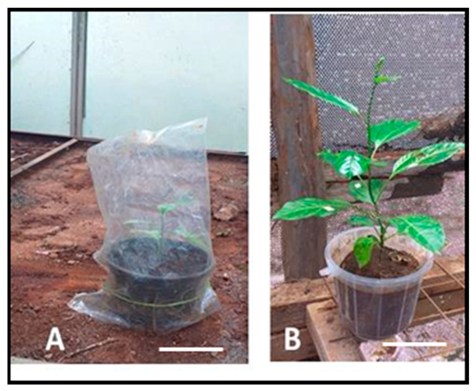
3.3. Conversion of Somatic Embryos to Form Plantlets

Only somatic embryos from immature seeds of KPF4 previously cultured on MS augmented with 8.0 mg L−1 2,4-D and 1 mg L−1 TDZ and then transferred onto MS supplemented with 1% (w/v) activated charcoal and thereafter into germination medium (MS with 0.5 mg L−1 BAP) formed plantlets. The micro shoots were from somatic embryos (Figure 4). A total of nine plantlets were obtained after 8 weeks of culture. The rest of the embryos retained their green color, for as long as they were subcultured into fresh medium, without further development. The plants were successfully hardened and grown in potted soil (Figure 5). Out of nine plantlets that were hardened for 4 weeks in small pots, seven plants survived—giving a survival rate of 77.8%.

4. Discussion

4.1. Responses of Leaf Explants of KPF4 and Purple Passion Fruit Genotypes to 2,4-D Concentrations

Callus was initiated from a few leaf explants of KPF4 genotype on hormone-free medium, an indication of endogenous hormones in the leaf explants. Auxins have been reported widely as being critical in regulating cell division and differentiation [10,21]. The findings from the present study show that the growth of callus on media containing increasing concentrations of 2,4-D can be attributed to the effect of auxins in eliciting production of endogenous cytokinins [22], while combinations of auxin and cytokinin, both endogenous and exogenous, stimulate cell division [23]. This is also in agreement with Braybrook and Kuhlemeier [24] that auxins and cytokinins are essential for callus induction.
For the purple variety of passion fruit, loose non-embryogenic callus without further development into somatic embryos was recorded on different concentrations of 2,4-D. The KPF4 leaf explants inoculated on MS medium supplemented with the different concentrations of 2,4-D formed friable callus which developed further into somatic embryos. These results show that callus induction and somatic embryogenesis in passion fruit is genotype-dependent. The genotypic effect on the response of passion fruit varieties to in vitro regeneration has also been reported by Amugune et al. [25] and Mukasa et al. [26].

4.2. Callus Induction and Somatic Embryogenesis Responses of Immature Seeds of KPF4 and Purple Passion Fruit Genotypes to 2,4-D Concentrations

Different concentrations of 2,4-D alone or in combination with TDZ were able to initiate callus and different stages of somatic embryos. Similar results were reported by Ferreira et al. [27] in wild species of passion fruit, Passiflora miniata and Passiflora speciosa, when media was supplemented with 2,4-D. In many in vitro embryogenic models, 2,4-D is necessary for the initiation of cell programming through somatic embryogenesis [16,18,28,29].
In most species of plants studied in which growth regulators are needed for induction of somatic embryogenesis, auxins and cytokinins are significant factors defining the embryogenic response, possibly due to their involvement in the regulation of the cell cycle, division and differentiation [29,30,31]. The utilization or interaction of 2,4-D with other auxins and cytokinins for the initiation of somatic embryogenesis through tissue culture of mature and immature zygotic embryos, seeds, leaves and cotyledons for several species has been reported [16,18,25,32]. Activated charcoal and abscisic acid possibly enhanced the differentiation of somatic embryos. This is in agreement with Pinto et al. [16] and da Silva et al. [33].

4.3. Conversion of Somatic Embryos to Form Plantlets

Conversion of somatic embryos, from leaf explants of KPF4 passion fruit, into plantlets was not achieved. However, nine somatic embryos from immature seeds of KPF4 passion fruit were converted into normal plantlets. This can be attributed to the effect of genotype, type of explant and type of vitamins on somatic embryogenesis. The findings of the present study are consistent with previous findings by Pinto et al. [16] reporting abnormal somatic embryos and no conversion of cotyledonary stage somatic embryos, obtained from zygotic embryo explants of Passiflora edulis Sims, into plantlets. The somatic embryos became necrotic after 10 days of culture. In contrast, Ferreira et al. [27] reported conversion of somatic embryos from immature zygotic embryos of Passiflora miniata and Passiflora speciosa into normal plantlets. Similarly, da Silva et al. [33] reported successful conversion of somatic embryos from Passiflora cincinnata generated using mature zygotic embryo explants, cultured on induction media containing MS basal salts and B5 vitamins, and supplemented with a combination of 18.1 μM of 2,4-D and 4.5 μM of BAP, to plants on hormone-free MS medium.

5. Conclusions

In the present study, somatic embryos were successfully induced from leaf and immature seed explants of KPF4 genotype of passion fruit. The type of explant and concentration of 2,4-D significantly influenced the number of somatic embryos, with 8 mg L−1 2,4-D and 1 mg L−1 TDZ giving the highest percentage of somatic embryos. Somatic embryos from KPF4 variety of passion fruit were successfully converted to plantlets. No somatic embryos were recorded in purple passion fruit in this study, an indication of the effect of genotype on somatic embryogenesis. The in vitro regeneration system through somatic embryogenesis for KPF4 passion fruits can be utilized for mass propagation of disease-free plants, genetic improvement and germplasm conservation of KPF4 variety grown in Kenya and beyond.
There is need for further research to optimize protocols for somatic embryogenesis of purple passion fruit and other genotypes grown in Kenya because the regeneration systems were genotype-specific.

Author Contributions

Conceptualization, L.K.A.; methodology, L.K.A., O.O., R.O.O. and E.N.N.; validation, O.O., R.O.O. and E.N.N.; formal analysis, L.K.A.; investigation, L.K.A.; data curation, L.K.A.; writing—original draft preparation, L.K.A.; writing—review and editing, L.K.A., O.O., R.O.O. and E.N.N.; supervision, O.O., R.O.O. and E.N.N.; project administration, O.O., R.O.O. and E.N.N.; funding acquisition, L.K.A. All authors have read and agreed to the published version of the manuscript.

Funding

This work was funded by the National Research Fund of Kenya through a PhD research grant (Financial Year 2016/2017) to the frst author.

Institutional Review Board Statement

Not applicable

Informed Consent Statement

Not applicable

Data Availability Statement

The datasets used and analyzed in this study are available from corresponding author on reasonable request.

Conflicts of Interest

The authors declare no conflict of interest.

References

  1. Ramaiya, S.D.; Bujang, J.S.; Zakaria, M.H.; King, W.S.; Shaffiq, S.M.A. Sugars, ascorbic acid, total phenolic content and total antioxidant activity in passion fruit (Passiflora) cultivars. J. Sci. Food Agric. 2013, 93, 1198–1205. [Google Scholar] [CrossRef] [PubMed]
  2. Pereira, Z.C.; dos Anjos Cruz, J.M.; Corrêa, R.F.; Sanches, E.A.; Campelo, P.H.; de Araújo Bezerra, J. Passion fruit (Passiflora spp.) pulp: A review on bioactive properties, health benefits and technological potential. Food Res. Int. 2023, 166, 112626. [Google Scholar] [CrossRef] [PubMed]
  3. Zas, P.; John, S. Diabetes and medicinal benefits of Passiflora edulis. World J. Pharm. Res. 2016, 5, 453–465. [Google Scholar]
  4. Kormelinck, A.G.; Janssen, I. Business Case Passion Fruit Contract Farming Migori County, South West-Kenya; Centre for Development Innovation: Wageningen, The Netherlands, 2012. [Google Scholar]
  5. Faleiro, F.G.; Junqueira, N.T.V.; Junghans, T.G.; Jesus, O.N.D.; Miranda, D.; Otoni, W.C. Advances in passion fruit (Passiflora spp.) propagation. Rev. Bras. de Frutic. 2019, 41, e-155. [Google Scholar] [CrossRef]
  6. Chen, Y.C.; Chang, C.; Lin, H.L. Topolins and red light improve the micropropagation efficiency of passion fruit (Passiflora edulis Sims)‘Tainung No. 1’. Hortscience 2020, 55, 1337–1344. [Google Scholar] [CrossRef]
  7. Huh, Y.S.; Lee, J.K.; Nam, S.Y. Effect of plant growth regulators and antioxidants on in vitro plant regeneration and callus induction from leaf explants of purple passion fruit (Passiflora edulis Sims). J. Plant Biotechnol. 2017, 44, 335–342. [Google Scholar] [CrossRef]
  8. Hieu, T.; Phong, T.H.; Khai, H.D.; Mai, N.T.N.; Cuong, D.M.; Luan, V.Q.; Tan, N.D. Efficient production of vigorous passion fruit rootstock for in vitro grafting. Plant Cell Tissue Organ Cult. 2022, 148, 635–648. [Google Scholar] [CrossRef]
  9. Ozarowski, M.; Thiem, B. Progress in micropropagation of Passiflora spp. to produce medicinal plants: A mini-review. Rev. Bras. de Farmacogn. 2013, 23, 937–947. [Google Scholar] [CrossRef]
  10. Chen, A.H.; Yang, J.L.; Niu, Y.D.; Yang, C.P.; Liu, G.F.; Yu, C.Y.; Li, C.H. High-frequency somatic embryogenesis from germinated zygotic embryos of Schisandra chinensis and evaluation of the effects of medium strength, sucrose, GA3, and BA on somatic embryo development. Plant Cell Tissue Organ Cult. 2010, 102, 357–364. [Google Scholar] [CrossRef]
  11. Asande, L.K.; Ombori, O.; Nyaboga, E.N.; Oduor, R.O. Efficient shoot organogenesis using leaf disc and nodal explants of passion fruit (Passiflora edulis Sims) and genetic fidelity assessment using sequence-related amplified polymorphism (SRAP) markers. Int. J. Agron. 2020, 2020, 1–10. [Google Scholar] [CrossRef]
  12. Phong, T.H.; Hieu, T.; Tung, H.T.; Mai, N.T.N.; Khai, H.D.; Cuong, D.M.; Luan, V.Q.; Nam, N.B.; Nhut, D.T. Silver nanoparticles: A positive factor for in vitro flowering and fruiting of purple passion fruit (Passiflora edulis Sim f. edulis). Plant Cell Tissue Organ Cult. 2022, 151, 401–412. [Google Scholar] [CrossRef]
  13. Tung, H.T.; Hieu, T.; Phong, T.H.; Khai, H.D.; Hanh, N.T.M.; Van, K.T.T.; Nhut, D.T. The application of thin cell layer culture technique in plant regeneration and micropropagation: Latest achievements. In Plant Tissue Culture: New Techniques and Application in Horticultural Species of Tropical Region; Springer: Singapore, 2022; pp. 231–257. [Google Scholar]
  14. Raza, G.; Singh, M.B.; Bhalla, P.L. Somatic embryogenesis and plant regeneration from commercial soybean cultivars. Plants 2019, 9, 38. [Google Scholar] [CrossRef] [PubMed]
  15. Phong, T.H.; Hieu, T.; Tung, H.T.; Mai, N.T.N.; Khai, H.D.; Luan, V.Q.; Nam, N.B.; Nhut, D.T. Somatic embryogenesis as potential method for commercial propagation in Passiflora edulis Sims f. edulis-an important horticultural crop. Sci. Hortic. 2023, 316, 112020. [Google Scholar] [CrossRef]
  16. Pinto, D.L.P.; de Almeida, A.M.R.; Rêgo, M.M.; da Silva, M.L.; de Oliveira, E.J.; Otoni, W.C. Somatic embryogenesis from mature zygotic embryos of commercial passion fruit (Passiflora edulis Sims) genotypes. Plant Cell Tissue Organ Cult. 2011, 107, 521–530. [Google Scholar] [CrossRef]
  17. Silva, T.C.R.; Carvalho, C.R. Vertical heterogeneity of DNA ploidy level assessed by flow cytometry in calli of Passiflora Cincinnata. Vitr. Cell. Dev. Biol.-Plant 2014, 50, 158–165. [Google Scholar] [CrossRef]
  18. Rocha, D.I.; Pinto, D.L.P.; Vieira, L.M.; Tanaka, F.A.O.; Dornelas, M.C.; Otoni, W.C. Cellular and molecular changes associated with competence acquisition during passion fruit somatic embryogenesis: Ultrastructural characterization and analysis of SERK gene expression. Protoplasma 2016, 253, 595–609. [Google Scholar] [CrossRef] [PubMed]
  19. Machado, M.D.; Souza, C.S.; Machado, M.; Reis, A.C.; de Sousa, S.M.; Matos, E.M.; Viccini, L.F.; Otoni, W.C.; de Carvalho, I.F.; Rocha, D.I.; et al. Novel avenues for passion fruit in vitro regeneration from endosperm culture, and morpho-agronomic and physiological traits of triploid Passiflora cincinnata Mast. emblings. Plant Cell Tissue Organ Cult. 2022, 150, 637–650. [Google Scholar] [CrossRef]
  20. Murashige, T.; Skoog, F. A revised medium for rapid growth and bio assays with tobacco tissue cultures. Physiol. Plant. 1962, 15, 473–497. [Google Scholar] [CrossRef]
  21. Jiménez, V.; Thomas, C. Participation of Plant Hormones in determination and progression of somatic embryogenesis. Somat. Embryog. 2006, 2, 103–118. [Google Scholar]
  22. Pernisová, M.; Klíma, P.; Horák, J.; Válková, M.; Malbeck, J.; Souček, P.; Za, E. Cytokinins modulate auxin-induced organogenesis in plants via regulation of the auxin efflux. Proc. Natl. Acad. Sci. USA 2009, 106, 3609–3614. [Google Scholar] [CrossRef]
  23. Mohr, H.; Schopfer, P. Physiology of hormone action. In Plant Physiology; Springer: Berlin/Heidelberg, Germany, 1995; pp. 383–408. [Google Scholar]
  24. Braybrook, S.A.; Kuhlemeier, C. How a plant builds leaves. Plant Cell 2010, 22, 1006–1018. [Google Scholar] [CrossRef] [PubMed]
  25. Amugune, N.O.; Gopalan, H.N.B.; Bytebier, B. Leaf disc regeneration of passion fruit. Afr. Crop Sci. J. 1993, 1, 99–104. [Google Scholar] [CrossRef]
  26. Mukasa, S.B.; Ssamula, A.; Asami, P.; Holton, T.A. In vitro propagation of three commercial passion fruit varieties in Uganda. Afr. Crop Sci. J. 2016, 24, 397–404. [Google Scholar] [CrossRef]
  27. Ferreira, D.A.T.; Sattler, M.C.; Carvalho, C.R.; Clarindo, W.R. Embryogenic potential of immature zygotic embryos of Passiflora: A new advance for in vitro propagation without plant growth regulators. Plant Cell Tissue Organ Cult. 2015, 122, 629–638. [Google Scholar] [CrossRef]
  28. Raghavan, V. Role of 2,4 dichlorophenoxyacetic acid (2,4-D) in somatic embryogenesis on cultured zygotic embryos of Arabidopsis: Cell expansion, cell cycling, and morphogenesis during continuous exposure of embryos to 2, 4 D. Am. J. Bot. 2004, 91, 1743–1756. [Google Scholar] [CrossRef] [PubMed]
  29. Fehér, A. Why somatic plant cells start to form embryos? In Somatic Embryogenesis; Mujib, A., Šamaj, J., Eds.; Springer: Berlin/Heidelberg, Germany, 2005; pp. 85–101. [Google Scholar] [CrossRef]
  30. Feher, A.; Pasternak, T.P.; Dudits, D. Transition of somatic plant cells to an embryogenic state. Plant Cell Tissue Organ Cult. 2003, 74, 201–228. [Google Scholar] [CrossRef]
  31. Gaaj, M.D. Factors influencing somatic embryogenesis induction and plant regeneration with particular reference to Arabidopsis thaliana (L.) Heynh. Plant Growth Regul. 2004, 43, 27–47. [Google Scholar] [CrossRef]
  32. Cerqueira-Silva, C.B.M.; Santos, E.S.L.; Conceição, L.D.H.C.S.; Cardoso-Silva, C.B.; Pereira, A.S.; Oliveira, A.C.; Corrêa, R.X. Genetic variation in a wild population of the ‘sleep’passion fruit (Passiflora setacea) based on molecular markers. Genet. Mol. Res. 2012, 11, 731–738. [Google Scholar] [CrossRef]
  33. da Silva, M.L.; Pinto, D.L.P.; Guerra, M.P. A novel regeneration system for a wild passion fruit species (Passiflora cincinnata Mast.) based on somatic embryogenesis from mature zygotic embryos. Plant Cell Tissue Organ Cult. 2009, 99, 47–54. [Google Scholar] [CrossRef]
Figure 1. A flow diagram illustrating the regeneration process for passion fruit genotypes grown in Kenya.
Ijpb 14 00086 g001
Figure 2. Effect of 2,4-D on callus induction and somatic embryogenesis on leaf discs of purple passion fruit and KPF4. (A) loose non-embryogenic cream yellow callus from purple passion fruit leaf explants on MS supplemented with 4.0 mg L−1 (Bar = 1 cm); (B) white friable embryogenic callus (EC) from leaf explants of KPF4 on MS supplemented with 8 mg L−1 2,4-D (Bar = 1 cm); (C) Globular (GS) and heart-shaped (HS) stages of somatic embryos of KPF4 on medium supplemented with 8 mg L−1 2,4-D (Bar = 1 mm); (D) torpedo stage (TS) of somatic embryos from KPF4 leaves on medium supplemented with 8 mg L−1 2,4-D (Bar = 1 mm).
Ijpb 14 00086 g002
Figure 3. Effect of 2,4-D and TDZ on the induction of somatic embryos from immature seeds of KPF4 and purple passion fruits. (A) Six-day-old callus from immature seeds of KPF4 on MS supplemented with 8 mg L−1 2,4-D and 1 mg L−1 TDZ (Bar = 1 cm); (B) Eight-week-old callus of KPF4 seeds on 1% activated charcoal showing the globular stage of somatic embryos (GE, globular embryos) (Bar = 1 mm); (C) Ten-week-old embryogenic callus of KPF4 passion displaying secondary somatic embryogenesis (Bar = 1 mm); (D) Eleven-week-old callus of KPF4 passion showing the further development of somatic embryos (EE, elongating embryos) (Bar = 1 mm); (E) Six-day-old little callus from immature seeds of purple passion on MS supplemented with 8 mg L−1 2,4-D and 1 mg L−1 TDZ (Bar = 1 cm); (F) Eight-week-old callus (without somatic embryos) from immature seed explants of purple passion on MS medium supplemented with 0.5 mg L−1 BAP (Bar = 1 mm).
Ijpb 14 00086 g003
Figure 4. Microshoot formation from somatic embryos originating from immature seeds of KPF4 cultured on MS augmented with 8.0 mg L−1 2,4-D and 1 mg L−1 TDZ (Bars = 1 cm). (A) Three-week-old callus with somatic embryos on MS medium supplemented with 1% activated charcoal; (B) Six-week-old shoots on MS medium supplemented with 0.5 mg L−1 BAP; (C) Eight-week-old plantlet on MS medium without plant growth regulators.
Ijpb 14 00086 g004
Figure 5. Hardening and growth of KPF4 passion fruit plantlets in the greenhouse. (A) Two-week-old plantlet in a plastic pot; (B) Four-week-old plantlet in a plastic pot containing garden soil mixed with sand (3:1). Bar = 10 cm.
Ijpb 14 00086 g005
Table 1. Effect of 2,4-D on callus induction and somatic embryogenesis from leaf explants of KPF4 and purple passion fruit genotypes after 6 weeks of culture.
PGRKPF4Purple
2,4-D
(mg/L)
Explants with Embryogenic Callus (%) X Callus with Somatic
Embryos (%) X
Explants
with Callus (%) X
Callus with Somatic Embryos (%) X
0.024.4 ± 4.4 d0.0 ± 0.0 d0.0 ± 0.0 c0.0 ± 0.0
0.575.6 ± 5.6 c0.0 ± 0.0 d51.1 ± 5.6 b0.0 ± 0.0
1.581.1 ± 4.9 bc0.0 ± 0.0 d60.0 ± 4.5 b0.0 ± 0.0
2.597.8 ± 1.5 a0.0 ± 0.0 d86.7 ± 3.9 a0.0 ± 0.0
4.096.7 ± 1.8 a46.7 ± 3.6 b85.5 ± 3.8 a0.0 ± 0.0
6.097.8 ± 2.2 a53.3 ± 6.6 b93.3 ± 2.3 a0.0 ± 0.0
8.0100.0 ± 0.0 a91.1 ± 4.3 a90 ± 0.7 a0.0 ± 0.0
10.92.2 ± 2.8 ab82.2 ± 4.5 a86.7 ± 2.7 a0.0 ± 0.0
12.093.3 ± 2.2 ab38.9 ± 6.3 bc90 ± 3.3 a0.0 ± 0.0
16.0100.0 ± 0.0 a20.0 ± 3.6 c86.7 ± 3.9 a0.0 ± 0.0
X Values represent the mean ± standard error of 6 replicates per treatment in three repeated experiments. Means having the same letter within a column are not significantly different according to Tukey’s honest significant difference at 5% level.
Table 2. Effect of 2,4-D and TDZ on somatic embryogenesis from immature seed explants of KPF4 and purple passion fruits.
PGRKPF4Purple
2,4-D
(mg/L)
TDZ (mg/L)Explant with Callus (%) XCallus with Somatic Embryos (%) XExplants with Callus (%) XCallus with Somatic Embryos (%) X
0.00.080.0 ± 3.9 c0.0 ± 0.0 d56.6 ± 4.0 c0.0 ± 0
4.01.086.6 ± 3.6 b,c77.8 ± 3.9 c72.0 ± 2.9 b,c0.0 ± 0
8.01.095.5 ± 2.0 a91.1 ± 1.8 a93.3 ± 2.3 a0.0 ± 0
16.01.090.0 ± 3.7 b85.6 ± 2.9 a,b74.4 ± 4.2 b0.0 ± 0
32.01.092.2 ± 2.8 b75.5 ± 4.7 b,c73.3 ± 3.9 b0.0 ± 0
X Values represent the mean ± standard error of 6 replicates per treatment in three repeated experiments. Means having the same letter within a column are not significantly different according to Tukey’s honest significant difference at 5% level.
Disclaimer/Publisher’s Note: The statements, opinions and data contained in all publications are solely those of the individual author(s) and contributor(s) and not of MDPI and/or the editor(s). MDPI and/or the editor(s) disclaim responsibility for any injury to people or property resulting from any ideas, methods, instructions or products referred to in the content.

