
Exposure of Dairy Cows to Coxiella burnetii in Greece: Surveillance Results and Association of Bacterial Presence with Environmental Variables

 , , , ,
Abstract
:1. Introduction
2. Materials and Methods
2.1. Study Area and Farm Selection
2.2. Sample Collection
2.3. Serological Assays
2.4. Molecular Assays
2.5. Geographical Information System
2.6. Environmental Parameters
2.7. Ecological Niche Model
3. Results
4. Discussion
Supplementary Materials
Author Contributions
Funding
Institutional Review Board Statement
Informed Consent Statement
Data Availability Statement
Conflicts of Interest
References
- Celina, S.S.; Cerný, J. Coxiella burnetii in Ticks, Livestock, Pets and Wildlife: A Mini-Review. Front. Vet. Sci. 2022, 9, 1068129. [Google Scholar] [CrossRef] [PubMed]
- Christodoulou, M.; Malli, F.; Tsaras, K.; Billinis, C.; Papagiannis, D. A Narrative Review of Q Fever in Europe. Cureus 2023, 15, e38031. [Google Scholar] [CrossRef] [PubMed]
- Ortega-Mora, L.-M. IS Q Fever a Significant Cause of Reproductive Failure in Cattle? Vet. Rec. 2012, 170, 257–258. [Google Scholar] [CrossRef] [PubMed]
- Garcia-Ispierto, I.; Tutusaus, J.; López-Gatius, F. Does Coxiella burnetii Affect Reproduction in Cattle? A Clinical Update. Reprod. Domest. Anim. 2014, 49, 529–535. [Google Scholar] [CrossRef] [PubMed]
- Saegerman, C.; Grégoire, F.; Delooz, L. Diagnosis of Coxiella burnetii Cattle Abortion: A One-Year Observational Study. Pathogens 2022, 11, 429. [Google Scholar] [CrossRef] [PubMed]
- Mioni, M.D.S.R.; Henker, L.C.; Teixeira, W.S.R.; Lorenzett, M.P.; Labruna, M.B.; Pavarini, S.P.; Driemeier, D.; Rousset, É.; Sidi-Boumedine, K.; Thiéry, R.; et al. Molecular Detection of Coxiella burnetii in Aborted Bovine Fetuses in Brazil. Acta Trop. 2022, 227, 106258. [Google Scholar] [CrossRef]
- Parisi, A.; Fraccalvieri, R.; Cafiero, M.; Miccolupo, A.; Padalino, I.; Montagna, C.; Capuano, F.; Sottili, R. Diagnosis of Coxiella burnetii-Related Abortion in Italian Domestic Ruminants Using Single-Tube Nested PCR. Vet. Microbiol. 2006, 118, 101–106. [Google Scholar] [CrossRef] [PubMed]
- Guatteo, R.; Beaudeau, F.; Joly, A.; Seegers, H. Coxiella burnetii Shedding by Dairy Cows. Vet. Res. 2007, 38, 849–860. [Google Scholar] [CrossRef]
- Pouquet, M.; Bareille, N.; Guatteo, R.; Moret, L.; Beaudeau, F. Coxiella burnetii Infection in Humans: To What Extent Do Cattle in Infected Areas Free from Small Ruminants Play a Role? Epidemiol. Infect. 2020, 148, e232. [Google Scholar] [CrossRef]
- España, P.P.; Uranga, A.; Cillóniz, C.; Torres, A. Q Fever (Coxiella burnetii). Semin. Respir. Crit. Care Med. 2020, 41, 509–521. [Google Scholar] [CrossRef] [PubMed]
- Bae, M.; Lee, H.J.; Park, J.H.; Bae, S.; Jung, J.; Kim, M.J.; Lee, S.-O.; Choi, S.-H.; Kim, Y.S.; Shin, Y.; et al. Molecular Diagnosis of Coxiella burnetii in Culture Negative Endocarditis and Vascular Infection in South Korea. Ann. Med. 2021, 53, 2258–2267. [Google Scholar] [CrossRef] [PubMed]
- Raoult, D.; Fenollar, F.; Stein, A. Q Fever during Pregnancy: Diagnosis, Treatment, and Follow-Up. Arch. Intern. Med. 2002, 162, 701. [Google Scholar] [CrossRef] [PubMed]
- Karageorgou, I.; Kogerakis, N.; Labropoulou, S.; Hatzianastasiou, S.; Mentis, A.; Stavridis, G.; Angelakis, E. Q Fever Endocarditis and a New Genotype of Coxiella burnetii, Greece. Emerg. Infect. Dis. 2020, 26, 2527–2529. [Google Scholar] [CrossRef] [PubMed]
- Pape, M.; Bouzalas, E.G.; Koptopoulos, G.S.; Mandraveli, K.; Arvanitidou-Vagiona, M.; Nikolaidis, P.; Alexiou-Daniel, S. The Serological Prevalence of Coxiella burnetii Antibodies in Sheep and Goats in Northern Greece. Clin. Microbiol. Infect. 2009, 15, 146–147. [Google Scholar] [CrossRef] [PubMed]
- Filioussis, G.; Theodoridis, A.; Papadopoulos, D.; Gelasakis, A.; Vouraki, S.; Bramis, G.; Arsenos, G. Serological Prevalence of Coxiella burnetii in Dairy Goats and Ewes Diagnosed with Adverse Pregnancy Outcomes in Greece. Ann. Agric. Environ. Med. 2017, 24, 702–705. [Google Scholar] [CrossRef] [PubMed]
- Valiakos, G.; Giannakopoulos, A.; Spanos, S.A.; Korbou, F.; Chatzopoulos, D.C.; Mavrogianni, V.S.; Spyrou, V.; Fthenakis, G.C.; Billinis, C. Use of Geographical Information System and Ecological Niche Model to Analyse Potential Exposure of Small Ruminants to Coxiella burnetii Infection in Central Greece. Small Rumin. Res. 2017, 147, 77–82. [Google Scholar] [CrossRef]
- Chochlakis, D.; Santos, A.S.; Giadinis, N.D.; Papadopoulos, D.; Boubaris, L.; Kalaitzakis, E.; Psaroulaki, A.; Kritas, S.K.; Petridou, E.I. Genotyping of Coxiella burnetii in Sheep and Goat Abortion Samples. BMC Microbiol. 2018, 18, 204. [Google Scholar] [CrossRef] [PubMed]
- Vourvidis, D.; Kyrma, A.; Linou, M.; Edouard, S.; Angelakis, E. Sero-epidemiology Investigation of Coxiella burnetii in Domestic Ruminants throughout Most Greek Regions. Vet. Med. Sci. 2021, 7, 99–104. [Google Scholar] [CrossRef] [PubMed]
- Kalaitzakis, E.; Fancello, T.; Simons, X.; Chaligiannis, I.; Tomaiuolo, S.; Andreopoulou, M.; Petrone, D.; Papapostolou, A.; Giadinis, N.D.; Panousis, N.; et al. Coxiella burnetii Shedding in Milk and Molecular Typing of Strains Infecting Dairy Cows in Greece. Pathogens 2021, 10, 287. [Google Scholar] [CrossRef] [PubMed]
- Hellenic Statistical Authority. Annual Report of Ariculture in Greece. 2021. Available online: https://elstat-outsourcers.statistics.gr/apografi_georgias_21_FINAL_web.pdf (accessed on 18 February 2024).
- Hijmans, R.J.; Cameron, S.E.; Parra, J.L.; Jones, P.G.; Jarvis, A. Very High Resolution Interpolated Climate Surfaces for Global Land Areas. Int. J. Climatol. 2005, 25, 1965–1978. [Google Scholar] [CrossRef]
- Phillips, S.J.; Anderson, R.P.; Schapire, R.E. Maximum Entropy Modeling of Species Geographic Distributions. Ecol. Model. 2006, 190, 231–259. [Google Scholar] [CrossRef]
- Gyuranecz, M.; Dénes, B.; Hornok, S.; Kovács, P.; Horváth, G.; Jurkovich, V.; Varga, T.; Hajtós, I.; Szabó, R.; Magyar, T.; et al. Prevalence of Coxiella burnetii in Hungary: Screening of Dairy Cows, Sheep, Commercial Milk Samples, and Ticks. Vector-Borne Zoonotic Dis. 2012, 12, 650–653. [Google Scholar] [CrossRef] [PubMed]
- Anastácio, S.; Carolino, N.; Sidi-Boumedine, K.; Da Silva, G.J. Q Fever Dairy Herd Status Determination Based on Serological and Molecular Analysis of Bulk Tank Milk. Transbound. Emerg. Dis. 2016, 63, e293–e300. [Google Scholar] [CrossRef] [PubMed]
- Rodolakis, A.; Berri, M.; Héchard, C.; Caudron, C.; Souriau, A.; Bodier, C.C.; Blanchard, B.; Camuset, P.; Devillechaise, P.; Natorp, J.C.; et al. Comparison of Coxiella burnetii Shedding in Milk of Dairy Bovine, Caprine, and Ovine Herds. J. Dairy Sci. 2007, 90, 5352–5360. [Google Scholar] [CrossRef] [PubMed]
- Nogareda, C.; Almería, S.; Serrano, B.; García-Ispierto, I.; López-Gatius, F. Dynamics of Coxiella burnetii Antibodies and Seroconversion in a Dairy Cow Herd with Endemic Infection and Excreting High Numbers of the Bacterium in the Bulk Tank Milk. Res. Vet. Sci. 2012, 93, 1211–1212. [Google Scholar] [CrossRef] [PubMed]
- Garcia-Ispierto, I.; Almería, S.; López-Gatius, F. Coxiella burnetii Seropositivity Is Highly Stable throughout Gestation in Lactating High-Producing Dairy Cows: Coxiella burnetii Titration in Dairy Cows. Reprod. Domest. Anim. 2011, 46, 1067–1072. [Google Scholar] [CrossRef] [PubMed]
- van Engelen, E.; Schotten, N.; Schimmer, B.; Hautvast, J.L.A.; van Schaik, G.; van Duijnhoven, Y.T.H.P. Prevalence and Risk Factors for Coxiella burnetii (Q Fever) in Dutch Dairy Cattle Herds Based on Bulk Tank Milk Testing. Prev. Vet. Med. 2014, 117, 103–109. [Google Scholar] [CrossRef] [PubMed]
- Nusinovici, S.; Frössling, J.; Widgren, S.; Beaudeau, F.; Lindberg, A. Q Fever Infection in Dairy Cattle Herds: Increased Risk with High Wind Speed and Low Precipitation. Epidemiol. Infect. 2015, 143, 3316–3326. [Google Scholar] [CrossRef] [PubMed]
- Efstratiou, A.; Karanis, G.; Karanis, P. Tick-Borne Pathogens and Diseases in Greece. Microorganisms 2021, 9, 1732. [Google Scholar] [CrossRef]
- Kersh, G.J.; Fitzpatrick, K.A.; Self, J.S.; Priestley, R.A.; Kelly, A.J.; Lash, R.R.; Marsden-Haug, N.; Nett, R.J.; Bjork, A.; Massung, R.F.; et al. Presence and Persistence of Coxiella burnetii in the Environments of Goat Farms Associated with a Q Fever Outbreak. Appl. Environ. Microbiol. 2013, 79, 1697–1703. [Google Scholar] [CrossRef] [PubMed]
- Nusinovici, S.; Hoch, T.; Brahim, M.L.; Joly, A.; Beaudeau, F. The Effect of Wind on Coxiella burnetii Transmission between Cattle Herds: A Mechanistic Approach. Transbound. Emerg. Dis. 2017, 64, 585–592. [Google Scholar] [CrossRef] [PubMed]
- Rodolakis, A. Q Fever in Dairy Animals. Ann. N. Y. Acad. Sci. 2009, 1166, 90–93. [Google Scholar] [CrossRef] [PubMed]
- Phillips, S.J.; Dudík, M.; Schapire, R.E. A Maximum Entropy Approach to Species Distribution Modeling. In Proceedings of the Twenty-First International Conference on Machine Learning—ICML ’04, Banff, AB, Canada, 4–8 July 2004; ACM Press: Banff, AB, Canada, 2004; p. 83. [Google Scholar]
- Phillips, S.J.; Dudík, M. Modeling of Species Distributions with Maxent: New Extensions and a Comprehensive Evaluation. Ecography 2008, 31, 161–175. [Google Scholar] [CrossRef]
- Papeş, M.; Gaubert, P. Modelling Ecological Niches from Low Numbers of Occurrences: Assessment of the Conservation Status of Poorly Known Viverrids (Mammalia, Carnivora) across Two Continents. Divers. Distrib. 2007, 13, 890–902. [Google Scholar] [CrossRef]
- Pearson, R.G.; Raxworthy, C.J.; Nakamura, M.; Townsend Peterson, A. ORIGINAL ARTICLE: Predicting Species Distributions from Small Numbers of Occurrence Records: A Test Case Using Cryptic Geckos in Madagascar. J. Biogeogr. 2007, 34, 102–117. [Google Scholar] [CrossRef]
- Sidi-Boumedine, K.; Rousset, E.; Henning, K.; Ziller, M.; Niemczuck, K.; Roest, H.; Thiéry, R. Development of Harmonised Schemes for the Monitoring and Reporting of Q-fever in Animals in the European Union. EFSA Support. Publ. 2010, 7, 48E. [Google Scholar] [CrossRef]


| Farms | Herd Size | BTM PCR | BTM ELISA | Animal Serum ELISA |
|---|---|---|---|---|
| 60 | Mean = 250 (Range = 80–1000) | 35/60 (58.3%, CI95% = 44.9–70.93%)) | 57/60 (95.0%, CI95% = 86.1–99.0%) | 97/257 (37.7%, CI95% = 31.8–44.0%) |
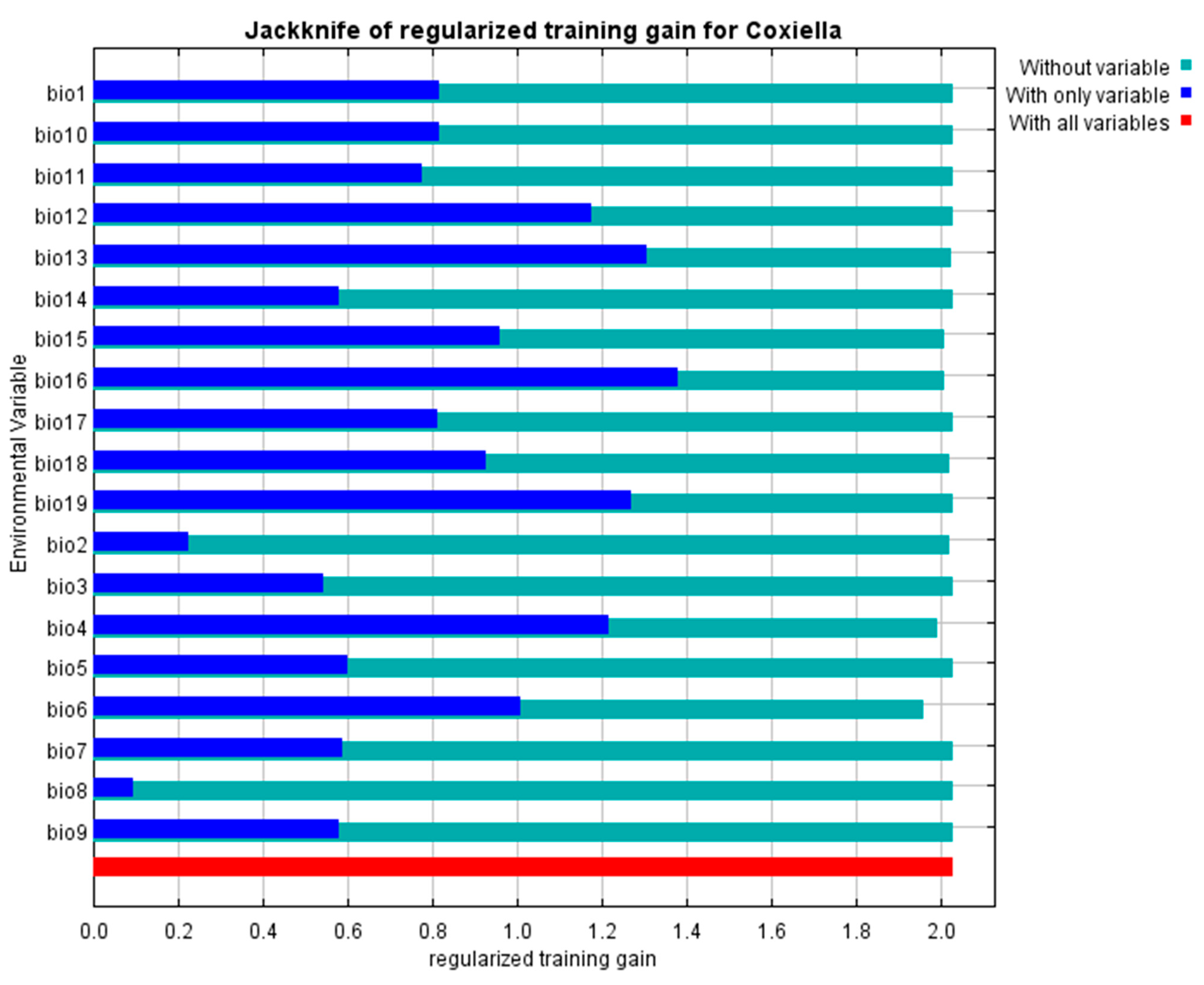
| Environmental Variable | Unit of Meas. | Code | % Contribution | Permutation Importance |
|---|---|---|---|---|
| Annual mean temperature | °C | bio1 | 0.1 | 0 |
| Mean diurnal temperature range (Mean of monthly (max temp–min temp)) | °C | bio2 | 4.4 | 3.8 |
| Isothermality (BIO2/BIO7) (×100) | percent | bio3 | 0 | 0 |
| Temperature Seasonality (standard deviation×100) | percent | bio4 | 4 | 0.1 |
| Maximal temperature of the warmest month | °C | bio5 | 0 | 0 |
| Minimal temperature of the coldest month | °C | bio6 | 6.9 | 64.1 |
| Temperature annual range (BIO5-BIO6) | °C | bio7 | 1.5 | 0 |
| Mean temperature of wettest quarter | °C | bio8 | 0 | 0 |
| Mean temperature of driest quarter | °C | bio9 | 0.9 | 0 |
| Mean temperature of warmest quarter | °C | bio10 | 0 | 0 |
| Mean temperature of coldest quarter | °C | bio11 | 0.7 | 0.9 |
| Annual precipitation | mm | bio12 | 1.3 | 0 |
| Precipitation of the wettest month | mm | bio13 | 40.2 | 15.4 |
| Precipitation of the driest month | mm | bio14 | 0 | 0 |
| Precipitation seasonality (coefficient of variation) | percent | bio15 | 7,8 | 2.2 |
| Precipitation of the wettest quarter | mm | bio16 | 28.3 | 0.3 |
| Precipitation of the driest quarter | mm | bio17 | 0 | 0 |
| Precipitation of the warmest quarter | mm | bio18 | 4 | 13.2 |
| Precipitation of the coldest quarter | mm | bio19 | 0 | 0 |
Disclaimer/Publisher’s Note: The statements, opinions and data contained in all publications are solely those of the individual author(s) and contributor(s) and not of MDPI and/or the editor(s). MDPI and/or the editor(s) disclaim responsibility for any injury to people or property resulting from any ideas, methods, instructions or products referred to in the content. |
© 2024 by the authors. Licensee MDPI, Basel, Switzerland. This article is an open access article distributed under the terms and conditions of the Creative Commons Attribution (CC BY) license (https://creativecommons.org/licenses/by/4.0/).
Share and Cite
Valiakos, G.; Gouvias, I.; Lysitsas, M.; Bouzalas, I.; Tampach, S.; Malissiova, E.; Giannakopoulos, A.; Tsokana, C.N.; Vourvidis, D.; Kyrma, A.; et al. Exposure of Dairy Cows to Coxiella burnetii in Greece: Surveillance Results and Association of Bacterial Presence with Environmental Variables. Microbiol. Res. 2024, 15, 655-666. https://doi.org/10.3390/microbiolres15020043
Valiakos G, Gouvias I, Lysitsas M, Bouzalas I, Tampach S, Malissiova E, Giannakopoulos A, Tsokana CN, Vourvidis D, Kyrma A, et al. Exposure of Dairy Cows to Coxiella burnetii in Greece: Surveillance Results and Association of Bacterial Presence with Environmental Variables. Microbiology Research. 2024; 15(2):655-666. https://doi.org/10.3390/microbiolres15020043
Chicago/Turabian StyleValiakos, George, Ioannis Gouvias, Marios Lysitsas, Ilias Bouzalas, Stefania Tampach, Eleni Malissiova, Alexis Giannakopoulos, Constantina N. Tsokana, Dimitrios Vourvidis, Anna Kyrma, and et al. 2024. "Exposure of Dairy Cows to Coxiella burnetii in Greece: Surveillance Results and Association of Bacterial Presence with Environmental Variables" Microbiology Research 15, no. 2: 655-666. https://doi.org/10.3390/microbiolres15020043