Article

Development of a Novel Emulsion Formulation of Trichoderma asperelloides PSU-P1 Conidia against Stem Canker on Dragon Fruit Caused by Neoscytalidium dimidiatum

1
School of Agricultural Technology and Food Industry, Walailak University, Nakhon Si Thammarat 80161, Thailand
2
Department of Plant Science, Faculty of Technology and Community Development, Thaksin University, Phatthalung Campus, Phatthalung 93210, Thailand
3
Agricultural Innovation and Management Division (Pest Management), Faculty of Natural Resources, Prince of Songkla University, Hatyai, Songkhla 90110, Thailand
*
Author to whom correspondence should be addressed.
Microbiol. Res. 2023, 14(3), 1139-1149; https://doi.org/10.3390/microbiolres14030076
Submission received: 17 July 2023 / Revised: 14 August 2023 / Accepted: 16 August 2023 / Published: 17 August 2023

Abstract

:
Stem canker on dragon fruit caused by Neoscytalidium dimidiatum causes severe losses in production of this fruit worldwide. Biological control by Trichoderma species is widely used to control several plant diseases. However, environmental conditions affect the use of biocontrol agents in the field. The development of a new formulation may offer an alternative way to address the problem of stem canker on dragon fruit caused by N. dimidiatum. In this study, we sought to develop a Trichoderma asperelloides PSU-P1 formulation that would be effective against N. dimidiatum. Three vegetable oils, two emulsifier-dispersing agents (Tween 20 and Tween 80), and one source of carbon (dextrose) were tested for carrier additives. We assessed the viability and antifungal ability of formulations incubated at ambient temperature and at 10 °C during a storage period of 1–6 months. The formulation composed of coconut oil, DW, and tween 20 in a ratio of 30:60:10 required a mixing time of 1.14 min; this was significantly faster than the mixing times of other formulations. Application of this formulation suppressed canker development; a canker area of 0.53 cm2 was recorded, compared with a control (pathogen only) area of 1.65 cm2. In terms of viability, this formulation stored at ambient temperature showed a surface area percentage of T. asperelloides PSU-P1 ranging from 64.43 to 75.7%; the corresponding range for the formulation stored at cool temperature was 70.59–75.6%. For both formulations, percentage inhibition gradually decreased from 1 to 6 months, with ranges of 59.21–77% and 60.65–76.19% for formulations incubated at ambient and cool temperatures, respectively. Our findings suggest that the formulation developed in this study prolongs the viability of T. asperelloides PSU-P1 conidia by up to 6 months, effectively inhibits N. dimidiatum in vitro, and reduces stem canker in vivo.

1. Introduction

Trichoderma species are commonly found in soil and are endophytic fungi which play an important role in ecosystems by degrading plant biomass, restricting plant pathogens, and promoting plant growth [1,2,3]. The main mechanisms of Trichoderma species are antibiosis, competition, induction of plant resistance, parasitism, and the promotion of plant growth [4]. Several Trichoderma species are highly adaptive to their environment, with high growth rates which enable them to compete for nutrients and space against fungal pathogens [5]. Trichoderma spp. have the ability to produce cell-wall-degrading enzymes such as chitinase and β-1,3-glucanase which degrade the cell walls of fungal pathogens [6,7]. They can also produce and release volatile organic compounds and/or secondary metabolites with fungicidal ability against plant pathogens [8,9]. Trichoderma species mainly reproduce by means of three asexual propagules: mycelia; conidia; and chlamydospores [10,11]. These exhibit typical characteristics with respect to production, stability and biocontrol activity; however, in nature, some species of this genus form ascospores in perithecia [12].
The use of antagonistic microorganisms to control several plant diseases has led to the manufacture and registration of many biofungicide products [13,14]. A number of Trichoderma species, such as Trichoderma asperellum, T. asperelloides, T. kiningiopsis, T. harzianum, T. viride and T. virens, have been used as biocontrol agents against several tropical plant diseases [14]. However, conditions in agricultural fields such as temperature, light, wind and moisture represent a major limitation on the use of biocontrol agents [15]. One potentially important way to overcome this problem is by the use of an appropriate formulation to create a microclimate that protects active spores from diverse environmental conditions [16].
Dragon fruit or pitaya are commonly planted throughout Thailand due to their rich micronutrients and vitamins [17]. To date, dragon fruits are classified into three varieties: red skin with white flesh, namely, Hylocereus undatus; red skin with red flesh, namely, H. costaricensis; and yellow skin with white flesh, namely, H. megalenthus [18]. Red-fleshed dragon fruit (Selenicereus costaricensis) or syn. H. costaricensis are tropical crops commonly cultivated for consumption. Thailand has a tropical and sub-tropical climate suitable for the cultivation of dragon fruit, and the climate is also suitable for pathogen germination and disease spread [19]. Several fungal diseases of dragon fruits, especially stem canker, are considered the most destructive and negatively impact dragon fruit production.
Stem canker of red-fleshed dragon fruit caused by the fungus Neoscytalidium sp. is a devastating disease which affects cultivation of this fruit worldwide, especially in tropical and sub-tropical regions [20]. In Thailand, the most widespread species is Neoscytalidium dimidiatum, which is endemic in the southern part of the country [21]. Control of fungal diseases has depended on the application on synthetic fungicides [22]. For example, the synthetic fungicides cyprodinil + fludioxonil, azoxystrobin + difenoconazole, and tebuconazole are all able to inhibit mycelial growth of N. dimidiatum, and metiram, trifloxystrobin, pyraclostrobin, azoxystrobin, azoxystrobin + difenoconazole, and iminoctadine are all effective in inhibiting spore germination of N. dimidiatum [23]. However, the excessive use of synthetic chemicals leads to pathogen resistance and causes harmful side effects on human health [24,25]. The use of fresh conidia of Trichoderma sp. has been shown to effectively reduce the severity of disease in many plant species [26,27]. However, preparation of viable conidia of Trichoderma may take time due to culturing on a substrate for sporulation. Nevertheless, the use of Trichoderma species as a biocontrol agent in a ready-to-use formulation may be considered an effective method for application in controlling plant diseases.
In this study, therefore, we sought to develop a novel emulsion formulation of T. asperelloides PSU-P1 whose viability and antifungal ability against N. dimidiatum could be tested in vitro, and whose capacity to reduce stem canker on dragon fruit could be tested in vivo. The objectives of this study were (i) to test the mixing time of various formulations and identify which is the most suitable, (ii) to test the recommended formulation against stem canker on dragon fruit in vivo, (iii) to test the viability of the recommended formulation stored at ambient temperature and cool temperature, and (iv) to test the antifungal ability of the recommended formulation against N. dimidiatum in vitro.

2. Materials and Methods

2.1. Fungal Materials and Spore Suspension Preparation

Trichoderma asperelloides PSU-P1 with multiple mechanisms against fungal pathogens [28,29] was obtained from the Culture Collection of Pest Management Division (CCPMD), Faculty of Natural Resources, Prince of Songkla University. T. asperelloides PSU-P1 was cultured on potato dextrose agar (PDA) and incubated at ambient temperature (28 ± 2 °C) for 7 days for sporulation. Conidia of T. asperelloides PSU-P1 were harvested and suspended in sterile distilled water (DW) for further formulation preparation. Neoscytalidium dimidiatum, which causes stem canker of dragon fruit [21], was also obtained from CCPMD and cultured on PDA for further bioassays.

2.2. Formulation Preparation

To develop the emulsion formulation, several carrier additives of three different types were tested: (1) three vegetable oils (coconut, palm and soybean); (2) two emulsifier-dispersing agents (Tween 20 and Tween 80); and (3) one source of carbon (dextrose). The compositions of the different oil dispersions tested are presented in Table 1. Emulsion-based formulations were prepared by mixing oils with emulsifier-dispersing agents and then adding dextrose. Finally, water was added. Conidia of T. asperelloides PSU-P1 were then progressively incorporated. The experiment comprised two sets in order to determine the most appropriate vegetable oil and achieve the fastest mixing time. Firstly, to determine the most appropriate vegetable oil, the viscosity of 12 formulations with varying amounts of vegetable oils in 12 formulations were tested, as shown in Table 1, and the mixing times were measured. The vegetable oil with the fastest mixing time was selected. Secondly, varying volumes of the selected vegetable oil were prepared and dextrose was added, resulting in 6 formulations, as shown in Table 2. The mixing time was then measured to identify the formulation with the fastest mixing time. The viscosity of the formulations was assessed by recording the mixing time. The formulation with the fastest mixing time was then selected for further evaluation.

2.3. In Vivo Testing of Formulation

In order to test the effectiveness of the emulsion formulation of T. asperelloides PSU-P1 in reducing stem canker, in vivo testing was conducted on healthy stems of dragon fruits. A total of ten cladodes from healthy dragon fruit were prepared for inoculation. The cladodes were surface-disinfected with 70% ethanol and wounded by fine needle. The emulsion formulation was diluted in DW at a ratio of 1:99. The experiment consisted of four treatments: (i) dropping of 1 mL distilled water alone (T1); (ii) dropping of 1 mL spore suspension of N. dimidiatum alone (T2); (iii) dropping of 1 mL spore suspension of N. dimidiatum for 24 h, followed by challenge with formulation (T3); and (iv) dropping of formulation alone (T4). The experiment was conducted in the form of a completely randomized design (CRD); each treatment involved three cladodes, and the experiment was repeated twice. The dragon fruit cladodes were then incubated in a moist box at ambient temperature. Lesion development was observed and measured by ImageJ software.

2.4. Viability of Trichoderma Conidia, Antifungal Ability of Formulation, and pH Evaluation

The viability of T. asperelloides PSU-P1 conidia in emulsion formulations stored at ambient temperature (28 ± 2 °C) and at 10 °C was assessed over a period of 6 months. The pH of each formulation was tested using a pH meter. Each month, an emulsion formulation was diluted at 1:99. Autoclaved sterilized filter paper discs (0.4 cm) were soaked in a 1% suspension of T. asperelloides PSU-P1. A T. asperelloides PSU-P1 filter paper disc was placed on one side of the PDA plate; on the opposite side was placed a N. dimidiatum agar plug cut from a 2-day-old culture. For the control plate, the agar plug of N. dimidiatum was placed on one side of the PDA plate only. The tested plates were incubated at 28 ± 2 °C for 7 days. Viability was analyzed on the third day by determining the area of the colonized surface of T. asperelloides PSU-P1; this was expressed as a percentage of the total surface area of the Petri dish. Assessment of the antifungal ability of T. asperelloides PSU-P1 was carried out on day seven using the tested plates. All the tested plates were photographed and subjected to surface area analysis by ImageJ software 167 version 1.53e (National Institutes of Health, Washington, DC, USA). Each test involved three PDA plates (three replicates), and the experiment was repeated twice. The radial growth of N. dimidiatum was also measured; this was converted to percentage inhibition (PI) by the following formula:
PI = ( R 1 R 2 R 1 ) ×   100
where R1 is the radial growth of N. dimidiatum in the control plate, and R2 is the radial growth of N. dimidiatum in the tested plate.
The pH of the formulation was measured from 1 to 6 months by pH meter, and data are expressed as average ± SD.

2.5. Statistical Analysis

Mixing time, disease development, viability and antifungal ability were assessed. The data met the parametric criteria for normality and homogeneity of variance necessary for the analysis of variance (ANOVA) and were subjected to one-way ANOVA. Tukey’s test and t-testing were used to analyze statistically significant differences at a 95% significance level (p < 0.05).

3. Results

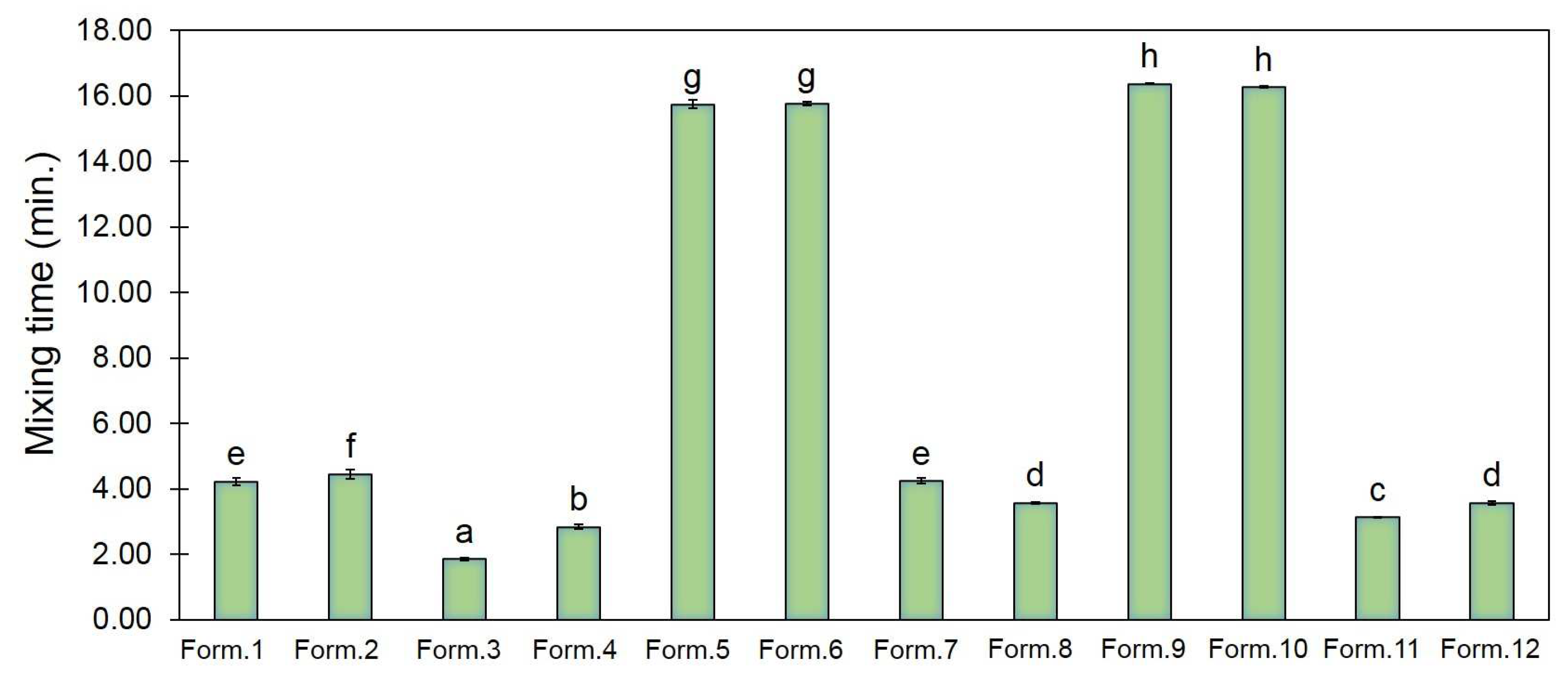
3.1. Selection of an Appropriate Formulation

The physico-chemical characteristics of the three oil dispersions tested, and of the two emulsifying agents, are shown in Table 1. We compared the mixing times of the twelve formulations and found that coconut-oil-based formulations showed more rapid mixing times than those based on palm oil or soybean oil (Figure 1). The results showed mixing times in the range 1.86 ± 0.05–16.37 ± 0.01 min. The coconut oil-based formulations had short mixing times in the range 1.86 ± 0.05–4.46 ± 0.14 min (Form. 1–4), whereas the soybean oil-based formulation showed the longest mixing time, ranging from 3.57 ± 0.06 to 16.37 ± 0.01 min (Form. 9–12). We also found that mixing time was impacted by the volume of oil in each formulation. Therefore, the coconut oil-based emulsion formulation was selected with varying amounts of the oil and DW phases to test the mixing time.
We selected coconut oil for further study and recorded the mixing times of various formulations, as shown in Table 2. The coconut oil-based emulsion formulations showed mixing times in the range 1.14 ± 0.06–3.58 ± 0.01 min (Table 3). We found that a ratio of 30:60:10 of coconut oil, DW, and tween 20, respectively, produced the fastest mixing time of 1.14 min, which was significantly faster than that of other formulations, p < 0.05 (Table 3). This formulation exhibited separation of oil and DW phases in the resting stage; after mixing, it was a slightly green milky lotion (Figure 2). This coconut-oil-based emulsion formulation was, therefore, considered most suitable for further bioassay testing.

3.2. In Vivo Test of Formulation

We tested the ability of the emulsion formulation of T. asperelloides PSU-P1 in suppressing the development of disease in dragon fruit cladodes. The results showed that application of this formulation resulted in a canker lesion area of 0.53 cm²; this was significantly smaller than the area of 1.65 cm² which resulted from inoculation with the pathogen alone (Figure 3). The control showed no canker lesions after inoculation with DW or with emulsion formulation alone. These results suggest that the emulsion formulation of T. asperelloides PSU-P1 suppressed the severity of canker on dragon fruit cladodes.

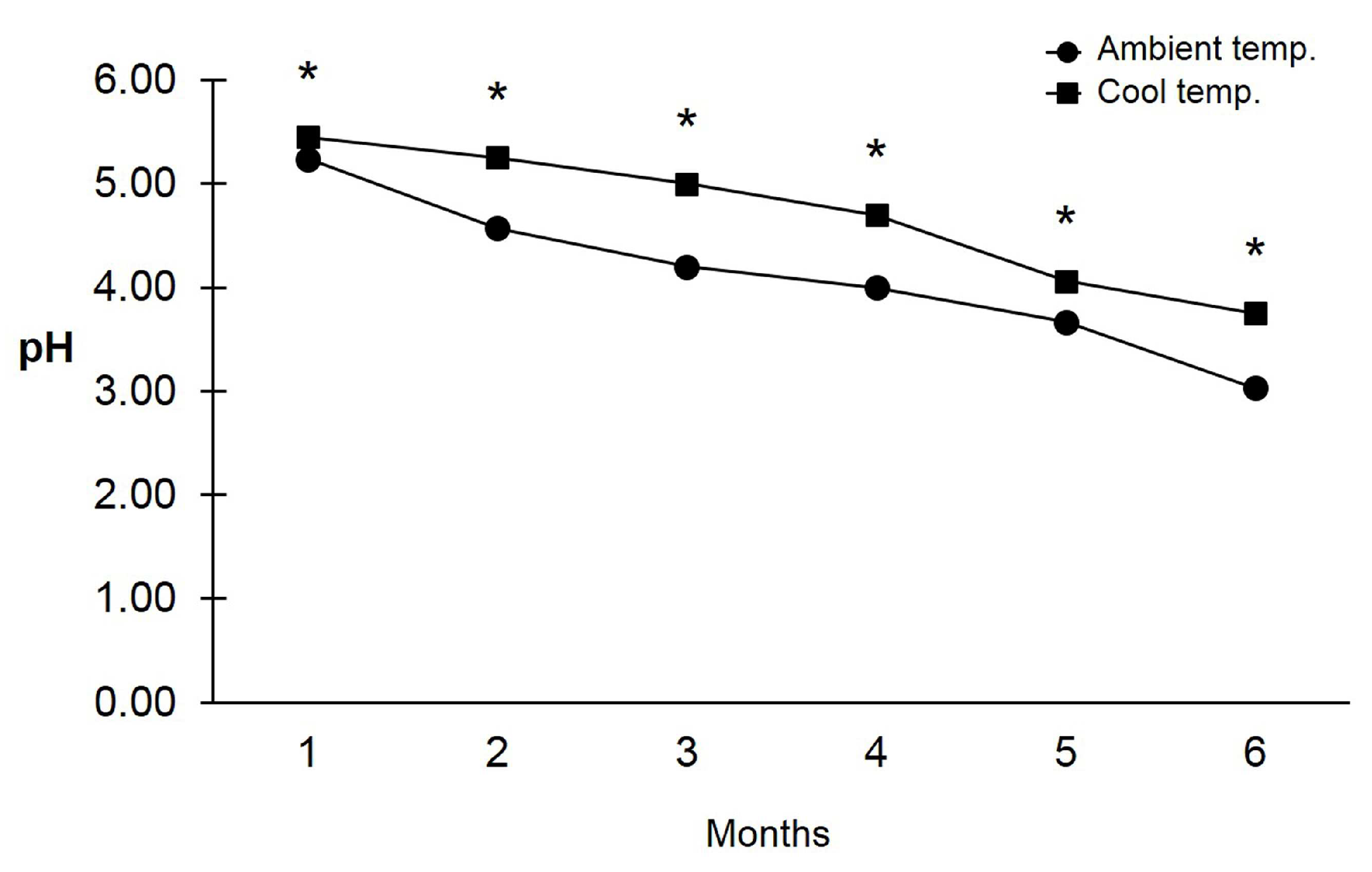
3.3. pH and Viability of Trichoderma asperelloides PSU-P1 Conidia in Formulation

The acidic and basic conditions of the emulsion formulation were measured from 1 to 6 months; the formulations were incubated at ambient and cool temperatures for comparison with the viability of T. asperelloides PSU-P1 conidia. The pH value of both formulations was below pH 6, indicating an acidic condition. The pH values ranged from 3.03 to 5.24 (mean ± SD; 4.12 ± 0.76) and from 3.75 to 5.45 (4.70 ± 0.67) for formulations incubated at ambient and cool temperatures, respectively (Figure 4).
The viability of T. asperelloides PSU-P1 conidia was assessed by observing colonization of the surface area in the PDA plates. We compared the viability of T. asperelloides PSU-P1 conidia in formulations incubated at ambient temperature (AT) and at a cool temperature of 10 °C (CT). The percentage surface area of T. asperelloides PSU-P1 gradually decreased between 1 and 6 months (Figure 5). After 3 days of incubation, T. asperelloides PSU-P1 conidia germinated in the PDA plate and covered more than 50% of the surface area (Figure 5). The percentage surface area of T. asperelloides PSU-P1 had ranges of 58.08–76% (68 ± 6.19) and 70.59–75.63% (73 ± 1.78) for formulations incubated at ambient and cool temperatures, respectively (Figure 5). In short, we found a greater viability of T. asperelloides PSU-P1 conidia in terms of growth for the formulation stored at cool temperature (10 °C), compared with the formulation stored at ambient temperature. The percentage of the surface area colonized by T. asperelloides PSU-P1 in the ambient-temperature formulation at 3–6 months was significantly lower than the corresponding area for the cool-temperature formulation.

3.4. Antifungal Ability of Trichoderma asperelloides PSU-P1 Conidia in Formulation against Neoscytalidium dimidiatum

An assessment of the fungicidal activity of T. asperelloides PSU-P1 conidia in the emulsion formulation was carried out by a dual-culture assay against N. dimidiatum in PDA plates. Mycelial growth which germinated from conidia covered more than 50% of the PDA plate surfaces and competed with the growth of N. dimidiatum. The percentage inhibition of the emulsion formulation against N. dimidiatum was also calculated from 1 to 6 months. The results showed that percentage inhibition gradually decreased for formulations incubated at both temperatures (Figure 6). The formulation incubated at cool temperature showed percentage inhibitions ranging from 60.25 to 76.19% (68 ± 6.65); those of the formulation incubated at ambient temperature ranged from 59.21 to 77% (70 ± 5.74) (Figure 6). The percentage inhibition of the formulation incubated at cool temperature was significantly higher than that of the formulation incubated at ambient temperature in months 4–6 (Figure 6).

4. Discussion

Our study is the first contribution to the development of an emulsion-based formulation of T. asperelloides PSU-P1 conidia for use as a biological control agent against N. dimidiatum, the pathogen of stem canker on dragon fruit. The goals of this study were to develop a formulation that can improve the protection of dragon fruit against stem canker. The emulsion-based formulation selected was composed of coconut oil (30%), tween 20 (10%) and T. asperelloides PSU-P1 conidia in DW (60%). In the course of in vivo testing, we found that application of the emulsion formulation reduced the severity of stem canker disease on dragon fruit cladodes. This result is in agreement with the findings of Syed-Ab-Rahman et al. [30], who reported that application of an emulsion formulation reduced the severity of brown root and bacterial leaf blight in rice by 55–61%.
In Thailand, farmers normally store biopesticides either at ambient temperature or in a refrigerator (approximately 10 °C). In this study, therefore, we tested the effects of temperature on the viability and antifungal ability of T. asperelloides PSU-P1 conidia in formulation. We found that the formulation stored at cool temperature seemed more effective against N. dimidiatum in terms of both viability and antifungal ability. The results of this study also showed that the viability and antifungal ability of the formulation both declined gradually from 1 to 6 months. Long periods of storage may, therefore, reduce the viability of the formulation. A similar previous study reported that formulations in invert emulsions (water-in-oil formulations) continued to exhibit viability at 36 months; however, viability also declined by 50% after 5.3 months at 20 °C [31]. Previous studies have also shown that an invert formulation of Trichoderma spp. may be used to prolong the shelf life of Trichoderma conidia and protect against subsurface contamination in the environment [32,33,34].
In the present study, we also found that, from 3 to 6 months, the formulation stored at cool temperature resulted in a surface area percentage of T. asperelloides PSU-P1 higher than that for the formulation stored at ambient temperature. Because of the hot and humid conditions in Thailand, Trichoderma conidia in formulation may have reduced viability and antifungal ability. It is known that Trichoderma sp. commonly grows at temperatures ranging from 12 to 37 °C, with maximum growth at 27 °C [35]. Furthermore, the present study showed that, while the pH of both formulations gradually decreased during the period from 1 to 6 months, the formulation stored at cool temperature showed pH values significantly higher than those of the formulation stored at ambient temperature. Although Trichoderma spp. commonly grows in acidic conditions, with pH ranging from 4 to 7 [36,37], a more strongly acidic environment (pH about 3) may not suitable for Trichoderma conidia; for this reason, the viability and percentage inhibition of the formulation stored at ambient temperature was significantly lower than that of the formulation stored at a cool temperature.
The shelf life of T. asperelloides PSU-P1 conidia in formulation may be associated with temperature. For the first 2 months, our results showed no significant differences in the surface area of T. asperelloides PSU-P1; however, significant differences were observed from 3 to 6 months. Our results indicate that active conidia of T. asperelloides PSU-P1 could be stored for up to half a year. This is in line with the findings of Batta et al. [31] who showed that liquid formulations could be preserved and stored without contamination or loss of viability. Several studies have shown that formulated conidia remain viable for longer periods [38,39]. Our results are, therefore, in agreement with those of previous studies which reported that formulated conidia of Trichoderma can remain viable for at least 6 months. The phenomenon may be due to the effect of certain ingredients in the formulation. As a basic food source, vegetable oils may help to boost conidial viability [40]. In the present study, the use of coconut oil as the main ingredient for the emulsion formulation may have helped to preserve conidia.
Approximately 90% of formulations of antagonistic microorganisms are made from active conidia of several Trichoderma species [14]. However, most formulations have been developed as granules [41] or as a wettable powder [13]. Only a few emulsion formulations have been developed from vegetable oils. In this study, we developed an emulsion formulation of T. asperelloides PSU-P1 conidia as an alternative formulation for agricultural application. One benefit of the emulsion formulation reported in this study is that it is better adapted for use in the sprayers commonly used by farmers, without resulting in coagulation. Conidia of T. asperelloides PSU-P1 as an emulsion dispersion can be mixed readily with water, so the formulation may be described as a ready-to-use product, and the conidia remain in suspension. Finally, during storage, this emulsion formulation separates into oil and water phases, as was previously reported in the work of Batta [31].

5. Conclusions

In the present study, T. asperelloides PSU-P1 conidia were incorporated in a water- and oil-based formulation and expressed as an emulsion formulation. The formulation composed of coconut oil, DW, and tween 20 in a ratio of 30:60:10 required the shortest mixing time. Application of coconut oil-based emulsion formulation to dragon fruit cladodes suppressed canker development, as observed by in vivo tests. Furthermore, this formulation prolonged the viability of T. asperelloides PSU-P1 conidia by up to 6 months, though viability decreased by about 20% over this period. The percentage inhibition of T. asperelloides PSU-P1 conidia against N. dimidiatum was also retained up to 6 months, though this also decreased over the period. These results indicate that the emulsion formulation ensures the prolonged viability and antifungal ability of T. asperelloides PSU-P1 conidia. The effectiveness of this formulation as an application should be verified by means of field trials in the near future.

Author Contributions

Conceptualization, W.I. and A.S.; methodology, K.S.D.; software, A.S.; validation, W.I., P.W. and A.S.; formal analysis, A.S.; investigation, K.S.D.; resources, A.S.; data curation, A.S.; writing—original draft preparation, W.I.; writing—review and editing, A.S.; visualization, A.S.; supervision, A.S.; project administration, A.S.; funding acquisition, A.S. and W.I. All authors have read and agreed to the published version of the manuscript.

Funding

This work was supported by the National Science, Research and Innovation Fund (NSRF) and Prince of Songkla University (grant no. NAT6505003M, NAT6505003b, NAT6601012M, NAT6601012b).

Institutional Review Board Statement

Not applicable.

Informed Consent Statement

Not applicable.

Data Availability Statement

Not applicable.

Acknowledgments

This work was partially supported by the Center of Excellence in Agricultural and Natural Resources Biotechnology (CoE-ANRB) Phase 3 and Walailak University.

Conflicts of Interest

The authors declare no conflict of interest.

References

  1. Tseng, Y.-H.; Rouina, H.; Groten, K.; Rajani, P.; Furch, A.C.U.; Reichelt, M.; Baldwin, I.T.; Nataraja, K.N.; Uma Shaanker, R.; Oelmüller, R. An Endophytic Trichoderma strain promotes growth of its hosts and defends against pathogen attack. Front. Plant Sci. 2020, 11, 573670. [Google Scholar] [CrossRef]
  2. Phoka, N.; Pornsuriya, C.; Sunpapao, A. High-throughput sequencing provides insight into soil fungal community structure and diversity in plant protected areas of Songkhla zoo in southern Thailand. Chiang Mai J. Sci. 2022, 49, 524–537. [Google Scholar] [CrossRef]
  3. Zhang, C.-I.; Zhang, B.; Yang, X.-Y.; Naicker, O.; Zhao, L. Brown rot disease caused by Trichoderma hamatum on the edible lily, Lilium leichtlinii var. maximowiczii. Chiang Mai J. Sci. 2022, 49, 1500–1508. [Google Scholar] [CrossRef]
  4. Benítez, T.; Rincón, A.M.; Limón, M.C.; Codón, A.C. Biocontrol mechanisms of Trichoderma strains. Int. Microbiol. 2004, 7, 249–260. [Google Scholar] [PubMed]
  5. Oszust, K.; Cybulska, J.; Frąc, M. How do Trichoderma genus fungi win a nutritional competition battle against soft fruit pathogens? A report on niche overlap nutritional potentiates. Int. J. Mol. Sci. 2020, 21, 4235. [Google Scholar] [CrossRef]
  6. Wonglom, P.; Daengsuwan, W.; Ito, S.; Sunpapao, A. Biological control of Sclerotium fruit rot of snake fruit and stem rot of lettuce by Trichoderma sp. T76-12/2 and the mechanism involved. Physiol. Mol. Plant Pathol. 2019, 107, 1–7. [Google Scholar] [CrossRef]
  7. Ruangwong, O.-U.; Pornsuriya, C.; Pitija, K.; Sunpapao, A. Biocontrol mechanisms of Trichoderma koningiopsis PSU3-2 against postharvest anthracnose of chili pepper. J. Fungi 2021, 7, 276. [Google Scholar] [CrossRef]
  8. Phoka, N.; Suwannarach, N.; Lumyong, S.; Ito, S.; Matsui, K.; Arikit, S.; Sunpapao, A. Role of volatiles from the endophytic fungus Trichoderma asperelloides PSU-P1 in biocontrol potential and in promoting the plant growth of Arabidopsis thaliana. J. Fungi 2020, 6, 341. [Google Scholar] [CrossRef]
  9. Intana, W.; Kheawleng, S.; Sunpapao, A. Trichoderma asperellum T76-14 released volatile organic compounds against postharvest fruit rot in muskmelons (Cucumis melo) caused by Fusarium incarnatum. J. Fungi 2021, 7, 46. [Google Scholar] [CrossRef]
  10. Gams, W.; Bissett, J. Morphology and identification of Trichoderma. In Trichoderma and Gliocladium: Basic Biology, Taxonomy and Genetics; Harman, G.E., Kubicek, C.P., Eds.; Taylor and Francis: London, UK, 1998; pp. 3–34. [Google Scholar]
  11. Verma, M.; Brar, S.K.; Tyagi, R.D.; Surampalli, R.Y.; Valero, J.R. Dissolved oxygen as principal parameter for conidia production of biocontrol fungi Trichoderma viride in non-Newtonian wastewater. J. Ind. Microbiol. Biotechnol. 2006, 33, 941–952. [Google Scholar] [CrossRef]
  12. Mukherjee, P.K.; Horwitz, B.A.; Herrera-Estrella, A.; Schmoll, M.; Kenerley, C.M. Trichoderma research in the genome era. Ann. Rev. Phytopathol. 2013, 51, 105–129. [Google Scholar] [CrossRef] [PubMed]
  13. Fravel, D.R. Commercialization and implementation of biocontrol. Annu. Rev. Phytopathol. 2005, 43, 337–359. [Google Scholar] [CrossRef] [PubMed]
  14. Kaewchai, S.; Soytong, K.; Hyde, K.D. Mycofungicides and fungal biofertilizers. Fungal Divers. 2009, 38, 25–50. [Google Scholar]
  15. Bashan, Y.; de Bashan, L.; Prabhu, S.; Hernandez, J.P. Advances in plant growth promoting bacterial inoculant technology: Formulations and practical perspectives (1998–2013). Plant Soil 2014, 378, 1–33. [Google Scholar] [CrossRef]
  16. Doni, F.; Isahak, A.; Zain, C.; Ariffin, S.; Mohamad, W.; Yusoff, W. Formulation of Trichoderma sp. SL2 inoculants using different carriers for soil treatment in rice seedling growth. SpringerPlus 2014, 3, 532. [Google Scholar] [CrossRef]
  17. Jaafar, R.A.; Abdul Rahman, A.R.B.; Mahnood, N.Z.C.; Vasudevan, R. Proximate analysis of dragon fruit (Hylocereus polyrhizus). Am. J. Appl. Sci. 2009, 6, 1341–1346. [Google Scholar]
  18. Abirami, K.; Swain, S.; Baskaran, V.; Sakthivel, K.; Bommayasamy, N. Distinguishing three dragon fruit (Hylocereus spp.) species grown in Andaman and Nicobar Islands of India using morphological, biochemical and molecular traits. Sci. Rep. 2021, 11, 2894. [Google Scholar] [CrossRef]
  19. Wonglom, P.; Thithuan, N.; Bunjongsiri, P.; Sunpapao, A. Plant-Parasitic algae (Cephaleuros spp.) in Thailand, including four new records. Pacific Sci. 2018, 72, 363–371. [Google Scholar] [CrossRef]
  20. Hong, C.F.; Gazis, R.; Crane, J.H.; Zhang, S. Prevalence and epidemics of Neoscytalidium stem and fruit canker on pitahaya (Hylocereus spp.) in South Florida. Plant Dis. 2020, 104, 1433–1438. [Google Scholar] [CrossRef]
  21. Dy, K.S.; Wonglom, P.; Pornsuriya, C.; Sunpapao, A. Morphological, molecular identification and pathogenicity of Neoscytalidium dimidiatum causing stem canker of Hylocereus polyrhizus in southern Thailand. Plants 2022, 11, 504. [Google Scholar] [CrossRef]
  22. Thaochan, N.; Pornsuriya, C.; Chairin, T.; Sunpapao, A. Roles of systemic fungicide in antifungal activity and induced defense responses in rubber tree (Hevea brasiliensis) against leaf fall disease caused by Neopestalotiopsis cubana. Physiol. Molec. Plant Pathol. 2020, 111, 101511. [Google Scholar] [CrossRef]
  23. Lin, C.; Ni, H.; Huang, C.; Yang, H. Pathogen characterization and chemical control of pitaya stem canker disease. Australasian Plant Dis. Notes 2017, 10, 1–4. [Google Scholar]
  24. Yoon, M.Y.; Cha, B.; Kim, J.C. Recent trends in studies on botanical fungicides in agriculture. Plant Pathol. J. 2013, 29, 1–9. [Google Scholar] [CrossRef] [PubMed]
  25. Fattahi, E.; Mousavi Moghadam, M.; Khanbabaei, R. The effect of fricyclazole on testosterone changes and testicular structure in mice. J. Babol. Univ. Med. Sci. 2015, 17, 43–49. [Google Scholar]
  26. Rees, H.J.; Drakulic, J.; Cromey, M.G.; Bailey, A.M.; Foster, G.D. Endophytic Trichoderma spp. can protect strawberry and privet plants from infection by the fungus Armillaria mellea. PLoS ONE 2022, 17, e0271622. [Google Scholar] [CrossRef] [PubMed]
  27. Tyśkiewicz, R.; Nowak, A.; Ozimek, E.; Jaroszuk-Ściseł, J. Trichoderma: The current status of its application in agriculture for the biocontrol of fungal phytopathogens and stimulation of plant growth. Int. J. Mol. Sci. 2022, 23, 2329. [Google Scholar] [CrossRef] [PubMed]
  28. Ruangwong, O.-U.; Wonglom, P.; Phoka, N.; Suwannarach, N.; Lumyong, S.; Ito, S.-I.; Sunpapao, A. Biological control activity of Trichoderma asperelloides PSU-P1 against gummy stem blight in muskmelon (Cucumis melo). Physiol. Mol. Plant Pathol. 2021, 115, 101663. [Google Scholar] [CrossRef]
  29. Intana, W.; Wonglom, P.; Suwannarach, N.; Sunpapao, A. Trichoderma asperelloides PSU-P1 induced expression of pathogenesis-related protein genes against gummy stem blight of muskmelon (Cucumis melo) in field evaluation. J. Fungi 2022, 8, 156. [Google Scholar] [CrossRef]
  30. yed-Ab-Rahman, S.F.; Carvalhais, L.C.; Omar, D. Development of plant-based emulsion formulation to control bacterial leaf blight and sheath brown rot of rice. Heliyon 2020, 6, e03151. [Google Scholar] [CrossRef]
  31. Batta, Y.A. Postharvest biological control of apple gray mold by Trichoderma harzianum Rifai formulated in an invert emulsion. Crop Prot. 2004, 23, 19–26. [Google Scholar] [CrossRef]
  32. Kumar, S.; Thakur, M.; Rani, A. Trichoderma: Mass production, formulation, quality control, delivery and its scope in commercialization in India for the management of plant disease. Afr. J. Agric. Res. 2014, 9, 3838–3852. [Google Scholar]
  33. Peeran, M.F.; Nagendran, K.; Gandhi, K.; Raguchander, T.; Prabakar, K. Water in oil based PGPR formulation of Pseudomonas fluorescens (FP7) showed enhanced resistance against Colletotrichum musae. Croop Protec. 2014, 65, 186–193. [Google Scholar] [CrossRef]
  34. Mbarka, J.B.; Begoude, B.A.D.; Ambang, Z.; Meboma, M.; Kuaté, J.; Schiffers, B.; Ten Hoopen, G.M. A new oil-based formulation of Trichoderma asperellum for the biological control of cacao black pod disease caused by Phytophthora megakarya. Biol. Control 2014, 77, 15–22. [Google Scholar] [CrossRef]
  35. Domingues, M.V.P.F.; de Moura, K.E.; Salomão, D.; Elias, L.M.; Patricio, F.R.A. Effect of temperature on mycelial growth of Trichoderma, Sclerotinia minor and S. sclerotiorum, as well as on mycoparasitism. Summa Phytopathol. Botucatu 2016, 42, 222–227. [Google Scholar] [CrossRef]
  36. Trushina, N.; Levin, M.; Mukherjee, P.K.; Horwitz, B.A. PacC and pH-dependent transcriptome of the mycotrophic fungus Trichoderma virens. BMC Genom. 2013, 14, 138. [Google Scholar] [CrossRef]
  37. Singh, A.; Shahid, M.; Srivastava, M.; Pandey, S.; Sharma, A.; Kumar, V. Optimal physical parameters for growth of Trichoderma species at varying pH, temperature and agitation. Virol Mycol 2014, 3, 127. [Google Scholar]
  38. Larena, I.; De Cal, A.; Melgarejo, P. Effects of stabilizers on shelf-life of Epicoccum nigrum formulations and their relationship with biocontrol of postharvest brown rot by Monilinia of peaches. J. Appl. Microbiol. 2007, 102, 570–582. [Google Scholar] [CrossRef]
  39. Wijesinghe, C.J.; Wilson Wijeratnam, R.S.; Samarasekara, J.K.R.R.; Wijesundera, R.L.C. Development of a formulation of Trichoderma asperellum to control black rot disease on pineapple caused by (Thielaviopsis paradoxa). Crop Prot. 2011, 30, 300–306. [Google Scholar] [CrossRef]
  40. Paau, A.S. Formulation of beneficial organisms applied to soil. In Formulation of Microbial Biopesticides: Beneficial Microorganisms, Nematodes and Seed Treatments; Burges, H.D., Ed.; Kluwer Academic Publishers: Dordrecht, The Netherlands, 1998; pp. 235–254. [Google Scholar]
  41. Jin, X.; Custis, D. Microencapsulating aerial conidia of Trichoderma harzianum through spray drying at elevated temperatures. Biol. Cont. 2011, 56, 202–206. [Google Scholar] [CrossRef]
Figure 1. Evaluation of viscosity of formulations in different forms as indicated by mixing time (minutes). Values are expressed as means ± standard deviation (SD) indicated by whiskers. Letters indicate significant differences among treatments according to Tukey’s test (p < 0.05).
Microbiolres 14 00076 g001
Figure 2. Characteristics of the novel emulsion formulation. Separation into oil and water phases: before mixing (A); and after mixing (B).
Microbiolres 14 00076 g002
Figure 3. Suppression of canker lesion by emulsion formulation: (A) T1 control; (B) T2 inoculated with Neoscytalidium dimidiatum alone; (C) T3 inoculated with N. dimidiatum for 24 h and then challenged with formulation; (D) T4 inoculated with formulation alone; and (E) lesion development under each treatment. Values are expressed as means ± standard deviation (SD) indicated by whiskers. Letters indicate significant differences among treatments according to Tukey’s test (p < 0.05).
Microbiolres 14 00076 g003
Figure 4. pH values of emulsion formulation incubated at ambient temperature and at cool temperature. Asterisks indicate significant differences among treatments according to Student’s t-test (p < 0.05).
Microbiolres 14 00076 g004
Figure 5. Viability of Trichoderma asperelloides PSU-P1 in emulsion formulation. (A) Observations of percentage surface area of PSU-P1 against Neoscytalidium dimidiatum from 1 to 6 months; (B,C) top and bottom views of colonies of T. asperelloides PSU-P1 in formulation incubated at cool temperature; (D,E) top and bottom views of formulations incubated at ambient temperature. Asterisks indicate significant differences among treatments according to Student’s t-test (p < 0.05).
Microbiolres 14 00076 g005
Figure 6. Percentage inhibition of Trichoderma asperelloides PSU-P1 conidia in formulation against Neoscytalidium dimidiatum in PDA plates from 1 to 6 months. Values are expressed as means ± standard deviation (SD) indicated by whiskers. Asterisks indicate significant differences among treatments according to Student’s t-test (p < 0.05).
Microbiolres 14 00076 g006
Table 1. Different compositions of oils and emulsifier-dispersing agents for emulsion formulation preparation.
Form. 1Ingredients (mL)
Coconut
Oil
Palm
Oil
Soybean
Oil
DWTween 20Tween 80Spore Suspension
140--4020-X 2
240--40-20X
320--6020-X
420--60-20X
5-40-4020-X
6-40-40-20X
7-20-6020-X
8-20-60-20X
9--404020-X
10--4040-20X
11--206020-X
12--2060-20X
1 Form. = formulation; 2 spore suspension at concentration ×108 conidia/mL, suspended in DW phase.
Table 2. Mixing times of different formulations with varying amounts of selected oil and dextrose additive.
Form. 1Ingredient
Selected Oil (mL)DW (mL)Dextrose (g)Tween 20
(mL)
Spore Suspension
(×108 Conidia/mL)
14040520X 2
24040-20X
34545510X
44545-10X
53060510X
63060-10X
1 Form. = formulation; 2 spore suspension at concentration ×108 conidia/mL, suspended in DW phase.
Table 3. Mixing times of different formulations (variations in amounts of coconut oil and dextrose additive).
Form. 1Ratio of
Coconut Oil:DW:Tween 20 (mL)
Dextrose (g)Mixing Time (min.)
140:40:2053.21 ± 0.02 d
240:40:20-2.29 ± 0.06 c
345:45:1053.58 ± 0.01 e
445:45:10-2.25 ± 0.02 c
530:60:1051.25 ± 0.06 b
630:60:10-1.14 ± 0.06 a
1 Form. = formulation; Values are expressed as means ± standard deviation (SD). Means within the same column followed by the same letters indicate significant differences among treatments according to Tukey’s test (p < 0.05).
Disclaimer/Publisher’s Note: The statements, opinions and data contained in all publications are solely those of the individual author(s) and contributor(s) and not of MDPI and/or the editor(s). MDPI and/or the editor(s) disclaim responsibility for any injury to people or property resulting from any ideas, methods, instructions or products referred to in the content.

